Embed Size (px)
Citation preview

1
IMP3 PROMOTES CHEMO-RESISTANCE IN BREAST CANCER CELLS
BY REGULATING BCRP (ABCG2) EXPRESSION*
Sanjoy Samanta, Bryan Pursell and Arthur M. Mercurio1
Department of Cancer Biology
University of Massachusetts Medical School
Worcester, MA 01605
Running title: IMP3 Promotes Chemo-Resistance
1To whom correspondence should be addressed: Department of Cancer Biology, University of
Massachusetts Medical School, LRB-408, 364 Plantation St., Worcester, MA 01605. Tel.: 508-856-8676;
Fax: 508-856-1310; E-mail: [email protected]
Keywords: Breast cancer; Chemo-resistance; mRNA; RNA binding protein; drug transporter
http://www.jbc.org/cgi/doi/10.1074/jbc.C112.442319The latest version is at JBC Papers in Press. Published on March 28, 2013 as Manuscript C112.442319
Copyright 2013 by The American Society for Biochemistry and Molecular Biology, Inc.
by guest on February 20, 2020http://w
ww
.jbc.org/D
ownloaded from

2
CAPSULE
Background: IMP3 is an mRNA binding protein
associated with aggressive cancers but whose
function is unknown in these cancers.
Results: IMP3 promotes chemo-resistance in
breast cancer cells by interacting directly with the
mRNA that encodes a specific drug transporter.
Conclusions: IMP3 has a causal role in the
chemo-resistance of breast cancer cells.
Significance: These data provide a mechanism for
how IMP3 contributes to breast cancer.
IMP3, a member of a family of insulin-like
growth factor II (IGF-II) mRNA binding
proteins (IMPs), is expressed preferentially in
triple-negative breast cancers, which are
resistant to many chemotherapeutics. However,
the mechanisms by which it impacts breast
cancer have not been elucidated. We
hypothesized a role for IMP3 in chemo-
resistance based on these observations.
Depletion of IMP3 expression in triple-negative
breast cancer cells increased their sensitivity to
doxorubicin and mitoxantrone significantly but
not to taxol. Given that doxorubicin and
mitoxantrone are effluxed by breast cancer
resistance protein (BCRP), we assessed whether
IMP3 regulates BCRP. The data obtained
demonstrate that IMP3 binds to BCRP mRNA
and regulates BCRP expression. These
findings are significant because they provide
insight into the mechanism by which IMP3
contributes to aggressive cancers and they
highlight the potential for targeting this mRNA
binding protein for the clinical management of
cancer. ________________________________________
The foundation for this study is the
compelling evidence that IMP3, a member of a
family of insulin-like growth factor II (IGF-II)
mRNA binding proteins (IMPs) that function in
RNA trafficking, stabilization and localization (1)
is expressed preferentially in triple-negative breast
cancer (2). Clinically, triple-negative breast
cancers are usually of high histological grade,
poorly differentiated and more aggressive
compared to other sub-types of breast cancer (3).
Most, if not all, breast tumors that contain
inactivating mutations in the BRCA1 gene, which
is a major determinant of hereditary breast cancer,
exhibit a triple-negative phenotype (4).
Importantly, treatment of triple-negative breast
cancer remains a challenge because of the lack of
targeted therapeutic options and resistance to
standard chemotherapy (3). What is not known is
whether there is a causal link between IMP3 and
the aggressive behavior of triple-negative breast
cancer and, if so, the mechanism by which this
RNA binding protein contributes to such behavior.
In pursuit of a causal role for IMP3 in
triple-negative breast cancer, we explored the
hypothesis that this mRNA binding protein
contributes to chemo-resistance. The results
obtained validate this hypothesis and they
establish a mechanism that involves IMP3-
mediated regulation of BCRP (breast cancer
resistance protein), also known as ABCG2, a
member of the ABC transporters and a major
effector of drug resistance in breast cancer (5).
EXPERIMENTAL PROCEDURES
Cells & reagents-The human breast cancer cell
line SUM-1315 was obtained from Dr. Stephen
Ethier (Medical College of South Carolina,
Charleston, SC). HEK293T and MDA-468 cell
lines were obtained from the American Type
Culture Collection (ATCC). SUM1315 cells were
maintained in F-12 medium supplemented with
5% fetal bovine serum, insulin (5µg/mL),
epidermal growth factor (10 ng/mL) and 1%
penicillin-streptomycin. MDA-468 cells were
maintained in DMEM supplemented with 10 %
fetal bovine serum and 1 % penicillin-
streptomycin. HEK293T cells were cultured in
DMEM (high glucose) supplemented with 10 %
fetal bovine serum, non-essential amino acids (1x,
Gibco), HEPES (pH 7.4, 1mM, Gibco), sodium
pyruvate (1mM, Gibco) and 1% penicillin-
streptomycin. All cell lines were grown at 370C
and 5 % CO2.
IMP3 specific shRNAs (TRCN0000074673 &
TRCN0000074675) were obtained from
Open Biosystems (Rockford). Doxorubicin,
mitoxantrone and taxol were procured from
Sigma-Aldrich. Doxorubicin was solubilized in
water whereas mitoxantrone and taxol were
by guest on February 20, 2020http://w
ww
.jbc.org/D
ownloaded from

3
solubilized in DMSO. A BCRP expression vector
(Plasmid ID: 25983) was obtained from Addgene.
The control vector was generated by excising
BCRP cDNA from the expression vector. IMP3
and BCRP antibodies were purchased from DAKO
and Abcam, respectively. Lipofectamine 2000 and
Fugene 6 were procured from Invitrogen and
Promega, respectively.
IMP3-depleted cell lines (SUM-1315 &
MDA-468) were generated by infecting them with
PLKO.1 based lentiviruses (produced in
HEK293T cells by transfecting plasmid DNA
using Lipofectamine) expressing shRNAs
targeting IMP3 mRNA and subsequent selection
under puromycin (2 µg/mL). Stable cell lines were
maintained regularly under puromycin (1 µg/mL).
BCRP expression was rescued by infecting IMP3-
depleted SUM-1315 cells with a lentivirus
expressing full-length BCRP cDNA and
subsequent selection under G418 (600 µg/mL).
The lentivirus was generated from HEK293T
cells.
MTT cytotoxicity assay-The chemo-sensitivity of
breast cancer cells was determined using standard
MTT (3-(4,5-Dimethylthiazol-2-yl)-2,5-
diphenyltetrazolium bromide) cytotoxicity assay
(6). The assay was performed 48 h after drug
treatment.
Immunoblotting- Cell extracts were prepared using
RIPA buffer containing EDTA and EGTA (Boston
Bioproducts). A protease and phosphatase
inhibitor cocktail was added separately (Roche
Applied Biosciences). Extracts (40-50 µg protein)
were blotted with the appropriate primary Abs and
then incubated with either mouse or rabbit IgG
horseradish peroxidase-conjugated secondary
antibody. An ECL kit (Thermo Scientific) was
used to develop the blots.
RNA isolation and real time PCR analysis- Total
RNA was isolated from cultured cells using Trizol
reagent (Invitrogen) following the manufacturer’s
protocol. cDNA was synthesized using
superscript-II reverse transcriptase (Invitrogen).
mRNAs were quantified by real-time PCR
analysis (ABI Prism, Applied Biosystems) using
Power Syber Green PCR master mix (Applied
Biosystems). Quantification was performed using
Ct method and GAPDH was used as reference
gene. The following primer pairs were used for
real time PCR analysis:
IMP3-forward:
5’-CCGCAGTTTGAGCAATCAGAA-3’
IMP3-reverse:
5’-CGAGAAAGCTGCTTGATGTGC-3’
IGFII-forward:
5’-CCGAAACAGGCTACTCTCCT-3’
IGFII-reverse:
5’-AGGGTGTTTAAAGCCAATCG-3’
BCRP-forward:
5’-GTTGTGATGGGCACTCTGAC-3’
BCRP-reverse:
5’-CCCTGTTAATCCGTTCGTTT-3’
ESR2-forward:
5’-AAGGTTAGTGGGAACCGTTG-3’
ESR2-reverse:
5’-ACATCCTTCACACGACCAGA-3’
Ribo-immunoprecipitation assay-The interaction
between IMP3 protein and BCRP mRNA was
determined using RIP-qPCR assay as described
previously (7). Briefly SUM-1315 cells (~2 × 10⁷)
were harvested and extracted for 15 min on ice in
250 μl of ice-cold lysis buffer (100 mm KCl, 5
mm MgCl2, 10 mm HEPES (pH 7.0), 0.5%
Nonidet P-40, 10 μm dithiothreitol) supplemented
with RNase and protease inhibitors. Extracts were
cleared by centrifugation for 15 min at 13,000 rpm
and supernatant was transferred to a fresh 1.5-ml
tube. To pre-clear the cytoplasmic extracts, 25 μg
of non-immune rabbit IgG (Sigma) was added to
the supernatant and kept on ice for 45 min, then
incubated with 50 μl of a 50% (v/v) suspension of
protein G-Sepharose beads (Biovision) for 3 h at
4°C with rotation. This was centrifuged at 13,000
rpm and the supernatant was recovered (pre-
cleared lysate). For immunoprecipitation, the pre-
cleared extract was incubated with 100 μl of a
50% suspension of protein G-Sepharose beads
(Sigma) pre-coated with the same amount of either
non-immune mouse IgG (Sigma) or anti-human
IMP-3 antibody (25 μg) in 800 μl of NT-2 buffer
(150 mm NaCl, 1 mm MgCl2, 50 mm Tris-HCl
(pH 7.4), 0.05% Nonidet P-40) containing RNase
inhibitor and protease inhibitors for over-night at 4
°C with rotation. Beads were washed 10 times
using ice-cold NT-2 buffer, digested with 20 units
of RNase-free DNase I (Promega) in 100 of μl of
NT-2 buffer for 20 min at 30 °C, washed with NT-
2 buffer, and further digested with 0.5 mg/ml
by guest on February 20, 2020http://w
ww
.jbc.org/D
ownloaded from

4
protease K (Ambion) in 100 μl of NT-2 buffer
containing 0.1% SDS at 55 °C for 30 min. RNA
was extracted with Trizol (Invitrogen). Glycogen
(Roche Applied Science) was added to facilitate
precipitation of RNA. Real-time PCR was
performed on equivalent amounts of sample to
quantify protein-bound mRNAs.
Generation of IMP3 expression construct resistant
to shIMP3-2-The IMP3 expression construct
resistant to shIMP3-2 was generated by mutating
two nucleotides within the target sequence
(located in the coding region of wild-type IMP3)
of shIMP3-2. The wild type IMP3 construct was
generated by cloning full length cDNA of IMP3 in
pCDH-CMV-MCS-EF1-GFP lentiviral vector
(System Biosciences, CA, USA) at EcoRI/NotI
sites. The desired mutation was carried out by site
directed muatagenesis (QuikChange XL Site-
Directed Mutagenesis Kit, Agilent
Technologies). The target sequence of
shIMP3-2 is 5’-CGGTGAATGAACTTCAGAATT and located
1782 bp downstream of transcription start site. The
sequence was mutated to
CGGTGAATGAATTGCAGAATT.
Primers used for mutagenesis:
For:5’GGAGGCAAAACGGTGAATGAATTGC
AGAATTTGTCAAGTGCAGAAG
Rev:5’CTTCTGCACTTGACAAATTCTGCAAT
TCATTCACCGTTTTGCCTCC
RESULTS AND DISCUSSION
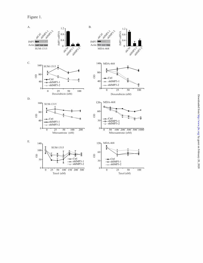
Depletion of IMP3 expression increases
chemosensitivity of triple-negative breast cancer
cells- To test the possible role of IMP3 in
promoting chemo-resistance, we depleted IMP3
expression in the triple-negative breast cancer cell
lines SUM-1315 and MDA-468 using two
different short hairpin RNAs (shRNAs) (Fig. 1A
& B). Control and IMP3-depleted cells were
assessed for their sensitivity to doxorubicin,
mitoxantrone and taxol, chemotherapeutic agents
used in breast cancer therapy (8-10). As shown in
Fig. 1C & D, depletion of IMP3 expression
increased the sensitivity of both cell lines to
doxorubicin and mitoxantrone significantly as
measured by the MTT assay. In contrast, IMP3-
depleted SUM-1315 cells were more resistant to
taxol than control cells (Fig. 1E). Similar results
were obtained with MDA-468 cells also. To
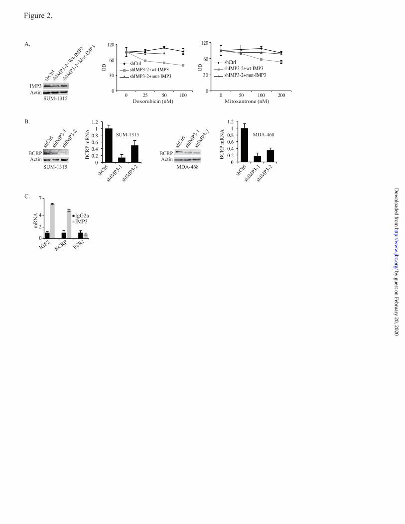
demonstrate the specificity of shRNA used to
deplete IMP3 expression, we rescued IMP3
expression in IMP3-depleted SUM-1315 cells
using a lentiviral construct that is resistant to
shIMP3-2 (Fig. 2A). As shown in Fig. 2A,
restoration of IMP3 expression decreases
sensitivity to both doxorubicin and mitoxantrone
compared to cells expressing wild-type IMP3,
which is targeted by shIMP3.
IMP3 promotes drug resistance by binding to
BCRP mRNA and regulating its expression- The
finding that IMP3-depleted cells are sensitive to
doxorubicin and mitoxantrone is noteworthy
because these drugs are effluxed by BCRP
(11,12). Interestingly, taxol is not effluxed by
BCRP (13). These observations prompted us to
examine the possible role of IMP3 in regulating
BCRP expression. To test this possibility, we
assessed BCRP mRNA and protein expression in
IMP3-depleted SUM-1315 and MDA-468 cells.
As shown in Fig. 2B, depletion of IMP3 reduced
the level of BCRP mRNA and protein
significantly in both cell lines.
An important consideration based on the
above findings is whether IMP3 interacts directly
with BCRP or regulates its expression indirectly.
To address this issue, we performed Ribo-
Immunoprecipitation-qPCR (RIP-qPCR), which
detects specific protein-RNA interactions. IGF2
was used as a positive control for this experiment
because IMP3 was defined initially as an IGF2
mRNA binding protein (14). Indeed, IMP3 binds
to BCRP mRNA at a level comparable to its
binding to IGF2 mRNA (Fig. 2C). We used ESR2
(ER) as negative control for this experiment
because IMP3 has not been reported to regulate its
expression or function. These data demonstrate
that IMP3 binds to BCRP mRNA and, as a
consequence, regulates its expression.
To demonstrate that IMP3 promotes chemo-
resistance by regulating BCRP, we rescued BCRP
expression in IMP3-depleted SUM-1315 cells by
transfecting them with a lentiviral-based BCRP
expression vector (Fig. 3A) and assayed for
chemo-resistance. As shown in Fig. 3B,
restoration of BCRP expression in IMP3-depleted
cells decreased their sensitivity to doxorubicin and
mitoxantrone significantly. To control for the
by guest on February 20, 2020http://w
ww
.jbc.org/D
ownloaded from

5
possibility that this result could be attributed
solely to BCRP over-expression and not linked to
IMP3, we over-expressed BCRP in parental SUM-
1315 cells and assayed chemo-resistance. As
shown in Fig. 3C, BCRP over-expression in
parental cells caused a slight, if any, increase in
resistance to doxorubicin and mitoxantrone in
comparison to the increase observed for IMP3-
depleted cells (Fig. 3B). We conclude from these
data that IMP3 promotes chemo-resistance in
triple-negative breast cancer cells by regulating
BCRP.
The findings presented in this report are
significant for several reasons. Although IMP3
expression correlates with the aggressive behavior
of many cancers and is used clinically for the
prognostic assessment of specific cancers (15,16),
the mechanism by which it functions in this
context had been elusive. Our demonstration that
IMP3 promotes the chemo-resistance of triple-
negative breast cancers by regulating a specific
drug transporter provides the first insight into this
mechanism. These findings are consistent with the
recent report that other IMPs contribute to the
initiation of glioblastomas (17) and highlight the
potential for targeting IMPs as a therapeutic
approach to cancer. Targeting IMP3 is a
potentially feasible and effective approach to the
clinical management of triple-negative breast
cancer for several reasons. IMP3 is not expressed
in normal breast (2), its mechanism of action is
known (binding to specific RNA sequences) and
its inhibition should increase susceptibility to
standard chemotherapy.
by guest on February 20, 2020http://w
ww
.jbc.org/D
ownloaded from

6
REFERENCES
1. Mueller-Pillasch, F., Pohl, B., Wilda, M., Lacher, U., Beil, M., Wallrapp, C., Hameister,
H., Knochel, W., Adler, G., and Gress, T. M. (1999) Expression of the highly conserved
RNA binding protein KOC in embryogenesis. Mechanisms of development 88, 95-99
2. Walter, O., Prasad, M., Lu, S., Quinlan, R. M., Edmiston, K. L., and Khan, A. (2009)
IMP3 is a novel biomarker for triple negative invasive mammary carcinoma associated
with a more aggressive phenotype. Human pathology 40, 1528-1533
3. Griffiths, C. L., and Olin, J. L. (2012) Triple negative breast cancer: a brief review of its
characteristics and treatment options. J Pharm Pract 25, 319-323
4. Turner, N., Tutt, A., and Ashworth, A. (2004) Hallmarks of 'BRCAness' in sporadic
cancers. Nat Rev Cancer 4, 814-819
5. Doyle, L. A., and Ross, D. D. (2003) Multidrug resistance mediated by the breast cancer
resistance protein BCRP (ABCG2). Oncogene 22, 7340-7358
6. Mosmann, T. (1983) Rapid colorimetric assay for cellular growth and survival:
application to proliferation and cytotoxicity assays. Journal of immunological methods
65, 55-63
7. Liao, B., Hu, Y., Herrick, D. J., and Brewer, G. (2005) The RNA-binding protein IMP-3
is a translational activator of insulin-like growth factor II leader-3 mRNA during
proliferation of human K562 leukemia cells. The Journal of biological chemistry 280,
18517-18524
8. Ibrahim, N. K., Buzdar, A. U., Asmar, L., Theriault, R. L., and Hortobagyi, G. N. (2000)
Doxorubicin-based adjuvant chemotherapy in elderly breast cancer patients: the M.D.
Anderson experience, with long-term follow-up. Annals of oncology : official journal of
the European Society for Medical Oncology / ESMO 11, 1597-1601
9. Petit, T., Borel, C., Theobald, S., Serin, D., Rodier, J. F., Prevot, G., Brettes, J. P., and
Klein, T. (2003) Randomized multicentric study of perioperative chemotherapy with
mitoxantrone in early breast cancer. Annals of surgical oncology 10, 369-375
10. Rowinsky, E. K., Onetto, N., Canetta, R. M., and Arbuck, S. G. (1992) Taxol: the first of
the taxanes, an important new class of antitumor agents. Seminars in oncology 19, 646-
662
11. Hazlehurst, L. A., Foley, N. E., Gleason-Guzman, M. C., Hacker, M. P., Cress, A. E.,
Greenberger, L. W., De Jong, M. C., and Dalton, W. S. (1999) Multiple mechanisms
confer drug resistance to mitoxantrone in the human 8226 myeloma cell line. Cancer
research 59, 1021-1028
12. Robey, R. W., Honjo, Y., Morisaki, K., Nadjem, T. A., Runge, S., Risbood, M.,
Poruchynsky, M. S., and Bates, S. E. (2003) Mutations at amino-acid 482 in the ABCG2
gene affect substrate and antagonist specificity. British journal of cancer 89, 1971-1978
13. Sharom, F. J. (2008) ABC multidrug transporters: structure, function and role in
chemoresistance. Pharmacogenomics 9, 105-127
14. Nielsen, J., Christiansen, J., Lykke-Andersen, J., Johnsen, A. H., Wewer, U. M., and
Nielsen, F. C. (1999) A family of insulin-like growth factor II mRNA-binding proteins
represses translation in late development. Molecular and cellular biology 19, 1262-1270
15. Jiang, Z., Chu, P. G., Woda, B. A., Rock, K. L., Liu, Q., Hsieh, C. C., Li, C., Chen, W.,
Duan, H. O., McDougal, S., and Wu, C. L. (2006) Analysis of RNA-binding protein
IMP3 to predict metastasis and prognosis of renal-cell carcinoma: a retrospective study.
The lancet oncology 7, 556-564
by guest on February 20, 2020http://w
ww
.jbc.org/D
ownloaded from

7
16. Ikeda, K., Tate, G., Suzuki, T., Kitamura, T., and Mitsuya, T. (2011) Diagnostic
usefulness of EMA, IMP3, and GLUT-1 for the immunocytochemical distinction of
malignant cells from reactive mesothelial cells in effusion cytology using cytospin
preparations. Diagnostic cytopathology 39, 395-401
17. Janiszewska, M., Suva, M. L., Riggi, N., Houtkooper, R. H., Auwerx, J., Clement-
Schatlo, V., Radovanovic, I., Rheinbay, E., Provero, P., and Stamenkovic, I. (2012) Imp2
controls oxidative phosphorylation and is crucial for preserving glioblastoma cancer stem
cells. Genes & development 26, 1926-1944
by guest on February 20, 2020http://w
ww
.jbc.org/D
ownloaded from

8
Acknowledgements- We thank Dr. Stephen Ethier for providing the SUM-1315 cell line.
Footnotes
* This work was supported by NIH Grant CA168464. 1To whom correspondence should be addressed: Department of Cancer Biology, University of
Massachusetts Medical School, LRB-408, 364 Plantation St., Worcester, MA 01605. Tel.:508856-8676;
Fax: 508-856-1310; E-mail: [email protected]
The abbreviations used are:
IMP3: Insulin-like growth factor-II mRNA binding protein
BCRP: Breast cancer resistance protein
RIP: Ribo-Immunoprecipitation
IGF-II: Insulin-like growth factor
GFP: Green fluorescent protein
ESR2: Estrogen receptor 2
Figure Legends:
FIGURE 1. IMP3 promotes chemo-resistance in triple-negative breast cancer cells. (A & B) IMP3 was
depleted using two shRNAs (shIMP3-1 & shIMP3-2) in SUM-1315 (A) and MDA-468 cells (B) and its
expression was analyzed by immunoblotting and real-time PCR. (C-E) Control (shRNA targeting GFP)
and IMP3-depleted SUM-1315 (C-E, left panel) and MDA-468 cells (C-E, right panel) were treated with
either vehicle or varying concentrations of doxorubicin, mitoxantrone or taxol for 48 h and cytotoxicity
was measured by the MTT assay. The optical density (OD) of the cells treated with vehicle was
normalized to 100. Data presented are the mean of three independent experiments.
FIGURE 2. IMP3 promotes chemo-resistance by regulating BCRP. (A) The immunoblot shows the
restoration of IMP3 expression in IMP3 depleted cells (shIMP3-2) using a lentiviral expression construct
that is resistant to shIMP3-2 (Mut-IMP3). The chemo-resistance of these cells was compared to either
control cells (cells expressing shRNA targeting GFP) or IMP3-depleted cells infected with wild-type
IMP3 expression construct, which can be targeted by shIMP3-2. (B) BCRP expression (protein and
mRNA) was assessed by immunoblotting and real-time PCR in IMP3-depleted SUM-1315 and MDA-468
cells. (C) IMP3-associated RNAs were isolated from the cytoplasmic extracts of SUM-1315 cells by
immunoprecipitation using an IMP3 antibody (25 ug). Non-immune mouse IgG was used as negative
control. Expression of IGF2, ESR2 and BCRP was analyzed by real-time PCR.
FIGURE 3. Restoration of BCRP expression in IMP3-depleted cells increases chemo-resistance. (A)
IMP3-depleted SUM-1315 cells were infected with a lentivirus expressing BCRP and its expression was
determined by immunoblotting and real-time PCR. Sh-1+BCRP and sh-2+BCRP designate BCRP
restoration in IMP3-depleted cells (shIMP3-1 and shIMP3-2). (B) Control (shGFP), IMP3-depleted and
IMP3-depleted cells with restored BCRP expression were treated with either vehicle or varying
concentrations of doxorubicin (left) and mitoxantrone (right), and cytotoxicity was measured using the
MTT assay. (C) SUM-1315 cells were infected with the lentivirus expressing BCRP and treated with
vehicle or varying concentrations of doxorubicin and mitoxantrone. Cytotoxicity was measured by MTT
assay. The immunoblot inside the graph (right) shows BCRP expression. Data are representative of three
independent experiments.
by guest on February 20, 2020http://w
ww
.jbc.org/D
ownloaded from
IMP3Actin
shCtrl
shIMP3-1
shIM
P3-2
IMP3Actin
shIMP3-1
shIM
P3-2
0
0.4
0.8
1.2
shIM
P3-1
shIM
P3-2
0
0.4
0.8
1.2
shIM
P3-1
shIM
P3-2
40
140
0 25 50 100
Ctrl shIMP3-1 shIMP3-2
60
120
0 50 100 200 300 500 1000
Ctrl shIMP3-1 shIMP3-2
80
160
0 25 50 100 200
Ctrl shIMP3-1 shIMP3-2
100
140
0 25 50 100 150 200 300
Ctrl shIMP3-1 shIMP3-2
120
0 25 50 100
Ctrl shIMP3-1 shIMP3-2
A. B.
C.
D.
E.
Doxorubicin
Mitoxantrone Mitoxantrone
SUM-1315
MDA-468
MDA-468
SUM-1315
MDA-468
Taxol Taxol
40
160
0 25 50 100
Ctrl shIMP3-1 shIMP3-2
SUM-1315
Doxorubicin
(nM) (nM)
(nM)(nM)
(nM)(nM)
OD
OD
Figure 1.
shCtrl
shCtrl
shCtrl
80 80
0
50
0
60
00
0 0
3040
30
IMP3
mR
NA
IMP3
mR
NA
SUM-1315 MDA-468
OD
OD
OD
OD
by guest on February 20, 2020http://w
ww
.jbc.org/D
ownloaded from
0 0.2 0.4 0.6 0.8
1 1.2
shIMP3-1
shIM
P3-2
shIMP3-1
shIM
P3-2
Figure 2.
A.
0 0.2 0.4 0.6 0.8
1 1.2
Actin ActinBCRP BCRP
shCtrl
shCtrl
shIM
P3-1
shIM
P3-1
shIM
P3-2
shIM
P3-2
SUM-1315 MDA-468
BC
RP
mR
NA SUM-1315
shCtrl
shCtrl
BC
RP
mR
NA MDA-468
IMP3Actin
shCtrl
shIM
P3-2+
Wt-I
MP3
shIM
P3-2+
Mut-
IMP3
0
60
120
0 25 50 100
shCtrlshIMP3-2+wt-IMP3shIMP3-2+mut-IMP3
0
60
120
0 50 100 200
shCtrlshIMP3-2+wt-IMP3shIMP3-2+mut-IMP330 30
Doxorubicin MitoxantroneSUM-1315 (nM) (nM)O
D
OD
0
2
4
7
IGF2 BCRP
IgG2a IMP3
mR
NA
ESR2
B.
C.
by guest on February 20, 2020http://w
ww
.jbc.org/D
ownloaded from
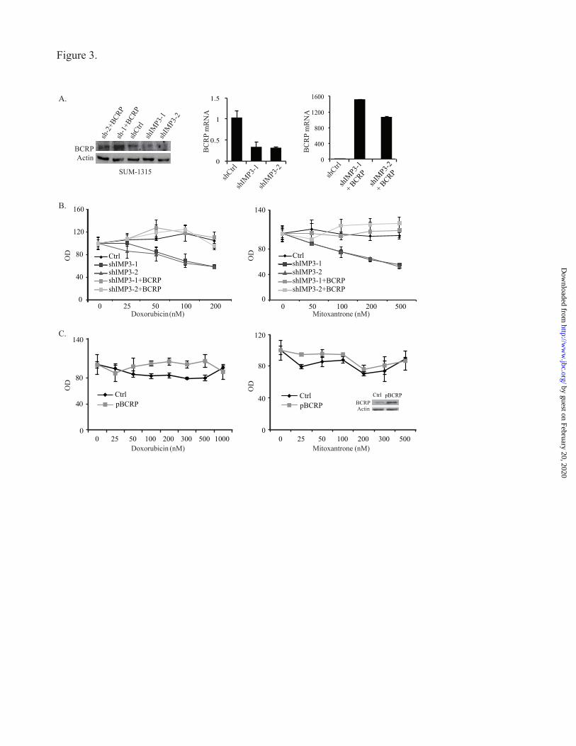
Figure 3.
0
400
800
1200
1600
shIM
P3-1
+ BCRP
shIM
P3-2
+ BCRP
shIMP3-1
shIM
P3-2
B.
C.
0
0.5
1
1.5
0
40
80
120
160
0 25 50 100 200
Ctrl shIMP3-1 shIMP3-2 shIMP3-1+BCRP shIMP3-2+BCRP
0
40
80
140
0 50 100 200 500
Ctrl shIMP3-1 shIMP3-2 shIMP3-1+BCRP shIMP3-2+BCRP
(nM) (nM)MitoxantroneDoxorubicin
shCtrl
shIM
P3-1
shIM
P3-2
sh-1
+BCR
P
sh-2
+BCR
P
ActinBCRP
shCtrl
shCtrl
BC
RP
mR
NA
BC
RP
mR
NA
OD
SUM-1315
0
40
80
140
0 25 50 100 200 300 500 1000
Ctrl pBCRP
0
40
80
120
0 25 50 100 200 300 500
Ctrl
OD
ODOD
pBCRP
(nM) (nM)
ActinBCRP
Doxorubicin Mitoxantrone
A.
Ctrl pBCRP
by guest on February 20, 2020http://w
ww
.jbc.org/D
ownloaded from

Sanjoy Samanta, Bryan Pursell and Arthur M. Mercurioexpression
IMP3 promotes chemo-resistance in breast cancer cells by regulating BCRP (ABCG2)
published online March 28, 2013J. Biol. Chem.
10.1074/jbc.C112.442319Access the most updated version of this article at doi:
Alerts:
When a correction for this article is posted•
When this article is cited•
to choose from all of JBC's e-mail alertsClick here
by guest on February 20, 2020http://w
ww
.jbc.org/D
ownloaded from