Embed Size (px)
Citation preview
ARTICLE
Breaking a dative bond with mechanical forcesPengcheng Chen1,9, Dingxin Fan2,9, Yunlong Zhang 3✉, Annabella Selloni 4, Emily A. Carter 5,6,
Craig B. Arnold 1,5, David C. Dankworth 3, Steven P. Rucker 3, James R. Chelikowsky 2,7,8✉ &
Nan Yao 1✉
Bond breaking and forming are essential components of chemical reactions. Recently, the
structure and formation of covalent bonds in single molecules have been studied by non-
contact atomic force microscopy (AFM). Here, we report the details of a single dative bond
breaking process using non-contact AFM. The dative bond between carbon monoxide and
ferrous phthalocyanine was ruptured via mechanical forces applied by atomic force micro-
scope tips; the process was quantitatively measured and characterized both experimentally
and via quantum-based simulations. Our results show that the bond can be ruptured either by
applying an attractive force of ~150 pN or by a repulsive force of ~220 pN with a significant
contribution of shear forces, accompanied by changes of the spin state of the system. Our
combined experimental and computational studies provide a deeper understanding of the
chemical bond breaking process.
https://doi.org/10.1038/s41467-021-25932-6 OPEN
1 Princeton Institute for the Science and Technology of Materials, Princeton University, Princeton, NJ 08540-8211, USA. 2McKetta Department of ChemicalEngineering, University of Texas at Austin, Austin, TX 78712-1589, USA. 3 ExxonMobil Research and Engineering Company, Annandale, NJ 08801-3096,USA. 4Department of Chemistry, Princeton University, Princeton, NJ 08544-0001, USA. 5Department of Mechanical and Aerospace Engineering, PrincetonUniversity, Princeton, NJ 08544-5263, USA. 6Office of the Chancellor and Department of Chemical and Biomolecular Engineering, University of California,Los Angeles, Los Angeles, CA 90095-1405, USA. 7Department of Physics, University of Texas at Austin, Austin, TX 78712-1192, USA. 8 Center forComputational Materials, Oden Institute for Computational Engineering and Sciences, University of Texas at Austin, Austin, TX 78712-1229, USA. 9Theseauthors contributed equally: Pengcheng Chen, Dingxin Fan. ✉email: [email protected]; [email protected]; [email protected].
NATURE COMMUNICATIONS | (2021) 12:5635 | https://doi.org/10.1038/s41467-021-25932-6 | www.nature.com/naturecommunications 1
1234
5678
90():,;

The ability to obtain images of organic molecules withatomic resolution was first demonstrated by Gross et al. in2009 by using a carbon monoxide (CO) molecule attached
to an Atomic Force Microscope (AFM) tip1 mounted on a qPlussensor2. This work inspired a wide range of applications,including directly characterizing molecular structures3–5, probingmolecular properties6–10, creating new structures11,12, and evenproviding a tool for studying various types of chemical bonds,such as hydrogen bonds and halogen bonds13,14. These studiesstimulated significant discussions on the contrast mechanism ofAFM images and on the extent to which the image couldrepresent a physical description of a chemical bond15,16. A similartechnique was used to directly manipulate individual chemicalbonds. Wagner et al.17 measured the binding energies (includingnonspecific interactions) between an organic molecule, 3,4,9,10-perylene-teracarboxylic-dianhydride, and a metal substrate,Au(111). Recently, Kawai et al. measured the C=O ∙ ∙ ∙H-Cbonding interaction between a CO tip and the C-H group of anaromatic hydrocarbon before the onset of Pauli repulsion18.Huber et al. studied the interactions between a CO tip with Fe,Cu, and Si adatoms and revealed the bond-forming processduring the transition from physisorption to chemisorption19.
These studies using AFM tips to manipulate chemical bondsprovided insights into the bond-forming process involved in manysurface interactions. However, the controlled breaking of a chemicalbond using mechanical forces, along with accurate measurements ofthese forces, is also important and its detailed process has not yetbeen fully understood. Understanding the process of rupturing abond is essential for obtaining insights into the physical nature of achemical bond and its role in many chemical and catalyticmechanisms. Dative bonds are commonly found in transition metalcomplexes and play vital roles in catalysis, organometallic chemistry,and biochemistry. Here, we focus on understanding the breaking of asingle chemical bond between a CO molecule and a ferrous phtha-locyanine (FePc) complex using AFM together with real-spacepseudopotential density functional theory (DFT) calculations20–22.Our results reveal detailed mechanisms of bond breaking by bothrepulsive and attractive forces. This work advances understanding ofthe origins of measured forces in dative bond breaking.
ResultsSTM/AFM topography and structure identification. We pre-pared a supported CO-FePc system by dosing CO molecules ontoFePc adsorbed on a Cu(111) surface at 4.8 K. The scanningtunneling microscope (STM) image in Fig. 1a shows two distinctfeatures for the FePc molecule and the complex (CO-FePc),which are similar to previously reported STM images on othersurfaces23. AFM images were obtained using a CO-terminatedtip, confirming their respective structures (Fig. 1b, c). The AFMimage in Fig. 1b of the CO-FePc complex featured a protrudingcenter due to the CO attached to Fe. This characterization isconfirmed by comparing with AFM images of FePc molecules onthe surface Fig. 1c, and further verified by our simulated images(Fig. 1d, e).
Breaking a dative bond with AFM tips. The dative CO-FePcbond is known to be formed via σ-donation from the CO 5σorbital and π-back donation from Fe dπ24–27. We studied therupture of this dative bond by applying mechanical forces usingthe AFM tip. The same CO-terminated tip employed for imagingwas used first because it is known to be chemically inert28. Bydecreasing the tip height, the repulsive interactions increased, asindicated by the increased contrast in the images (Fig. 2b–d). At atip height of about +40 pm, the peripheral aromatic rings of FePcbecame visible, while the center of the image became distorteddue to strong repulsions with the tip. Upon further reducing thetip height (+30 pm), a sudden change of the image occurredduring scanning, as indicated by a line created with a differentcontrast. Subsequent scans showed the repulsion had dis-appeared, indicating that the CO attached to FePc was dislodgeddue to the strong repulsion with the tip. The chemical structure ofFePc revealed from subsequent scanning of the lower part of themolecule confirmed that a free FePc was left after CO removaland that the tip remained intact during the dissociation. Com-parison of the contrast in the lower part to the upper part of thesame AFM image Fig. 2d obtained at the same tip height reveals adownward shift of FePc by ~30 pm upon CO removal. This shiftindicates a trans effect by the Cu substrate on the FePccomplex24–27,29, whereby the binding or removal of one ligand
Fig. 1 STM and AFM images of FePc and the datively bonded CO-FePc complex. a STM image of the FePc molecule with (indicated by the arrow) andwithout adsorption of CO, with the insert showing the chemical structure of FePc (set point: Vsample=+100mV, I= 100 pA, scale bar: 3 nm).b, c Experimental AFM images of FePc with and without adsorbed CO, obtained using a CO-terminated tip (V= 0 V, A= 100 pm, scale bar: 0.5 nm) at tipheights z of +160 pm and -10 pm, respectively. The tip height z was set with respect to a reference height given by the STM set point (100mV, 100 pA)above the bare Cu(111) substrate in the vicinity of the molecule. The minus sign of tip height z indicates a decrease of tip height. d, e Simulated AFM imagescorresponding to CO-FePc and FePc at tip heights of 554 pm and 300 pm. The tip height in the simulation is defined as the distance between the frontatom of the tip and the average height of the FePc complex (excluding the decorated CO) (scale bar: 0.5 nm).
ARTICLE NATURE COMMUNICATIONS | https://doi.org/10.1038/s41467-021-25932-6
2 NATURE COMMUNICATIONS | (2021) 12:5635 | https://doi.org/10.1038/s41467-021-25932-6 | www.nature.com/naturecommunications

respectively reduces or enhances the strength of the bond to theligand on the opposite side30. This observation confirms therupture of the dative bond between the CO and FePc induced bythe increased interactions during tip scanning.
To elucidate CO-FePc bond rupture, we performed detailedmeasurements of the interaction forces during the entire bondrupture process. Figure 2e shows a 3D force map representing thefrequency shift (Δf) obtained at different tip heights (z) byscanning across the center of the CO-FePc complex (shown in theinset). The dislodging of the CO was indicated by a breakpoint(x= 0) with decreasing the tip height during scanning and by adiscontinuity in the frequency shift (Δf) curve (red curve inFig. 2f). The interaction force and energy were calculated fromthe measured frequency shift with the method and formulasproposed by Sader31. The force curve along with the tip height (z)at the breaking point (x= 0), in Fig. 2g, shows that the dativebond ruptured with a force of 220 ± 30 pN, after passing amaximal force at ~300 pN.
Dislodging experiments were also performed with a bare metaltip, which was terminated by a Cu atom under similarexperimental conditions as for the CO-terminated tip. The Cu
atom tip is known to be a chemically active tip32. When we usedthe Cu tip, only attractive interactions between the tip and CO-FePc were detected (Fig. 2h), until the rupture of the dative bondtook place. At this point, the attractive force reached 150 ± 30 pNby reducing the tip height (Fig. 2i, j).
Real-space DFT calculations. The surprising observation that bothan attractive force of −150 pN and a repulsive force of +220 pN arecapable of breaking the same dative bond highlights the importantrole of probe tips, although this result is consistent with findings byBerwanger et al. that the CO-terminated tip can exert forces of up to450 pN without breaking off33,34. We employed real-space DFTcalculations to address the role of the AFM tip and to shed light onthe details of the bond-breaking process.
To understand interactions between the AFM tips and CO-FePc before the bond is broken, we computed frequency shifts ofthe probe tips at relatively large heights (z~5 Å) using optimizedgeometries22 (Supplementary Figure 1). We modeled the Cu tipusing a Cu2 cluster and the CO tip using a Cu-CO cluster. Wetested more complex tip conformations and found the effect in
Fig. 2 Rupturing the dative CO-FePc bond using AFM tips. a Schematic of a CO-AFM tip interacting with CO-FePc (Cu: yellow; C: black; O: red; Fe:brown). b–d Non-contact AFM images obtained at different tip heights (z); the final dislodging of CO occurs at z=+30 pm. e 3D force map of thefrequency shift (Δf) vs. AFM tip heights (z) and horizontal position (x), with a CO tip. Step size is 5 pm in z, and the scan path in x is across the center ofthe Fe, as shown in the inset. The tip position at bond rupture is indicated by the breakpoint (arrow). f Frequency shift (Δf) obtained in the horizontal (x)direction before, during (indicated by the disjointed curve), and after the bond rupture. g The force curve deconvoluted from Δf at the breakpoint in thevertical (z) direction. h 3D force map of the frequency shift (Δf) showing quantitative rupture of the dative bond, obtained using a Cu tip; the insert showsschematic of interaction between a Cu tip and CO-FePc. i Frequency shift (Δf) obtained using a Cu tip scanned in the horizontal (x) direction. j Thedeconvoluted force curve at the breakpoint in the vertical (z) direction using a Cu tip. (Red arrows indicate the bond rupture point. Long-range backgroundforces are subtracted in Figures g and j).
NATURE COMMUNICATIONS | https://doi.org/10.1038/s41467-021-25932-6 ARTICLE
NATURE COMMUNICATIONS | (2021) 12:5635 | https://doi.org/10.1038/s41467-021-25932-6 | www.nature.com/naturecommunications 3
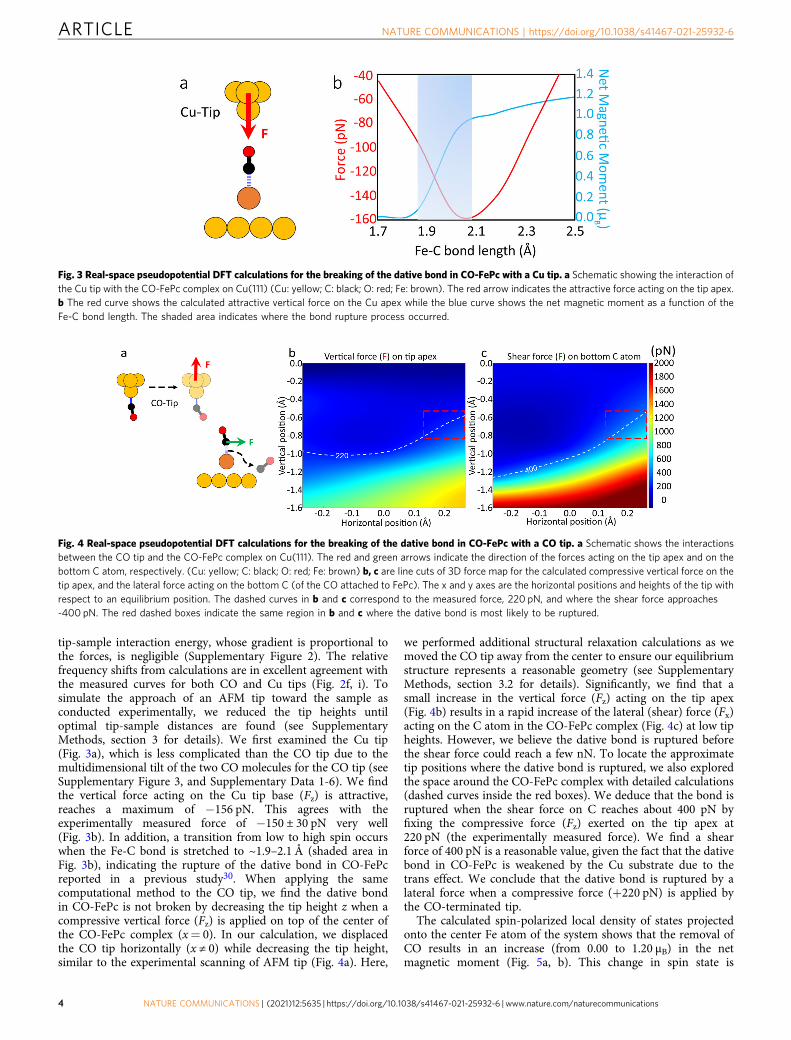
tip-sample interaction energy, whose gradient is proportional tothe forces, is negligible (Supplementary Figure 2). The relativefrequency shifts from calculations are in excellent agreement withthe measured curves for both CO and Cu tips (Fig. 2f, i). Tosimulate the approach of an AFM tip toward the sample asconducted experimentally, we reduced the tip heights untiloptimal tip-sample distances are found (see SupplementaryMethods, section 3 for details). We first examined the Cu tip(Fig. 3a), which is less complicated than the CO tip due to themultidimensional tilt of the two CO molecules for the CO tip (seeSupplementary Figure 3, and Supplementary Data 1-6). We findthe vertical force acting on the Cu tip base (Fz) is attractive,reaches a maximum of −156 pN. This agrees with theexperimentally measured force of −150 ± 30 pN very well(Fig. 3b). In addition, a transition from low to high spin occurswhen the Fe-C bond is stretched to ~1.9–2.1 Å (shaded area inFig. 3b), indicating the rupture of the dative bond in CO-FePcreported in a previous study30. When applying the samecomputational method to the CO tip, we find the dative bondin CO-FePc is not broken by decreasing the tip height z when acompressive vertical force (Fz) is applied on top of the center ofthe CO-FePc complex (x= 0). In our calculation, we displacedthe CO tip horizontally (x ≠ 0) while decreasing the tip height,similar to the experimental scanning of AFM tip (Fig. 4a). Here,
we performed additional structural relaxation calculations as wemoved the CO tip away from the center to ensure our equilibriumstructure represents a reasonable geometry (see SupplementaryMethods, section 3.2 for details). Significantly, we find that asmall increase in the vertical force (Fz) acting on the tip apex(Fig. 4b) results in a rapid increase of the lateral (shear) force (Fx)acting on the C atom in the CO-FePc complex (Fig. 4c) at low tipheights. However, we believe the dative bond is ruptured beforethe shear force could reach a few nN. To locate the approximatetip positions where the dative bond is ruptured, we also exploredthe space around the CO-FePc complex with detailed calculations(dashed curves inside the red boxes). We deduce that the bond isruptured when the shear force on C reaches about 400 pN byfixing the compressive force (Fz) exerted on the tip apex at220 pN (the experimentally measured force). We find a shearforce of 400 pN is a reasonable value, given the fact that the dativebond in CO-FePc is weakened by the Cu substrate due to thetrans effect. We conclude that the dative bond is ruptured by alateral force when a compressive force (+220 pN) is applied bythe CO-terminated tip.
The calculated spin-polarized local density of states projectedonto the center Fe atom of the system shows that the removal ofCO results in an increase (from 0.00 to 1.20 µB) in the netmagnetic moment (Fig. 5a, b). This change in spin state is
Fig. 3 Real-space pseudopotential DFT calculations for the breaking of the dative bond in CO-FePc with a Cu tip. a Schematic showing the interaction ofthe Cu tip with the CO-FePc complex on Cu(111) (Cu: yellow; C: black; O: red; Fe: brown). The red arrow indicates the attractive force acting on the tip apex.b The red curve shows the calculated attractive vertical force on the Cu apex while the blue curve shows the net magnetic moment as a function of theFe-C bond length. The shaded area indicates where the bond rupture process occurred.
Fig. 4 Real-space pseudopotential DFT calculations for the breaking of the dative bond in CO-FePc with a CO tip. a Schematic shows the interactionsbetween the CO tip and the CO-FePc complex on Cu(111). The red and green arrows indicate the direction of the forces acting on the tip apex and on thebottom C atom, respectively. (Cu: yellow; C: black; O: red; Fe: brown) b, c are line cuts of 3D force map for the calculated compressive vertical force on thetip apex, and the lateral force acting on the bottom C (of the CO attached to FePc). The x and y axes are the horizontal positions and heights of the tip withrespect to an equilibrium position. The dashed curves in b and c correspond to the measured force, 220 pN, and where the shear force approaches-400 pN. The red dashed boxes indicate the same region in b and c where the dative bond is most likely to be ruptured.
ARTICLE NATURE COMMUNICATIONS | https://doi.org/10.1038/s41467-021-25932-6
4 NATURE COMMUNICATIONS | (2021) 12:5635 | https://doi.org/10.1038/s41467-021-25932-6 | www.nature.com/naturecommunications
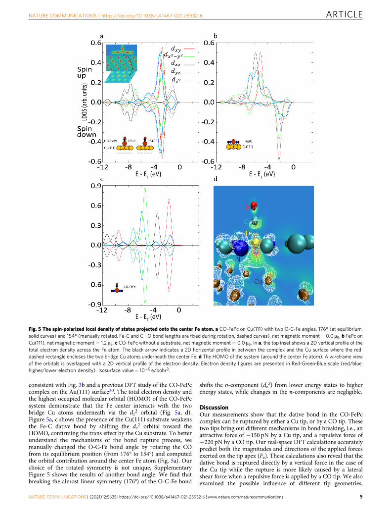
consistent with Fig. 3b and a previous DFT study of the CO-FePccomplex on the Au(111) surface30. The total electron density andthe highest occupied molecular orbital (HOMO) of the CO-FePcsystem demonstrate that the Fe center interacts with the twobridge Cu atoms underneath via the dz2 orbital (Fig. 5a, d).Figure 5a, c shows the presence of the Cu(111) substrate weakensthe Fe-C dative bond by shifting the dz2 orbital toward theHOMO, confirming the trans effect by the Cu substrate. To betterunderstand the mechanisms of the bond rupture process, wemanually changed the O-C-Fe bond angle by rotating the COfrom its equilibrium position (from 176° to 154°) and computedthe orbital contribution around the center Fe atom (Fig. 5a). Ourchoice of the rotated symmetry is not unique, SupplementaryFigure 5 shows the results of another bond angle. We find thatbreaking the almost linear symmetry (176°) of the O-C-Fe bond
shifts the σ-component (dz2) from lower energy states to higherenergy states, while changes in the π-components are negligible.
DiscussionOur measurements show that the dative bond in the CO-FePccomplex can be ruptured by either a Cu tip, or by a CO tip. Thesetwo tips bring out different mechanisms in bond breaking, i.e., anattractive force of −150 pN by a Cu tip, and a repulsive force of+220 pN by a CO tip. Our real-space DFT calculations accuratelypredict both the magnitudes and directions of the applied forcesexerted on the tip apex (Fz). These calculations also reveal that thedative bond is ruptured directly by a vertical force in the case ofthe Cu tip while the rupture is more likely caused by a lateralshear force when a repulsive force is applied by a CO tip. We alsoexamined the possible influence of different tip geometries,
Fig. 5 The spin-polarized local density of states projected onto the center Fe atom. a CO-FePc on Cu(111) with two O-C-Fe angles, 176° (at equilibrium,solid curves) and 154° (manually rotated, Fe-C and C=O bond lengths are fixed during rotation, dashed curves), net magnetic moment= 0.0 µB. b FePc onCu(111), net magnetic moment= 1.2 µB. c CO-FePc without a substrate, net magnetic moment= 0.0 µB. In a, the top inset shows a 2D vertical profile of thetotal electron density across the Fe atom. The black arrow indicates a 2D horizontal profile in between the complex and the Cu surface where the reddashed rectangle encloses the two bridge Cu atoms underneath the center Fe. d The HOMO of the system (around the center Fe atom). A wireframe viewof the orbitals is overlapped with a 2D vertical profile of the electron density. Electron density figures are presented in Red-Green-Blue scale (red/blue:higher/lower electron density). Isosurface value= 10−5 e/bohr3.
NATURE COMMUNICATIONS | https://doi.org/10.1038/s41467-021-25932-6 ARTICLE
NATURE COMMUNICATIONS | (2021) 12:5635 | https://doi.org/10.1038/s41467-021-25932-6 | www.nature.com/naturecommunications 5

finding that more complex tips do not affect significantly the tip-sample interaction energy. As for the equilibrium conformationof the CO tip, our calculation is in agreement with previousstudies35,36.
Our calculations confirm that the dative CO-FePc bond isweakened by the presence of the Cu(111) substrate due to thetrans effect29,30. However, the CO-FePc bond remains a chemicalbond, instead of a physisorption19, based on the computed Fe-Cbond length (~1.7 Å). Furthermore, upon the dislodging of theCO by the AFM tip, a ~30 pm downward shift of FePc observedin the experiment revealed that the FePc molecule was lifted~30 pm upward from Cu(111) surface by attaching a single COmolecule to Fe. This observation is consistent with our calculatedshortening of the distance between the Fe atom and the surface ofthe Cu substrate from 273 to 248 pm, further confirming therupture of the chemical bond.
When a CO tip is used, the dative bond ruptures at 220 pN,after passing a maximal force of about 300 pN, as shown inFig. 2g. This trend is consistent with a sequential cleavage of theσ-donation and π-back donation of the dative bond as Fig. 5ashows. The σ-bond has a higher symmetry than the π-bond andhence a higher force is required to tilt and weaken the σ-bondbefore the dative bond is completely ruptured by the lateral force.
Metal-Pc molecules are widely used model catalysts system forelectrochemical reduction of CO2
37,38. Our study is important forunderstanding how the activities of Metal-Pc molecules can bemanipulated and controlled with single atomic level engineering,and for the design of new FePc-based catalysts. In addition, ourresults clarify the different mechanisms in bond breaking inducedby inert and active tips. This detailed information on the ruptureof the bond between CO and FePc will allow us to betterunderstand other dative bonds, such as CO-heme interactions inbiochemistry24,39, as well as chemical reactions of materials undermechanical stress40,41.
MethodsExperimental parameters. Our experiments were performed with a CreaTecTM
STM/AFM system under ultrahigh vacuum conditions of ~10−10 mbar and atemperature of approximately 5 K. The qPlus sensor has a resonance frequency of30 kHz with a spring constant k= 1800 N/m. In our measurements, the qualityfactor of the sensor is about 20,000. To minimize crosstalk between the qPlus signaland the STM channel, no voltage was applied on the tip during the force mea-surement process. The oscillation amplitude was set to be 100 pm.
Chemicals and sample preparation. The iron(II) phthalocyanine (FePc, dyecontent ~90%, Sigma–Aldrich) molecules were evaporated from a silicon chip viadirect heating, and the vapor was subsequently deposited on a Cu(111) substrateheld at 5 K. The AFM Cu tip apex was functionalized by controlled pickup of a COmolecule from the substrate1. All the experiments were conducted using a pure Cutip or a CO-functionalized tip.
DFT modelling and computations. We computed ground state energies using areal-space pseudopotential DFT code, PARSEC42. We employed the local densityapproximation (LDA) by Perdew-Wang (PW92)43 for the exchange-correlationfunctional together with Troullier-Martins norm-conserving pseudopotentials44.We also tested another exchange-correlation functional by Ceperley-Alder45, thedifferences were negligible. In addition, a previous study showed that LDA and thegeneralized gradient approximation (GGA) gave similar results for properties ofFePc and CO-FePc on Au(111)30. We employed boundary conditions that requirethe electron wave functions to vanish outside a spherical or a slab domain, of whichthe boundary is at least ~300 pm from the outermost atom. We set the distancebetween neighbor points in the real-space grid to be 16 pm. The density-weightedself-consistent residual error was less than 10−4 Ry. We employed a finite differ-ence method to approximate the relative frequency shift profiles based on thecomputed ground state energies across the middle line of the FePc complex, asindicated in Fig. 2f, i at relatively large tip heights. We performed further structuralrelaxations when the tips were close to the specimen, as the assumption that themovement of the tip had negligible influence on the electronic structure of thespecimen may not be valid. We then applied the Hellmann–Feynman theorem tothe total ground state energies to compute the net forces acting on each atom. Weemployed the frozen density embedding theory and a finite difference method for
AFM image simulations22 (see Supplementary Information—Image Simulationssection and Supplementary Fig. 4 for details).
Data availabilityThe data supporting our results are available within this article and the SupplementaryInformation. The Supplementary Information contains a more detailed description offorce calculation, structural relaxation, AFM image simulations and tip conformationtests. In addition, we provide the relaxed atomic coordinates of the systems inSupplementary Figure 3 and in Supplementary Data 1-6.
Received: 4 March 2021; Accepted: 2 September 2021;
References1. Gross, L., Mohn, F., Moll, N., Liljeroth, P. & Meyer, G. The chemical structure
of a molecule resolved by atomic force microscopy. Science 325, 1110LP–1111114 (2009).
2. Giessibl, F. J. High-speed force sensor for force microscopy and profilometryutilizing a quartz tuning fork. Appl. Phys. Lett. 73, 3956–3958 (1998).
3. Gross, L. et al. Organic structure determination using atomic-resolutionscanning probe microscopy. Nat. Chem. 2, 821–825 (2010).
4. Schuler, B. et al. Heavy oil based mixtures of different origins andtreatments studied by atomic force microscopy. Energy Fuels 31, 6856–6861(2017).
5. Hanssen, K. Ø. et al. A combined atomic force microscopy and computationalapproach for the structural elucidation of breitfussin A and B: highly modifiedhalogenated dipeptides from Thuiaria breitfussi. Angew. Chem. Int. Ed. 51,12238–12241 (2012).
6. Gross, L. et al. Bond-order discrimination by atomic force microscopy. Science337, 1326–1329 (2012).
7. Fatayer, S. et al. Molecular structure elucidation with charge-state control.Science 365, 142–145 (2019).
8. Gross, L. et al. Measuring the charge state of an adatom with noncontactatomic force microscopy. Science 324, 1428–1431 (2009).
9. Mohn, F., Gross, L., Moll, N. & Meyer, G. Imaging the charge distributionwithin a single molecule. Nat. Nanotechnol. 7, 227–231 (2012).
10. de Oteyza, D. G. et al. Noncovalent dimerization after enediyne cyclization onAu(111). J. Am. Chem. Soc. 138, 10963–10967 (2016).
11. Pavliček, N. et al. Synthesis and characterization of triangulene. Nat.Nanotechnol. 12, 308–311 (2017).
12. Kaiser, K. et al. An sp-hybridized molecular carbon allotrope, cyclo[18]carbon. Science 365, 1299–1301 (2019).
13. Zhang, J. et al. Real-Space identification of intermolecular bonding withatomic force microscopy. Science 342, 611–614 (2013).
14. Han, Z. et al. Imaging the halogen bond in self-assembled halogenbenzenes onsilver. Science 358, 206–210 (2017).
15. Ellner, M., Pou, P. & Pérez, R. Molecular identification, bond orderdiscrimination, and apparent intermolecular features in atomic forcemicroscopy studied with a charge density based method. ACS Nano 13,786–795 (2019).
16. Hämäläinen, S. K. et al. Intermolecular contrast in atomic force microscopyimages without intermolecular bonds. Phys. Rev. Lett. 113, 186102 (2014).
17. Wagner, C. et al. Measurement of the binding energies of the organic-metalperylene-teracarboxylic-dianhydride/Au(111) bonds by molecularmanipulation using an atomic force microscope. Phys. Rev. Lett. 109, 076102(2012).
18. Kawai, S. et al. Direct quantitative measurement of the C=O∙∙∙H-C bond byatomic force microscopy. Sci. Adv. 3, e1603258 (2017).
19. Huber, F. et al. Chemical bond formation showing a transition fromphysisorption to chemisorption. Science 366, 235–238 (2019).
20. Sakai, Y., Lee, A. J. & Chelikowsky, J. R. First-principles atomic forcemicroscopy image simulations with density embedding theory. Nano Lett. 16,3242–3246 (2016).
21. Chelikowsky, J. R., Fan, D., Lee, A. J. & Sakai, Y. Simulating noncontactatomic force microscopy images. Phys. Rev. Mater. 3, 110302 (2019).
22. Fan, D., Sakai, Y. & Chelikowsky, J. R. Chemical and steric effects insimulating noncontact atomic force microscopy images of organic moleculeson a Cu (111) substrate. Phys. Rev. Mater. 4, 53802 (2020).
23. Tsukahara, N., Minamitani, E., Kim, Y., Kawai, M. & Takagi, N. Controllingorbital-selective Kondo effects in a single molecule through coordinationchemistry. J. Chem. Phys. 141, 54702 (2014).
24. Stynes, D. V. & James, B. R. Kinetics and equilibriums for carbon monoxidebinding to ferrous phthalocyanine complexes. J. Am. Chem. Soc. 96,2733–2738 (1974).
ARTICLE NATURE COMMUNICATIONS | https://doi.org/10.1038/s41467-021-25932-6
6 NATURE COMMUNICATIONS | (2021) 12:5635 | https://doi.org/10.1038/s41467-021-25932-6 | www.nature.com/naturecommunications
25. Calderazzo, F. et al. Synthesis and Moessbauer spectroscopic studies ofcarbonyl derivatives of (phthalocyaninato)iron(II). Inorg. Chem. 21,2302–2306 (1982).
26. Li, J., Noll, B. C., Schulz, C. E. & Scheidt, W. R. Comparison of cyanide andcarbon monoxide as ligands in iron(II) porphyrinates. Angew. Chem. Int. Ed.48, 5010–5013 (2009).
27. Collman, J. P., Brauman, J. I. & Doxsee, K. M. Carbon monoxide binding toiron porphyrins. Proc. Natl Acad. Sci. 76, 6035–6039 (1979).
28. Sang, H. et al. Identifying tips for intramolecular NC-AFM imaging via in situfingerprinting. Sci. Rep. 4, 6678 (2014).
29. Hieringer, W. et al. The surface trans effect: influence of axial ligands on thesurface chemical bonds of adsorbed metalloporphyrins. J. Am. Chem. Soc. 133,6206–6222 (2011).
30. Isvoranu, C. et al. Comparison of the carbonyl and nitrosyl complexes formedby adsorption of CO and NO on monolayers of iron phthalocyanine onAu(111). J. Phys. Chem. C. 115, 24718–24727 (2011).
31. Sader, J. E. & Jarvis, S. P. Accurate formulas for interaction force and energy infrequency modulation force spectroscopy. Appl. Phys. Lett. 84, 1801 (2004).
32. Mönig, H. Copper-oxide tip functionalization for submolecular atomic forcemicroscopy. Chem. Commun. 54, 9874 (2018).
33. Berwanger, J. et al. Atomically resolved chemical reactivity of small Fe clusters.Phys. Rev. Lett. 124, 096001 (2020).
34. Berwanger, J., Huber, F., Stilp, F. & Giessibl, F. J. Lateral manipulation of singleiron adatoms by means of combined atomic force and scanning tunnelingmicroscopy using CO-terminated tips. Phys. Rev. B 98, 195409 (2018).
35. Weymouth, A. J., Hofmann, T. & Giessibl, F. J. Quantifying molecular stiffnessand interaction with lateral force microscopy. Science 343, 1120–1122 (2014).
36. Salmeron, M. CO meets CO, one at a time. Science 343, 1083–1084 (2014).37. Kim, H. et al. Identification of single-atom Ni site active toward
electrochemical CO2 conversion to CO. J. Am. Chem. Soc. 143, 925–933(2021).
38. Lin, L. et al. Enhancing CO2 electroreduction to methane with a cobaltphthalocyanine and zinc-nitrogen-carbon tandem catalyst. Angew. Chem. Int,Ed. 59, 22408–22413 (2020).
39. Collman, J. P., Brauman, J. I., Halbert, T. R. & Suslick, K. S. Nature of O2 andCO binding to metalloporphyrins and heme proteins. Proc. Natl Acad. Sci. U.S. A 73, 3333–3337 (1976).
40. Caruso, M. M. et al. Mechanically-induced chemical changes in polymericmaterials. Chem. Rev. 109, 5755–5798 (2009).
41. Boucly, A. et al. Soft X-ray heterogeneous radiolysis of pyridine in thepresence of hydrated strontium-hydroxyhectorite and its monitoring by near-ambient pressure photoelectron spectroscopy. Sci. Rep. 8, 6164 (2018).
42. Liou, K.-H., Yang, C. & Chelikowsky, J. R. Scalable implementation ofpolynomial filtering for density functional theory calculation in PARSEC.Comput. Phys. Commun. 254, 107330 (2020).
43. Perdew, J. P. & Wang, Y. Accurate and simple analytic representation of theelectron-gas correlation energy. Phys. Rev. B 45, 13244–13249 (1992).
44. Troullier, N. & Martins, J. L. Efficient pseudopotentials for plane-wavecalculations. Phys. Rev. B 43, 1993–2006 (1991).
45. Ceperley, D. M. & Alder, B. J. Ground state of the electron gas by a stochasticmethod. Phys. Rev. Lett. 45, 566–569 (1980).
AcknowledgementsThe authors gratefully acknowledge Yeju Zhou, Dan Gregory, Michele L. Sarazen, andGuangming Cheng for help with data processing and general discussion. This work was
partially supported by ExxonMobil through its membership in the Princeton E-filliatesPartnership of the Andlinger Center for Energy and the Environment. This researchmade use of the Imaging and Analysis Center operated by the Princeton Institute for theScience and Technology of Materials at Princeton University, which is supported in partby the Princeton Center for Complex Materials, a National Science Foundation MaterialsResearch Science and Engineering Center (Grant No. DMR-2011750). D.F. and J.R.C.acknowledge support from the Welch Foundation under grant F-1837 and the U.S.Department of Energy under DOE/DE-FG02-06ER46286. The National Energy ResearchScientific Computing (NERSC) and the Texas Advanced Computing Center (TACC)provided computational resources.
Author contributionsP.C. and N.Y. designed and carried out the experiments. D.F. and J.R.C. performed theDFT calculations. A.S. and E.A.C. provided theoretical insight. P.C., N.Y., Y.Z., D.F.,drafted the manuscript with the input from A.S., J.R.C., E.A.C., C.B.A., D.C.D and S.P.R.N.Y. directed the project. All authors discussed the results and contributed to theinterpretation and conclusions.
Competing interestsThe authors declare no competing interests.
Additional informationSupplementary information The online version contains supplementary materialavailable at https://doi.org/10.1038/s41467-021-25932-6.
Correspondence and requests for materials should be addressed to Nan Yao, YunlongZhang or James R. Chelikowsky.
Peer review information Nature Communications thanks Yongsheng Leng, Jan vanRuitenbeek and the other, anonymous, reviewer for their contribution to the peer reviewof this work. Peer reviewer reports are available.
Reprints and permission information is available at http://www.nature.com/reprints
Publisher’s note Springer Nature remains neutral with regard to jurisdictional claims inpublished maps and institutional affiliations.
Open Access This article is licensed under a Creative CommonsAttribution 4.0 International License, which permits use, sharing,
adaptation, distribution and reproduction in any medium or format, as long as you giveappropriate credit to the original author(s) and the source, provide a link to the CreativeCommons license, and indicate if changes were made. The images or other third partymaterial in this article are included in the article’s Creative Commons license, unlessindicated otherwise in a credit line to the material. If material is not included in thearticle’s Creative Commons license and your intended use is not permitted by statutoryregulation or exceeds the permitted use, you will need to obtain permission directly fromthe copyright holder. To view a copy of this license, visit http://creativecommons.org/licenses/by/4.0/.
© The Author(s) 2021
NATURE COMMUNICATIONS | https://doi.org/10.1038/s41467-021-25932-6 ARTICLE
NATURE COMMUNICATIONS | (2021) 12:5635 | https://doi.org/10.1038/s41467-021-25932-6 | www.nature.com/naturecommunications 7