Embed Size (px)
Citation preview
CLINICAL MICROBIOLOGY REVIEWS, Oct. 1990, p. 293-320 Vol. 3, No. 40893-8512/90/040293-28$02.00/0Copyright © 1990, American Society for Microbiology
Branhamella catarrhalis: an Organism GainingRespect as a Pathogen
B. WESLEY CATLINtDepartment of Microbiology, The Medical College of Wisconsin, Milwaukee, Wisconsin 53226
INTRODUCTION ......................................................................... 293PATHOGENIC POTENTIAL OF B. CATARRHALIS ...................................................................294
Critique of Sampling Procedures ......................................................................... 294Importance of Adequate Descriptions of Isolates .......................................................................295Bacteremia, Endocarditis, and Meningitis ......................................................................... 295Otitis Media, Sinusitis, and Keratitis......................................................................... 295Pseudogonococcal Conjunctivitis and Urogenital Infections..........................................................297Lower Respiratory Tract Infections......................................................................... 297Analogy between B. catarrhalis and N. meningitidis Infections .....................................................298Healthy Carriers of B. catarrhalis ....................................................................... 298
Isolation rates......................................................................... 298Selective culture media for B. catarrhalis...................................................................... 299
Virulence......................................................................... 299Immunity ......................................................................... 300
IDENTIFICATION AND BIOCHEMICAL CHARACTERISTICS OF B. CATARRHALIS ...................301Differential Tests ......................................................................... 301
Action on carbohydrates ......................................................................... 301Reduction of nitrate and nitrite ......................................................................... 301Hydrolysis of DNA ......................................................................... 301Hydrolysis of tributyrin and other esterase substrates .............................................................302Hydrolysis of aminopeptidase substrates......................................................................... 302Response to superoxol (30% H202).....................................................................................302Other differential tests......................................................................... 302
Stages in the Identification Process......................................................................... 303Strain Differentiation: Typing ......................................................................... 303
Restriction endonuclease analysis of DNA......................................................................... 303OMP and LOS analyses......................................................................... 303
SUSCEPTIBILITY TO ANTIBACTERIAL AGENTS ...................................................................303Non-p-Lactam Agents ......................................................................... 303Unequal Susceptibility of P-Lactamase-Negative Strains to P-Lactam Agents ..................................304P-Lactamase-Positive Strains ......................................................................... 305
Detection methods ......................................................................... 305Prevalence ......................................................................... 305Problems in determination of resistance to P-lactam antibiotics.................................................305Genetic features ......................................................................... 307Possible influence in mixed infections: indirect pathogenicity ....................................................308
Characteristics of the P-Lactamases ......................................................................... 308STRUCTURE AND COMPOSITION OF B. CATARRHALIS .........................................................309
Capsule and Pili......................................................................... 309Envelope ......................................................................... 309DNA......................................................................... 311
WHAT'S IN A NAME?......................................................................... 311CONCLUSIONS AND PROSPECTS ......................................................................... 312ACKNOWLEDGMENTS ......................................................................... 312LITERATURE CITED ......................................................................... 312
INTRODUCTION present in the normal pharynx (117, 209, 232, 316). Conse-
Branhamella catarrhalis received little respectful atten- quently, medical technologists usually elected not to identifytion until recently. Strains isolated from clinical material organisms referred to as nongonococcal, nonmeningococcalwere often disregarded because of the repeated, confident neisseriae or normal flora (58, 221, 248, 262, 268). In aassertion that the organism is nonpathogenic and commonly discussion during a symposium on B. catarrhalis held in
1985, C. D. Bluestone [Drugs 31(Suppl. 3):38, 1986] statedhis impression that, in non-university hospitals in the United
t Present address: 3550 Lebon Drive, no. 6420, San Diego, CA States, "the organism is still frequently called Neisseria92122. catarrhalis and is not considered pathogenic." On the other
293
on July 8, 2018 by guesthttp://cm
r.asm.org/
Dow
nloaded from

CLIN. MICROBIOL. REV.
TABLE 1. Chronology of differential biochemical tests developed for Neisseria spp. and B. catarrhalis
Test Date Authors (reference)
Acid production from carbohydrates inRich basal medium 1905 Dunn and Gordon (108)Low-concn peptone medium 1983 Knapp and Holmes (178)Buffered salts solution 1970 Catlin (61)
Polysaccharide synthesis from sucrose 1948 Hehre and Hamilton (142)1961 Berger (20)
Nitrate and nitrite reduction 1959 VWron et al. (308)1961 Berger (22)
Butyrate esterase action onTributyrinReduced opacity in agar 1962 Berger and Paepcke (25); Pike et al. (236)Acid production in fluid 1981 Riou et al. (254)
4-Methylumbelliferyl butyrate 1988 Vaneechoutte et al. (301)Indoxyl butyrate 1989 Dealler et al. (88)
Deoxyribonucleate hydrolysis 1964 Catlin and Cunningham (69)1977 Riou and Guibourdenche (256)
-y-Glutamyl-,-naphthylamide hydrolysis 1967 Mannheim and Burger (199)1978 D'Amato et al. (84)
o-Nitrophenyl-p-D-galactopyranoside hydrolysis 1968 Corbett and Catlin (80)Superoxol (30% H202) decomposition 1982 Saginur et al. (266)
hand, a developing appreciation of the pathogenic potentialof B. catarrhalis and concern over the increasing prevalenceof P-lactamase-producing (Pla') strains in clinically signifi-cant infections are reflected in numerous recent reports.These are summarized in several valuable reviews (e.g., seereferences 140, 158, 305, and 321). The 1989 symposium,"Branhamella catarrhalis: a Microbiological and ClinicalUpdate," illustrates the rising interest in this organism.
Originally described in German as Mikrokokkus catarrha-lis by R. Pfeiffer (cited by Frosch and Kolle [125]) and thenin English as Micrococcus catarrhalis (e.g., see reference108), the species was subsequently classified in the genusNeisseria (26, 41, 149). In 1970, it was transferred to the newgenus Branhamella (60), named for Sara E. Branham. Herstudies (described in a biographical sketch by Pittman [239])greatly contributed to knowledge of the neisseriae. B. ca-tarrhalis was accepted in the 1974 edition of Bergey'sManual (245), but was reduced to a subgenus within thegenus Moraxella in the 1984 edition of Bergey's Manual (37).My reasons for retaining the name Branhamella catarrhaliswill be discussed in a later section. In describing the numer-ous studies of this organism, however, I shall use the namethat was given to the bacteria in each report.
B. catarrhalis has been regarded as one of the commonestinhabitants of the pharynx (195). A current textbook (324)estimates its presence as 12% in the nose and nasopharynxand 10 to 97% in the oropharynx. Early studies had found"Micrococcus catarrhalis" in 33% of nasal cultures (9), 18%of nasopharyngeal cultures (112), and 46% of nose and throatcultures from healthy persons and 45% of subjects with thecommon cold (130). "M. catarrhalis" was recognized as agram-negative diplococcus, capable of growth at 22°C,which displays certain well-described colony characteristicsand has no action on "fermentable substances" (9, 108, 112).
Significant advances in our understanding ofB. catarrhalisawaited the development of additional tests (Table 1). Theseenabled Berger (23, 25) to show that the common, nonsac-charolytic, gram-negative cocci comprised two distinct spe-cies, Neisseria cinerea and "N. catarrhalis." Described inGerman and later in French (256), N. cinerea usually wasoverlooked until 1984, when it was further characterized byKnapp et al. (180). Two studies of bacteria isolated fromthroat cultures on semiselective media showed that the
ratios of N. cinerealB. catarrhalis were 26:1 (23) and 6:1(179). Accurate species identification together with the ap-plication of methods for typing strains and determining theirvirulence are needed in further studies of carrier and caseisolates.The intent of this review is to provide a critical evaluation
of B. catarrhalis and its relation to disease, susceptibility toantibiotics, basic biological and differential features, andareas of fundamental research in need of further investiga-tion.
PATHOGENIC POTENTIAL OF B. CATARRHALIS
Virulence is not an exclusive characteristic of the bacte-rium, but involves bacterium-host interactions. The processof infection starts with bacterial attachment to mucosal cellsand may proceed through several distinguishable steps,possibly ending in dissemination (204, 285). The outcomedepends on numerous variables, such as the number andcharacteristics of bacteria and nonspecific and specific im-mune defenses. Immunodeficiency states and certainchronic debilitating diseases are predisposing factors. Myevaluation of the pathogenic potential of B. catarrhalisconsiders the methods used for collecting and processingspecimens and bacterial identification as well as clinicalobservations.
Critique of Sampling Procedures
The early view that B. catarrhalis has no significance inbronchopulmonary infections is due in part to a failure toappreciate that expectorated sputum is contaminated withindigenous pharyngeal bacteria. Transtracheal puncture wasintroduced as a procedure for collecting uncontaminatedmaterial from the lower respiratory tract (127, 158, 224, 270),but it causes patient discomfort. Therefore, Geckler et al.(127) and Aitken and Thornley (5) compared transtrachealaspirates with good-quality sputum specimens plated quan-titatively and found good agreement between the results. Itis essential to prepare and Gram stain a smear of theexpectorated sputum and observe the numbers of host cellsand bacteria. Good-quality specimens contain at least 25leukocytes and no more than 10 squamous epithelial cells per
294 CATLIN
on July 8, 2018 by guesthttp://cm
r.asm.org/
Dow
nloaded from

BRANHAMELLA CATARRHALIS 295
low-power (x 100 magnification) field (269). The presence ofmore than 25 squamous epithelial cells indicates that aspecimen is unsatisfactory since these cells are derived fromoropharyngeal mucous membranes (73, 127, 241). The spu-tum is liquefied with buffered N-acetyl-L-cysteine (28, 211),dithiothreitol (5), or Sputasol (Oxoid Ltd., London, En-gland) (200, 279) and mixed to permit a uniform sampling.For research purposes, many workers then prepare serialdilutions for plating on agar medium since they attachetiological significance to B. catarrhalis when its numbers ofCFU are >107/ml (28, 93, 211, 218, 267) or 108/ml (5, 200,277).
Middle-ear fluid (MEF) obtained by tympanocentesis hasbeen the preferred specimen for research on otitis mediabecause the tympanic cavity is normally sterile, but thisinvasive procedure is not done routinely in pediatric practice(272, 274). Appropriate nasopharyngeal cultures (but notthroat cultures) in which B. catarrhalis constitutes themajority of colonies were considered by Kamme et al. (168)and Schwartz et al. (272) to be of some help in predicting themiddle-ear pathogen in acute otitis media. For maxillarysinus infections, on the other hand, the results of nasopha-ryngeal cultures from children (309) or nasal cultures fromadults (46, 163) were of little predictive value; the reliabilitycompared with sinus aspirates was 20% for B. catarrhalis(163).
Importance of Adequate Descriptions of Isolates
Chapters in the 1974 editions of three widely used manuals(62, 82, 245) for the first time listed nitrate reduction as acharacteristic of B. catarrhalis. This test and others devel-oped more recently are needed to differentiate B. catarrhalisfrom other species of gram-negative cocci, such as N.cinerea, that do not form acid from carbohydrates.The characteristics of the isolates from the first five cases
of systemic disease listed in Table 2 were compatible with"N. catarrhalis" as it was known, but did not include nitratereduction. For these strains and for others reported in laterpapers that did not specify the differential tests performed,or at least cite a pertinent reference, I have noted that aparticular characteristic was not stated. Such strains arereferred to in this review as presumptively identified B.catarrhalis since the descriptions were insufficient to estab-lish that the identifications satisfy the criteria currentlyaccepted for determining the species.
This critical approach probably underestimates the per-centage of strains adequately characterized in the labora-tory, but focuses attention on the necessity that publishedreports provide sufficient detail to confirm the identification.
Bacteremia, Endocarditis, and Meningitis
At least 21 cases of meningitis (cited in references 77, 113,126, and 287) and 1 case of endocarditis (76) which occurredbefore 1965 were caused by nonsaccharolytic, gram-negativediplococci resembling "N. catarrhalis." Confirmed B. ca-tarrhalis strains were isolated from patients with bacteremia(13, 50, 72, 98, 140, 197, 218, 268) and endocarditis (300), andpresumptively identified strains were recovered from otherpatients with systemic disease (Table 2). Six of the isolateswere said to be penicillin resistant (13, 242, 268) and sevenproduced P-lactamase (11, 72, 75, 98, 138, 140, 306). Identi-cal characteristics of the isolates from two or more relevantspecimens established their role in the diseases afflicting atleast 17 of these patients. Suppurative arthritis in a 3-month-
old girl which affected two joints, knee and hip, was causedby a penicillin-susceptible strain (confirmed by nitrate reduc-tion and DNase production) (151); dissemination of thebacteria through the bloodstream probably occurred, al-though a blood culture was sterile. Similarly, a hematoge-nous route of infection was presumed for a case of acutepurulent pericarditis in a 65-year-old man (183). A case of B.catarrhalis septicemia with disseminated intravascular coag-ulation leading to death in 1983 was cited by McLeod et al.(205).
Petechial rash, ecchymoses, or purpura fulminans (indi-cated by P in Table 2), occurred in nine children. Thesecutaneous manifestations are characteristic of meningococ-cal septicemia (illustrated in Youmans et al. [324]). Twochildren with B. catarrhalis bacteremia, including one whodeveloped purpura, did not appear to be seriously ill andtheir bacteremia was unsuspected (13).
Immunodeficiency/suppression or neutropenia was con-ductive to many of these infections (11, 31, 50, 52, 72, 75, 98,144, 268). Septicemia in an infant with acquired immunode-ficiency syndrome was caused by B. catarrhalis (presump-tive identification) (320). Other predisposing conditions men-tioned were neurosurgery (113), gynecological surgery (105),inguinal hernia surgery (218), otolaryngological surgery(219), aortic prosthesis and dental extractions (242), hemo-dialysis (300), chronic obstructive pulmonary disease (140,287), and mouth-to-mouth resuscitation and type A influenza(306). No predisposing condition was evident in seven chil-dren (13, 31, 75, 77, 118, 232) or two adults (138, 197).
Otitis Media, Sinusitis, and Keratitis
"Neisseria sp." (not identified) was considered a sufficientidentification of strains isolated from MEF from 15 childrenin a study published in 1979 (272) and from 18 children in a1982 report (129). By 1985, however, a textbook (324) couldstate that B. catarrhalis is "convincingly associated" withacute suppurative otitis media and sinusitis.Evidence for this comes from bacteriological studies of
MEF obtained by needle aspiration through the tympanicmembrane. Many cultures yielded "N. catarrhalis" (78,135, 141, 166, 168) and B. catarrhalis (30, 184, 188, 189, 194,265, 274, 290, 305, 310). The organism also was isolated fromaspirates obtained by puncture of the maxillary sinus (30, 46,163, 225, 309).The identity of B. catarrhalis was confirmed by nitrate
reduction or DNase production by the strains reported ineight of the foregoing studies (46, 163, 166, 168, 188, 225,274, 305), but the other identifications were presumptive.The middle ear and sinuses normally are sterile. Cultures ofthe external auditory canal did not contain B. catarrhalis(141, 166, 168, 184, 274), thereby excluding this site as alikely source of MEF specimen contamination (189). Exter-nal auditory canal cultures from 4 of 72 children werereported as "Neisseria sp." (43); these isolates which hadnot been identified further were erroneously listed as B.catarrhalis in a later review (44).
Additional evidence of the pathogenic role of B. catarrh-alis in many of these infections was the observation ofgram-negative diplococci occurring intracellularly in poly-morphonuclear leukocytes in MEF. Also, immunoglobulin A(IgA) class antibodies specific for B. catarrhalis were de-tected in MEF and in sera from some children with otitismedia (189).
Otitis media is the most frequent diagnosis made byphysicians who provide health care for children (30, 274).
VOL. 3, 1990
on July 8, 2018 by guesthttp://cm
r.asm.org/
Dow
nloaded from
CLIN. MICROBIOL. REV.
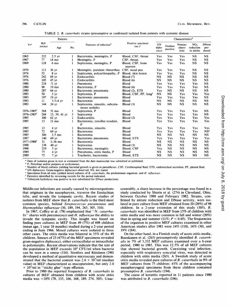
TABLE 2. B. catarrhalis strains (presumptive or confirmed) isolated from patients with systemic disease
Patients Characteristics'
Yr' Ref- Features of infection' Positive specimen GN Nonsac- NO3 DNaseerence Age No. (no.) diplo- Oxidase charo- reduction pro-cocci positive lytic to nitrite duced
1965 232 2.5 yr 1 Bacteremia, meningitis, P Blood, CSF, throat Yes Yes Yes NS NS1967 77 14 mo 1 Meningitis, P CSF, throat Yes Yes Yes NS NS1969 118 6 mo 1 Septicemia, meningitis, P Blood, CSF, bone Yes Yes Yes NS NS
marrow1970 113 36 yr 1 Meningitis, purulent rhinorrhea CSF, nasal pus Yes Yes Yes NS NS1974 52 8 yr 1 Septicemia, polyarthropathy, P Blood, skin lesion Yes Yes Yes NS NS1976 242 69 yr 1 Endocarditis Blood (7) NS NS NS NS NS1976 105 45 yr 1 Endocarditis Blood (6) NS NS NS NS NS1980 218 /1 yr 1 Pneumonia Blood Yes Yes Yes Yes NS1980 98 19 mo 1 Bacteremia, P Blood (6) Yes Yes Yes Yes NS1980 287 64 yr 1 Bacteremic pneumonia Blood (2), ETS Yes NS NS NS NS1983 50 3 yr 1 Septicemia, P Blood, CSF, PF, lunge NS NS Yes Yes Yes1983 197 19 yr 1 Bacteremic pneumonia Blood Yes Yes Yes Yes NS1983 11 3.5-4 yr 3 Bacteremia Blood NS NS NS NS NS1983 144 24 yr 1 Septicemia, sinusitis, subcuta- Blood (3) NS NS NS NS NS
neous nodules1976-1985f 268 31 mo 1 Septicemia, P Blood Yes Yes Yes Yes NS1976-1985f 268 15, 39, 41 yr 3 Septicemia Blood Yes Yes Yes Yes NS1985 300 62 yr 1 Endocarditis Blood (2) Yes Yes Yes Yes Yes1985 13 11 mo 1 Bacteremia, tonsillar exudate, Blood Yes Yes Yes Yes Yes
P1985 13 4 yr 1 Bacteremia, sinusitis Blood Yes Yes Yes Yes Yes1985 72 69 yr 1 Bacteremia Blood Yes Yes Yes Yes NS'1985 306 2.5 mo 1 Bacteremia Blood NS NS NS NS NS1987 140 69 yr 1 Bacteremic pneumonia Blood, ETS Yes Yes Yes Yes Yes1977-1988f' 31 1-36 mo 5 Bacteremia Blood NS NS NS NS NS1988 138 40 yr 1 Septicemia Blood (3) NS NS NS NS NS1988 219 5 yr 1 Bacteremia, meningitis Blood, CSF Yes NS NS NS NS1989 75 4 yr 1 Pneumonia, otitis media, P Blood NS NS NS NS NS1989 75 7 yr 1 Tracheitis, bacteremia Blood, ETS NS NS NS NS NS
U Date of isolation given in text or estimated from the date manuscript was submitted or published.b p, Petechiae and/or purpura or ecchymoses.Number of blood cultures yielding bacterial growth is given in parentheses. CSF, Cerebrospinal fluid; ETS, endotracheal secretion; PF, pleural fluid.dGN diplococci, Gram-negative diplococci observed; NS, not stated.Specimens from all sites yielded mixed cultures of B. catarrhalis, the predominant organism, and H. influenzae.
f Patient(s) identified by reviewing records for the period indicated.g Tributyrin hydrolysis was positive (a test substituted for DNase production).
Middle-ear infections are usually caused by microorganismsthat originate in the nasopharynx, traverse the Eustachiantube, and invade the tympanic cavity. Recent studies ofisolates from MEF show that B. catarrhalis is the third mostcommon species, behind Streptococcus pneumoniae andHaemophilus influenza (30, 189, 194, 265, 305, 310).
In 1967, Coffey et al. (78) emphasized that "N. catarrha-lis" shares with pneumococci and H. influenzae the ability toinvade the tympanic cavity. This insight was based onfinding pure cultures in MEF from 49 (7%) of 698 children(mean age, 1 year 10 months) studied during a 2-year periodending in June 1966. Mixed cultures were isolated in threeother cases. The otitis media was bilateral in 35% of the 49children. Smears of 23 (47%) of the MEF specimens revealedgram-negative diplococci, either extracellular or intracellularin polymorphs. Recent observations indicate that the size ofthe population in MEF exceeds 104 CFU/ml when bacteriacan be seen in a smear (310). Stenfors and Raisanen (290)developed a method of quantitative microscopy and demon-strated that the bacterial content was 2.6 x 10⁷/ml (medianvalue) in MEF characterized as mucopurulent, but only 2.0x 10⁵/ml in "sticky glue"-type effusion.Prior to 1980 the reported frequency of B. catarrhalis in
cultures of MEF obtained from children with acute otitismedia was <10% (78, 135, 166, 168, 189, 274, 305). Unac-
countably, a sharp increase in the percentage was found in astudy conducted by Shurin et al. (274) in Cleveland, Ohio,between October 1980 and February 1982. Strains, con-firmed by nitrate reduction and DNase activity, were iso-lated in pure culture from MEF obtained from 20 (20%) of 98children. In a 2-year extension of this study (305), B.catarrhalis was identified in MEF from 15% of children withotitis media and was more common in fall and winter (20%)than in spring and summer (11%; P < 0.05). The frequenciesof the organism in positive MEF cultures examined in otherAmerican studies after 1981 were 14% (310), 16% (30), and19% (184).On the other hand, in a Finnish study of acute otitis media,
Ruuskanen et al. (265) presumptively identified B. catarrh-alis in 7% of 3,332 MEF cultures examined over a 6-yearperiod, 1980 to 1985. This was 12.5% of all MEF culturesthat showed bacterial growth. Coexisting viral infection,especially with respiratory syncytial virus, was detected inchildren with otitis media (265). A Swedish study of acuteotitis media revealed pure cultures of B. catarrhalis in 9% ofMEF cultures from 75 children; 43% of the accompanyingnasopharyngeal specimens from these children containedpresumptive B. catarrhalis (194).The cause of keratitis reported in 11 patients since 1980
was attributed to B. catarrhalis (196).
296 CATLIN
on July 8, 2018 by guesthttp://cm
r.asm.org/
Dow
nloaded from

BRANHAMELLA CATARRHALIS 297
Pseudogonococcal Conjunctivitis and Urogenital Infections
A case of conjunctivitis in a 5-day-old girl was described in1987 by Romberger et al. (260). A strain isolated from thepurulent eye discharge was reported as N. gonorrhoeaeresistant to penicillin. The diagnosis of gonorrhea was re-ported to the public health department and the parents weresubjected to intramuscular injections of cefotaxime. Thisstrain subsequently was identified as B. catarrhalis by a statelaboratory and the Centers for Disease Control in Atlanta,Ga. The stressful implications of the erroneous identificationcan be imagined.
Historically, ophthalmia neonatorum was attributed to N.gonorrhoeae, but the identification often was based solely onthe characteristics of bacteria seen in Gram-stained prepa-rations of conjunctival exudate (248, 284, 297). B. catarrhalis(confirmed by nitrate reduction) as a cause of ophthalmianeonatorum was first reported by Spark et al. (284) in 1979 inan infant with fever of 3-week duration and a conjunctivitisthat was partially suppressed by short-term parenteral anti-biotic therapy. Righter and Nicol (248) described anothercase of conjunctivitis in an infant who received only ineffec-tive topical therapy for months because cultures of purulentdischarge from both eyes were reported as growing normalflora, not N. gonorrhoeae or N. meningitidis. Thirteenadditional cases of conjunctivitis in infants were reportedlycaused by B. catarrhalis (cited in references 260, 297, and317). Furthermore, a confirmed strain was isolated fromconjunctival discharge obtained from a previously healthy,58-year-old woman with acute bilateral conjunctivitis (172).Confirmed B. catarrhalis strains were isolated from ure-
thral exudates from four men with urethritis (96, 278) andfrom urine specimens collected 5 days apart from a womanwith chronic pyelonephritis (153). The organism was theonly pathogen isolated from a urinary tract infection in a13-year-old girl (2). Strains provisionally identified as "N.catarrhalis" were recovered from four anal canal specimenscultured on selective medium (29). Johnson (158) citedadditional cases of urogenital infections associated with "N.catarrhalis" and pointed out that their pathogenic roleremained speculative because of the possibility of infectionwith Chlamydia trachomatis or Ureaplasma urealyticumthat would not have been detected by conventional bacteri-ology.
Lower Respiratory Tract Infections
Pneumonia, acute exacerbations of chronic bronchitis,tracheitis, and empyema are caused by B. catarrhalis. Theisolates were regarded as primarily opportunistic in manycases. The factors predisposing to infection include under-lying disease of the bronchial tree or lung parenchyma,especially chronic obstructive pulmonary disease, and animmunodeficiency state due to chemotherapeutic agents orabnormalities involving the cellular or humoral immunesystems (58, 134, 140, 277). Thus, B. catarrhalis pneumoniawas reported in patients with multiple myeloma (91, 209),chronic lymphocytic leukemia (287), or quantitative abnor-malities of serum immunoglobulins (171, 192). Advanced ageappears to be a factor, possibly due to the significantreductions of IgG and IgM seen after age 60 years (51). Viraldamage to respiratory tract epithelium, for example, byrespiratory syncytial virus infection (6, 319), may promoteinvasion by B. catarrhalis. A high risk of aspiration from theoropharynx has been identified as a predisposing factor insome patients (259, 277).
B. catarrhalis pneumonia may present a clinical picturesimilar to other bacterial pneumonias, but generally is amilder illness except in the immunocompromised patient(321). An indication of its relative mildness is found inreports that most blood cultures are negative (58, 85, 118,209, 221, 241, 259, 313).The earlier reluctance to accept a pathogenic role for B.
catarrhalis has diminished in view of evidence of its pres-ence in pure cultures in sites that normally are sterile. Thus,it has been isolated from pericardiocentesis fluid aspiratedfrom a patient with acute pericarditis (183). Pleural effusionspecimens obtained by thoracentesis from each of twopatients with empyema yielded B. catarrhalis on two occa-sions (83, 313). The organism was isolated from transtra-cheal or endotracheal aspirates from 47 patients with pneu-monia or acute bronchopulmonary infections (5, 73, 99, 171,173, 192, 209, 224, 270, 279, 287, 316). Sections of lung tissuefrom two patients who died with untreated pneumoniarevealed intracellular and extracellular gram-negative diplo-cocci (209, 277), and B. catarrhalis was isolated in pureculture from postmortem lung secretions (277). Further-more, elevated antibody titers were detected in convalescentsera from patients with B. catarrhalis bronchopulmonaryinfections (28, 71, 197).A sputum culture usually yields clinically significant re-
sults if the specimen is of good quality and is inoculated byusing quantitative methods (described above). Nevertheless,mixed cultures of B. catarrhalis with H. influenzae orpneumococci or both are isolated from substantial numbersof sputum specimens (73, 200, 241, 277). Some authorsquestioned the role of B. catarrhalis as a respiratory patho-gen in cases of mixed infection with acknowledged patho-gens (134, 259). Others omitted such cases from consider-ation (207, 279). As expected, sputa from patients withaspiration pneumonia often yielded mixed cultures typical oforopharyngeal flora (259).
Seeking further evidence of the clinical significance of B.catarrhalis in bronchopulmonary infection, Yuen et al. (328)conducted a case control study of 26 patients ("cases")whose expectorated sputum yielded pure cultures of con-firmed B. catarrhalis and patients ("controls") whose spu-tum cultures contained only commensal Neisseria spp.(DNase negative). Proportionately more cases than controls(P < 0.05) showed three or more clinical features of bron-chopulmonary infection (cough with increased purulence ofthe sputum, fever of >37.40C, leukocytosis, chest X-raysigns, and crepitations and/or dullness of the chest). Further-more, intracellular gram-negative diplococci were observedin 13 of the 26 sputum specimens containing B. catarrhalisand none were detected in the control sputa.Lower respiratory tract-infections caused by B. catarrha-
lis in children tend to differ from those in adults by theabsence of underlying respiratory disease (200). Thus, B.catarrhalis bronchopneumonia developed in three childrenwho were intubated and on mechanical ventilation followingelective surgery or trauma (173) and also was reported in a15-day-old, prematurely born infant whose lungs had ap-peared normal 5 days earlier (228). On the other hand,preexisting lung disease was recognized in five children, 6weeks to 6 months of age, who suffered from B. catarrhalispneumonia. All had been born prematurely, entered a hos-pital in Phoenix, Ariz., during winter months, and experi-enced precipitous clinical deterioration requiring emergencyintubation and mechanical ventilation (19).
Tracheitis caused by B. catarrhalis developed in a previ-ously healthy 11-month-old girl (137) and also in a 4-year-old
VOL. 3, 1990
on July 8, 2018 by guesthttp://cm
r.asm.org/
Dow
nloaded from
CLIN. MICROBIOL. REV.
girl following local injury to the trachea by a cuffed endot-racheal tube (114). Epithelial damage by respiratory syncy-tial virus infection was considered a predisposing factor inthe development of B. catarrhalis tracheitis in a 9-year-oldboy (319) and a 6-month-old boy (6).A nonhealing, thoracic, surgical wound infection was
described by Gray et al. (132) in a 59-year-old woman whohad undergone an open lung biopsy procedure. B. catarrh-alis was isolated from deep thoracotomy wound specimensas well as from tracheal secretions and sputum, and smearsof each revealed numerous polymorphonuclear leukocytesand intracellular and extracellular gram-negative diplococci.The peak incidence of B. catarrhalis respiratory infections
occurs in winter months. This was found south of theequator in Western Australia (93) and New Zealand (279), aswell as in Denmark (269), The Netherlands (85, 86), England(200), Scotland (57), Japan (203), and the United States(241).Davies and Maesen (86) described a remarkable 10-year
study at the Regional Public Health Laboratory in Heerlen,The Netherlands. Approximately 1,700 positive culturesfrom respiratory tract specimens were obtained per year.The frequency of B. catarrhalis strains, which in 1977 was5%, steadily increased until in 1982 it reached 20% and by1986 it accounted for 26% of the respiratory tract pathogens.This inexplicable rise occurred in a relatively constantantibiotic climate and was not merely the result of increasedawareness in the laboratory.
Person-to-person transmission of B. catarrhalis wasthought responsible for some hospital-acquired infections.Pulmonary infections in three premature infants were tracedto an attending nurse (139). Acute bronchitis caused by aP3la' strain developed in a previously healthy, 22-year-oldnurse who worked in a chest ward (207). Calder et al. (57)undertook an environmental study of chest wards in whichclusters of cases were found adjacent to index patients.Nasopharyngeal carriage of B. catarrhalis was identified in7% of staff members and 8% of patients. Six B. catarrhaliscultures were isolated from 117 settle plates that wereexposed for 6 to 8 h in a duty room frequented by carriersand in a chest ward. Also, experiments showed that theorganism survived for at least 3 weeks in sputum (57). Thiscircumstantial evidence of environmental spread illustratedthe need for a means of determining strain relatedness.The development of restriction endonuclease typing (See
subsection, "Strain Differentiation") made it possible totrace the source of an outbreak of respiratory illness due toPla' B. catarrhalis that occurred in an intermediate careunit. Patterson et al. (230) analyzed isolates from three staffmembers and ten patients, four with pneumonia, four withbronchitis, and two who were asymptomatic (hospitalizedfor nonrespiratory conditions). The typing results indicatedthat the isolates from five patients (including one asympto-matic) and two staff probably had a common origin.
Analogy between B. catarrhalis and N. meningitidisInfections
B. catarrhalis resembles N. meningitidis in many respectsin spite of their very significant genotypic differences. Theirnatural habitat is believed to be human beings exclusively,and the nasopharynx is the site of initial mucosal coloniza-tion (37, 145, 204, 212, 307). Presumably this is due to thedistribution of specific host cell receptors (16). Both organ-isms often are carried asymptomatically (212, 292). Thecarriage rates are lower than formerly believed, however,
owing to the recognition of distinctions between B. catarrh-alis and N. cinerea and the differentiation of nontypablemeningococci from N. lactamica and N. polysaccharea(177, 258). N. meningitidis may be more pathogenic and lessdependent than B. catarrhalis on predisposing conditions forthe manifestation of disease. Nevertheless, both possesscapsules and endotoxin which may facilitate later stages ofinfection.
Systemic infections with both organisms may be accom-panied by petechial rashes, purpura, or ecchymoses. Suchlesions were found in nine patients (Table 2), all of whomwere .8 years of age, and in a 64-year-old man withpneumonia from whom B. catarrhalis was isolated in pureculture from a transtracheal aspirate (316). Disease causedby either organism may display a rapid onset of symptoms(118, 205) or be the cause of an unsuspected bacteremia inyoung children who present with fever and upper respiratoryinfections (13).An unknown number of infections with B. catarrhalis
have been attributed to N. meningitidis (52, 72, 77, 192, 205,232). This error would result from the failure to use adequatedifferential tests because of the belief that gram-negativediplococci seen in purulent spinal fluid or good-qualitysputum must be the established pathogen.The term commensal, which refers to the nondetrimental
coexistence of organisms, has frequently been applied to B.catarrhalis but not to N. meningitidis. Characterizing oneorganism as a commensal (205, 324) or a nonpathogen (117)and another as a pathogen carries unwarranted implicationsof qualitative differences. Actually, the distinction betweentheir potential pathogenicity appears to be primarily quanti-tative.
Healthy Carriers of B. catarrhalisIsolation rates. Various culture media containing selective
agents, together with differential tests sufficient to confirmthe identity of B. catarrhalis, were used in investigations ofcarrier rates (Table 3). Also, in two of these studies theprevalence of N. cinerea was determined as 14.5% (23) and28.2% (177) compared with <5% for B. catarrhalis. Na-sopharyngeal colonization of healthy children in Cleveland,Ohio, was 46% in fall and winter, but only 9% (P < 0.005) inspring and summer, overall 18% (305). Higher percentages ofB. catarrhalis were isolated from children younger than 24months than from older children (Table 3).A corresponding age relationship was observed by Ark-
wright (9) in 1907. "Micrococcus catarrhalis" was isolated"from a much larger proportion of infants under one yearthan of older children or of adults: the last furnished propor-tionately the fewest strains" (9). Further evidence camefrom three studies in the 1980s of nasopharyngeal specimenstaken in fall or winter months from Swedish children whoshowed no symptoms of respiratory infections. B. catarrh-alis was isolated from 45% of children 2 years or younger,but from only 17% aged 6 to 7 years (194) and, similarly,from 36% of children 2 years or younger, but from only 13%who were 6 to 9 years old (48). Molstad et al. (210) studied191 children aged 1 to 7 (mean, 4.5) years who attendedday-care nurseries and isolated B. catarrhalis from 58%.Selective media were not used in these investigations, andsome of the strains possibly were N. cinerea since not allidentifications were confirmed. In contrast to results withchildren, B. catarrhalis was recovered from only 3% ofblood agar cultures of posterior nasal specimens from 286healthy men (mean age, 20 years) who were military servicerecruits in Finland (163).
298 CATLIN
on July 8, 2018 by guesthttp://cm
r.asm.org/
Dow
nloaded from

BRANHAMELLA CA7ARRHALIS 299
TABLE 3. Isolation of confirmed B. catarrlhalis strains on semiselective and selective media
Author(s) Yr Place Subjects (no. in study) Culture site B. catarrhalis (%)
Berger (21) 1961 Hamburg Adults (172) Throat' 0.6Knapp (177) 1982-1983 Seattle Adults (202) Oropharynxb 4.5Calder et al. (57) 1984 Edinburgh Adults (148)c Nasopharynx 7.4DiGiovanni et al. (93) 1985 Perthd Adults (123) Nasopharynx 2.4Van Hare et al. (305) 1982-1983 Cleveland Children <24 mo (37) Nasopharynx 24.0
Children .24 mo (14) Nasopharynx 0Vaneechoutte et al. (302) 1988 Ghent Children 3-12 yr (178) Saliva 48.9
Children 3-12 yr (75) Throat 30.7
aCarriage of "N. catarrhalis" was reported by Berger (21) as 15% (26 of 172 subjects); however, after N. cinerea was defined in a later study (25), retests ofthe 26 strains identified 25 (14.5%) as N. cinerea and 1 (0.6%) as 'N. catarrhalis" (23)."N. cinerea was identified in 28.2% of the cultures.Hospital staff and patients whose illness was not attributed to B. catarrhalis infection. All other study groups were made up of subjects without respiratory
symptoms.' Western Australia.
Selective culture media for B. catarrhalis. Selective mediadesigned to improve the detection of gonococci and menin-gococci are inhibitory for many B. catarrhalis strains prin-cipally due to the action of colistin (73, 81, 177, 325). Thus,only 12 strains were isolated from 1,204 throat cultures on amedium containing colistin (6 pLg/ml) and other selectiveagents (325). However, a good recovery of B. caitarrhalisand inhibition of various other species are obtained onsemiselective media containing vancomycin and trimetho-prim, each at final concentrations of 3 tFg/ml (81, 179, 305) orhigher (93, 283). Other semiselective media contained 25 ,ugof ristocetin per ml (21) or 10 U of bacitracin per ml (207).Some investigators added maltose (21), sucrose (179), orDNA (283) to facilitate differentiation of colonies. Corkilland Makin (81) initially added DNA to their semiselectivemedium, but found that the zone representing DNase activ-ity around single colonies of B. catarrhalis was not alwayseasy to detect. Vaneechoutte et al. (302) improved theselective action of a sheep blood agar containing vancomy-cin (10 pRg/ml), trimethoprim (5 ,ug/ml), and amphotericin B(2 ,ug/ml) by adding sodium acetazolamide (10 pLg/ml). Incu-bation of this medium in air greatly reduced the growth ofNeisseria species due to inhibition of the activity of carbonicanhydrase, an enzyme typically produced by Neisseriaspecies, but not by B. catarrhalis (24, 37, 302, 307). Theenhanced selectivity of the medium was considered respon-sible for the high rates of B. catarrhalis isolation (Table 3)since rates were far lower on semiselective media or onblood agar (302).
VirulenceFeatures associated with virulence in other bacteria are
also present in B. c atarrhalis. Pili may facilitate adherence tomucosal surfaces. An outer coat analogous to a capsule andthe tendency of cells to form clusters may reduce theeffective actions of host defenses. The spontaneous releaseof outer membrane blebs provides a mechanism for thedispersal of endotoxin into peripheral tissues (285).
Studies of the virulence of clinical isolates are hamperedby the low virulence of B. catarrhalis for laboratory animals(113, 125, 130). Intraperitoneal inoculation of mice withenormous doses proved lethal, but the bacteria were notfound in the heart blood, and death was thought to be due tothe "toxic effect of the bacterial protein" (130).Animal models of otitis media secondary to bacterial
infection make use of chinchillas and gerbils. Middle-earinoculations of B. caatarrhalis resulted in the development ofeffusions. Nevertheless, viable bacteria were not recovered
after 24 h (95, 106), although elevated levels of neutralphosphatase and hexosaminidase were found in 4-day sam-ples of chinchilla effusions (95). Killed B. catarrhalis cul-tures also produced effusions (106), suggesting the involve-ment of endotoxin in inflammatory changes seen in themiddle ear (89). Experiments in which five B. catarrhalisisolates were added to specimens of mucoid effusion mate-rial aspirated from middle ears of young children showedthat viability was retained for 18 to 36 days (291). However,conditions affecting bacterial viability in MEF from livingchinchillas and in MEF following experimental inoculationare hardly comparable.The mechanisms enabling B. catarrhalis to colonize the
human nasopharyngeal mucous membrane are virtually un-known, but must involve adherence. Nonadherent bacteriaare bathed by secretions containing antibacterial enzymesand products of local immune systems and, moreover, arelikely to be swept away by the flushing effects of mucus andciliary beating (195). Studies of adherence have demon-strated specific interactions between adhesins on bacterialsurfaces and receptors on mucosal surfaces (16). The adhes-ins of gonococci and meningococci include pili and heat-modifiable outer membrane proteins, for example, gonococ-cal protein 11 (285, 292).
Investigators at Nagasaki University described interestingresearch on B. catarrhalis adherence. The text of one paper(249) is in Japanese, but the abstract, technique, and thelabels and legends for figures are in English. A second study(203) is described in English. For the adherence assay,squamous epithelial cells scraped from the oropharynx werewashed twice in phosphate-buffered saline to remove looselyattached bacteria and then suspended in phosphate-bufferedsaline containing Ca24 and Mg24 to give 105 human cells perml. Bacteria from overnight cultures in Mueller-Hintonbroth were washed, and a suspension was adjusted to anoptical density of 0.1 at 620 nm. "These suspensions con-tained from 1-6 x 10⁷ CFU/ml'' (249). The attachmentmixture contained equal volumes (249) or a ratio of 1 partcells to 100 bacteria (203). After incubation for 30 min in ashaking 37°C water bath, this mixture was washed four timesto eliminate nonadherent bacteria and then collected on acover glass and Gram stained. Counts were made at x 1,000magnification of the number of attached bacteria per cell for50 consecutive epithelial cells. The B. catarrhalis strains(identity confirmed by nitrate reduction) were recent isolatesfrom respiratory infections.
Rikitomi et al. (249) studied 60 samples of oropharyngealcells obtained from 25 patients with chronic bronchopulmo-
VOL . 3 , 1990
on July 8, 2018 by guesthttp://cm
r.asm.org/
Dow
nloaded from
CLIN. MICROBIOL. REV.
nary infections. B. catarrhalis adhered in greater numbers tothese cells than to cells from healthy persons (P < 0.001); themean number of bacteria per cell ranged between seven andless than one per oropharyngeal cell from patients comparedwith less than one per cell from healthy adult controls. Incontrast, adherence to cells from patients and control sub-jects was low and approximately equal for control bacteriacharacterized as Neisseria sp.Mbaki et al. (203) investigated the seasonal frequency of
B. catarrhalis infections in experiments with oropharyngealcells from 48 patients who suffered from various types ofchronic bronchopulmonary disease involving B. catarrhalisand other bacteria. The adherence of B. catarrhalis, in termsof mean number of bacteria per oropharyngeal cell, washighest in winter (3.11 + 1.30) and lowest in summer (0.400.25), with intermediate values in spring (1.07 0.45) andautumn (1.84 + 1.30). The value was 0.70 0.16 foradherence to oropharyngeal cells obtained in December(winter) from nine infection-free control subjects. Mbaki etal. (203) considered that high adherence levels in winter wererelated to low environmental temperatures. However, lowhumidity in heated rooms may be more relevant since someoropharyngeal cell samples were from hospitalized patients.A provocative observation by Rikitomi et al. (249) was the
possibly greater adherence to oropharyngeal cells of Ila+cells (two strains tested) than P-lactamase-negative cells(one strain). Unfortunately, the technical difficulty in adjust-ing suspensions of these B. catarrhalis cells to contain equalnumbers of CFU per milliliter raises doubts about thevalidity of comparisons between strains when data areexpressed as mean bacteria per cell. In view of the possiblysignificant differences between numbers of CFU per millili-ter in the initial attachment mixtures, it would have beenpreferable to express results in terms of (mean number ofbacteria per human cell)/(number of bacterial CFU permilliliter).
Studies of the activities of B. catarrhalis in human cellcultures are needed to elucidate the complex process ofinfection. Adherence is required, but is not sufficient forcytotoxicity and invasion (292). N. lactamica cells, forexample, adhere irreversibly to human cells but are notinternalized (273). Upon introduction into human nasopha-ryngeal organ cultures, piliated meningococci attach to mi-crovilli of nonciliated cells and in 6 to 12 h become trappedand pulled into membrane-bound vesicles in a manner re-
sembling phagocytosis (204). Nearby ciliated epithelial cells,to which meningococci do not attach, soon lose ciliaryactivity and undergo sloughing believed due to the action oflipopolysaccharide and peptidoglycan monomers releasedby the cocci (204, 292).
B. catarrhalis was shown to produce histamine duringgrowth in broth supplemented with histidine (1 mg/ml).Devalia et al. (90) speculated that this ability could contrib-ute to the pathogenicity of such strains in the lower respira-tory tract.A mechanism for iron acquisition is a requirement for
bacterial growth and, therefore, for pathogenicity (285).Iron-binding proteins present at mucosal surfaces (lactofer-rin) and in blood serum (transferrin) of the human host havehigh association constants for iron resulting in insufficientfree iron for bacterial growth. Schryvers and Lee (271)showed that B. catarrhalis cells possess membrane-associ-ated proteins that bind human lactoferrin and human trans-ferrin, thereby enabling the bacteria to utilize these sources
of iron for growth. On the other hand, lactoferrin andtransferrin from nonhuman species apparently are not
bound, which may contribute to the specificity of B. catarrh-alis for human beings.Some clinical isolates are resistant to killing by normal
human serum and others are susceptible (46, 71, 282). Themolecular basis of this complement-dependent bactericidalaction in B. catarrhalis has not been investigated, but ispresumably multifactoral as in N. gonorrhoeae (246).
Prellner (243) demonstrated that B. catarrhalis can bindthe complement component Clq with high efficiency withoutthe participation of antibodies. She speculated that, in pa-tients with acute otitis media, Clq binding might contributeto abnormal C1 complexes leading to impaired complementactivation and defective opsonization of bacteria.IgAl protease was not produced by any of 45 confirmed
strains of B. catarrhalis (208, 305). Koomey and Falkow(182) showed that this is due to the absence of the IgAlprotease-encoding gene in B. catarrhalis rather than to thepresence of a silent or defective allele. IgAl protease hasbeen considered a virulence factor since it specificallycleaves and inactivates human IgAl, but Sparling (285) hasquestioned its clinical importance. Furthermore, an IgAlprotease-deficient N. gonorrhoeae mutant is unimpaired inits attachment to and invasion of human mucosal cells inorgan culture (79).
ImmunityCircumstantial evidence of the importance of immuno-
globulins as mediators of defense against B. catarrhalis isfound in the strong clinical association discussed earlierbetween various immunodeficiency states and systemic dis-ease or bronchopulmonary infections. Furthermore, thegreater susceptibility of older individuals may be due in partto the significant decline of serum IgG and IgM concentra-tions by the sixth decade of life (51).The development of antibodies reactive with B. catarrha-
lis has been investigated in patients suffering from maxillarysinusitis (46), acute bronchopulmonary infections (28, 71,197), and acute otitis media (189). The serological methodsinvolved immunodiffusion (46), complement fixation (46), anenzyme-linked immunoassay (189, 197), a bactericidal assay(71), and an immunofluorescent-antibody test (28). Modestincreases of antibody titers between acute and convalescentsera occurred in most, but not all, patients. Antibodies werealso detected in control sera from substantial numbers ofhealthy persons (28, 46, 189), possibly due to immunogenicstimulation by B. catarrhalis present in the pharyngealnormal flora (189).Leinonen et al. (189) found that IgA class antibody di-
rected against B. catarrhalis developed in the sera of eight ofnine children, 2 to 6 years old, who suffered from acute otitismedia caused by B. catarrhalis. IgA antibodies were notdetected in control sera from children with otitis mediacaused by pneumococci and H. influenzae. Although half ofthese control sera contained IgG reactive with B. catarrha-lis, the titers did not increase during convalescence. Middle-ear aspirates from which B. catarrhalis was cultured con-tained IgA and IgG antibodies directed against the pathogen.The presence and titers of both immunoglobulins dependedon the age of the child. Using the same enzyme-linkedimmunoassay, Malkamaki et al. (197) detected high andrising titers of IgG and IgA antibodies in serum samples froma 19-year-old man with bacteremic pneumonia caused by B.catarrhalis.
B. catarrhalis is recognized as a T-cell-independent mito-gen which stimulates DNA synthesis in human B lympho-
300 CATLIN
on July 8, 2018 by guesthttp://cm
r.asm.org/
Dow
nloaded from

BRANHAMELLA CATARRHALIS 301
TABLE 4. Differential biochemical reactions of B. catarrhalis and possibly confusing Neisseria spp.a
Acid formation from: Polysaccharide Reduction Hydrolysis of:Species
Glucose Maltose Lactose Sucrose from sucrose of nitrate DNA Tributyrin y-GNA"
B. catarrhalis 0 0 0 0 0 + + + 0N. cinerea 0 0 0 0 0 0 0 0 0N. flavescens 0 0 0 0 + 0 0 0 0N. gonorrhoeae + 0 0 0 0 0 0 0 0N. meningitidis + + 0 0 0 0 0 0 +N. caviaec 0 0 0 0 0 + + + 0
a +, Most strains (>90%) give positive reactions; 0, negative reactions in tests with adequate controls.b y-GNA, N-y-Glutamyl-o3-naphthylamide.c This organism, designated Neisseria caviae (257, 307)) or Moraxella (Branhamella) caviae (37), is isolated from guinea pigs; if isolated from human sources,
it might be called B. catarrhalis in spite of differences in DNA base composition, DNA-DNA hybridization reactions (174), genetic transformation frequencies(148), serological (24) and biochemical (15, 74, 257, 264, 298) characteristics, and major cellular fatty acids (298).
cytes. This B-cell activation is related to the binding ofbacteria to IgD present on the B cell. Forsgren et al. (120)offered the suggestion that B-cell binding may alter theimmune response of the host and confer a selective advan-tage upon B. catarrhalis.
IDENTIFICATION AND BIOCHEMICALCHARACTERISTICS OF B. CATARRHALIS
The appearance of bacteria in a properly Gram-stainedpreparation should be evaluated carefully. B. catarrhalis isgram negative, but has a tendency to resist decolorization (9,23, 60, 245, 257). This interaction with the crystal violet dyemay be responsible for the inhibition of growth on crystalviolet medium found for B. catarrhalis but not N. cinerea(4).The bacteria characterized in Table 4 are oxidase-positive,
nonmotile, gram-negative cocci commonly arranged in pairswith their abutting sides flattened. Division occurs in twoplanes at right angles to one another, thereby preventingchain formation which occurs when cell division is restrictedto one plane (15). The potentially confusing coccobacillarycells of Moraxella and Acinetobacter species elongate intosnakelike cells when division is blocked by sublethal con-centrations of penicillins or certain other antibacterialagents. Since elongation does not occur with B. catarrhalisor Neisseria species, this is an easy way to distinguish themfrom nonsaccharolytic, short paired rods (63).
B. catarrhalis colonies on blood agar are nonhemolytic,circular, grayish white, opaque, convex, and _2 mm indiameter after 48 h. Colonies when pushed tend to remainintact and slide across the agar like a hockey puck on ice[P. A. Shurin, Drugs 31(Suppl. 3):38-39, 1986].
Differential TestsAction on carbohydrates. B. catarrhalis does not produce
acid from any of 9 (69) or 10 (112) carbohydrates. Negativeresults are observed uniformly with various methods (9, 61,178) including the rapid carbohydrate degradation testsdesignated RIM-N (94, 155, 185), Gonobio-Test (94), andAPI QuadFERM+ (109, 156). Minitek test reactions werecorrect in some instances (94), but not all (233).
Aberrant reactions in carbohydrate tests more often con-fuse the identification of Neisseria species. False-positivereactions in rapid glucose utilization tests were recorded forsome strains of N. cinerea (40, 94, 177) which led to theirpresumptive identification as N. gonorrhoeae in some cases(317). Rare meningococcal isolates are maltose negative(293). Glucose-negative reactions have been observed with
meningococci and gonococci (73, 82, 109, 177, 178, 245).Such strains often, but not always, give positive reactionsafter a few subcultures on a favorable medium (212) or in atest with 10% (wt/vol) glucose, which facilitates transport ofthe sugar (62).N. flavescens possesses an amylosucrase that builds su-
crose into a polysaccharide which reacts with iodine (20, 61,142). This reaction differentiates N. flavescens from N.cinerea (Table 4) and N. polysaccharea from N. meningitidis(177, 258).
Reduction of nitrate and nitrite. B. catarrhalis reducesNO3 to NO2 and beyond (22, 24, 37, 69, 73, 99, 256). Theabsence of both NO3 and NO2 is confirmed by the negativereaction that persists after addition of the NO2 test reagentsfollowed by zinc dust, which reduces NO3 nonenzymati-cally. Berger (23) found that 0.03 to 0.01% (wt/vol) NO3,rather than the customary 0.1%, provides a more sensitivetest useful for revealing weak NO3 reduction. The test forNO3 reduction, when modified for use as a rapid method,was found difficult to interpret (315) or unreliable (177).NO2 is denitrified by all species given in Table 4 provided
that growth occurs. Because of NO2 toxicity, the customary0.1% (wt/vol) concentration should be reduced for tests of B.catarrhalis, N. cinerea, N. gonorrhoeae, and N. meningiti-dis. Seven B. catarrhalis strains were able to grow in thepresence of 0.005% (wt/vol) KNO2, but not 0.03%; the typestrain ATCC 25238 (Ne 11) tolerated 0.01% (69). Denitrifi-cation of NO2 by B. catarrhalis occurs without the formationof gas (24, 25, 69), whereas nitrogen gas is produced by N.cinerea and N. flavescens (24, 25, 307). The test for gasproduction requires a large volume of broth (e.g., 25 ml)when NO2 is used at 0.005 or 0.01% so that enough gas willaccumulate in the inverted collection vial.
Hydrolysis of DNA. The original finding of DNase produc-tion by B. catarrhalis (69) was confirmed by subsequentinvestigators who also demonstrated the differential value ofthe characteristic (4, 73, 81, 226, 256). The customary DNasetest medium contains 0.2% (wt/vol) DNA. Various reagentsdetect the zone of hydrolysis that develops around an area ofdense bacterial growth. The Frazier reagent used in threestudies (69, 73, 256) contains mercuric chloride, 15 g dis-solved in 100 ml of distilled water, plus 20 ml of concentratedhydrochloric acid. Alternatively, a solution of 0.1 N HCl wasused (4, 81) or toluidine blue, 0.1% (wt/vol) (226).The time of incubation can significantly affect the DNase
test results. For example, one B. catarrhalis strain examinedby Corkill and Makin (81) produced no detectable DNaseactivity after 24 h, whereas an 18-mm-diameter zone wasfound with a duplicate culture incubated for 48 h. In my
VOL. 3, 1990
on July 8, 2018 by guesthttp://cm
r.asm.org/
Dow
nloaded from
CLIN. MICROBIOL. REV.
experience, the type strains N. cinerea ATCC 14685, N.flavescens ATCC 13120, and N. gonorrhoeae ATCC 19424produced 1 + zones of DNase detected by the addition of theFrazier reagent after incubation of cultures for 4 days; 2+zones were found with B. catarrhalis ATCC 25238 and 4+zones were found with Staphylococcus aureus (unpublishedobservations). For definitive results, control cultures of B.catarrhalis and a typically negative Neisseria species shouldbe incubated in parallel with test cultures to enable directcomparisons of DNase zone diameters.
Hydrolysis of tributyrin and other esterase substrates.Tributyrin hydrolysis is a valuable differential characteristic(73, 154, 201, 247, 250, 257). The original test was relativelyunsatisfactory, however, because the hydrolysis of 1% (vol/vol) tributyrin incorporated in an agar medium (25) producedonly a partial reduction of opacity that was difficult to see(256). A micromethod with 0.1% (vol/vol) tributyrin in agarrevealed hydrolysis by Neisseria species as well as B.catarrhalis (238). Recent methods take advantage of the10-fold-greater amount of butyric acid liberated from tribu-tyrin by an esterase of B. catarrhalis than by enzymes ofNeisseria species likely to be isolated from human sources(254). In one method a disk containing tributyrin and phenolred indicator is added to a dense bacterial suspensionprepared in buffer solution. A positive reaction is seen as thechange from red to yellow (acid pH). Disk tests usually givepositive results (154, 201, 257), but negative reactions werereported for 2 of 31 isolates (109) and 1 of 28 (247) B.catarrhalis strains.
4-Methylumbelliferyl butyrate and indoxyl butyrate areused in place of tributyrin in other tests for esterase. Releaseof the 4-methylumbelliferone moiety by hydrolysis is de-tected by its fluorescence under UV light (301). Hydrolysisof indoxyl butyrate releases indoxyl, which spontaneouslyforms indigo blue (88).
Hydrolysis of aminopeptidase substrates. N-y-Glutamyl-f-naphthylamide and L-hydroxyproline-p3-naphthylamide aresubstrates used in rapid tests of -y-glutamylaminopeptidaseand hydroxyprolylaminopeptidase, respectively. Mannheimand Burger (199) first demonstrated that N-y-glutamyl-,B-naphthylamide was hydrolyzed by N. meningitidis but notby B. catarrhalis. This was confirmed by other investigators(84, 147, 255), who also reported negative reactions for N.gonorrhoeae. A purified arylamidase from "N. catarrhalis"which lacked activity on N--y-glutamyl-3-naphthylamide hadstrong activity for alanine P-naphthylamine and severalother amino acid P-naphthylamines (18). The production ofhydroxyprolylaminopeptidase by N. cinerea, but not by B.catarrhalis, is of possible differential value (147, 177). Thisenzyme was produced by all N. gonorrhoeae strains exam-ined (84, 147, 150) and by various percentages of N. menin-gitidis (84, 147, 177).
Response to superoxol (30% H202). All species listed inTable 4 produce catalase detected with 3% H202. The use of30% H202 was recommended as a differential test (266). Apositive superoxol test is seen as brisk bubbling immediatelyafter dropping 30% H202 onto a young (s24-h) culture.Weak bubbling or a reaction delayed for 3 s or longer isconsidered negative. Positive reactions were obtained with99.9% of N. gonorrhoeae isolates (8, 266, 326). Most N.meningitidis strains gave negative reactions, but 98% ofgroup A meningococci were positive (8). The superoxol testappears to lack differential value for B. catarrhalis sinceboth negative and positive reactions have been reported (4,8, 177, 326).Other differential tests. Serological tests involving classic
agglutination methods were unsuccessful due to the notori-ous spontaneous agglutination of B. catarrhalis in 0.85%NaCl solution (9, 24, 112, 130, 143, 245). Precipitin testswere used to distinguish B. catarrhalis from N. cinerea (24,25). An immunodiffusion technique showed that one B.catarrhalis strain (ATCC 8176) was serologically unrelatedto N. cinerea or to strains of eight other Neisseria species(263). Eliasson (110) investigated a trypsin-sensitive, heat-stable P antigen located on the surface of B. catarrhalis. Hesuggested that an antibody specific for P antigen might serveas a reagent for the identification of B. catarrhalis.A DNA probe of chromosomal origin was developed and
found to be 100% specific for B. catarrhalis (D. Beaulieu andP. H. Roy, Abstr. Annu. Meet. Am. Soc. Microbiol. 1989,D249, p. 124). Such a probe will reduce the time required foridentification since it can be used on primary cultures withonly a short pretreatment to release DNA.Growth at 220C and growth on nutrient agar have long
been considered typical of B. catarrhalis (4, 25, 82, 108, 117,212, 245). Unfortunately, the components of nutrient agarare poorly defined and nutritionally variable (62). Additionalvariables (often unstated) are the inoculum density, whichshould be low enough to minimize spurious reactions due tocarry-over of nutrients in cells grown on an adequate me-dium, time of incubation, and atmospheric humidity. A dryincubation atmosphere deters growth. B. catarrhalis ATCC25238 did not grow on nutrient agar (Difco Laboratories)during incubation in an atmosphere of humidified air at either22 or 360C for 6 days, although sparse growth occurred onblood agar at 220C. N. cinerea ATCC 14685 failed to grow oneither nutrient or blood agar at 22TC, but moderate growthoccurred on nutrient agar at 360C (unpublished findings). Thefailure of 11 clinical isolates of B. catarrhalis to grow onnutrient agar incubated at 22°C led Doern and Morse (99) torecommend that this test not be included among the criteriafor identification.Some B. catarrhalis strains are able to grow on modified
Thayer-Martin agar and other colistin-containing selectivemedia (29, 94, 99, 109, 177, 257, 325). Thus, contrary toearlier findings (212), growth inhibition on these media is nota reliable differential characteristic.
Nutritional requirements were investigated by Riou andGuibourdenche (257). They found that all of 178 strains of B.catarrhalis required arginine for growth in Catlin's chemi-cally defined medium (64). On the other hand, N. cinerearequired cysteine, proline, and arginine (180, 257) and alsohistidine and biotin (unpublished findings). Juni et al. (164)independently showed the arginine requirement in a simplerdefined medium and found that most strains could utilizeornithine in place of arginine. An attempt to demonstratemultiple nutritional biotypes used ion-exchange chromatog-raphy to detect the use and production of amino acids by B.catarrhalis grown in nutrient broth (27). The utilization ofvarious carbon sources and additional physiological reac-tions were reported (15, 73).Weiner and Penha (314) recently reported the value of
Bacto TB Hydrolysis Reagent (Difco) as a means of detect-ing Tween 80 hydrolysis by B. catarrhalis. Negative resultswere obtained with 152 strains of Neisseria species and 41strains of Moraxella species. On the other hand, lipaseproduction detected in complex medium supplemented with0.01% (wt/vol) Tween 80 was reported as positive for Mor-axella lacunata, M. liquefaciens, M. osloensis, and Acine-tobacter strains, as well as for B. catarrhalis (15). Althougha potentially useful differential test, Tween 80 hydrolysisneeds further evaluation.
302 CATLIN
on July 8, 2018 by guesthttp://cm
r.asm.org/
Dow
nloaded from

BRANHAMELLA CATARRHALIS 303
Stages in the Identification Process
Various commercial enzyme substrate kits for rapid testshave been described (94, 150, 155, 177, 212, 233, 315).Unfortunately, some rapid tests yield only negative reac-tions for B. catarrhalis or, worse, false identifications (40,94, 109, 233). Rapid tests may be justified provided that theresults are rapidly communicated and the physicians fullyrecognize the possible provisional nature of a bacterialidentification. In a survey described by Staneck (289), phy-sicians indicated their willingness to sacrifice some degree ofaccuracy in return for the prompt delivery of test results.However, it is unacceptable to give a genus and speciesname to an isolate solely on the basis of negative reactions.A second stage in the identification process is needed.The identity of B. catarrhalis is best confirmed by positive
reactions in three differential tests (Table 4) since none ofthem is 100% sensitive or specific. Before undertaking thesetests, it is important to verify the microscopic appearanceand purity of the culture. The coccobacillary form of severalMoraxella species that reduce NO3 (37) must be recognized(63). The possible difficulties associated with the use ofinocula from a primary culture are well known, but a furtherexample is instructive. A mixed culture obtained from atranstracheal aspirate contained B. catarrhalis which pro-vided growth factors that enabled H. influenzae, the unde-tected strain, to grow and form acid in a glucose-containingmedium. Accordingly, the diplococcus was initially identi-fied as N. gonorrhoeae in three different laboratories (53).The identification of atypical strains is a third stage. This
may require methods used primarily in research. An organ-ism isolated from blood cultures gave aberrant results in fourtests, but was identified as an atypical strain of B. catarrhalison the basis of cell wall fatty acid and lipopolysaccharideanalyses (322). DNA base composition was determined in aneffort to identify oxidase-positive, nonsaccharolytic, gram-negative cocci isolated from urogenital cultures (240). Theguanine-plus-cytosine content (69 mol%) proved that theisolates were not related to Branhamella or Neisseria spe-cies. Isolates of interest like these should be preserved forpossible use in further studies (64).Survey specimens were sent to "qualified reference labo-
ratories" as part of the proficiency testing program of theCollege of American Pathologists. B. catarrhalis was cor-rectly identified by 74% of 685 laboratories in 1983 and 82%of 688 laboratories in 1985 (162). This represents 125 failuresto recognize B. catarrhalis in 1985. (Greater respect isneeded!)
Strain Differentiation: Typing
The profiles of electrophoretic mobilities of soluble pro-teins are of potential value for identifying and differentiatingB. catarrhalis strains (122). Following their separation byelectrophoresis through gels, proteins were stained nonspe-cifically with amido black (122, 123) or were visualizedspecifically with substrates for 12 metabolic enzymes (74) orwith five substrates specific for esterases (235). Analysis ofthe profiles of 80 B. catarrhalis strains enabled Picard et al.(235) to identify 34 esterase zymotypes and trace the spreadof strains between patients with nosocomial bronchopulmo-nary infections.
Restriction endonuclease analysis of DNA. In restrictionendonuclease typing, DNA obtained by lysis of whole cellsis purified and digested with a restriction endonuclease, anenzyme that recognizes and attacks a specific DNA base
sequence. This target sequence is relatively uncommon inthe DNA of any given strain and is likely to differ for DNAsof unrelated strains. Agarose gel electrophoresis separatesthe DNA fragments produced by cleavage, and their patternof bands is revealed by reaction with ethidium bromide.
Patterson et al. (230) used three restriction enzymes (PstI,HindIII, and HaeIII) to analyze isolates from a nosocomialoutbreak of B. catarrhalis respiratory infections. The DNAfragment patterns were similar for strains isolated from fivepatients and two staff members, indicating the clonal originof these seven isolates; different patterns were displayed byfour other isolates. The same investigators (229) later exam-ined 14 Pla' B. catarrhalis strains collected over a 16-monthperiod from other patients in the same hospital. Twelvedistinct DNA fragment patterns were identified, 10 in epide-miologically unrelated strains. Identical fragment patternswere displayed by isolates from two patients located on thesame ward for over 2 months.Dickinson et al. (92) used PstI and ClaI restriction endo-
nucleases in a study of B. catarrhalis isolates from thenasopharynx and MEF of seven children who experiencedone or two episodes of otitis media. Strains from six of thesechildren were unique in their DNA fragment patterns. How-ever, in seven of eight episodes, the pair of isolates from thepharynx and middle ear had identical DNA fragment pat-terns, which suggested that the populations had a commonorigin and that these pharyngeal isolates were virulent.A method is needed for classifying the DNA fragment
patterns and identifying the variable sequences. This willfacilitate the consolidation of findings from different studiesand evaluation of nucleotide sequence diversity.OMP and LOS analyses. Eight major outer membrane
proteins (OMPs) of B. catarrhalis were characterized byBartos and Murphy (14). The patterns of electrophoreticmobilities were similar for each of 50 strains of diverseorigins. Further investigation (216) may show whether theantigenic determinants of these OMPs have the diversityrequired for serotyping in the way meningococci and gono-cocci are typed (124). In a preliminary study of lipooligosac-charides (LOS), Murphy (215, 216) detected antigenic differ-ences by use of an immunoblot assay. Vaneechoutte et al.(303) developed an inhibition enzyme-linked immunosorbentassay to determine LOS antigenic differences. Three LOStypes were distinguished which represented 61, 29, and 5%of 302 strains tested. No relation was discerned between theLOS type and the clinical or geographical origin of thestrains.
SUSCEPTIBILITY TO ANTIBACTERIAL AGENTSSusceptibility implies that the antibacterial agent gains
access to a cellular target in a concentration sufficient tosuppress a particular vital function. Antibiotics penetratewild-type gram-negative bacteria by two routes. Outer mem-brane porin channels are permeable to hydrophilic solutes ofrelatively low molecular weight (<600 in enteric bacteria)(223). Tetracyclines, chloramphenicol, and most,-lactamagents diffuse through porin channels, although passage isimpaired by bulky side chains and hydrophobicity (222, 223).Diffusion through the phospholipid bilayer regions of theouter membrane is the principal means of entry of rifampin,erythromycin, vancomycin, fatty acids, and various otherhydrophobic compounds (213, 223).
Non-13-Lactam AgentsThe high susceptibility to erythromycin and rifampin
typical of B. catarrhalis is related to the considerable hydro-
VOL. 3, 1990
on July 8, 2018 by guesthttp://cm
r.asm.org/
Dow
nloaded from

CLIN. MICROBIOL. REV.
phobicity of its cell surface (131). Rifampin MICs are 0.007to 0.25 pug/ml (49, 103). The range of erythromycin MICs,which was 0.02 to 0.3 jig/ml in 1970 (166), was nearly thesame in some recent studies (7, 107, 259); in two others (103,119), the range extended to 1.0 jig/ml. A few erythromycin-resistant strains (MICs not stated) were reported (93, 221,277, 313). Differences of technique, such as medium pH andincubation atmosphere (47, 107), can account for minordifferences between erythromycin test results. However,genuine resistance indicated by MICs in the range 4 to 16[Lg/ml was recorded for a few B. catarrhalis strains isolatedin Japan (218), The Netherlands (296), Sweden (165, 225),and the United States (49).
Gram-negative bacteria can gain increased resistance toerythromycin by at least two mechanisms: alteration of aprotein in the 50S ribosomal subunit (324) and reducedpermeability of the outer membrane which provides a pro-tective barrier against various antibiotics (191, 222). Thecharacteristics of two erythromycin-resistant B. catarrhalisstrains described by Kallings (165) (her strains 1 and 7)suggest to me that their mechanisms of resistance differed.Strain 1 displayed low MICs (micrograms per milliliter) ofpenicillin V (<0.125), ampicillin (.0.064), cefaclor (.0.25),doxycycline (.0.125), and trimethoprim (TMP)-sulfameth-oxazole (SMX; 0.5), but the MIC of erythromycin was 4pig/ml. These characteristics are compatible with the ideathat strain 1 carries a mutation that specifically affects theerythromycin target. Strain 7, which is also P-lactamasenegative, displays high MICs (micrograms per milliliter) ofpenicillin V (>8), ampicillin (0.5), cefaclor (4), doxycycline(0.5), and TMP-SMX (16), in addition to erythromycin (>8).Such multiple resistances suggest that the strain may carryone or more mutations that result in reduced permeability.Unaccountably, these levels of resistance of strain 7 aresimilar to those of strain 2 which produces P-lactamase. Inthis connection, it is important to recognize that P-lactamaseproduction is a trait that may be superimposed upon genet-ically atypical strains.
Differences in susceptibility to fatty acids and lipids alsoindicate outer membrane permeability differences. Free fattyacids naturally present in culture media are inhibitory to thegrowth of many N. gonorrhoeae strains, and this toxicity isneutralized by starch (64, 213). Arko and Odugbemi (8)examined seven B. catarrhalis strains and observed that thegrowth of three was suppressed on an area of a starch-containing medium that had been exposed to alpha-amylase.They suggested that digestion of the starch eliminated itsdetoxifying action. It would be interesting to determine theresponse to fatty acids in a more controlled study of addi-tional strains.
Tetracycline resistance (MIC, 16 jig/ml) was detected in1988 in two B. catarrhalis isolates from Texans with chroniclung disease (49). Previously, the highest MICs were 0.5,ug/ml reported in three surveys (7, 251, 259) and 1.0 ,ug/ml inanother (103). For the related drug doxycycline, MICsranged between <0.125 and 4 ,ug/ml in Sweden (165, 225),but were <1 jxg/ml in Spain (119) and 0.06 to 0.25 pig/ml inAustralia (251).Chloramphenicol MICs fell within the range of 0.25 to 1.0
,ug/ml for isolates in the United States (7, 103, 259) and Spain(119), but were as high as 4 ,ug/ml in Sweden (165).TMP MICs have ranged between 4 and >64 jg/ml (3, 10,
99, 252, 294, 302) and SMX MICs were reported to be 2 to 32,ug/ml (252). The target of TMP is dihydrofolate reductase,and in B. catarrhalis the affinity ofTMP for enzyme is weak(10). The TMP/SMX combination is more effective than
either agent alone (3, 252) due to the inhibitory actions ofTMP and SMX on different steps in the vital pathway offolate biosynthesis (reviewed in reference 67). Most strainsare susceptible (103, 121, 193) and display TMP/SMX MICswithin the range 0.02/0.4 to 1.6/30 pug/ml (7, 49, 259, 299).Rare strains were considered resistant (86, 93, 313) andMICs of 4.0/76 txg/ml were reported (299). Since TMP is lessactive than SMX, the optimum ratio for most B. catarrhalisstrains is not 1:20, the ratio for a commercially availableTMP/SMX combination, but is closer to 2: 1 or 1: 1 (252) or1:4 (165).Vancomycin can be used in selective media for B. catarrh-
alis because the typical range of MICs is 32 to 128 t±g/ml (99,302). Reports from one group (7, 140), however, gave thelower limit as a MIC of <0.5 pug/ml. This suggests that someisolates may represent hypersusceptibility mutants, likethose of N. gonorrhoeae (181), which would fail to grow onvancomycin-containing selective media.
Colistin susceptibility was considered typical of B. ca-tarrhalis in the 1970s (99). Although many recent isolates aresusceptible (73, 283), others are sufficiently resistant toenable them to grow on selective medium containing colistinat 7.5 ,ug/ml (81, 99, 109). A correlation between increasedresistance to colistin and 3-lactamase production was ob-served (81, 99), but was not confirmed in another study (3).The inhibitory concentrations of 11 heavy-metal ions for
56 strains of B. catarrhalis were determined by an agardilution method. Riley and Taylor (253) found that only 10%of the strains were inhibited by 0.1 M molybdenum, whereas100% were inhibited by 0.0001 M cadmium ions. The strainswere not equally susceptible to a given metal ion, but allstrains were inhibited by 0.001 M each silver and mercury,0.01 M cobalt, and 0.1 M each arsenate, chromium, iron,lead, manganese, and nickel ions. Resistance to multiplemetal ions which characterized many strains was unrelatedto P-lactamase production.
Unequal Susceptibility of P-Lactamase-Negative Strains toP-Lactam Agents
As with equality (some individuals are more equal thanothers), so it is also true of susceptibility that some strainsare more susceptible than others. Thus, Kamme (166) found60-fold differences between ampicillin MICs (range, 0.005 to0.3 pug/ml) and penicillin G MICs (range, 0.01 to 0.6 pug/ml)for confirmed "N. catarrhalis" strains isolated in 1969 fromchildren with otitis media and purulent rhinitis. More recentstudies of strains lacking r-lactamase found ampicillin MICs(micrograms per milliliter) as high as 0.5 (165), 0.78 (267), 1.0(225), and >4 (85), and Davies et al. (87) recorded acefuroxime MIC of 16 for one isolate in a study thatestablished the range as 0.25 to 1 pug/ml for 17 other isolatesfrom patients with bronchitis.The targets of P-lactam antibiotics are penicillin-binding
proteins that catalyze the final stages of peptidoglycanbiosynthesis (286). Mutations that reduce the affinity ofbinding proteins for penicillins lead to increased resistance.Other mutations that affect the outer membrane may in-crease resistance to penicillin by reducing permeability (191,222). Although the levels of resistance may not be dramati-cally increased by these structural modifications, theyshould not be disregarded. The chromosomally mediatedresistance in N. gonorrhoeae is a clinically important form ofpenicillin resistance in ,-lactamase-negative strains (286).Therefore, when analyzing results of B. catarrhalis suscep-tibility tests, it is important to separate P-lactamase-negative
304 CATLIN
on July 8, 2018 by guesthttp://cm
r.asm.org/
Dow
nloaded from

BRANHAMELLA CATARRHALIS 305
strains from those producing P3-lactamase in view of theirdiffering mechanisms of resistance. Furthermore, potentiallysignificant genetic differences between strains may be con-cealed by MICs considered for clinical purposes to indicatesusceptibility.
This is illustrated in the interesting study by Sweeney etal. (299) of 10 B. catarrhalis isolates from patients withpneumonia (referred to as P strains) and 15 isolates thatcolonized healthy adults (C strains). Two P and three Cstrains were P-lactamase negative. The MICs of six penicil-lins and seven cephalosporins, determined by a standardbroth macrodilution method, showed that the five strainswere susceptible according to the accepted interpretativebreakpoints. Combining the results for the 1-lactamase-negative and P3la' strains led to the conclusion that thesusceptibilities of the 10 P strains did not differ significantlyfrom those of the 15 C isolates (299).On the other hand, if the data for the P3-lactamase-negative
strains are considered separately, the MICs (expressed asgeometric means) reveal susceptibility differences of poten-tial genetic significance. For example, the penicillin G MICsfor the P and C isolates were 0.06 and 2.52 ,xg/ml, respec-tively, a 42-fold difference. For both ampicillin and piper-acillin, the MICs were 0.06 txg/ml for the P isolates and 1.59pg/ml for C isolates, a 26-fold difference. The best exampleis the 53-fold difference between the MICs of mezlocillin,0.06 [ig/ml for P and 3.17 jxg/ml for C isolates. Investigationsof many more P-lactamase-negative strains are needed as abasis for definitive conclusions. However, real differencesbetween the geometric mean MICs might indicate differ-ences between the outer membrane compositions of P and Cisolates that could be of potential medical importance, forexample, by affecting the adhesions responsible for adher-ence to host mucosal surfaces (249, 292). It should be notedthat Soto-Hernandez et al. (282) reported ampicillin MICs of0.008 jig/ml for six P-lactamase-negative isolates: three frompatients who met the criteria for bronchopulmonary diseaseand three from patients who did not meet these criteria(so-called colonizing strains). However, the strains coloniz-ing patients may differ from C strains isolated from healthyadults.
I-Lactamase-Positive Strains
Detection methods. Biological methods detect P-lactamaseby its reduction of the antibacterial effectiveness of a peni-cillin or cephalosporin for a susceptible indicator strain.Biochemical methods detect enzymatic cleavage of the13-lactam ring (101). Nitrocefin and PADAC are cephalospo-rins that undergo color changes from yellow to red andpurple to yellow, respectively, in positive tests for P-lacta-mase. An acidometric method detects the acid pH thatdevelops when penicillin is hydrolyzed to penicilloic acid. Apositive reaction in the iodometric test with penicillin is seenas a color change of the starch-iodine indicator from blue tocolorless when iodine is bound by the P-lactam cleavageproduct.The results obtained with these methods are not equally
reliable (101, 156, 234). For example, one B. catarrhalisstrain that produces a low level of P-lactamase was sent tolaboratories as part of the 1983 proficiency survey by theCollege of American Pathologists. Only the nitrocefin assaygave an acceptable percentage (96%) of positive results. Thepercentages detected in other tests were as follows: acido-metric, 78%; PADAC, 69%; iodometric, 64% (162). Thesuperior performance of the nitrocefin test is related to the
higher rate of hydrolysis of nitrocefin compared with that ofpenicillin by B. catarrhalis.
Prevalence. Strains isolated before 1970 were susceptibleto penicillin (12, 15, 33, 69, 78, 245) and did not produce1-lactamase (4). P-Lactamase also was not detected in 46 B.catarrhalis strains isolated between 1970 and 1976 in France(234), 18 isolates from patients in the United States between1952 and 1975 (311), or 5 from Japanese patients in 1976 to1979 (218).Two strains isolated in 1976 (Table 5) produced an enzyme
identified as the BRO-1 P-lactamase (311). In Sweden (198),England (231), France (56), and Belgium (224), jila' strainswere detected in 1977. Interestingly, in southern Nether-lands, which lies next to Belgium, a Ala+ strain was notisolated until February 1980 in spite of a "constant search"(86), but the frequency rose to over 60% by 1985 to 1986.P-Lactamase was detected in 46% of 250 isolates fromSwedish outpatients in 1983 (121). In Nagasaki, Japan, thePla+ strains reached 50% in 1980 and 1981, 61% in 1982, and74% in 1983 (218). In Beijing, China, 25 (83%) of 30 isolateswere reported in 1988 (329), and in Western Australia thepercentage of B. catarrhalis Pla' isolates from bronchopul-monary infections doubled between 1982 and 1985 (93).Furthermore, although the prevalence of B. catarrhalis inacute otitis media of Finnish children remained approxi-mately 7%, the Ala+ isolates increased from 30 to 71%between 1980 and 1985 (265). A national collaborative studyof B. catarrhalis strains submitted by 15 U.S. medicalcenters found that 84% of 378 respiratory isolates were pla+in 1987 to 1988 (J. H. Jorgensen, L. A. Maher, A. W.Howell, J. S. Redding, and G. V. Doern, Progr. Abstr. 29thIntersci. Conf. Antimicrob. Agents Chemother., abstr. no.1286, 1989).The rising frequency of Ala+ strains has been attributed to
the selective action of P-lactam agents used for treatment.However, community-acquired Ila' strains are the sourcesof some bronchopulmonary infections (86, 200, 207, 229) andmost cases of acute otitis media. Furthermore, two recentstudies show that Ala+ B. catarrhalis strains are carried byhealthy children. P-Lactamase was detected in 78% of 94isolates from Belgian school children (302) and in 57 (54%) of105 isolates from day-care children in Sweden (210). Thirty-one of these Swedish Ila' carriers had not received antibi-otics during the previous 6 months.Problems in determination of resistance to jP-lactam antibi-
otics. The formation of bacterial clusters is a source oftechnical difficulty in quantitative susceptibility tests. Forexample, aberrant killing curves were obtained in a study ofthe activity of a new cephalosporin against 10 strains of Ala+B. catarrhalis. A massive decline of CFU per milliliteroccurred during 6 h of incubation, but this was followed after24 and 48 h by "regrowth," i.e., an increase ofCFU to reachthe number attained by the drug-free control culture (327).One possible explanation for regrowth (66) is that vortexmixing of the broth deposited clusters of cells on the glassabove the fluid level, out of reach of the cephalosporin.Subsequent vortexing reintroduced viable cells into thebroth in which the effective cephalosporin concentration wasreduced by P-lactamase.The results of susceptibility tests represent the outcome of
complex interactions among (i) the cell, its physiologicalstate, relative permeability, and penicillin-binding proteins;(ii) the concentration and physicochemical characteristics ofthe f3-lactamase; and (iii) the particular antibiotic.The outer membrane of B. catarrhalis is not an efficient
permeability barrier (116). However, the barrier function
VOL. 3, 1990
on July 8, 2018 by guesthttp://cm
r.asm.org/
Dow
nloaded from

CLIN. MICROBIOL. REV.
TABLE 5. Prevalence of P-lactamase-producing (Pla+) strains confirmed as B. catarrhalis
No. of strainsReference Place Yr Source of cultures Detection method
Total pla+ %
311 Arizona 1976 Sputum Ncef NSC 1311 Puerto Rico 1976 Conjunctiva Ncef NS 1198 Sweden 1977 Nasopharynx, children Ncef, iodo 53 2 3.8224 Belgium 1977 TA, BPI Ncef 15 1 6.7294 The Netherlands 1980 Sputum, BPI Ncef or biol 27 2 7.4
1981 50 19 38.01982 42 20 47.6
85 The Netherlands 1981 Sputum, BPI Ncef 307 66 21.51983-1984 737 355 48.2
86 The Netherlands 1985-1986 Sputum, BPI Ncef NS NS 63-70206 Scotland 1981-1982 Sputum, BPI Ncef 96 42 43.8
1983 116 68 58.61984 44 34 77.3
269 Denmark 1986 TA, sputum, BPI Biol or Ncef 106 52 49.1234 France 1981-1985 Sputum, pharynx Ncef, iodo, acid 134 NS 56.8277 New Zealand 1980-1982 Sputum, BPI Acid, Ncef 99 38 38.4279 New Zealand 1983 Sputum, BPI Acid NS 2 2.7
1984 NS 13 18.61985 22 11 50.0
313 Australia 1984 Sputum, BPI Ncef 13 7 53.8267 Japan 1983-1984 Sputum, BPI Ncef, acid 47 29 61.7160 California 1978-1979 Sputum, BPI Ncef 7 5 71.4274 Ohio 1980-1982 MEF, children Biol 26 20 76.9305 Ohio 1982-1984 MEF, children Ncef 61 46 75.4
Throat, children 63 46 73.07 Tennessee 1982-1983d TA, sputum, BPI Ncef 53 46 86.8283 Tennessee 1983-1986 Sputum Ncef 31 24 77.4241 Texas 1983-1985 TA, sputum, BPI Ncef 220 158 71.8119 Spain 1986-1987d TA, sputum, BPI Iodo 61 47 77.0302 Belgium 1987 BPI Ncef 83 NS 69.8
1988 Oral, school children 94 73 77.7
a TA, Transtracheal or endotracheal aspirates; BPI, bronchopulmonary infections; MEF, middle-ear fluid.b Ncef, Nitrocefin; iodo, iodometric; biol, biological; acid, acidometric.NS, Not stated.
d Date of isolation given in related paper or estimated from the date manuscript was submitted.
may be increased by mutation, and the ensuing reducedpermeability will limit entry of antibiotics into the periplas-mic space and lead to increased resistance. A second barrieris provided by 1-lactamase which amplifies the resistance toP-lactam agents (222). The result is the maintenance of aperiplasmic concentration low enough to permit the continu-ing action of peptidoglycan-synthesizing enzymes in spite ofa potentially inhibitory P-lactam concentration in the exter-nal environment.
Susceptibility test results are affected by the physiologicalstate of the inoculum and the composition of the antibiotic-containing medium. These factors may partly explain thedifferences shown in Table 6 between the ranges of benzyl-penicillin MICs in tests with media containing or lackingblood which were recorded by Doern and colleagues (100,103) and Dunkin et al. (107). The use of media supplementedwith cysteine resulted in reduced activity of amoxicillin/clavulanic acid (281).Inoculum density is another factor that significantly affects
the MIC determined for Ala+ strains (102, 111, 160, 184,231). Differences between the number of cells spot inocu-lated on agar media may account to some extent for differ-ences between the results reported by Doern and Tubert(103) (Table 6) and Yokota et al. (323) (Table 7) for antibi-otics that are readily hydrolyzed. A better correspondence isseen in the values reported by these authors for MICs ofcefotaxime and other antibiotics that are comparativelyresistant to inactivation by P-lactamase. The inoculum effect
in broth (2-ml final volume) dilution tests was most conspic-uous with relatively susceptible Pla+ strains tested at highdensities (102). For example, in tests of five strains withampicillin, inocula of 104 CFU/ml gave MICs of 0.025 pig/ml,whereas with 107 CFU/ml the MICs ranged between 0.4 and3.2 jxg/ml.
B. catarrhalis strains characterized by an ampicillin MICof s0.25 jig/ml were considered susceptible (162) or, morerecently, MICs ranging between 0.125 and 0.5 jig/ml weredefined as moderately susceptible (97). A peculiarity of Ala+strains is that some appear to be susceptible. AmpicillinMICs of 0.03 (103), 0.06 (101), <0.125 (7), and -0.125 (85,184, 259, 267) jig/ml were reported for Ala+ strains. Signifi-cantly, two strains with ampicillin MICs of 0.064 Rxg/mlhydrolyzed nitrocefin at only 60% of the mean value foundfor 13 strains with ampicillin MICs of .0.25 jig/ml (295).Two other aberrant B. catarrhalis strains were 13-lactamasenegative by the nitrocefin disk test, but were recognized as
Ila' when cell extracts were tested with nitrocefin solution(193). Both strains produced a ,B-lactamase identified as the1908 type (now designated BRO-2; see below) and weremore susceptible to six P-lactam antibiotics than strains thatproduced 1-lactamases of the Ravasio (BRO-1) type.The idea (193) that cellular production of BRO-2 activity
might be less than that of BRO-1 was confirmed by Wallaceet al. (311). The mean activity of BRO-1 P-lactamase incell-free extracts from 11 clinical strains was 149.1 ± 56.5(standard deviation) nmol of nitrocefin per min per mg of
306 CATLIN
on July 8, 2018 by guesthttp://cm
r.asm.org/
Dow
nloaded from
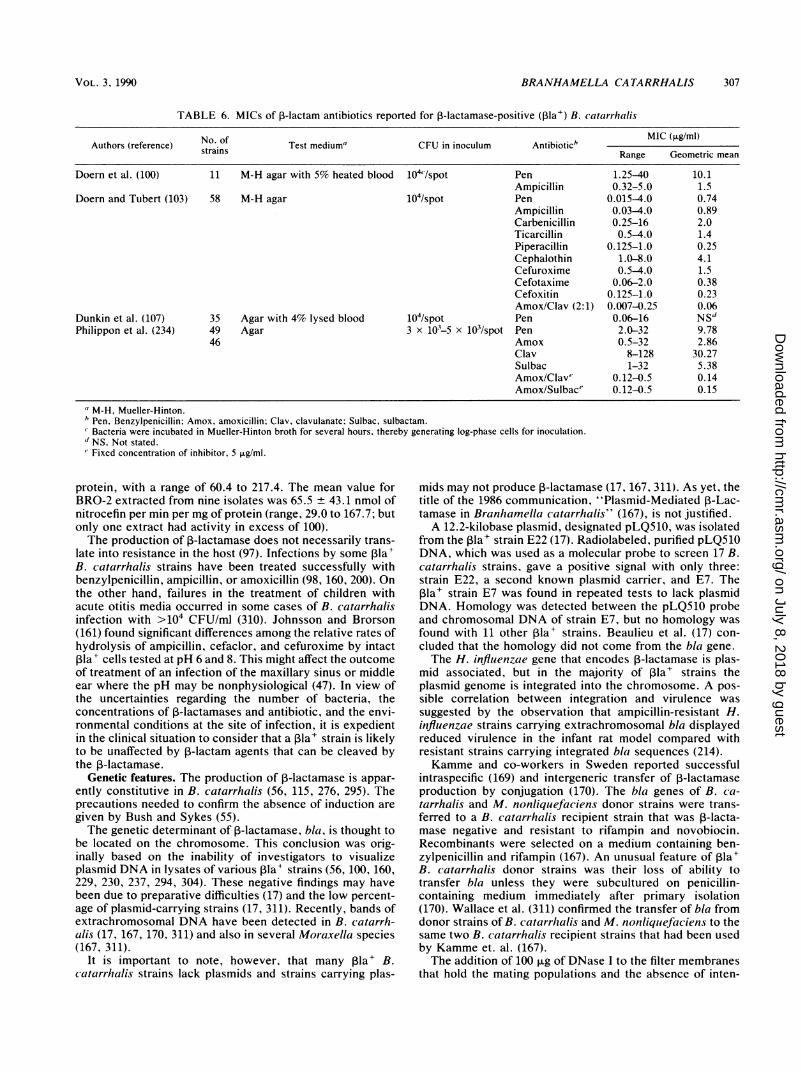
BRANHAMELLA CATARRHALIS 307
TABLE 6. MICs of J3-lactam antibiotics reported for P-lactamase-positive (Pla') B. catarrihalis
Authors (reference) No. of Test medium" CFU in inoculum Antibiotic' MIC (,ug/ml)strains Range Geometric mean
Doern et al. (100) 11 M-H agar with 5% heated blood 104'/spot Pen 1.25-40 10.1Ampicillin 0.32-5.0 1.5
Doern and Tubert (103) 58 M-H agar 104/spot Pen 0.015-4.0 0.74Ampicillin 0.03-4.0 0.89Carbenicillin 0.25-16 2.0Ticarcillin 0.5-4.0 1.4Piperacillin 0.125-1.0 0.25Cephalothin 1.0-8.0 4.1Cefuroxime 0.5-4.0 1.5Cefotaxime 0.06-2.0 0.38Cefoxitin 0.125-1.0 0.23Amox/Clav (2:1) 0.007-0.25 0.06
Dunkin et al. (107) 35 Agar with 4% lysed blood 104/spot Pen 0.06-16 NSdPhilippon et al. (234) 49 Agar 3 x 10³-5 x 10³/spot Pen 2.0-32 9.78
46 Amox 0.5-32 2.86Clav 8-128 30.27Sulbac 1-32 5.38Amox/Clav" 0.12-0.5 0.14Amox/Sulbace 0.12-0.5 0.15
'M-H, Mueller-Hinton."Pen, Benzylpenicillin; Amox, amoxicillin; Clav, clavulanate; Sulbac, sulbactam.Bacteria were incubated in Mueller-Hinton broth for several hours, thereby generating log-phase cells for inoculation.
d NS, Not stated." Fixed concentration of inhibitor, 5 ,L.g/ml.
protein, with a range of 60.4 to 217.4. The mean value forBRO-2 extracted from nine isolates was 65.5 + 43.1 nmol ofnitrocefin per min per mg of protein (range, 29.0 to 167.7; butonly one extract had activity in excess of 100).The production of 1-lactamase does not necessarily trans-
late into resistance in the host (97). Infections by some ,Bla+B. catarrhalis strains have been treated successfully withbenzylpenicillin, ampicillin, or amoxicillin (98, 160, 200). Onthe other hand, failures in the treatment of children withacute otitis media occurred in some cases of B. catarrhalisinfection with >104 CFU/ml (310). Johnsson and Brorson(161) found significant differences among the relative rates ofhydrolysis of ampicillin, cefaclor, and cefuroxime by intactIla+ cells tested at pH 6 and 8. This might affect the outcomeof treatment of an infection of the maxillary sinus or middleear where the pH may be nonphysiological (47). In view ofthe uncertainties regarding the number of bacteria, theconcentrations of 1-lactamases and antibiotic, and the envi-ronmental conditions at the site of infection, it is expedientin the clinical situation to consider that a rla+ strain is likelyto be unaffected by 3-lactam agents that can be cleaved bythe P-lactamase.
Genetic features. The production of P-lactamase is appar-ently constitutive in B. catarrhalis (56, 115, 276, 295). Theprecautions needed to confirm the absence of induction aregiven by Bush and Sykes (55).The genetic determinant of P-lactamase, bla, is thought to
be located on the chromosome. This conclusion was orig-inally based on the inability of investigators to visualizeplasmid DNA in lysates of various Pla' strains (56, 100, 160,229, 230, 237, 294, 304). These negative findings may havebeen due to preparative difficulties (17) and the low percent-age of plasmid-carrying strains (17, 311). Recently, bands ofextrachromosomal DNA have been detected in B. catarrh-alis (17, 167, 170, 311) and also in several Moraxella species(167, 311).
It is important to note, however, that many Pla+ B.catarrhalis strains lack plasmids and strains carrying plas-
mids may not produce 1-lactamase (17, 167. 311). As yet, thetitle of the 1986 communication, "Plasmid-Mediated 1-Lac-tamase in Branhamella catarrhalis" (167), is not justified.A 12.2-kilobase plasmid, designated pLQ510, was isolated
from the 3la+ strain E22 (17). Radiolabeled, purified pLQ510DNA, which was used as a molecular probe to screen 17 B.catarrhalis strains, gave a positive signal with only three:strain E22, a second known plasmid carrier, and E7. ThePla+ strain E7 was found in repeated tests to lack plasmidDNA. Homology was detected between the pLQ510 probeand chromosomal DNA of strain E7, but no homology wasfound with 11 other Ila+ strains. Beaulieu et al. (17) con-cluded that the homology did not come from the bla gene.The H. influenzae gene that encodes 1-lactamase is plas-
mid associated, but in the majority of I3la' strains theplasmid genome is integrated into the chromosome. A pos-sible correlation between integration and virulence wassuggested by the observation that ampicillin-resistant H.influenzae strains carrying extrachromosomal bla displayedreduced virulence in the infant rat model compared withresistant strains carrying integrated bla sequences (214).Kamme and co-workers in Sweden reported successful
intraspecific (169) and intergeneric transfer of ,B-lactamaseproduction by conjugation (170). The bla genes of B. ca-tarrhalis and M. nonliquefaciens donor strains were trans-ferred to a B. catarrhalis recipient strain that was ,3-lacta-mase negative and resistant to rifampin and novobiocin.Recombinants were selected on a medium containing ben-zylpenicillin and rifampin (167). An unusual feature of Pla+B. catarrhalis donor strains was their loss of ability totransfer bla unless they were subcultured on penicillin-containing medium immediately after primary isolation(170). Wallace et al. (311) confirmed the transfer of bla fromdonor strains of B. catarrhalis and M. nonliquefaciens to thesame two B. catarrhalis recipient strains that had been usedby Kamme et. al. (167).The addition of 100 ,ug of DNase I to the filter membranes
that hold the mating populations and the absence of inten-
VOL. 3 , 1990
on July 8, 2018 by guesthttp://cm
r.asm.org/
Dow
nloaded from

CLIN. MICROBIOL. REV.
tionally added DNA were considered sufficient to identifythe mechanism of transfer as conjugation. I suggest thealternative possibility, transformation. Transformation ofstreptomycin resistance is known to occur between B.catarrhalis and strains originally designated M. nonlique-faciens (33, 59) but more recently assigned to the new
species M. osloensis (15, 37).Three testable considerations support my argument that
the mechanism of the reported bla transfers could be trans-formation. (i) DNase activity may be lost due to proteolysisand diffusion from the filter membrane into the underlyingmedium. A fraction of the cells crowded on the membranemay remain competent for transformation which can occurbefore the end of the overnight incubation period allowed formating and phenotypic expression. (ii) DNA released fromcells spontaneously can elicit transformation. I (65) showedthat the cell-to-cell transfer of N. gonorrhoeae chromosomalgenes occurred during cocultivation of a competent recipientstrain with a donor strain that lacked a conjugative plasmid;DNA was not added experimentally, but DNase abolishedthis transfer. Dorward et al. (104) found that a vehicle forDNA transfer was naturally elaborated membrane vesicles(blebs). Such blebs, which are also produced by B. catarrh-alis during growth (143, 159, 217), may provide a naturalmechanism for transfer of transforming DNA between cells.(iii) The tendency of donor cells to transfer DNA may beenhanced by growth on penicillin-containing medium.1-Lactam antibiotics are known to disorganize the wallstructure of gram-negative bacteria, thereby increasing per-meability (191) and, possibly, the tendency for the formationof membrane blebs.
Is the worldwide increase of Pla' B. catarrhalis since thelate 1970s due to the dissemination of one highly communi-cable progenitor strain? Apparently it is not. Differencesbetween Pla+ strains were demonstrated by restriction en-
donuclease typing (229, 230). Patterson et al. (229) identified12 distinct HaeIII digestion patterns of chromosomal DNAamong a group of Pla' strains.How, then, did B. catarrhalis gain the ability to produce
P-lactamase when historically the species was penicillinsusceptible? Mutations of a preexisting, functional bla genecan produce minor changes of the enzyme product. How-ever, interbacterial transfer must be responsible for therecent, sudden appearance of an efficient P-lactamase. Thelarge increase in Pla' strains does not imply that largenumbers of bla gene transfers occurred. Continued multipli-cation of the recipients of occasional transfers, especially inthe presence of P-lactam agents, the genetic stability of blain its chromosomal location, and person-to-person spreadwithin the community and between regions by internationaltravel can account for the high prevalence of Pla+ strains.
Possible influence in mixed infections: indirect pathogenic-ity. In localized, mixed infections with a penicillin-suscepti-ble pathogen and a Ala+ commensal, the P-lactamase isthought to protect the pathogen by inactivating a penicillinused for therapy. This effect has been referred to as indirectpathogenicity (44, 85, 165, 205, 312). Wardle (312) foundsuggestive evidence of protection in a few patients withbronchopulmonary infections who showed no clinical or
radiological improvement during treatment with ampicillinor amoxicillin. Penicillin-susceptible strains of pneumococcior H. influenzae together with Ala+ B. catarrhalis were
identified in sputum specimens and 13-lactamase was de-tected, but the antibacterial activity expected of the penicil-lin was absent. Subsequently, favorable therapeutic re-
sponses were obtained with antibiotics to which each of theorganisms was susceptible.A mouse model was used by Renneberg and Walder (244)
to study the survival of a penicillin-susceptible pneumococ-cal strain mixed with either of two B. catarrhalis strains, oneAla+ and the other (control) P-lactamase negative. Threadsimpregnated with bacteria were implanted subcutaneouslyon the backs of mice, which were then treated with ampicil-lin by periodic injection into the hind leg. Threads wereremoved after 8 h and immersed in phosphate-bufferedsaline, which was immediately used for assays of CFU andampicillin concentrations. The P-lactamase produced duringthe mixed infection greatly reduced the antibacterial effect ofthe ampicillin.The probability of ampicillin failure in mixed infections
depends in part on the characteristics and the magnitude ofthe populations and the P-lactamase. B. catarrhalis 1-lac-tamases are membrane associated and released only inminimal amounts (100, 115, 116). Thus, many Pla' strainsmay not release enough P-lactamase to protect other bacte-ria in a localized site.
Characteristics of the P-LactamasesA recent classification placed the Ravasio and BRO-1
P-lactamases of B. catarrhalis in group 2c (54). Enzymes in2c hydrolyze carbenicillin at least 75% as fast as benzylpen-icillin and hydrolyze cloxacillin and many cephalosporins atrelatively low rates; the isoelectric point (pl) typically fallsbelow neutrality. Investigators have characterized P-lactam-ases of strains isolated from patients in England (231, 276),France (56, 234), Belgium (115), The Netherlands (294, 295),Sweden (111), the United States (220, 305, 311), and Japan(323). These enzymes are distinct from the plasmid genome-encoded TEM-1 P-lactamase found in N. gonorrhoeae, H.influenzae, and Escherichia coli (56, 111, 115, 234, 294, 311).A large proportion of the ,B-lactamase remains associated
with the cell debris obtained by ultrasonication ofB. catarrh-alis cells (111, 115). However, the presence of 0.5% TritonX-100 during ultrasonication results in >90% solubilizationof enzyme (116). This indicates that this 13-lactamase isstrongly membrane associated.
Different methods used for enzyme extraction yield dif-ferent amounts of 13-lactamase (275) and other cellular pro-teins. Crude extracts are known to contain substances thataffect enzyme activity either by interacting with the enzymeitself or by interfering with its reaction with substratecompounds (276). The use of crude extracts with weakenzyme activity in many of the studies may account in partfor the diversity of substrate profiles and pIs reported and forthe inference by some investigators that there are a numberof distinct types of B. catarrhalis P-lactamase, perhaps asmany as five (294) or six (295). On the other hand, with theexception of strain 939 described by Stobberingh et al. (294),Kamme et al. (167) found no evidence that B. catarrhalispossesses more than the one type of P-lactamase which theydesignated BRO-1. More recently, Wallace et al. (311)classified the Ravasio-type P-lactamase as BRO-1 and therelated 1908 type as BRO-2.
Isoelectric focusing separates proteins by aligning them assharp bands at their isoelectric points in a pH gradientproduced electrophoretically. The 13-lactamase bands aredetected with nitrocefin overlays (275). Multiple, "satellite"bands often appear which are believed to represent biologi-cally active molecules with altered net charges resultingfrom the loss and/or modification of specific amino acid
308 CATLIN
on July 8, 2018 by guesthttp://cm
r.asm.org/
Dow
nloaded from

BRANHAMELLA CATARRHALIS 309
TABLE 7. P-Lactam antibiotics and the P-lactamase ofB. catarrhalis NNBR-8303a
Substrate Vmaxb Km (>LM) MIC (,Lg/ml)c
Benzylpenicillin 100 75 100Ampicillin 66 11 50Carbenicillin 77 3.7 50Ticarcillin 51 26 25Piperacillin 85 29 100Cloxacillin 6 2.2 50Cephaloridine 4 1.5 12.5Cephalothin 8 2.9 25Cefuroxime 18 155 0.78Cefotaxime 8 147 0.20Cefoxitin 1 7.4 0.20
a The P-lactamase was purified by column chromatography on DEAE-Sepharose. (Data taken from Yokota et al. [323] with permission of thepublisher.)
b Maximum rate of hydrolysis of P-lactam substrate relative to that ofbenzylpenicillin, which is assigned a value of 100.
c Determined in tests of one loopful (approximately 5 ,ul) of an overnightbroth culture on sensitivity disk agar-N containing twofold dilutions ofantibiotic; inoculum estimated as 108 CFU/ml.
residues, possibly by enzymatic action (276). The 1-lacta-mase in a crude extract (parent enzyme) of B. catarrhalis2001E focused as four major bands, A, B, C, and D, with pIvalues of 7.2, 6.4, 6.0, and 5.6, respectively (276). Theactivity of band A was not sufficient to permit furtheranalyses, but B, C, and D were isolated and found to havesimilar substrate profiles. These differed somewhat, how-ever, from the profile displayed by the less purified parentenzyme. Gel filtration of the parent enzyme gave a singlepeak of activity, corresponding to a molecular weight of41,000. Molecular weights estimated for bands B, C, and Dwere 41,000, 41,000, and 36,500, respectively.
Kinetic constants of the P-lactamase purified from strainNNBR-8303 are given in Table 7. The data also show thecorrelation between the antibacterial activity (MIC) of thevarious P-lactam substrates and their resistance to cleavageby the P-lactamase. Not shown are diffusibility differencesbetween the antibiotics which may also affect the MIC (222).The enzyme efficiently inactivates ampicillin as shown bythe high rate of hydrolysis (Vmax of 66) and the fairly highaffinity for this substrate (Km of 11 ,uM). With cefotaxime assubstrate, on the other hand, both the maximum rate ofhydrolysis (Vmax of 8) and the substrate affinity (Km of 147,uM) are low, indicating the relative inability of the enzymeto attack cefotaxime. This is consistent with the low MIC ofcefotaxime (0.2 ,ug/ml) compared with that of ampicillin (50,ug/ml). This purified P-lactamase had a pI of 5.4 and anapproximate molecular weight of 33,000 (323).M. nonliquefaciens strains isolated in Sweden (111, 170)
and the United States (311) also produce P-lactamase, a traitthat appears to have been acquired in the recent past. The,-lactamases from M. nonliquefaciens and B. catarrhalisresemble each other (111). Wallace et al. (311) identifiedisoelectric focusing patterns characteristic of BRO-1 andBRO-2 in both organisms and found that relatively similarsubstrate profiles were displayed by the ,B-lactamases fromboth organisms. Benzylpenicillin, ampicillin, and methicillinwere hydrolyzed at approximately equal rates. The hydrol-ysis rates for cefaclor and nitrocefin were higher and that forcephaloridine was much lower than that for benzylpenicillin.
Clavulanic acid and sulbactam are examples of active-site-directed, irreversible inhibitors called suicide inhibitors (55).Low concentrations of these agents inactivate B. catarrhalis
P-lactamases (115, 234, 294, 323), including both BRO-1 andBRO-2 (116). Since suicide inhibitors protect amoxicillinfrom the hydrolytic action of P-lactamase, the MIC ofamoxicillin plus clavulanate is lower than the MIC of eitheragent alone (Table 6).The important work of sequencing the gene encoding the
BRO-1 P-lactamase is under way (D. Beaulieu, L. Piche, andP. H. Roy, Progr. Abstr. 29th Intersci. Conf. Antimicrob.Agents Chemother., abstr. no. 1120, 1989). Comparisons ofthis sequence with those of genes encoding BRO-2 and otherpossibly different P-lactamases will show the extent of theirdivergence.
STRUCTURE AND COMPOSITION OF B. CATARRHALIS
Capsule and Pili
B. catarrhalis, like N. gonorrhoeae (227), produces cap-sules visible by electron microscopy as a fibrillar coat. Thefuzzy appearance is seen in an ultrathin section of B.catarrhalis stained with magnesium uranyl acetate and leadcitrate (illustrated in plate 10.2, Fig. 1, of the 1974 edition ofBergey's Manual [245]). Hellio et al. (143) used rutheniumred staining to show the polysaccharide composition of thisfibrillar coat, which is seen here (Fig. ic and d) as spicule-like material (sp) projecting from the outer membrane. Theaggregated (autoagglutinated) cells appear to be held by theirspicules.
Pili, also called fimbriae, are filamentous appendages thatextend from the outer membrane and serve as adhesins (16).Pili were not observed in electron micrographs of B. catarrh-alis ATCC 25238T (143), ATCC 8176 (318), or ATCC 8193(318). Preliminary observations of four clinical isolates re-vealed piliation (215). Wistreich and Baker (318) reported in1971 that a strain identified as "N. catarrhalis" was piliatedand agglutinated erythrocytes from rabbit and mouse, butnot human (0, Rho+), sources. Scanning electron micros-copy revealed adhesion of these cells and their pili toagglutinated mouse erythrocytes. Soto-Hernandez et al.(282) observed 4+ hemagglutination of human group 0 Rh+erythrocytes by 16 B. catarrhalis clinical isolates. Furtherwork is needed to determine whether this disagreementconcerning erythrocytes is due to structural differences ofpili or of human erythrocyte receptors.
Hemagglutination was not observed with strain ATCC25238 (282) as expected from its lack of pili (143). A featureof strain ATCC 25238 (earlier designated Ne 11) which isvaluable for genetic studies is its high frequency of intraspe-cific transformation (33, 68). The absence of piliation isinteresting in view of the association between piliation(indicated by distinctive colony types [39, 64]) and trans-formability of N. gonorrhoeae (65, 181) and M. nonlique-faciens (39).
EnvelopeThe envelope or cell wall (Fig. la and b) resembles that of
N. gonorrhoeae (227). Two trilaminar structures (cm andom) can be seen between which lie a periplasmic space andthe dense peptidoglycan-containing layer (pg). The cytoplas-mic membrane (cm), which is the innermost layer, carriespenicillin-binding proteins (PBPs) at its outer face (286).PBPs 1, 2, and 3 of B. catarrhalis have molecular weights of77,000, 68,000, and 39,000, respectively (128). PBP 3, like E.coli PBP 5/6, possesses a weak ,-lactamase activity.
Proteins, LOS, and phospholipids are found in the outer
VOL. 3, 1990
on July 8, 2018 by guesthttp://cm
r.asm.org/
Dow
nloaded from

CLIN. MICROBIOL. REV.
bbV
A....*1at-om
..'It
.6
Lcm a,.B. .q
-pg
a
d iAV
FIG. 1. Ultrastructure of B. catarrhalis ATCC 25238T seen in thin sections. (a and b) Fixation with OSO4; (c and d) ruthenium red staining.Abbreviations: bl, blebs; om, outer membrane; cm, cytoplasmic membrane; pg. peptidoglycan-containing layer; sp, spicule-like material.Bars, 0.2 ptm. (Reprinted from Annales de l'Institut PasteurlMicrobiology [143] with permission of the publisher.)
membrane. It should be noted that LOS is a variety oflipopolysaccharide that lacks the long, surface-exposedpolysaccharide constituting the 0 antigen of smooth entericbacteria (133, 159, 303). The absence of 0 antigen increasesouter membrane permeability, resulting in enhanced suscep-tibility to erythromycin, rifampin, and other hydrophobicagents (222, 223).
Outer membrane vesicles or blebs are released spontane-ously during growth (159, 217, 264). Their membranousappearance is evident in Fig. 1 (bl) and in the electronmicrograph of outer membrane vesicles that Murphy andLoeb (217) obtained from the supernatant of a B. catarrhalisbroth culture. Such preparations contain LOS and have aprotein-binding pattern similar to that of outer membranesprepared by other methods (159, 217, 264).
N. gonorrhoeae blebs are highly toxic for embryonatedeggs (227). In the early literature, blebs were referred to asfree endotoxin (227, 264) and, indeed, they function as apackaging system for the distribution of endotoxin. Thetoxicity is due to the biological activity of lipid A, theglycolipid component of LOS. Lipid A is anchored in theouter membrane and is covalently bound to core oligosac-charides (133). The compositions of the B. catarrhalis "li-popolysaccharide" and its lipid A and core oligosaccharideswere reported (1, 159).
Eight major OMPs (OMP-A to OMP-H) whose approxi-mate molecular weights ranged between 98,000 and 21,000were identified in B. catarrhalis (14, 216, 217). OMP-F has amolecular weight of 43,000 that is not altered by 100'C
310 CATLIN
on July 8, 2018 by guesthttp://cm
r.asm.org/
Dow
nloaded from

BRANHAMELLA CATARRHALIS 311
TABLE 8. DNA base compositions
G+C content (mol%) determined by:
Species Chromatography Buoyant density in CsCl Tm (thermal denaturation midpoint)(68,69)a (38) 202b 148 261 258
B. catarrhalisStrain ATCC 25238Tc 40.8d 41.0 41 41.3 41.0Other strained 40.0-43.0 41.0-42.0 41.6
N. cinerea 49.0 50.9 50.5N. flavescens 50.2d 46.5-47.5 49 49.6 50.1N. gonorrhoeae 53.3 53.2N. meningitidis 51.3d 51.5 52.0 52.8 53.2N. caviae 44.5 47.3 46.9
a Numbers in parentheses are reference numbers.b Reference number.c Type strain of the species, originally designated Ne 11.d G+C content was derived from the A+T/G+C ratio.eExcludes "N. catarrhalis" NCTC 4103, which deviates enough to suggest that the species designation may be incorrect (15, 33, 59, 69).
treatment, in contrast to OMP-C, -D, -E, -G, and -H whichare heat modifiable (217).
Porins are proteins that typically exist as trimers formingchannels through the outer membrane for passive diffusionof small hydrophilic nutrients and antibiotics (223). OMP 2 ofN. meningitidis is a porin characterized by a molecularweight of 40,000 to 42,000 (124). In view of the relationshipbetween response to hydrophilic antibiotics and porin func-tion, it will be of interest to investigate the B. catarrhalisporin(s). I speculate that OMP-F may be a porn.The fatty acid profiles of members of the family Neisser-
iaceae have been determined in various laboratories (45,147, 157, 187, 190, 298), but caution is needed in comparingthe results. The fatty acid content is greatly affected by theculture medium components and incubation temperature(157, 187). For B. catarrhalis cells grown on supplementedGC medium base agar, 20% of the total fatty acids wererecorded as 17:1 acid (where the number left of the colonindicates the number of carbon atoms and that on the right isthe number of double bonds). However, 17:1 acid was notdetected in cells grown on Trypticase soy agar (187). Variousauthors (147, 157, 187, 298) agree that the 16:0, 16:1, and18: 1 acids are abundant in B. catarrhalis and several speciesfrom animals, including N. caviae (this species is describedin Table 4, footnote c). Nevertheless, the fatty acid profilesdiffer. By determining the double-bond positions (A) in 18:1acids, Sugimoto et al. (298) distinguished B. catarrhalis (All18:1, 64% of total fatty acids) from N. caviae (A9 18:1,42%).
DNA
The guanine-plus-cytosine (G+C) contents of DNA prep-arations from B. catarrhalis and possibly confusing Neis-seria species are given in Table 8. The findings for a givenspecies were in agreement, except for several values (espe-cially for N. flavescens) determined by the CsCl gradientcentrifugation method. Also, divergent G+C values deter-mined by a chromatographic method were reported (186) for11 strains of N. caviae (range, 47.7 to 50.4 mol%; notincluded in Table 8).
Differences between the G+C contents of B. catarrhalisand N. meningitidis DNA, for example, indicate base se-quence differences sufficient to explain the absence of DNA-mediated transformation of streptomycin resistance betweentransformable strains of these species (68), their low degree
of DNA relatedness shown by DNA-DNA hybridization(174), and their lack of stable DNA duplexes (176). The "N.catarrhalis" genome contains approximately 2.3 x 10⁶ nu-cleotide pairs (175). They must encode several thousandgene products. In view of the DNA differences, it is notsurprising that a two-dimensional polyacrylamide gel elec-trophoresis system which separated 200 polypeptides fromcellular extracts revealed only 9% similarity between thelocations of polypeptide spots on electrophoretograms of aB. catarrhalis strain and a reference N. gonorrhoeae strain(152).
WHAT'S IN A NAME?
A scientific bacterial name should be unambiguous, rec-ognizable in all languages, and stable (42, 280). "One of theannoying problems facing the practicing physician is theconstant reclassification and changing names of microorgan-isms" (324). Although aware of the inconvenience of thename change, investigators working before 1970 recognizedthat substantial differences between "N. catarrhalis" andthe "true neisseriae" mandated their separation into dif-ferent genera (1, 15, 33, 68, 146, 174, 190). This separationwas validated by subsequent studies (147, 152, 159, 261,288).
Ideally, a genus is composed of species that are relativelysimilar phenotypically and show 50 to 65% DNA relatednessdetermined under conditions optimal for DNA reassociation(42). The "true neisseriae" associated with human hostsinclude N. gonorrhoeae, N. meningitidis, and the less patho-genic N. flavescens, N. cinerea, N. sicca, N. subflava, N.mucosa, and N. polysaccharea (177, 258, 307). Their levelsof DNA relatedness are appropriate for membership in asingle genus. On the other hand, relations between theseorganisms and B. catarrhalis are minimal or absent asdetermined by (i) genetic transformation of streptomycinresistance (33, 68, 148); (ii) DNA base composition (Table 8);(iii) DNA homologies determined by several techniques(136, 148, 174, 176); and (iv) rRNA binding to DNA (261).
In spite of the widespread agreement that it is inappropri-ate to include "N. catarrhalis" in the genus Neisseria, thename of the genus to which the species should be transferredis controversial. Two names have been proposed: Bra-nhamella catarrhalis (Catlin in 1970 [60]) and Moraxella(Branhamella) catarrhalis (B0vre in 1979 [35]). The latter isan emended version of the name Moraxella catarrhalis
VOL. 3, 1990
on July 8, 2018 by guesthttp://cm
r.asm.org/
Dow
nloaded from
CLIN. MICROBIOL. REV.
which was suggested in 1968 (146) in consideration of thegenetic relatedness demonstrated independently by B0vre(32, 34) and by Catlin (59, 70). The extensive work of theOslo group and their rationale for including bacterial rodsand cocci in the genus Moraxella are summarized in tworeviews (39, 145). B0vre found it expedient to divide thegenus Moraxella into the subgenus Branhamella (to containcoccal organisms) and the subgenus Moraxella (to containrod-shaped organisms). This is an awkward arrangement,however, since subgenus names are now rarely used (280),Moraxella (Branhamella) catarrhalis is unnecessarily long,and deletion of the subgenus name leaves the possiblyconfusing M. catarrhalis. The name Branhamella catarrha-lis served to distinguish the organism from the so-callednonpathogenic neisseriae and focused attention on theirdiffering potentials for pathogenicity. The proposed associ-ation with moraxellae, most of which possess only limitedpathogenicity (39, 145), would relegate B. catarrhalis to anunfamiliar conglomerate of cocci and rods.The differences revealed by DNA-rRNA hybridization are
so great that Rossau et al. (261) recommended the removal ofBranhamella and Moraxella from the family Neisseriaceaein which they were classified (36, 39, 146). The genusNeisseria is now placed in the beta group of the new classProteobacteria (a taxonomic category higher than the fami-ly), whereas Branhamella and Moraxella are in the gammagroup (288). The genetic relatedness between Branhamellaand Moraxella is genuine, but it can be acknowledgedadequately, I believe, by assigning these genera to a newfamily, Branhamaceae.
CONCLUSIONS AND PROSPECTS
B. catarrhalis has finally gained respect as a pathogenthanks to the many recent reports of its causal role in avariety of infections. Much has been learned about itsstructural, biochemical, and differential characteristics, re-sponses to antibacterial agents, and 1-lactamases. Futurestudies must use strains precisely identified by standardmethods. Research is needed to enhance the molecular andgenetic respectability of the organism. Initial strategies mayuse transformation to develop isogenic strains carryingauxotrophic and antibacterial resistance markers. The pos-sible cell-to-cell transmission of chromosomal loci mediatedby DNA associated with blebs should be investigated.Knowledge of the genes and their organization in B. catarrh-alis, N. caviae, and M. nonliquefaciens is needed. This willprovide a sounder basis for judgment concerning the relat-edness and evolution of these organisms, permit compari-sons with the genetic markers of N. gonorrhoeae, and aidthe identification of gene products.A multifactoral problem centers around the genetic loci
encoding bla, their origin, mode of transfer, features ofchromosomal integration, and phenotypic effects, in additionto P-lactamase production, possibly associated with theiracquisition. The rapid, worldwide increase in the percentageof IBla' strains is remarkable. In some regions this has beenaccompanied by rising numbers of B. catarrhalis infections.The increasing incidence of bronchopulmonary infectionsmay be due in part to the greater use of immunosuppressiveagents. However, this could not account for the documentedincreases of childhood otitis media caused by B. catarrhalis.Possibly the success of ,Bla+ strains is associated with animproved mechanism of adherence to mucous membranes,especially of children, or some other virulence-associatedproperty. Further investigations of this problem as well as
the virulence of case and carrier strains and the molecularbasis of pathogenesis await the development of organ cul-tures or animal models. The recent logarithmic increase ofpublications illustrates the increased awareness of B. ca-tarrhalis and growth of interest in the multitude of significantquestions now apparent.
ACKNOWLEDGMENTS
Information and preprints received from D. Beaulieu, G. V.Doern, T. F. Murphy, and R. J. Wallace, Jr., are gratefully ac-knowledged. I especially thank Jean-Yves Riou for providing meoriginal photographs for Fig. 1.
LITERATURE CITED
1. Adams, G. A., T. G. Tornabene, and M. Yaguchi. 1969. Cellwall lipopolysaccharides from Neisseria catarrhalis. Can. J.Microbiol. 15:365-374.
2. Ahmad, F., M. A. Calder, M. J. Croughan, and T. G. Marshall.1985. Urinary tract infection caused by Branhamella catarrh-alis. J. Infect. 10:176-177.
3. Ahmad, F., D. T. McLeod, M. J. Croughan, and M. A. Calder.1984. Antimicrobial susceptibility of Branhamella catarrhalisisolates from bronchopulmonary infections. Antimicrob.Agents Chemother. 26:424-425.
4. Ahmad, F., H. Young, D. T. McLeod, M. J. Croughan, andM. A. Calder. 1987. Characterisation of Branhamella catarrh-alis and differentiation from Neisseria species in a diagnosticlaboratory. J. Clin. Pathol. 40:1369-1373.
5. Aitken, J. M., and P. E. Thornley. 1983. Isolation of Bra-nhamella catarrhalis from sputum and tracheal aspirate. J.Clin. Microbiol. 18:1262-1263.
6. Alligood, G. A., and J. F. Kenny. 1989. Tracheitis and supra-glottis associated with Branhamella catarrhalis and respiratorysyncytial virus. Pediatr. Infect. Dis. J. 8:190-191.
7. Alvarez, S., M. Jones, S. Holtsclaw-Berk, J. Guarderas, andS. L. Berk. 1985. In vitro susceptibilities and P-lactamaseproduction of 53 clinical isolates of Branhamella catarrhalis.Antimicrob. Agents Chemother. 27:646-647.
8. Arko, R. J., and T. Odugbemi. 1984. Superoxol and amylaseinhibition tests for distinguishing gonococcal and nongonococ-cal cultures growing on selective media. J. Clin. Microbiol.20:1-4.
9. Arkwright, J. A. 1907. On the occurance of the Micrococcuscatarrhalis in normal and catarrhal noses and its differentiationfrom other Gram-negative cocci. J. Hyg. 7:145-154.
10. Averett, D. R., B. Roth, J. J. Burchall, and D. P. Baccanari.1979. Dihydrofolate reductase from Neisseria sp. Antimicrob.Agents Chemother. 15:428-435.
11. Bannatyne, R. M., and V. Kolodej. 1985. Branhamella catarrh-alis bacteremia and immunosuppression-part of a larger prob-lem? Diagn. Microbiol. Infect. Dis. 3:65-67.
12. Barber, M., and P. M. Waterworth. 1962. Antibacterial activityof the penicillins. Br. Med. J. i:1159-1164.
13. Baron, J., and E. D. Shapiro. 1985. Unsuspected bacteremiacaused by Branhamella catarrhalis. Pediatr. Infect. Dis. J.4:100-101.
14. Bartos, L. C., and T. F. Murphy. 1988. Comparison of theouter membrane proteins of 50 strains of Branhamella catarrh-alis. J. Infect. Dis. 158:761-765.
15. Baumann, P., M. Doudoroff, and R. Y. Stanier. 1968. Study ofthe Moraxella group. I. Genus Moraxella and the Neisseriacatarrhalis group. J. Bacteriol. 95:58-73.
16. Beachey, E. H. 1981. Bacterial adherence: adhesin-receptorinteractions mediating the attachment of bacteria to mucosalsurfaces. J. Infect. Dis. 143:325-345.
17. Beaulieu, D., M. Quellette, M. G. Bergeron, and P. H. Roy.1988. Characterization of a plasmid isolated from Branhamella
312 CATLIN
on July 8, 2018 by guesthttp://cm
r.asm.org/
Dow
nloaded from

BRANHAMELLA CATARRHALIS 313
catarrhalis and detection of plasmid sequences within thegenome of a Branhamella catarrhalis strain. Plasmid 20:158-162.
18. Behal, F. J., and J. D. Folds. 1967. Arylamidase of Neisseriacatarrhalis. Arch. Biochem. Biophys. 121:364-371.
19. Berg, R. A., and D. L. Bartley. 1987. Pneumonia associatedwith Branhamella catarrhalis in infants. Pediatr. Infect. Dis. J.6:569-573.
20. Berger, U. 1961. Polysaccharidbildung durch saprophytischeNeisserien. Zentralbl. Bakteriol. Parasitenkd. Infektionskr.Hyg. Abt. 1 Orig. 181:345-349.
21. Berger, U. 1961. Ein Elektivnahrboden fur Neisseria catarrh-alis. Zentralbl. Bakteriol. Parasitenkd. Infektionkr. Hyg. Abt.1 Orig. 183:135-138.
22. Berger, U. 1961. Reduktion von Nitrat und Nitrit durch Neis-seria. Z. Hyg. Infektionskr. 148:45-50.
23. Berger, U. 1963. Die anspruchslosen Neisserien. Ergeb. Mi-krobiol. Immunitaetsforsch. Exp. Ther. 36:97-167.
24. Berger, U. 1984. Serology of non-gonococcal, non-meningo-coccal Neisseria and Branhamella species. Methods Micro-biol. 16:225-245.
25. Berger, U., and E. Paepcke. 1962. Untersuchungen uber dieasaccharolytischen Neisserien des menschlichen Nasophar-ynx. Z. Hyg. Infektionskr. 148:269-281.
26. Bergey, D. H., F. C. Harrison, R. S. Breed, B. W. Hammer,and F. M. Huntoon. 1923. Bergey's manual of determinativebacteriology, p. 43. The Williams & Wilkins Co., Baltimore.
27. Bezjak, V., H. N. Shah, and J. Barley. 1988. Profiles ofamino-acid utilisation and production amongst strains of Bra-nhamella catarrhalis. J. Med. Microbiol. 26:143-146.
28. Black, A. J., and T. S. Wilson. 1988. Immunoglobulin G (IgG)serological response to Branhamella catarrhalis in patientswith acute bronchopulmonary infections. J. Clin. Pathol. 41:329-333.
29. Blackwell, C., H. Young, and S. S. R. Bain. 1978. Isolation ofNeisseria meningitidis and Neisseria catarrhalis from thegenitourinary tract and anal canal. Br. J. Vener. Dis. 54:41-44.
30. Bluestone, C. D. 1986. Otitis media and sinusitis in children:role of Branhamella catarrhalis. Drugs 31(Suppl. 3):132-141.
31. Bonadio, W. A. 1988. Branhamella catarrhalis bacteremia inchildren. Pediatr. Infect. Dis. J. 7:738-739.
32. B0vre, K. 1963. Affinities between Moraxella spp. and a strainof Neisseria catarrhalis as expressed by transformation. Briefreport. Acta Pathol. Microbiol. Scand. 58:528.
33. B0vre, K. 1965. Studies on transformation in Moraxella andorganisms assumed to be related to Moraxella. 4. Streptomy-cin resistance transformation between asaccharolytic Neis-seria strains. Acta Pathol. Microbiol. Scand. 64:229-242.
34. B0vre, K. 1967. Transformation and DNA base composition intaxonomy, with special reference to recent studies in Morax-ella and Neisseria. Acta Pathol. Microbiol. Scand. 69:123-144.
35. B0vre, K. 1979. Proposal to divide the genus Moraxella Lwoff1939 emend. Henriksen and B0vre 1968 into two subgenera,subgenus Moraxella (Lwoff 1939) B0vre 1979 and subgenusBranhamella (Catlin 1970) B0vre 1979. Int. J. Syst. Bacteriol.29:403-406.
36. B0vre, K. 1984. Family VIII. Neisseriaceae Prevot 1933, p.288-290. In N. R. Krieg and J. G. Holt (ed.), Bergey's manualof systematic bacteriology, vol. 1. The Williams & WilkinsCo., Baltimore.
37. B0vre, K. 1984. Genus II. Moraxella Lwoff 1939, emend.Henriksen and B0vre 1968, p. 296-303. In N. R. Krieg andJ. G. Holt (ed.), Bergey's manual of systematic bacteriology,vol. 1. The Williams & Wilkins Co., Baltimore.
38. B0vre, K., M. Fiandt, and W. Szybalski. 1969. DNA basecomposition of Neisseria, Moraxella, and Acinetobacter, asdetermined by measurement of buoyant density in CsCl gradi-ents. Can. J. Microbiol. 15:335-338.
39. B0vre, K., and N. Hagen. 1981. The family Neisseriaceae:rod-shaped species of the genera Moraxella, Acinetobacter,Kingella, and Neisseria, and the Branhamella group of cocci,p. 1506-1529. In M. P. Starr, H. Stolp, H. G. Truper, A.Balows, and H. G. Schlegel (ed.), The prokaryotes, vol. 2.
Springer-Verlag, Berlin.40. Boyce, J. M., and E. B. Mitchell, Jr. 1985. Difficulties in
differentiating Neisseria cinerea from Neisseria gonorrhoeaein rapid systems used for identifying pathogenic Neisseriaspecies. J. Clin. Microbiol. 22:731-734.
41. Branham, S. E., and M. J. Pelczar. 1957. Family VIII. Neisser-iaceae Prdvot 1933, p. 480-485. In R. S. Breed, E. G. D.Murray, and N. R. Smith (ed.), Bergey's manual of determi-native bacteriology, 7th ed. The Williams & Wilkins Co.,Baltimore.
42. Brenner, D. J. 1985. Taxonomy, classification, and nomencla-ture of bacteria, p. 1-7. In E. H. Lennette, A. Balows, W. J.Hausler, Jr., and H. J. Shadomy (ed.), Manual of clinicalmicrobiology, 4th ed. American Society for Microbiology,Washington, D.C.
43. Brook, I. 1981. Microbiological studies of the bacterial flora ofthe external auditory canal in children. Acta Oto Laryngol.91:285-287.
44. Brook, I. 1986. Direct and indirect pathogenicity of Bra-nhamella catarrhalis. Drugs 31(Suppl. 3):97-102.
45. Brooks, J. B., D. S. Kellogg, L. Thacker, and E. M. Turner.1971. Analysis by gas chromatography of fatty acids found inwhole cultural extracts of Neisseria species. Can. J. Microbiol.17:531-543.
46. Brorson, J.-E., A. Axelsson, and S. E. Holm. 1976. Studies onBranhamella catarrhalis (Neisseria catarrhalis) with specialreference to maxillary sinusitis. Scand. J. Infect. Dis. 8:151-155.
47. Brorson, J.-E., and P. Larsson. 1985. pH and incubationatmosphere influence erythromycin activity against Bra-nhamella catarrhalis. J. Antimicrob. Chemother. 15:644-645.
48. Brorson, J.-E., and B.-E. Malmvall. 1981. Branharnella ca-tarrhalis and other bacteria in the nasopharynx of children withlongstanding cough. Scand. J. Infect. Dis. 13:111-113.
49. Brown, B. A., R. J. Wallace, Jr., C. W. Flanagan, R. W.Wilson, J. I. Luman, and S. D. Redditt. 1989. Tetracycline anderythromycin resistance among clinical isolates of Bra-nhamnella catarrhalis. Antimicrob. Agents Chemother. 33:1631-1633.
50. Bryant, J. 1983. Fatal sepsis due to Branhamella catarrhalisand Haernophilus influenzae in a child with selective IgGdeficiency. Arch. Pathol. Lab. Med. 107:442.
51. Buckley, C. E., III, and F. C. Dorsey. 1970. The effect of agingon human serum immunoglobulin concentrations. J. Immunol.105:964-972.
52. Burnett, A. K., D. J. Easton, A. M. Gordon, and R. M. Rowan.1975. Neisseria catarrhalis septicaemia in acute lymphoblasticleukaemia. Scot. Med. J. 20:37-38.
53. Busch, D. F., D. M. Citron, and D. A. Casciato. 1979. Falseidentification of Neisseria gonorrhoeae in lower respiratorytract secretions-result of mixed culture. Curr. Microbiol.2:299-300.
54. Bush, K. 1989. Classification of P-lactamases: groups 2c, 2d,2e, 3, and 4. Antimicrob. Agents Chemother. 33:271-276.
55. Bush, K., and R. B. Sykes. 1986. Methodology for the study ofP-lactamases. Antimicrob. Agents Chemother. 30:6-10.
56. Buu Hoi-Dang Van, A., C. Brive-Le Bouguenec, M. Barthelemy,and R. Labia. 1978. Novel P-lactamase from Branhamellacatarrhalis. Ann. Microbiol. (Paris) 129B:397-406.
57. Calder, M. A., M. J. Croughan, D. T. McLeod, and F. Ahmad.1986. The incidence and antibiotic susceptibility of Bra-nhamella catarrhalis in respiratory infections. Drugs 31(Suppl.3):11-16.
58. Capewell, S., D. T. McLeod, M. J. Croughan, F. Ahmad, M. A.Calder, and A. Seaton. 1988. Pneumonia due to Branhamellacatarrhalis. Thorax 43:929-930.
59. Catlin, B. W. 1964. Reciprocal genetic transformation betweenNeisseria catarrhalis and Moraxella nonliquefaciens. J. Gen.Microbiol. 37:369-379.
60. Catlin, B. W. 1970. Transfer of the organism named Neisseriacatarrhalis to Branhamella gen. nov. Int. J. Syst. Bacteriol.20:155-159.
61. Catlin, B. W. 1970. Neisseria ineningitidis (meningococcus), p.
VOL. 3, 1990
on July 8, 2018 by guesthttp://cm
r.asm.org/
Dow
nloaded from

CLIN. MICROBIOL. REV.
76-81. In J. E. Blair, E. H. Lennette, and J. P. Truant (ed.),Manual of clinical microbiology. American Society for Micro-biology, Washington, D.C.
62. Catlin, B. W. 1974. Neisseria meningitidis (meningococcus), p.116-123. In E. H. Lennette, E. H. Spalding, and J. P. Truant(ed.), Manual of clinical microbiology, 2nd ed. AmericanSociety for Microbiology, Washington, D.C.
63. Catlin, B. W. 1975. Cellular elongation under the influence ofantibacterial agents: way to differentiate coccobacilli fromcocci. J. Clin. Microbiol. 1:102-105.
64. Catlin, B. W. 1978. Characteristics and auxotyping of Neis-seria gonorrhoeae, p. 345-380. In T. Bergan and J. R. Norris(ed.), Methods in microbiology, vol. 10. Academic Press, Inc.(London), Ltd., London.
65. Catlin, B. W. 1982. Cell-to-cell transmission of chromosomalloci in Neisseria gonorrhoeae, p. 311-321. In U. N. Streips,S. H. Goodgal, W. R. Guild, and G. A. Wilson (ed.), Geneticexchange. Marcel Dekker, Inc., New York.
66. Catlin, B. W. 1984. Probenecid: antibacterial action againstNeisseria gonorrhoeae and interaction with benzylpenicillin.Antimicrob. Agents Chemother. 25:676-682.
67. Catlin, B. W. 1989. Genetic basis of the association of sulpho-namide resistance with methionine auxotrophy in Neisseriagonorrhoeae. J. Gen. Microbiol. 135:1101-1111.
68. Catlin, B. W., and L. S. Cunningham. 1961. Transformingactivities and base contents of deoxyribonucleate preparationsfrom various neisseriae. J. Gen. Microbiol. 26:303-312.
69. Catlin, B. W., and L. S. Cunningham. 1964. Genetic transfor-mation of Neisseria catarrhalis by deoxyribonucleate prepara-tions having different average base compositions. J. Gen.Microbiol. 37:341-352.
70. Catlin, B. W., and L. S. Cunningham. 1964. Transformingactivities and base composition of deoxyribonucleates fromstrains of Moraxella and Mima. J. Gen. Microbiol. 37:353-367.
71. Chapman, A. J., Jr., D. M. Musher, S. Jonsson, J. E. Clar-ridge, and R. J. Wallace, Jr. 1985. Development of bactericidalantibody during Branhamella catarrhalis infection. J. Infect.Dis. 151:878-882.
72. Christensen, J. J., and B. Bruun. 1985. Bacteremia caused bya beta-lactamase producing strain of Branhamella catarrhalis.Acta Pathol. Microbiol. Immunol. Scand. Sect B 93:273-275.
73. Christensen, J. J., 0. Gadeberg, and B. Bruun. 1986. Bra-nhamella catarrhalis: significance in pulmonary infections andbacteriological features. Acta Pathol. Microbiol. Immunol.Scand. Sect. B 94:89-95.
74. Chun, P. K., G. F. Sensabaugh, and N. A. Vedros. 1985.Genetic relationships among Neisseria species assessed bycomparative enzyme electrophoresis. J. Gen. Microbiol. 131:3105-3115.
75. Cimolai, N., and R. J. Adderley. 1989. Branhamella catarrhalisbacteremia in children. Acta Pediatr. Scand. 78:465-468.
76. Clarke, R. M., and R. B. Haining. 1936. Neisseria catarrhalisendocarditis. Ann. Intern. Med. 10:117-121.
77. Cocchi, P., and A. Ulivelli. 1968. Meningitis caused by Neis-seria catarrhalis. Acta Pediatr. Scand. 57:451-453.
78. Coffey, J. D., Jr., A. D. Martin, and H. N. Booth. 1967.Neisseria catarrhalis in exudate otitis media. Arch. Otolaryn-gol. 86:403-406.
79. Cooper, M. D., Z. A. McGee, M. H. Mulks, J. M. Koomey, andT. L. Hindman. 1984. Attachment to and invasion of humanfallopian tube mucosa by an IgAl protease-deficient mutant ofNeisseria gonorrhoeae and its wild-type parent. J. Infect. Dis.150:737-744.
80. Corbett, W. P., and B. W. Catlin. 1968. Galactosidase activityof lactose-positive Neisseria. J. Bacteriol. 95:52-57.
81. Corkill, J. E., and T. Makin. 1982. A selective medium fornon-pathogenic aerobic Gram negative cocci from the respira-tory tract: with special reference to Branhamella catarrhalis.Med. Lab. Sci. 39:3-10.
82. Cowan, S. T. 1974. Cowan and Steel's manual for the identifi-cation of medical bacteria, 2nd ed., p. 80-83. University Press,Cambridge.
83. Cox, P. M., Jr., and E. Colloff. 1979. Neisseria catarrhalis
empyema in an immunodeficient host. Am. Rev. Respir. Dis.120:471-472.
84. D'Amato, R. F., L. A. Eriquez, K. M. Tomfohrde, and E.Singerman. 1978. Rapid identification of Neisseria gonor-rhoeae and Neisseria meningitis by using enzymatic profiles. J.Clin. Microbiol. 7:77-81.
85. Davies, B. I., and F. P. V. Maesen. 1986. Epidemiological andbacteriological findings on Branhamella catarrhalis respiratoryinfections in The Netherlands. Drugs 31(Suppl. 3):28-33.
86. Davies, B. I., and F. P. V. Maesen. 1988. The epidemiology ofrespiratory tract pathogens in Southern Netherlands. Eur.Respir. J. 1:415-420.
87. Davies, B. I., F. P. V. Maesen, and J. P. Teengs. 1987.Cefuroxime axetil in acute purulent exacerbations of chronicbronchitis. Infection 15:253-256.
88. Dealler, S. F., M. Abbott, M. J. Croughan, and P. M. Hawkey.1989. Identification of Branhamella catarrhalis in 2.5 min withan indoxyl butyrate strip test. J. Clin. Microbiol. 27:1390-1391.
89. DeMaria, T. F. 1988. Endotoxin and otitis media. Ann. Otol.Rhinol. Laryngol. 97:31-33.
90. Devalia, J. L., D. Grady, Y. Harmanyeri, S. Tabaqchali, andR. J. Davies. 1989. Histamine synthesis by respiratory tractmicro-organisms: possible role in pathogenicity. J. Clin.Pathol. 42:516-522.
91. Diamond, L. A., and B. Lorber. 1984. Branhamella catarrhalispneumonia and immunoglobulin abnormalities: a new associa-tion. Am. Rev. Respir. Dis. 129:876-878.
92. Dickinson, D. P., B. G. Loos, D. M. Dryja, and J. M. Bernstein.1988. Restriction fragment mapping of Branhamella catarrha-lis: a new tool for studying the epidemiology of this middle earpathogen. J. Infect. Dis. 158:205-208.
93. DiGiovanni, C., T. V. Riley, G. F. Hoyne, R. Yeo, and P.Cooksey. 1987. Respiratory tract infections due to Branhamellacatarrhalis: epidemiological data from Western Australia. Epi-demiol. Infect. 99:445-453.
94. Dillon, J. R., M. Carballo, and M. Pauze. 1988. Evaluation ofeight methods for identification of pathogenic Neisseria spe-cies: Neisseria-Kwik, RIM-N, Gonobio-Test, Minitek, Gono-chek II, GonoGen, Phadebact Monoclonal GC OMNI Test,and Syva MicroTrak Test. J. Clin. Microbiol. 26:493-497.
95. Diven, W. F., W. J. Doyle, and B. Vietmeier. 1988. Hydrolyticenzymes in otitis media pathogenesis. Ann. Otol. Rhinol.Laryngol. 97:6-9.
96. Doern, G. V., and N. M. Gantz. 1982. Isolation of Branhamella(Neisseria) catarrhalis from men with urethritis. Sex. Transm.Dis. 9:202-204.
97. Doern, G. V., and R. N. Jones. 1988. Antimicrobial suscepti-bility testing of Haemophilus influenzae, Branhamella catarrh-alis, and Neisseria gonorrhoeae. Antimicrob. Agents Chemo-ther. 32:1747-1753.
98. Doern, G. V., M. J. Miller, and R. E. Winn. 1981. Branhamella(Neisseria) catarrhalis systemic disease in humans. Case re-ports and review of the literature. Arch. Intern. Med. 141:1690-1692.
99. Doern, G. V., and S. A. Morse. 1980. Branhamella (Neisseria)catarrhalis: criteria for laboratory identification. J. Clin. Mi-crobiol. 11:193-195.
100. Doern, G. V., K. G. Siebers, L. M. Hallick, and S. A. Morse.1980. Antibiotic susceptibility of beta-lactamase-producingstrains of Branhamella (Neisseria) catarrhalis. Antimicrob.Agents Chemother. 17:24-29.
101. Doern, G. V., and T. A. Tubert. 1987. Detection of P-lactamaseactivity among clinical isolates of Branhamella catarrhaliswith six different f-lactamase assays. J. Clin. Microbiol.25:1380-1383.
102. Doern, G. V., and T. A. Tubert. 1987. Effect of inoculum sizeon results of macrotube broth dilution susceptibility tests withBranhamella catarrhalis. J. Clin. Microbiol. 25:1576-1578.
103. Doern, G. V., and T. A. Tubert. 1988. In vitro activities of 39antimicrobial agents for Branhamella catarrhalis and compar-ison of results with different quantitative susceptibility testmethods. Antimicrob. Agents Chemother. 32:259-261.
104. Dorward, D. W., C. F. Garon, and R. C. Judd. 1989. Export
314 CATLIN
on July 8, 2018 by guesthttp://cm
r.asm.org/
Dow
nloaded from

BRANHAMELLA CATARRHALIS 315
and intercellular transfer of DNA via membrane blebs ofNeisseria gonorrhoeae. J. Bacteriol. 171:2499-2505.
105. Douer, D., Y. Danziger, and J. Pinkhas. 1977. Neisseriacatarrhalis endocarditis. Ann. Intern. Med. 86:116.
106. Doyle, W. J. 1989. Animal models of otitis media: otherpathogens. Pediatr. Infect. Dis. J. 8(Suppl.):S45-S47.
107. Dunkin, K. T., S. Jones, and A. J. Howard. 1988. The in-vitroactivity of CP-62,993 against Haemophilus influenzae, Bra-nhamella catarrhalis, staphylococci and streptococci. J. Anti-microb. Chemother. 21:405-411.
108. Dunn, R. A., and M. H. Gordon. 1905. Clinical and bacterio-logical aspects of an epidemic simulating influenza. Br. Med. J.ii:421-427.
109. Durussel, C., and H. H. Siegrist. 1988. Evaluation of threecommercial systems for the identification of pathogenic Neis-seria and Branhamella species against the conventionalmethod. Zentralbl. Bakteriol. Mikrobiol. Hyg. Reihe A 268:318-324.
110. Eliasson, I. 1980. A protein antigen characteristic of Bra-nhamella catarrhalis. Acta Pathol. Microbiol. Scand. Sect. B88:281-286.
111. Eliasson, I., and C. Kamme. 1985. Characterization of theplasmid-mediated P-lactamase in Branhainella catarrhalis,with special reference to substrate affinity. J. Antimicrob.Chemother. 15:139-149.
112. Elser, W. J., and F. M. Huntoon. 1909. Studies on meningitis.J. Med. Res. 20:373-541.
113. Elston, H. R., H. J. Quigley, Jr., and D. M. Fitch. 1970.Meningitis due to Neisseria catarrhalis. Nebr. State Med. J.55:369-371.
114. Ernst, T. N., and M. Philp. 1987. Bacterial tracheitis caused byBranhamella catarrhalis. Pediatr. Infect. Dis. J. 6:574.
115. Farmer, T., and C. Reading. 1982. P-Lactamases of Bra-nhamella catarrhalis and their inhibition by clavulanic acid.Antimicrob. Agents Chemother. 21:506-508.
116. Farmer, T., and C. Reading. 1986. Inhibition of the,B-lactam-ases of Branhamella catarrhalis by clavulanic acid and otherinhibitors. Drugs 31(Suppl. 3):70-78.
117. Feder, H. M., Jr., and R. A. Garibaldi. 1984. The significanceof nongonococcal, nonmeningococcal Neisseria isolates fromblood cultures. Rev. Infect. Dis. 6:181-188.
118. Feigin, R. D., V. San Joaquin, and J. N. Middelkamp. 1969.Pupura fulminans associated with Neisseria catarrhalis sep-ticema and meningitis. Pediatrics 44:120-123.
119. Fernandez-Roblas, R., M. J. Arriero, J. L. Rodriguez-Tudela,and F. Soriano. 1988. In-vitro activity of amoxycillin/clavulanicacid and five other oral antibiotics against clinical isolates ofHaemophilus influenzae and Branhamella catarrhalis. J. An-timicrob. Chemother. 22:867-872.
120. Forsgren, A., A. Penta, S. F. Schlossman, and T. F. Tedder.1988. Branhamella catarrhalis activates human B lymphocytesfollowing interactions with surface IgD and class 1 majorhistocompatibility complex antigens. Cell. Immunol. 112:78-88.
121. Forsgren, A., and M. Walder. 1984. Activity of commonantibiotics against Branhamella catarrhalis, Haemophilus in-fluenzae, pneumococci, group A streptococci and Staphylo-coccus aureus in 1983. Acta Oto Laryngol. Suppl. 407:43-49.
122. Fox, R. H., and D. E. McClain. 1974. Evaluation of thetaxonomic relationship of Micrococcus crvophilus, Bra-nhamella catarrhalis, and neisseriae by comparative poly-acrylamide gel electrophoresis of soluble proteins. Int. J. Syst.Bacteriol. 24:172-176.
123. Fox, R. H., and D. E. McClain. 1975. Enzyme electrophoreto-grams in the analysis of taxon relatedness of Micrococcuscryophilus, Branhamella catarrhalis and atypical neisserias. J.Gen. Microbiol. 86:210-216.
124. Frasch, C. E., W. D. Zollinger, and J. T. Poolman. 1985.Proposed scheme for identification of serotypes of Neisseriameningitidis, p. 519-524. In G. K. Schoolnik, G. F. Brooks, S.Falkow, C. E. Frasch, J. S. Knapp, J. A. McCutchan, andS. A. Morse (ed.), The pathogenic neisseriae. American Soci-ety for Microbiology, Washington, D.C.
125. Frosch, P., and W. Kolle. 1896. Die Mikrokokken, p. 154-155.In C. Flugge (ed.), Die Mikroorganismen. vol. 2. Verlag vonVogel, Leipzig.
126. Garland, J. 1923. A case of meningitis due to Micrococcuscatarrhalis. Am. J. Dis. Child. 26:600-602.
127. Geckler, R. W., D. H. Gremillion, C. K. McAllister, and C.Ellenbogen. 1977. Microscopic and bacteriological comparisonof paired sputa and transtracheal aspirates. J. Clin. Microbiol.6:396-399.
128. Georgopapadakou, N. H., and R. B. Sykes. 1983. Bacterialenzymes interacting with 13-lactam antibiotics. p. 1-77. InA. L. Demain and N. A. Soloman (ed.), Antibiotics containingthe beta-lactam structure, part Il. Springer-Verlag, Berlin.
129. Giebink, G. S., S. K. Juhn, M. L. Weber, and C. T. Le. 1982.The bacteriology and cytology of chronic otitis media witheffusion. Pediatr. Infect. Dis. 1:98-103.
130. Gordon, J. E. 1921. The Gram-negative cocci in "colds" andinfluenza. J. Infect. Dis. 29:462-494.
131. Gotoh, N., S. Tanaka, and T. Nishino. 1989. Supersusceptibil-ity to hydrophobic antimicrobial agents and cell surface hydro-phobicity in Branhatnella catarrhalis. FEMS Microbiol. Lett.59:211-214.
132. Gray, L. D., R. E. Van Scoy, J. P. Anhalt, and P. K. W. Yu.1989. Wound infection caused by Branhamella catarrhalis. J.Clin. Microbiol. 27:818-820.
133. Griffiss, J. M., H. Schneider, R. E. Mandrell, R. Yamasaki,G. A. Jarvis, J. J. Kim, B. W. Gibson, R. Hamadeh, and M. A.Apicella. 1988. Lipooligosaccharides: the principal glycolipidsof the neisserial outer membrane. Rev. Infect. Dis. 10(Suppl.2):S287-S298.
134. Griffiths, L. R., S. Purohit, and P. A. Leonard. 1986. Increasein bronchopulmonary infection due to Branhamnella catarrha-lis. Br. Med. J. 292:1461.
135. Gronroos, J. A., A. E. Kortekangas, L. Ojala, and M. Vuori.1964. The aetiology of acute middle ear infection. Acta Oto-laryngol. 58:149-158.
136. Guibourdenche, M., M. Y. Popoff, and J. Y. Riou. 1986.Deoxyribonucleic acid relatedness among Neisseria gonor-rhoeae, N. meningitidis. N. lactarnica, N. cinerea, and "Neis-seria polysaccharea." Ann. Inst. Pasteur (Paris) 137B:177-185.
137. Gunnarsson, M., and L. Grip. 1982. Atypical croup andBranhamella catarrhalis. Lancet ii:156.
138. Guthrie, R., K. Bakenhaster, R. Nelson, and R. Woskobnick.1988. Branhatnella catarrhalis sepsis: a case report and reviewof the literature. J. Infect. Dis. 158:907-908.
139. Haddad, J., A. Lefaou, U. Simeoni, and J. Messer. 1985.Branhaamella catarrizalis en pathologie infectieuse pulmonaireneonatale. Pediatrie 40:553-556.
140. Hager, H., A. Verghese, S. Alvarez, and S. L. Berk. 1987.Branhamella catarrhalis respiratory infections. Rev. Infect.Dis. 9:1140-1149.
141. Halsted, C., M. L. Lepow, N. Balassanian, J. Emmerich, and E.Wolinsky. 1968. Otitis media: clinical observations, microbiol-ogy, and evaluation of therapy. Amer. J. Dis. Child. 115:542-551.
142. Here, E. J., and D. M. Hamilton. 1948. The conversion ofsucrose to a polysaccharide of the starch-glycogen class byNeisseria from the pharynx. J. Bacteriol. 55:197-208.
143. Hellio, R., M. Guibourdenche, E. Collatz, and J. Y. Riou. 1988.The envelope structure of Branhamnella catarrhalis as studiedby transmission electron microscopy. Ann. Inst. Pasteur(Paris) 139:515-525.
144. Henny, F. C., C. J. J. Mulder, A. S. Lampe, and J. W. M. vander Meer. 1984. Branhamella catarrhalis septicemia in a gran-ulocytopenic patient. Infection 12:208-209.
145. Henriksen, S. D. 1976. Moraxella, Neisseria, Branhamella,and Acinetobacter. Annu. Rev. Microbiol. 30:63-83.
146. Henriksen, S. D., and K. Bovre. 1968. The taxonomy of thegenera Moraxella and Neisseria. J. Gen. Microbiol. 51:387-392.
147. Hoke, C., and N. A. Vedros. 1982. Taxonomy of the neisseriae:fatty acid analysis, aminopeptidase activity, and pigment ex-
VOL. 3, 1990
on July 8, 2018 by guesthttp://cm
r.asm.org/
Dow
nloaded from

CLIN. MICROBIOL. REV.
traction. Int. J. Syst. Bacteriol. 32:51-56.148. Hoke, C., and N. A. Vedros. 1982. Taxonomy of the neisseriae:
deoxyribonucleic acid base composition, interspecific transfor-mation, and deoxyribonucleic acid hybridization. Int. J. Syst.Bacteriol. 32:57-66.
149. Holland, D. F. 1920. Generic index of the commoner forms ofbacteria. J. Bacteriol. 5:215-229.
150. Hosmer, M. E., M. A. Cohenford, and P. D. Ellner. 1986.Preliminary evaluation of a rapid colorimetric method foridentification of pathogenic Neisseria. J. Clin. Microbiol. 24:141-142.
151. Izraeli, S., B. Flasterstein, R. Shamir, A. Rachmel, M. Nitzan,M. Drucker, and Z. Samra. 1989. Branhamella catarrhalis as acause of suppurative arthritis. Pediatr. Infect. Dis. J. 8:256-257.
152. Jackson, P., M. J. Thornley, and R. J. Thompson. 1984. Astudy by high-resolution two-dimensional polyacrylamide gelelectrophoresis of relationships between Neisseria gonor-rhoeae and other bacteria. J. Gen. Microbiol. 130:3189-3201.
153. Jacobson, S. H., and A. Bjorklind. 1989. Symptomatic bacte-riuria caused by Branhamella catarrhalis. J. Infect. 18:192-193.
154. Janda, W. M., and P. Ruther. 1989. B.CAT CONFIRM, arapid test for confirmation of Branhamella catarrhalis. J. Clin.Microbiol. 27:1130-1131.
155. Janda, W. M., M. G. Ulanday, M. Bohnhoff, and L. J. LeBeau.1985. Evaluation of the RIM-N, Gonochek II, and Phadebactsystems for the identification of pathogenic Neisseria spp. andBranhamella catarrhalis. J. Clin. Microbiol. 21:734-737.
156. Janda, W. M., K. L. Zigler, and J. J. Bradna. 1987. APIQuadFERM+ with rapid DNase for identification of Neisseriaspp. and Branhamella catarrhalis. J. Clin. Microbiol. 25:203-206.
157. Jantzen, E. 1984. Analysis of cellular components in bacterialclassification and diagnosis, p. 257-302. In G. Odham, L.Larsson, and P.-A. Mardh (ed.), Gas chromatography, massspectrometry: applications in microbiology. Plenum PublishingCorp., New York.
158. Johnson, A. P. 1983. The pathogenic potential of commensalspecies of Neisseria. J. Clin. Pathol. 36:213-223.
159. Johnson, K. G., I. J. McDonald, and M. B. Perry. 1976. Studieson the cellular and free lipopolysaccharides from Branhamellacatarrhalis. Can. J. Microbiol. 22:460-467.
160. Johnson, M. A., W. L. Drew, and M. Roberts. 1981. Bra-nhamella (Neisseria) catarrhalis-a lower respiratory tractpathogen? J. Clin. Microbiol. 13:1066-1069.
161. Johnsson, J., and J.-E. Brorson. 1983. Influence of P-lacta-mase-producing strains of Branhamella catarrhalis and Hae-mophilus influenzae on certain f-lactam antibiotics. J. Antimi-crob. Chemother. 12:269-271.
162. Jones, R. N., and H. M. Sommers. 1986. Identification andantimicrobial susceptibility testing of Branhamella catarrhalisin United States laboratories, 1983-1985. Drugs 31(Suppl.3):34-37.
163. Jousimies-Somer, H. R., S. Savolainen, and J. S. Ylikoski. 1989.Comparison of the nasal bacterial floras in two groups ofhealthy subjects and in patients with acute maxillary sinusitis.J. Clin. Microbiol. 27:2736-2743.
164. Juni, E., G. A. Heym, and M. Avery. 1986. Defined medium forMoraxella (Branhamella) catarrhalis. Apple. Environ. Micro-biol. 52:546-551.
165. Kallings, I. 1986. Sensitivity of Branhamella catarrhalis to oralantibiotics. Drugs 31(Suppl. 3):17-22.
166. Kamme, C. 1970. Evaluation of the in vitro sensitivity ofNeisseria catarrhalis to antibiotics with respect to acute otitismedia. Scand. J. Infect. Dis. 2:117-120.
167. Kamme, C., I. Eliasson, B. K. Knutson, and M. Vang. 1986.Plasmid-mediated P-lactamase in Branhamella catarrhalis.Drugs 31(Suppl. 3):55-63.
168. Kamme, C., K. Lundgren, and P.-A. Mardh. 1971. The aetiol-ogy of acute otitis media in children. Scand. J. Infect. Dis.3:217-223.
169. Kamme, C., M. Vang, and S. Stahl. 1983. Transfer of P-lacta-
mase production in Branhamella catarrhalis. Scand. J. Infect.Dis. 15:225-226.
170. Kamme, C., M. Vang, and S. Stahl. 1984. Intrageneric andintergeneric transfer of Branhamella catarrhalis 3-lactamaseproduction. Scand. J. Infect. Dis. 16:153-155.
171. Karnad, A., S. Alvarez, and S. L. Berk. 1986. Branhamellacatarrhalis pneumonia in patients with immunoglobulin abnor-malities. South. Med. J. 79:1360-1362.
172. Kawakami, Y., K. Segawa, and M. Kanai. 1983. A case of acutecatarrhal conjunctivitis due to Branhamella catarrhalis. Micro-biol. Immunol. 27:641-643.
173. Keren, G., B. Bogokowsky, and Z. Barzilay. 1989. Branhamellacatarrhalis pneumonia in non-immunocompromised pediatricpatients: report of three cases and review of the literature. J.Med. 20:65-71.
174. Kingsbury, D. T. 1967. Deoxyribonucleic acid homologiesamong species of the genus Neisseria. J. Bacteriol. 94:870-874.
175. Kingsbury, D. T. 1969. Estimate of the genome size of variousorganisms. J. Bacteriol. 98:1400-1401.
176. Kingsbury, D. T., G. R. Fanning, K. E. Johnson, and D. J.Brenner. 1969. Thermal stability of interspecies NeisseriaDNA duplexes. J. Gen. Microbiol. 55:201-208.
177. Knapp, J. S. 1988. Historical perspectives and identification ofNeisseria and related species. Clin. Microbiol. Rev. 1:415-431.
178. Knapp, J. S., and K. K. Holmes. 1983. Modified oxidation-fermentation medium for detection of acid production fromcarbohydrates by Neisseria spp. and Branhamella catarrhalis.J. Clin. Microbiol. 18:56-62.
179. Knapp, J. S., and E. W. Hook III. 1988. Prevalence andpersistence of Neisseria cinerea and other Neisseria spp. inadults. J. Clin. Microbiol. 26:896-900.
180. Knapp, J. S., P. A. Totten, M. H. Mulks, and B. H. Minshew.1984. Characterization of Neisseria cinerea, a nonpathogenicspecies isolated on Martin-Lewis medium selective for patho-genic Neisseria spp. J. Clin. Microbiol. 19:63-67.
181. Koelbl, J. A., and B. W. Catlin. 1986. Vancomycin hypersus-ceptibility in Neisseria gonorrhoeae isolated from patientsinvolves diverse mutations. Antimicrob. Agents Chemother.29:687-695.
182. Koomey, J. M., and S. Falkow. 1984. Nucleotide sequencehomology between the immunoglobulin Al protease genes ofNeisseria gonorrhoeae, Neisseria meningitidis, and Haemoph-ilus influenzae. Infect. Immun. 43:101-107.
183. Kostiala, A. A. I., and T. Honkanen. 1989. Branhamellacatarrhalis as a cause of acute purulent pericarditis. J. Infect.19:291-292.
184. Kovatch, A. L., E. R. Wald, and R. H. Michaels. 1983.P-Lactamase-producing Branhamella catarrhalis causing otitismedia in children. J. Pediatr. 102:261-264.
185. Lairscey, R. C., and M. T. Kelly. 1985. Evaluation of aone-hour test for identification of Neisseria species. J. Clin.Microbiol. 22:238-240.
186. LaMacchia, E. H., and M. J. Pelezar, Jr. 1966. Analyses ofdeoxyribonucleic acid of Neisseria caviae and other Neisseria.J. Bacteriol. 91:514-516.
187. Lambert, M. A., D. G. Hollis, C. W. Moss, R. E. Weaver, andM. L. Thomas. 1971. Cellular fatty acids of nonpathogenicNeisseria. Can. J. Microbiol. 17:1491-1502.
188. Lee, W.-S., T. Fordham, and J. Alban. 1981. Otitis mediacaused by beta-lactamase-producing Branhamella (Neisseria)catarrhalis. J. Clin. Microbiol. 13:222-223.
189. Leinonen, M., J. Luotonen, E. Herva, K. Valkonen, and P. H.Makela. 1981. Preliminary serologic evidence for a pathogenicrole of Branhamella catarrhalis. J. Infect. Dis. 144:570-574.
190. Lewis, V. J., R. E. Weaver, and D. G. Hollis. 1968. Fatty acidcomposition of Neisseria species as determined by gas chro-matography. J. Bacteriol. 96:1-5.
191. Livermore, D. M. 1988. Permeation of P-lactam antibiotics intoEscherichia coli, Pseudomonas aeruginosa, and other gram-negative bacteria. Rev. Infect. Dis. 10:691-698.
192. Louie, M. H., E. L. Gabay, G. E. Mathisen, and S. M.Finegold. 1983. Branhamella catarrhalis pneumonia. West. J.Med. 138:47-49.
316 CATLIN
on July 8, 2018 by guesthttp://cm
r.asm.org/
Dow
nloaded from

BRANHAMELLA CATARRHALIS 317
193. Luman, I., R. W. Wilson, R. J. Wallace, Jr., and D. R. Nash.1986. Disk diffusion susceptibility of Branhamella catarrhalisand relationship of P-lactam zone size to 1-lactamase produc-tion. Antimicrob. Agents Chemother. 30:774-776.
194. Lundgren, K., and L. Ingvarsson. 1986. Acute otitis media inSweden: role of Branhamella catarrhalis and the rationale forchoice of antimicrobial therapy. Drugs 31(Suppl. 3):125-131.
195. Mackowiak, P. A. 1982. The normal microbial flora. N. Engl. J.Med. 307:83-93.
196. Macsai, M. S., D. S. Hillman, and J. B. Robin. 1988. Bra-nhamella keratitis resistant to penicillin and cephalosporins.Arch. Ophthalmol. 106:1506-1507.
197. MalkamAki, M., E. Honkanen, M. Leinonen, and P. H. Makela.1983. Branhamella catarrhalis as a cause of bacteremic pneu-monia. Scand. J. Infect. Dis. 15:125-126.
198. Malmvall, B.-E., J.-E. Brorsson, and J. Johnsson. 1977. In vitrosensitivity to penicillin V and P-lactamase production of Bra-nhamella catarrhalis. J. Antimicrob. Chemother. 3:374-375.
199. Mannheim, W., and H. Burger. 1967. The taxonomic signifi-cance of some bacterial hydrolase reactions-preliminary re-sults, p. 199-202. In Reports from the conference on thetaxonomy of bacteria held in the Czechoslovak Collection ofMicroorganisms, September 1967, Brno. Spisy, University J.E. Purkyne Publications, Brno, Czechoslovakia.
200. Mannion, P. T. 1987. Sputum microbiology in a district generalhospital: the role of Branhamella catarrhalis. Br. J. Dis. Chest81:391-396.
201. Mannion, P. T. 1989. Tributyrin hydrolysis for identifyingBranhamella catarrhalis. J. Clin. Pathol. 42:115.
202. Marmur, J., and P. Doty. 1962. Determination of the basecomposition of deoxyribonucleic acid from its thermal dena-turation temperature. J. Mol. Biol. 5:109-118.
203. Mbaki, N., N. Rikitomi, T. Nagatake, and K. Matsumoto. 1987.Correlation between Branhamella catarrhalis adherence tooropharyngeal cells and seasonal incidence of lower respira-tory tract infections. Tohoku J. Exp. Med. 153:111-121.
204. McGee, Z. A., D. S. Stephens, L. H. Hoffman, W. F. Schlech,Ill, and R. G. Horn. 1983. Mechanisms of mucosal invasion bypathogenic Neisseria. Rev. Infect. Dis. 5(Suppl. 4):S708-S714.
205. McLeod, D. T., F. Ahmad, and M. A. Calder. 1985. Infectionand Branhamella catarrhalis. Biomed. Pharmacother. 39:170-173.
206. McLeod, D. T., F. Ahmad, S. Capewell, M. J. Croughan, M. A.Calder, and A. Seaton. 1986. Increase in bronchopulmonaryinfection due to Branhamella catarrhalis. Br. Med. J. 292:1103-1105.
207. McLeod, D. T., F. Ahmad, J. T. Power, M. A. Calder, and A.Seaton. 1983. Bronchopulmonary infection due to Branhamellacatarrhalis. Br. Med. J. 287:1446-1447.
208. McLeod, D. T., M. J. Croughan, F. Ahmad, R. P. Brettle, andM. A. Calder. 1986. Lack of immunoglobulin Al proteaseproduction by Branhamella catarrhalis. Infect. Immun. 52:631-632.
209. McNeely, D. J., C. S. Kitchens, and R. M. Kluge. 1976. FatalNeisseria (Branhamella) catarrhalis pneumonia in an immun-odeficient host. Am. Rev. Respir. Dis. 114:399-402.
210. Molstad, S., I. Eliasson, B. Hovelius, C. Kamme, and C.Schalen. 1988. Beta-lactamase production in the upper respira-tory tract flora in relation to antibiotic consumption: a study inchildren attending day nurseries. Scand. J. Infect. Dis. 20:329-334.
211. Monroe, P. W., H. G. Muchmore, F. G. Felton, and J. K.Pirtle. 1969. Quantitation of microorganisms in sputum. Appl.Microbiol. 18:214-220.
212. Morello, J. A., W. M. Janda, and M. Bohnhoff. 1985. Neisseriaand Branhamella, p. 176-192. In E. H. Lennette, A. Balows,W. J. Hausler, Jr., and H. J. Shadomy (ed.). Manual of clinicalmicrobiology, 4th ed. American Society for Microbiology,Washington, D.C.
213. Morse, S. A., P. G. Lysko, L. McFarland, J. S. Knapp, E.Sandstrom, C. Critchlow, and K. K. Holmes. 1982. Gono-coccal strains from homosexual men have outer membraneswith reduced permeability to hydrophobic molecules. In-
fect. Immun. 37:432-438.214. Murphey-Corb, M., M. Nolan-Willard, and R. S. Daum. 1984.
Integration of plasmid DNA coding for P-lactamase productionin the Haemophilus influenzae chromosome. J. Bacteriol.160:815-817.
215. Murphy, T. F. 1989. The surface of Branhamella catarrhalis: asystematic approach to the surface antigens of an emergingpathogen. Pediatr. Infect. Dis. J. 8(Suppl.):S75-S77.
216. Murphy, T. F., and L. C. Bartos. 1989. Surface-exposed andantigenically conserved determinants of outer membrane pro-teins of Branhamella catarrhalis. infect. Immun. 57:2938-2941.
217. Murphy, T. F., and M. R. Loeb. 1989. Isolation of the outermembrane of Branhamella catarrhalis. Microb. Pathogen.6:159-174.
218. Nagatake, T. 1985. Clinical significance of respiratory infectioncaused by Branhamella catarrhalis with special reference toP-lactamase producing strains. Tohoku J. Exp. Med. 147:1-13.
219. Naqvi, S. H., B. Kilpatrick, and J. Bouhasin. 1988. Bra-nhamella catarrhalis meningitis following otolaryngologic sur-gery. APMIS Suppl. 3:74-75.
220. Nash, D. R., R. J. Wallace, Jr., V. A. Steingrube, and P. A.Shurin. 1986. Isoelectric focusing of P-lactamases from sputumand middle ear isolates of Branhamella catarrhalis recoveredin the United States. Drugs 31(Suppl. 3):48-54.
221. Nicotra, B., M. Rivera, J. I. Luman, and R. J. Wallace, Jr.1986. Branhamella catarrhalis as a lower respiratory tractpathogen in patients with chronic lung disease. Arch. Intern.Med. 146:890-893.
222. Nikaido, H. 1989. Outer membrane barrier as a mechanism ofantimicrobial resistance. Antimicrob. Agents Chemother. 33:1831-1836.
223. Nikaido, H., and M. Vaara. 1985. Molecular basis of bacterialouter membrane permeability. Microbiol. Rev. 49:1-32.
224. Ninane, G., J. Joly, and M. Kraytman. 1978. Bronchopulmo-nary infection due to Branhamella catarrhalis: 11 cases as-sessed by transtracheal puncture. Br. Med. J. 1:276-278.
225. Nord, C. E. 1988. Efficacy of penicillin treatment in purulentmaxillary sinusitis: a European multicenter trial. Infection16:209-214.
226. Norrod, E. P., L. Bartenstein, and S. A. Morse. 1981. Compar-ison of the superoxide dismutases of Branhamella catarrhalis,Neisseria ovis, and Neisseria caviae. Int. J. Syst. Bacteriol.31:392-395.
227. Novotny, P., J. A. Short, and P. D. Walker. 1975. An electron-microscope study of naturally occurring and cultured cells ofNeisseria gonorrhoeae. J. Med. Microbiol. 8:413-427.
228. Ohlsson, A., and T. Bailey. 1985. Neonatal pneumonia causedby Branhamella catarrhalis. Scand. J. Infect. Dis. 17:225-228.
229. Patterson, J. E., T. F. Patterson, P. Farrel, W. J. Hierholzer,Jr., and M. J. Zervos. 1989. Evaluation of restriction endonu-clease analysis as an epidemiologic typing system for Bra-nhamella catarrhalis. J. Clin. Microbiol. 27:944-946.
230. Patterson, T. F., J. E. Patterson, B. L. Masecar, G. E. Burden,W. J. Hierholzer, Jr., and M. J. Zervos. 1988. A noscomialoutbreak of Branhamella catarrhalis confirmed by restrictionendonuclease analysis. J. Infect. Dis. 157:996-1001.
231. Percival, A., J. E. Corkill, J. Rowlands, and R. B. Sykes. 1977.Pathogenicity of and,B-lactamase production by Branhamella(Neisseria) catarrhalis. Lancet ii:1175.
232. Pfister, L. E., M. V. Gallagher, T. G. Potterfield, and D. W.Brown. 1965. Neisseria catarrhalis bacteremia with meningitis.J. Am. Med. Assoc. 193:399-401.
233. Philip, A., and G. C. Garton. 1985. Comparative evaluation offive commercial systems for the rapid identification of patho-genic Neisseria species. J. Clin. Microbiol. 22:101-104.
234. Philippon, A., J. Y. Riou, M. Guibourdenche, and F. Sotolongo.1986. Detection, distribution and inhibition of Branhamellacatarrhalis P-lactamases. Drugs 31(Suppl. 3):64-69.
235. Picard, B., P. Goullet, E. Denamur, and G. Suermondt. 1989.Esterase electrophoresis: a molecular tool for studying theepidemiology of Branhamella catarrhalis nosocomial infec-tion. Epidemiol. Infect. 103:547-554.
VOL. 3, 1990
on July 8, 2018 by guesthttp://cm
r.asm.org/
Dow
nloaded from

CLIN. MICROBIOL. REV.
236. Pike, E. B., J. H. Freer, G. H. G. Davis, and K. A. Bisset. 1962.The taxonomy of micrococci and neisseriae of oral origin.Arch. Oral. Biol. 7:715-726.
237. Pintado, C., C. Salvador, R. Rotger, and C. Nombela. 1985.Multiresistance plasmid from commensal Neisseria species.Antimicrob. Agents Chemother. 27:120-124.
238. Piotrowski, H. D., and U. Berger. 1973. Weitere Untersuchun-gen uber die lipolytische Aktivitat der Neisserien. Zentralbl.Bakteriol. Parasitenkd. Infektionskr. Hyg. Abt. 1 Orig. ReiheA 225:466-470.
239. Pittman, M. 1986. Sara Elizabeth Branham (Matthews). Abiographical sketch. Drugs 31(Suppl. 3):ix-xi.
240. Platt, D. J., and J. J. S. Snell. 1976. Isolation of oxidase-positive Gram-negative cocci not belonging to the genus Neis-seria from the urogenital tract. J. Clin. Pathol. 29:537-542.
241. Pollard, J. A., R. J. Wallace, Jr., D. R. Nash, J. I. Luman, andR. W. Wilson. 1986. Incidence of Branhamella catarrhalis inthe sputa of patients with chronic lung disease. Drugs31(Suppl. 3):103-108.
242. Pollock, A. A., and R. S. Holzman. 1976. Neisseria catarrhalisendocarditis. Ann. Intern. Med. 85:206-207.
243. Prellner, K. 1980. Bacteria associated with acute otitis mediahave high Clq binding capacity. Acta Pathol. Microbiol.Scand. Sect. C 88:187-190.
244. Renneberg, J., and M. Walder. 1989. The role of,-lactamase inmixed infections in mice in relation to treatment with ampicil-lin. J. Infect. Dis. 160:337-341.
245. Reyn, A. 1974. Family I. Neisseriaceae Prdvot 1933, p. 427-433, 442-443. In R. E. Buchanan and N. E. Gibbons (ed.),Bergey's manual of determinative bacteriology, 8th ed. TheWilliams & Wilkins Co., Baltimore.
246. Rice, P. A. 1989. Molecular basis for serum resistance inNeisseria gonorrhoeae. Clin. Microbiol. Rev. 2(Suppl.):S112-S117.
247. Richards, J. 1988. Evaluation of a rapid method for identifyingBranhamella catarrhalis. J. Clin. Pathol. 41:462-464.
248. Righter, J., and G. Nicol. 1983. Branhamella catarrhalis con-junctivitis. Can. Med. Assoc. J. 128:955-956.
249. Rikitomi, N., N. Mbaki, T. Nagatake, and K. Matsumoto. 1986.Adherence of Branhamella catarrhalis to human pharyngealcells: relationship between adherence in vitro and chronicrespiratory infections. Nippon Kyobu Shikkan Gakkai Zasshi24:633-638. (In Japanese.)
250. Riley, T. V. 1987. A note on hydrolysis of tributyrin byBranhamella and Neisseria. J. Appi. Bacteriol. 62:539-542.
251. Riley, T. V., and C. DiGiovanni. 1989. Susceptibility of Bra-nhamella catarrhalis to tetracyclines. J. Antimicrob. Chemo-ther. 23:802-804.
252. Riley, T. V., C. DiGiovanni, and G. F. Hoyne. 1987. Suscepti-bility of Branhamella catarrhalis to sulphamethoxazole andtrimethoprim. J. Antimicrob. Chemother. 19:39-43.
253. Riley, T. V., and M. L. Taylor. 1989. A note on susceptibilityofBranhamella catarrhalis to heavy metals. J. Appl. Bacteriol.67:185-189.
254. Riou, J. Y., J. Buissiere, M. Guibourdenche, G. Brault, andJ. P. Carlier. 1981. Hydrolyse de la tributyrine pas les Neis-seria et les Branhamella. Ann. Microbiol. (Paris) 132A:159-169.
255. Riou, J. Y., J. Buissiere, C. Richard, and M. Guibourdenche.1982. Interet de la recherche de la -y-glutamyl-transfdrase chezles Neisseriaceae. Ann. Microbiol. (Paris) 133A:387-392.
256. Riou, J. Y., and M. Guibourdenche. 1977. Diagnostic bacter-iologique des especes des genres Neisseria et Branhamella.Ann. Biol. Clin. 35:73-87.
257. Riou, J. Y., and M. Guibourdenche. 1986. Branhamella ca-tarrhalis: new methods of bacterial diagnosis. Drugs 31(Suppl.3):1-6.
258. Riou, J.-Y., and M. Guibourdenche. 1987. Neisseria polysac-charea sp. nov. Int. J. Syst. Bacteriol. 37:163-165.
259. Robinson, A., P. C. Johnson, and S. B. Griffith. 1987. Clinicalinterpretation of beta-lactamase-producing strains of Bra-nhamella catarrhalis in sputum Gram's stain and culture. Am.J. Clin. Pathol. 87:498-503.
260. Romberger, J. A., E. R. Wald, and P. F. Wright. 1987.Branhamella catarrhalis conjunctivitis. South. Med. J. 80:926-928.
261. Rossau, R., A. Van Landschoot, W. Mannheim, and J. De Ley.1986. Inter- and intrageneric similarities of ribosomal ribonu-cleic acid cistrons of the Neisseriaceae. Int. J. Syst. Bacteriol.36:323-332.
262. Roth, R. M., R. A. Gleckman, and D. M. Hibert. 1984.Branhamella catarrhalis: an unappreciated pulmonary patho-gen. Am. Fam. Physician 30:169-173.
263. Russell, R. R. B., K. G. Johnson, and I. J. McDonald. 1978.Immunological relationships of some non-pathogenic Neis-seria. Can. J. Microbiol. 24:189-191.
264. Russell, R. R. B., and I. J. McDonald. 1976. Comparison of thecell envelope proteins of Micrococcus cryophilus with those ofNeisseria and Branhlamella species. Can. J. Microbiol. 22:309-312.
265. Ruuskanen, O., M. Arola, A. Putto-Laurila, J. Mertsola, 0.Meurman, M. K. Viljanen, and P. Halonen. 1989. Acute otitismedia and respiratory virus infections. Pediatr. Infect. Dis. J.8:94-99.
266. Saginur, R., B. Clecner, J. Portnoy, and J. Mendelson. 1982.Superoxol (catalase) test for identification of Neisseria gonor-rhoeae. J. Clin. Microbiol. 15:475-477.
267. Saito, A., K. Yamaguchi, Y. Shigeno, S. Kohno, H. Shigeno, N.Kusano, Y. Dotsu, and K. Hara. 1986. Clinical and bacterio-logical evaluation of Branhamella catarrhalis in respiratoryinfections. Drugs 31(Suppl. 3):87-92.
268. Saito, H., E. J. Anaissie, N. Khardori, and G. P. Bodey. 1988.Branhamella catarrhalis septicemia in patients with leukemia.Cancer 61:2315-2317.
269. Sch0nheyder, H., and T. Ejlertsen. 1989. Branhamella catarrh-alis in lower respiratory tract secretions in adults. Eur. J. Clin.Microbiol. Infect. Dis. 8:299-300.
270. Schreiner, A., G. Bjerkestrand, A. Digranes, F. J. Halvorsen,and T. M. Kommedal. 1978. Bacteriological findings in thetranstracheal aspirate from patients with acute exacerbation ofchronic bronchitis. Infection 6:54-56.
271. Schryvers, A. B., and B. C. Lee. 1989. Comparative analysis ofthe transferrin and lactoferrin binding proteins in the familyNeisseriaceae. Can. J. Microbiol. 35:409-415.
272. Schwartz, R., W. J. Rodriguez, R. Mann, W. Kahn, and S.Ross. 1979. The nasopharyngeal culture in acute otitis media: areappraisal of its usefulness. J. Am. Med. Assoc. 421:2170-2173.
273. Shaw, J. H., and S. Falkow. 1988. Model for invasion of humantissue culture cells by Neisseria gonorrhoeae. Infect. Immun.56:1625-1632.
274. Shurin, P. A., C. D. Marchant, C. H. Kim, G. F. Van Hare,C. E. Johnson, M. A. Tutihasi, and L. J. Knapp. 1983. Emer-gence of beta-lactamase-producing strains of Branhamella ca-tarrhalis as important agents of acute otitis media. Pediatr.Infect. Dis. J. 2:34-38.
275. Simpson, I. N., and P. B. Harper. 1981. Isoelectric focusing ofP-lactamases. J. Antimicrob. Chemother. 7:307.
276. Simpson, I. N., and S. J. Plested. 1983. The origin and proper-ties of P-lactamase satellite bands seen in isoelectric focusing.J. Antimicrob. Chemother. 12:127-131.
277. Slevin, N. J., J. Aitken, and P. E. Thornley. 1984. Clinical andmicrobiological features of Branhamella catarrhalis broncho-pulmonary infections. Lancet i:782-783.
278. Smith, G. L. 1987. Branhamella catarrhalis infection imitatinggonorrhea in a man. N. Engl. J. Med. 316:1277.
279. Smith, J. M. B., and B. M. Lockwood. 1986. A 2-year survey ofBranhamella catarrhalis in a general hospital. J. Hosp. Infect.7:277-283.
280. Sneath, P. H. A. 1984. Bacterial nomenclature, p. 19-23. InN. R. Krieg and J. G. Holt (ed.), Bergey's manual of system-atic bacteriology, vol. 1. The Williams & Wilkins Co., Balti-more.
281. Soriano, F., M. Jimenez-Arriero, and R. Fernandez-Roblas.1987. The influence of culture medium on the comparativein-vitro activity of amoxycillin and amoxycillin/clavulanic acid
318 CATLIN
on July 8, 2018 by guesthttp://cm
r.asm.org/
Dow
nloaded from

BRANHAMELLA CATARRHALIS 319
against Branhamella catarrhalis. J. Antimicrob. Chemother.20:285-286.
282. Soto-Hernandez, J. L., S. Holtsclaw-Berk, L. M. Harvill, andS. L. Berk. 1989. Phenotypic characteristics of Branhamellacatarrhalis strains. J. Clin. Microbiol. 27:903-908.
283. Soto-Hernandez, J. L., D. Nunley, S. Holtsclaw-Berk, and S. L.Berk. 1988. Selective medium with DNase test agar and amodified toluidine blue 0 technique for primary isolation ofBranhamella catarrhalis in sputum. J. Clin. Microbiol. 26:405-408.
284. Spark, R. P., P. W. Dahlberg, and J. W. LaBelle. 1979.Pseudogonococcal ophthalmia neonatorum: Branhamella(Neisseria) catarrhalis conjunctivitis. Am. J. Clin. Pathol.72:471-473.
285. Sparling, P. F. 1983. Bacterial virulence and pathogenesis: anoverview. Rev. Infect. Dis. 5(Suppl. 4):S637-S646.
286. Spratt, B. G., and K. D. Cromie. 1988. Penicillin-bindingproteins of gram-negative bacteria. Rev. Infect. Dis. 10:699-711.
287. Srinivasan, G., M. J. Raff, W. C. Templeton, S. J. Givens,R. C. Graves, and J. C. Melo. 1981. Branhamella catarrhalispneumonia. Report of two cases and review of the literature.Am. Rev. Respir. Dis. 123:553-555.
288. Stackebrandt, E., R. G. E. Murray, and H. G. Truper. 1988.Proteobacteria classis nov., a name for the phylogenetic taxonthat includes the "purple bacteria and their relatives." Int. J.Syst. Bacteriol. 38:321-325.
289. Staneck, J. L. 1985. Screening tests and "rapid" identification:is anybody out there listening? Diagn. Microbiol. Infect. Dis.3(Suppl.):51S-57S.
290. Stenfors, L.-E., and S. Raisanen. 1988. Quantification of bac-teria in middle ear effusions. Acta Oto Laryngol. 106:435-440.
291. Stenfors, L.-E., and S. Raisanen. 1989. How long do middle earpathogens survive in mucoid effusion material? Acta OtoLaryngol. 107:244-248.
292. Stephens, D. S. 1989. Gonococcal and meningococcal patho-genesis as defined by human cell, cell culture, and organculture assays. Clin. Microbiol. Rev. 2(Suppl.):S104-S111.
293. Stirland, R. M., J. A. Tooth, and J. A. Meigh. 1984. Acutebacterial conjunctivitis and maltose negative meningococci. J.Clin. Pathol. 37:1416.
294. Stobberingh, E. E., B. I. Davies, and C. P. A. van Boven. 1984.Branhamella catarrhalis: antibiotic sensitivities and P-lactam-ases. J. Antimicrob. Chemother. 13:55-64.
295. Stobberingh, E. E., H. J. van Eck, A. W. Houben, and C. P. A.van Boven. 1986. Analysis of the relationship between ampicil-lin resistance and ,-lactamase production in Branhamellacatarrhalis. Drugs 31(Suppl. 3):23-27.
296. Stobberingh, E. E., M. Wunderink, M. Philips, and A. Houben.1987. Branhamella catarrhalis P-lactamase production andsensitivity to oral antibiotics, including new cephalosporins. J.Antimicrob. Chemother. 20:765-766.
297. Stull, T. L., and E. J. Stanford. 1986. Pseudogonococcalophthalmia neonatorum caused by Branhamella catarrhalis.Pediatr. Infect. Dis. 5:104-105.
298. Sugimoto, C., E. Miyagawa, M. Nakazawa, K. Mitani, andY.Isayama. 1983. Cellular fatty acid composition comparisons ofHaemophilus equigenitalis and Moraxella species.Int. J. Syst.Bacteriol. 33:181-187.
299. Sweeney, K. G., A. Verghese, and C. A. Needham. 1985. Invitro susceptibilities of isolates from patients with Bra-nhamella catarrhalis pneumonia compared with those of col-onizing strains. Antimicrob. Agents Chemother. 27:499-502.
300. Turner, H. R., M. R. Taylor, and W. R. Lockwood. 1985.Branhamella catarrhalis endocarditis in a patient receivinghemodialysis. South. Med. J. 78:1021-1022.
301. Vaneechoutte, M., G. Verschraegen, G. Claeys, and P. Flamen.1988. Rapid identification of Branhamella catarrhalis with4-methylumbelliferyl butyrate. J. Clin. Microbiol. 26:1227-1228.
302. Vaneechoutte, M., G. Verschraegen, G. Claeys, and A.-M. VanDen Abeele. 1988. Selective medium for Branhamella catarrh-alis with acetazolamide as a specific inhibitor of Neisseria spp.
J. Clin. Microbiol. 26:2544-2548.303. Vaneechoutte, M., G. Verschraegen, G. Claeys, and A.-M. Van
Den Abeele. 1990. Serological typing of Branhamella catarrh-alis strains on the basis of lipopolysaccharide antigens. J. Clin.Microbiol. 28:182-197.
304. van Embden, J. D. A., B. van Klingeren, M. Dessens-Kroon,and L. J. van Wijngaarden. 1980. Penicillinase-producingNeisseria gonorrhoeae in The Netherlands: epidemiology andgenetic and molecular characterization of their plasmids. An-timicrob. Agents Chemother. 18:789-797.
305. Van Hare, G. F., P. A. Shurin, C. D. Marchant, N. A. Cartelli,C. E. Johnson, D. Fulton, S. Carlin, and C. H. Kim. 1987.Acute otitis media caused by Branhamella catarrhalis: biologyand therapy. Rev. Infect. Dis. 9:16-27.
306. Van Lierde, S., J. M. Puck, J. M. Campos, and S. A. Plotkin.1985. Branhamella catarrhalis sepsis. Pediatr. Infect. Dis. J.4:562-563.
307. Vedros, N. A. 1984. Genus I. Neisseria Trevisan 1885, p.290-296. In N. R. Krieg and J. G. Holt (ed.), Bergey's manualof systematic bacteriology, vol. 1. The Williams & WilkinsCo., Baltimore.
308. Veron, M., P. Thibault, and L. Second. 1959. Neisseria mucosa(Diplococcus mucosus Lingelsheim). 1. Description bactdr-iologique et dtude du pouvoir pathogene. Ann. Inst. Pasteur(Paris) 97:497-510.
309. Wald, E. R., G. J. Milmoe, A. D. Bowen, J. Ledesma-Medina,N. Salamon, and C. D. Bluestone. 1981. Acute maxillary sinusi-tis in children. N. Engl. J. Med. 304:749-754.
310. Wald, E. R., D. D. Rohn, D. M. Chiponis, M. M. Blattner,K. S. Reisinger, and F. P. Wucher. 1983. Quantitative culturesof middle-ear fluid in acute otitis media. J. Pediatr. 102:259-261.
311. Wallace, R. J., Jr., V. A. Steingrube, D. R. Nash, D. G. Hollis,C. Flanagan, B. A. Brown, A. Labidi, and R. E. Weaver. 1989.BRO P-lactamases of Branhamella catarrhalis, and Moraxellasubgenus Moraxella, including evidence for chromosomalP-lactamase transfer by conjugation in B. catarrhalis, M.nonliquefaciens, and M. lacunata. Antimicrob. Agents Che-mother. 33:1845-1854.
312. Wardle, J. K. 1986. Branhamella catarrhalis as an indirectpathogen. Drugs 31(Suppl. 3):93-96.
313. Waters, M. J., C. L. Steinfort, and J. H. Andrew. 1985.Branhamella catarrhalis, a respiratory tract pathogen. Aust.N. Z. J. Med. 15:579-584.
314. Weiner, M., and P. D. Penha. 1990. Evaluation of Bacto TBHydrolysis Reagent (Tween 80) for the identification of Bra-nhamella catarrhalis. J. Clin. Microbiol. 28:126-127.
315. Welborn, P. P., C. T. Uyeda, and N. Ellison-Birang. 1984.Evaluation of Gonochek-IT as a rapid identification system forpathogenic Neisseria species. J. Clin. Microbiol. 20:680-683.
316. West, M., S. L. Berk, and J. K. Smith. 1982. Branhamellacatarrhalis pneumonia. South. Med. J. 75:1021-1023.
317. Whittington, W. L., R. J. Rice, J. W. Biddle, and J. S. Knapp.1988. Incorrect identification of Neisseria gonorrhoeae frominfants and children. Pediatr. Infect. Dis. J. 7:3-10.
318. Wistreich, G. A., and R. F. Baker. 1971. The presence offimbriae (pili) in three species of Neisseria. J. Gen. Microbiol.65:167-173.
319. Wong, V. K., and W. H. Mason. 1987. Branhamella catarrhalisas a cause of bacterial tracheitis. Pediatr. Infect. Dis. J.6:945-946.
320. Wong, V. K., and L. A. Ross. 1988. Branhamella catarrhalissepticemia in an infant with AIDS. Scand. J. Infect. Dis.20:559-560.
321. Wright, P. W., and R. J. Wallace, Jr. 1989. Pneumonia due toMoraxella (Branhamella) catarrhalis. Semin. Respir. Infect.4:40-46.
322. Wust, J., G. V. Doern, and A. von Graevenitz. 1988. Bra-nhamella catarrhalis: fatty acid and lipopolysaccharide analy-sis of an atypical strain from blood culture. Diagn. Microbiol.Infect. Dis. 10:131-134.
323. Yokota, E., T. Fujii, K. Sato, M. Inoue, and S. Mitsuhashi.1986. Purification and properties of P-lactamase produced by
VOL. 3, 1990
on July 8, 2018 by guesthttp://cm
r.asm.org/
Dow
nloaded from
CLIN. MICROBIOL. REV.
Branhamella catarrhalis. Antimicrob. Agents Chemother. 29:696-698.
324. Youmans, G. P., P. Y. Paterson, and H. M. Sommers. 1985.The biologic and clinical basis of infectious diseases, 3rd ed.,p. 67, 71-72; 237; 572-579; 756-780. The W. B. Saunders Co.,Philadelphia.
325. Young, H., and S. S. R. Bain. 1983. Neisserial colonisation ofthe pharynx. Br. J. Vener. Dis. 59:228-231.
326. Young, H., A. B. Harris, and J. W. Tapsall. 1984. Differentia-tion of gonococcal and non-gonococcal neisseriae by the
superoxol test. Br. J. Vener. Dis. 60:87-89.327. Yourassowsky, E., M. P. Van der Linden, M. J. Lismont, and
F. Crokaert. 1988. Bactericidal activity of BMY-28100 versusamoxicillin/clavulanic acid against Branhamella catarrhalis.Eur. J. Clin. Microbiol. Infect. Dis. 7:433-434.
328. Yuen, K. Y., W. H. Seto, and S. G. Ong. 1989. The significanceof Branhamella catarrhalis in bronchopulmonary infection-acase-control study. J. Infect. 19:251-256.
329. Zheng, X., and Y. Cao. 1988. Beta-lactamase-producing Bra-nhamella in Beijing, China. Pediatr. Infect. Dis. J. 7:744.
320 CATLIN
on July 8, 2018 by guesthttp://cm
r.asm.org/
Dow
nloaded from


![Moraxella (Branhamella)catarrhalis BRO β-Lactamase: a … · BRO-1 (1), we did not succeed in isolating [3H]palmitate-labeled BRO-2. Considering the lower level of BRO-2 produc-tion,](https://img.dokumen.tips/doc/110x75/5fde2baa87bc80396c591fc4/moraxella-branhamellacatarrhalis-bro-lactamase-a-bro-1-1-we-did-not-succeed.jpg)