Embed Size (px)
Citation preview

MANAGEMENT OF CUTANEOUS LESIONS - HEAD AND NECK
Dissertation Submitted to
THE TAMIL NADU DR. M.G.R. MEDICAL UNIVERSITY
in partial - fulfillment of the regulations
for the award of the degree of
M.Ch. Plastic Surgery
BRANCH III
AUGUST’ 2012.
THE TAMILNADU DR.MGR MEDICALUNIVERSITY,
CHENNAI, TAMILNADU.

CERTIFICATE
This is to certify that this dissertation entitled “MANAGEMENT
OF CUTANEOUS LESIONS - HEAD AND NECK” submitted by
Dr.Sutha S Sellamoni S.K.S, to the Department of Burns,Plastic and
Reconstructive Surgery, Kilpauk Medical College, Chennai - 600010 of
the Tamilnadu Dr.MGR Medical University, Guindy , Chennai - 600032,
in partial fulfillment of the requirement for the award of MCh Degree
Branch III (Plastic and Reconstructive Surgery) is a bonafide work
carried out by her under my direct supervision and guidance.
Prof.V.JayaramanMS, MCh,MNAMS,Dip.N.B,MNAS,PhD., FICS.Prof & HOD,Dept of Burns - PlasticAnd Reconstructive SurgeryChennai – 600010.
Dr.P. Ramakrishnan,MD,DLODean,Govt Kilpauk Medical College,Kilpauk, Chennai – 600010.
ACKNOWLEDGEMENT
At the outset, I wish to express my thanks to our Dean
Dr.P.Ramakrishnan, Govt Kilpauk Medical College, Kilpauk,
Chennai - 600010 and Prof.V.Jayaraman , Professor and HOD, Dept of
Burns and Plastic Surgery, Govt Kilpauk Medical College, Chennai -
600010 for permitting me to utilize the clinical materials from this
hospital.
I wish to express my sincere thanks and gratitude to my Professor
Dr.V.Jayaraman, Head of the Department of Burns, Plastic and
Reconstructive Surgery, Govt Kilpauk Medical College, for his
meticulous guidance and constant encouragement throughout the study.
I express my extreme gratitude to my Associate Professors
Dr.K.Gopalakrishnan and Dr.J Jagan Mohan and for constantly
guiding me with timely advice and help.
My sincere thanks to Former Professors of Department for their
guidance and suggestion throughout my study period.
I am extremely thankful to my Asst Professors for their consistent
support, help and eminent guidance throughout the study.
I thank all the faculties and staff of the department for their support
and help.
I thank all the patients who have accepted to undergo this study
without which this study would not have seen the light of the day.

CONTENTS
S.
NO.
TITLE PAGE.
NO.
1. INTRODUCTION 1
2. REVIEW OF LITERATURES 2
3. MATERIALS AND METHODS 33
4. OBSERVATIONS AND RESULTS 35
5. DISCUSSION 47
6. CONCLUSION 52
7. APPENDICES
a. PROFORMA OF THE STUDY
b. MASTER CHART
8. BIBLIOGRAPHY
ABSTRACT
MANAGEMENT OF CUTANEOUS LESIONS HEAD AND NECK
AIM OF THE STUDY
1. To identify the various cutaneous lesions of the head and neck reporting to our
department.
2. To analyse the various treatment and reconstructive options available and their
applications in the above patients.
3. To analyse the outcome of the surgical procedure.
STUDY PERIOD
September 2009 to February 2012
MATERIALS AND METHODS
This work includes the study of patients with the diagnosis of cutaneous lesions
of head and neck who were subjected to surgical management and their defects
reconstructed. Inpatients and outpatients in all age groups were studied.
METHODOLOGY
This includes obtaining information from patients, thorough clinical
examinations, necessary investigation, preoperative assessment and operability.
No grants or external funds were used for this study. Permission to carry out
the study was obtained from the Ethical Committee.
FOLLOW UP
The patients were discharged after surgical management and advised to follow
up after fifteen days, one month, three months, six months and one year.

RESULT
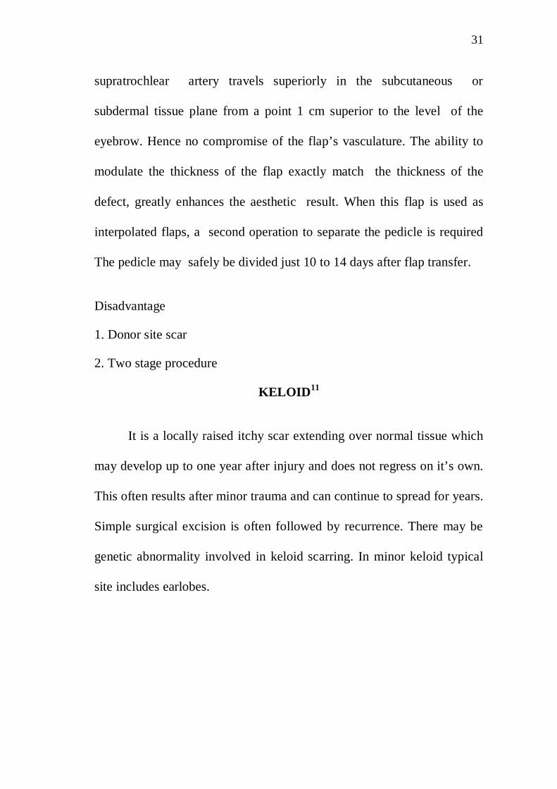
In the cutaneous lesions of head and neck region seen in our study
majority were benign (88%) and malignant lesions were only 12%. Benign
lesions occur in the age group of 10 – 40 years (72%) and malignant lesions
were above 40 years. Majority of the malignant cutaneous lesions were basel
cell carcinoma and majority occurs between 40 – 70 years with female
preponderance. Overall surgical procedures done for benign and malignant
lesions were as follows. 75% of patients had excision and primary closure. 8%
of the patients had excision and local flap cover. 2% of the patients had excision
and regional flap cover. 2% of the patients had excision and skin grafting. 4% of
patients had diathermy excision for wart. 5% of the patients had intralesional
sclerosant injection for hemangiomas and 4% of the patients had intralesional
steroids for keloid of earlobule.
Outcome
Satisfactory aesthetic outcome observed in 88% of patients and
unsatisfactory in 12 patients.
Key words
Benign, carcinoma, graft, flap, cutaneous
AIM AND OBJECTIVES
1. To identify the various cutaneous lesions of the head and neck
reporting to our department.
2. To analyse the various treatment and reconstructive options
available and their applications in the above patients.
3. To analyse the outcome of the surgical procedure done
1
INTRODUCTION
The patients with cutaneous lesions in head and neck region seek
reconstruction to cover the defect with good result. The priority of
reconstruction is complete tumor resection at the first instant and
followed with possible reconstruction.
Tumor biology is understood to get the best possible anatomic
functional and aesthetic results.
Face and it’s features have been subjects of poetic and artistic
endeavors throughout the ages. Because a person’s face is highly visible
and difficult to camouflage any lesions, scars or imperfections which are
obvious to others may be distressing to the affected individual. Surgical
planning and skill will have physical and psychological implications for
the patients. So in these patients, a surgeon’s goal is to achieve tumor free
margin, to avoid unsightly scar while using the simplest and most
effective reconstructive approach.

2
REVIEW OF LITERATURE
HISTORY
In 1861, LANGER12 published his “Proceedings of the Society for
Natural History of Vienna”, his monumental work on skin tension lines.
If the incisions were practiced across these lines, the scars were more
visible and broader, concluding thereby that all planned incisions should
follow the direction of those lines. In March 1948, RUBIN9 published the
work entitled “Langer’s lines and facial scars”, where, after an
introduction in which he reviews LANGER’s theory, he insists on the
fact, dubious in RUBIN’s opinion, that “the muscle is arranged in the
same direction as the lines, cancelling the tension on the wound’s border,
resulting in a finer scar (LANGER)”.
“Moreover, in a normal linear scar, collagen is oriented in the
longitudinal direction of the scar, whether it is parallel to the wrinkle
lines or not. In 1961, A.W. BULACIO NUNEZ, presented “New
procedure for researching the skin’s tension lines”.
In July 1962, BORGES and ALEXANDER1 publish a study on
“Relaxed skin tension lines, Zetaplasties on scars and fusiform excisions
of lesions” which starts by saying “It is a well known fact that a scar, the
3
more it follows relaxed skin tension lines (RSTLs), commonly known as
“the skin’s tension lines”, the better the functional and aesthetic. Results
are. The 1961 study by HOLMSTRAND, LONGACRE and
DeSTEFANO, which we have already commented on, supporting their
decision to follow the wrinkle lines.
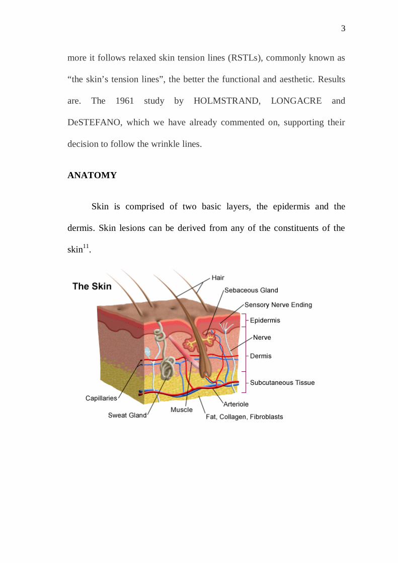
ANATOMY
Skin is comprised of two basic layers, the epidermis and the
dermis. Skin lesions can be derived from any of the constituents of the
skin11.
4
EPIDERMIS
Composed of stratified squamous epithelium, it is derived from
ectoderm. It is composed of five layers from deep to superficial stratum
germinatum, stratum spinosum, stratum granulosum, stratum lucidum,
stratum corneum.
SKIN LESIONS OF EPIDERMAL ORIGIN
1. Papilloma
a. Basal Cell Papilloma – known as Seborrheic Keratosis
Treatment – Curettage
b. Squamous Pailloma – known as skin tags
Treatment – Excision
2. Viral Wart – caused by HPV
Treatment – cryotherapy, curettage
DERMIS
Accounts for 95 % of thickness of skin.
Dermis is composed of collagen fibers, elastin fibers, ground substance
and vascular plexus

5
1. Papillary Dermis is superficial and contains more cells and finer
collagen fibers.
2. Reticular Dermis is deeper and contains fewer cells and coarser
collagen fibers.
HISTOLOGICAL CLASSIFICATION AND NOMENCLATURE
OF TUMOURS OF THE SKIN4
I. EPITHELIAL TUMOURS AND TUMOUR- LIKE LESIONS
A. BASAL CELL TUMOUR(BASAL CELL
CARCINOMA)
B. SQUAMOUS CELL CARCINOMA
C. PAPILLOMA
1. Squamous cell papilloma
2. Fibropapilloma
D. Sebaceous Gland Tumor
1. Sebaceous adenoma
2. Sebaceous carcinoma
3. Tumor- like hyperplasia
E. TUMOUR OF HEPATOID (PERIANAL) GLANDS
1. Adenoma of hepatoid glands
6
2. Carcinoma of hepatoid glands
3. Tumor-like hyperplasia
F. SWEAT GLAND TUMOUR
1. Papillary syring adenoma
2. Cystadenoma of apocrine sweat glands
3. Spiradenoma
4. Mixed tumor of apocrine sweat glands
5. Carcinoma of apocrine sweat glands
a. Papillary carcinoma
b. Tubular carcinoma
c. Solid carcinoma
d. Signet- ring- like carcinoma
G. TUMOUR OF HAIR FOLLICLE
1. Trichoepithelioma
2. Necrotizing and calcifying epithelioma(Mal-
herbe)
H. INTRACUTANEOUS CORNIFYING
EPITHELIOMA (“KERATOACANTHOMA”)
I. CYSTS
7
1. Epidermal cyst
2. Dermoid cyst
3. Follicular cyst
4. Cyst with epithelial proliferation
II. TUMOURS OF THE MELANOGENIC SYSTEM
A. BENIGN MELANOMA
1. Benign melanoma with junctional activity
2. Benign dermal melanoma
a. Cellular type
b. Fibromatous type
B. MALIGNANT MELANOMA
1. Epitheliod type
2. Spindle cell type
3. Epitheliod and spindle cell type
4. Dentritic and whorled type
III. TUMOURS OF SOFT (MESENCHYMAL) TISSUES
IV. SECONDARY TUMOURS
V. UNCLASSIFIED TUMOURS

8
BENIGN SKIN LESIONS OF DERMAL ORIGINS.
1. Trichoepithelioma – lesions from hair follicle
Transluscent, pinky white nodules located around nose and
mouth.
Treatment – Excision
2. Cylindroma – lesions from eccrine glands.
Known as turban tumor – occurs in scalp of elderly.
Treatment - Excision
BENIGN PIGMENTED SKIN LESIONS.
MELANOCYTIC NEVI - may be benign or malignant.
BENIGN MELANOCYTIC LESIONS
1. Congenital – giant hairy nevus
2. Acquired – junctional nevus, compound nevus and intradermal
nevus
3. Special nevi – Dysplastic nevus, halo nevus
MALIGNANT MELANOCYTIC LESIONS11
1. Melanoma accounts for 3% of all cancers.
9
2. Risk factors – premalignant lesions, previous melanoma,
atypical nevus syndrome, sun burn, Fitz Patrick type 1 skin
3. Classification
a. Clinical and histological
b. Breslow thickness
c. Clark level
d. TNM staging
e. Clinical staging
4. Prognosis
a. Males, elderly have worst prognosis.
b. Lesions on trunk, scalp, mucosa and perineum have worst
prognosis.
c. Acral lentiginous melanoma has poor prognosis.
d. Mackie’s checklist to identify melanoma
Major signs – change in size, shape and colour.
Minor signs – Diameter more than 5mm, inflammation,
itching, crusting or bleeding.
5. Treatment
a. Excision
10
I. Melanoma in situ – excision margin 5 to 10 mm
II. Melanoma with Breslow depth less than1 mm –
excision margin1cm.
III. Melanoma with Breslow depth 1 to 2mm – excision
margin 1 to 2 cms.
IV. Melanoma with Breslow depth more than 2mm.
Excision margin minimum 1cm.
Once the excision margin has been established the
skin and subcutaneous tissue should be excised
vertically. All tissues superficial to the deep fascia
should then be removed.
b. Reconstruction may be
I. Direct closure
II. Skin graft
III. Flap
c. Patients with clinically palpable nodes
I. FNAC
II. Open biopsy
d. Patients with histological positive nodes
I. Staging investigations.
11
II. Block dissection
III. Elective lymph node dissection.
MALIGNANT NONMELANO CYTIC SKIN LESIONS
BASAL CELL CARCINOMA (BCC)11
95% occurs between 40 and 80 years.
85% occurs in the head and neck.
Classification
1. Localised
a. Nodular
b. Nodulo Cystic
c. Micro nodular
d. Pigmented
2. Superficial
a. Superficial spreading
b. Multi focal
3. Infiltrative
a. Morpheaform
12
Of these nodular, nodulo cystic, superficial and morphic BCC are
the most common and accounts for more than 90 % lesions.
Treatment
Excision with 2 to 3 mm margin.
Lesions with indistinct margins require wider excision.
5% of BCC will be incompletely excised.
Tumors located on the ear periauricular region, nose, temporal
region, periocular region, mesolabial sulcus and upper lip (H-zone) have
high recurrence rates and are cosmetically sensitive areas with the limited
amount of surrounding tissues3.
Excision and reconstruction in these areas are more complicated –
insufficient resection.
1. Mohs Micro graphic surgery.
2. Radio therapy
3. Photodynamic therapy
4. Cryo therapy
5. Curettage
6. Intralesional interferon or BCG Vaccine.
13
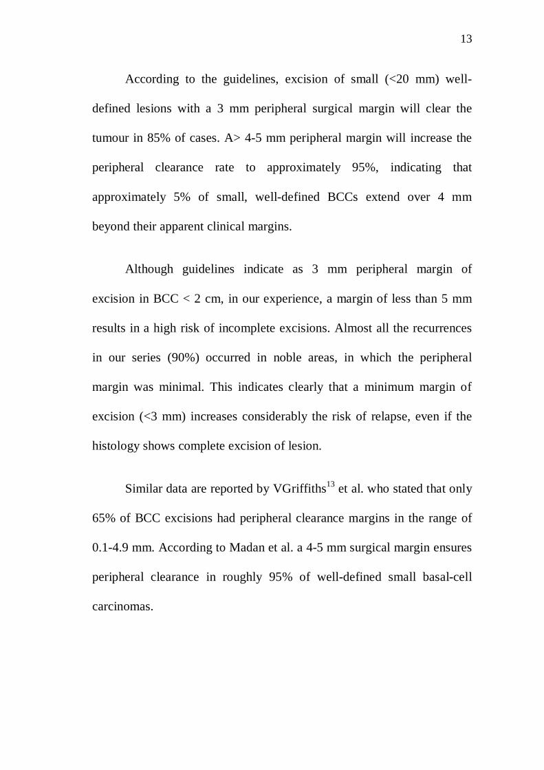
According to the guidelines, excision of small (<20 mm) well-
defined lesions with a 3 mm peripheral surgical margin will clear the
tumour in 85% of cases. A> 4-5 mm peripheral margin will increase the
peripheral clearance rate to approximately 95%, indicating that
approximately 5% of small, well-defined BCCs extend over 4 mm
beyond their apparent clinical margins.
Although guidelines indicate as 3 mm peripheral margin of
excision in BCC < 2 cm, in our experience, a margin of less than 5 mm
results in a high risk of incomplete excisions. Almost all the recurrences
in our series (90%) occurred in noble areas, in which the peripheral
margin was minimal. This indicates clearly that a minimum margin of
excision (<3 mm) increases considerably the risk of relapse, even if the
histology shows complete excision of lesion.
Similar data are reported by VGriffiths13 et al. who stated that only
65% of BCC excisions had peripheral clearance margins in the range of
0.1-4.9 mm. According to Madan et al. a 4-5 mm surgical margin ensures
peripheral clearance in roughly 95% of well-defined small basal-cell
carcinomas.

14
So no way a margin of 3 mm can be considered adequate in BCC
excision. This minimum margin may be considered for some particular
areas as nose, eyelid, and lips, for a better functional and aesthetic result.
SQUAMOUS CELL CARCINOMA11 (SCC)
SCC originates in the stratum spinosum of the epidermis.
The incidence of SCC is one quarter of BCC.
SCC often occurs in areas of abnormal skin containing – evidence
of sun damage, keratin horns, areas of Leukoplakia areas of Bowens
disease.
Presence of Keratin pearls is characteristic.
SCC may be well, moderately or poorly differentiated.
Bad prognostic indicators include increased depth of invasion,
vascular invasion, perineural invasion and lymphocytic infiltration.
SCCs are prone to local recurrence and metastases.
Patients should be followed up regularly to check for local and
regional recurrence.
Treatment
Excision - 0.5 to 1cm margin
15
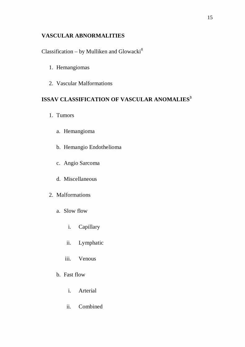
VASCULAR ABNORMALITIES
Classification – by Mulliken and Glowacki8
1. Hemangiomas
2. Vascular Malformations
ISSAV CLASSIFICATION OF VASCULAR ANOMALIES3
1. Tumors
a. Hemangioma
b. Hemangio Endothelioma
c. Angio Sarcoma
d. Miscellaneous
2. Malformations
a. Slow flow
i. Capillary
ii. Lymphatic
iii. Venous
b. Fast flow
i. Arterial
ii. Combined
16
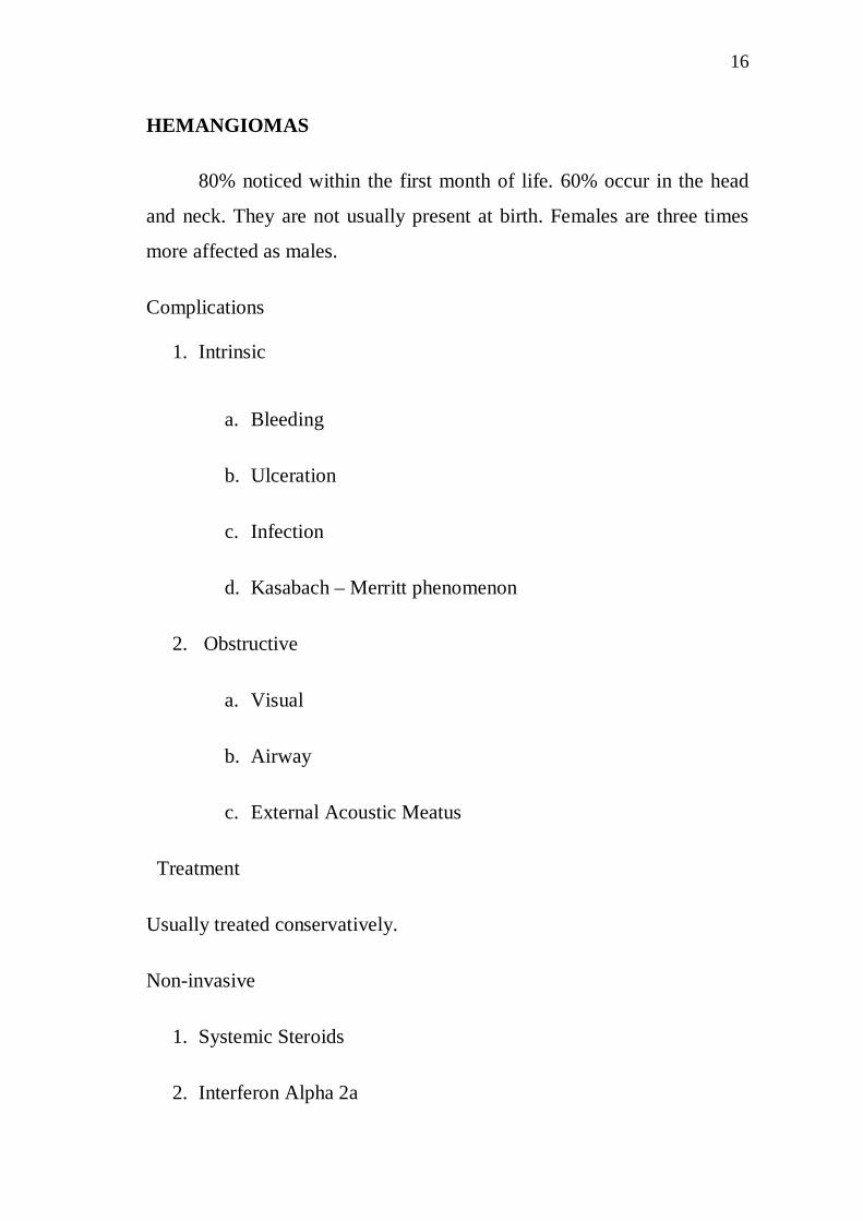
HEMANGIOMAS
80% noticed within the first month of life. 60% occur in the head
and neck. They are not usually present at birth. Females are three times
more affected as males.
Complications
1. Intrinsic
a. Bleeding
b. Ulceration
c. Infection
d. Kasabach – Merritt phenomenon
2. Obstructive
a. Visual
b. Airway
c. External Acoustic Meatus
Treatment
Usually treated conservatively.
Non-invasive
1. Systemic Steroids
2. Interferon Alpha 2a
17
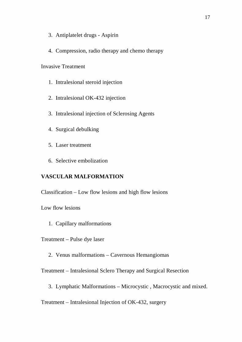
3. Antiplatelet drugs - Aspirin
4. Compression, radio therapy and chemo therapy
Invasive Treatment
1. Intralesional steroid injection
2. Intralesional OK-432 injection
3. Intralesional injection of Sclerosing Agents
4. Surgical debulking
5. Laser treatment
6. Selective embolization
VASCULAR MALFORMATION
Classification – Low flow lesions and high flow lesions
Low flow lesions
1. Capillary malformations
Treatment – Pulse dye laser
2. Venus malformations – Cavernous Hemangiomas
Treatment – Intralesional Sclero Therapy and Surgical Resection
3. Lymphatic Malformations – Microcystic , Macrocystic and mixed.
Treatment – Intralesional Injection of OK-432, surgery
18
High Flow Lesions
1. Arterial Malformations
2. Arterio Venus Malformations
Treatment
Embolization followed by resection within 24 to 72 hours.
TREATMENT OPTIONS
Excision - Healing by secondary intention
Primary closure
Skin grafting
Local flaps
Distant pedicled flaps
Free tissue transfer
EXCISION AND DIRECT CLOSURE
Orientation of Elective Incision
In the nineteenth century Langer showed that circular wounds
produced elliptical defects in cadaver skin. He believed that this occurred
because the skin tension along the longitudinal axis of the ellipse
exceeded that along the transverse axis.

19
Borges2 has provided 36 descriptive terms for skin lines. They
include RSTL - (Relaxed Skin Tension Lines) are parallel to the natural
skin wrinkles and tend to be perpendicular to the fibers of the underlying
muscle LMES (Line of Maximum Extensibility –these lie perpendicular
to the RSTL and parallel to the fibers of the underlying muscle.
Elective incision or excision of lesion is planned when possible so
that the final scars will be parallel to the RSTL5. Wrinkle lines are
generally same as RSTL and lie perpendicular to the long axis of the
underlying muscles. The direction of excision determines the eventual
appearance of scar.
Incisions and scars can be hidden by placing them at the junction of
aesthetic units.
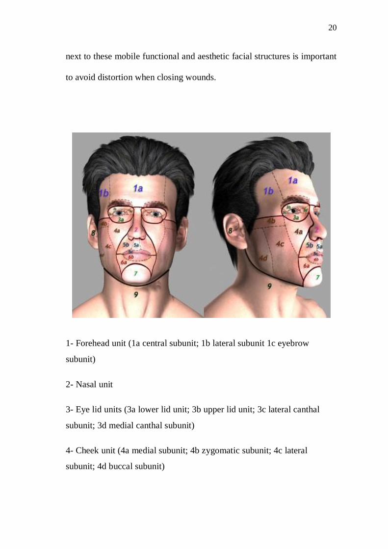
FACIAL AESTHETIC UNITS12
The face consists of 6 major aesthetic units comprised of: forehead,
eye/eyebrow, nose, lips, chin, and cheek. These aesthetic units can be
subdivided into additional anatomical subunits. For example, the nose can
be divided into nasal tip, dorsum, columella, soft-tissue triangles,
sidewalls, and nasal alar regions. Correct orientation of planned incisions
20
next to these mobile functional and aesthetic facial structures is important
to avoid distortion when closing wounds.
1- Forehead unit (1a central subunit; 1b lateral subunit 1c eyebrow
subunit)
2- Nasal unit
3- Eye lid units (3a lower lid unit; 3b upper lid unit; 3c lateral canthal
subunit; 3d medial canthal subunit)
4- Cheek unit (4a medial subunit; 4b zygomatic subunit; 4c lateral
subunit; 4d buccal subunit)
21
5- Upper lip unit (5a philtrum subunit; 5b lateral subunit; 5c mucosal
subunit)
6- Lower lip unit (6a central subunit; 6b mucosal subunit)
7- Mental unit
8- Auricular unit
9- Neck unit
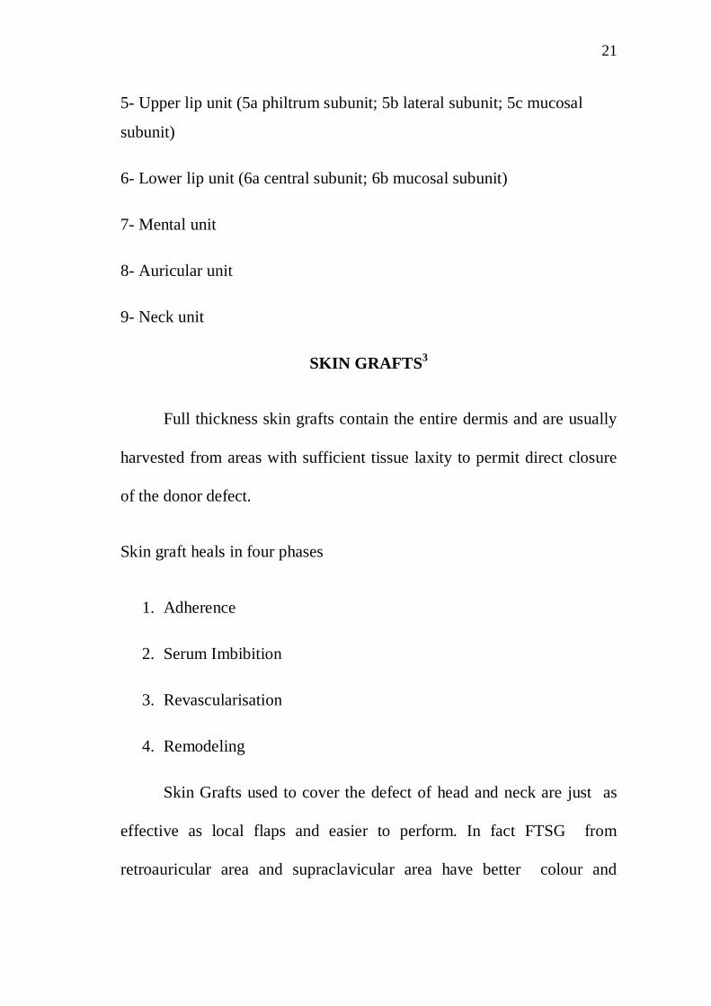
SKIN GRAFTS3
Full thickness skin grafts contain the entire dermis and are usually
harvested from areas with sufficient tissue laxity to permit direct closure
of the donor defect.
Skin graft heals in four phases
1. Adherence
2. Serum Imbibition
3. Revascularisation
4. Remodeling
Skin Grafts used to cover the defect of head and neck are just as
effective as local flaps and easier to perform. In fact FTSG from
retroauricular area and supraclavicular area have better colour and

22
texture for use in face. FTSG do not have 100% survival rate. They often
become paler or more pigmented than the surrounding skin and hence
aesthetically not good. Cutaneous malignancies are best treated by
excision and skin grafting. In FTSG colour and scar contracture related
complications are the disadvantages.
LOCAL FLAPS6
In full thickness defect of the facial structures, flap reconstruction
is mandatory. Functionally flaps are much better than grafts because little
or no scar contractures occur. Thus ectropion, epiphora and loss of oral
competence are prevented.
A Flap provides additional blood supply which is important for any
reconstruction. Local flaps are easy to use in older patients because of lax
skin availability. But much less so in children.
Advantages of using local flaps in head and neck
1. Similar colour and texture of the skin for the site of the defect.
2. Donor site frequently can be closed directly.
3. No scar contractures.
4. Survival not affected due to the underlying blood supply.
Disadvantage of local flaps

23
1. Requires planning and experience.
2. Flap may be too thick and bulky.
3. Flap should be of the same size and thickness as the defect.Otherwise problems will develop.
4. Preservation of local landmarks such as temporal hairline, eyebrows and symmetry.
5. Use of local flap is more difficult in children because of lack ofskin laxity.
LOCAL FLAPS – GENERAL PRINCIPLES
ROTATION FLAP6
In the rotation flap the surgeon triangulates the defect making the
shortest side the base of the triangle. The base then forms a portion of the
circumference of a circle and a flap is constructed so that it’s leading tip
will rotate around the circumference of the circle on which the triangular
defect lies.
The base of the flap is the radius of the large circle. When the flap
is elevated it can be rotated to close the defect. If rotation is not possible a
back cut is necessary. This rotation flap works very well on convexities
such as scalp or malar area. The edge of the flap is four to five times the
length of the base of the defect triangle.

24
TRANSPOSITION FLAP6
It is a rectangle or square of skin and subcutaneous tissue that also
is rotated about a pivot point into an immediately adjacent defect. The
flap donor site is closed by skin grafting. The maximum possible
transposition is 90 degrees from it’s original position. A back cut can be
used if the flap is under excessive tension. It can be used to close defect
on the anterior cheek.
For small defect single transposition cheek flap, and for large
defect double transposition flap can be used.

25
ADVANCEMENT FLAP3
It moves directly forward into a defect without any rotation or
lateral movement. Advancement is done by taking advantage of skin
elasticity, excising burrows triangles and pantographic expansion. Single
pedicle advancement flap, bi pedicle advancement flap and VY
advancement flap are the modifications. VY advancement flap used to
lengthen structures like Columella, to eliminate minor notches of the lip
and to close the donor site of skin flap.

26
27
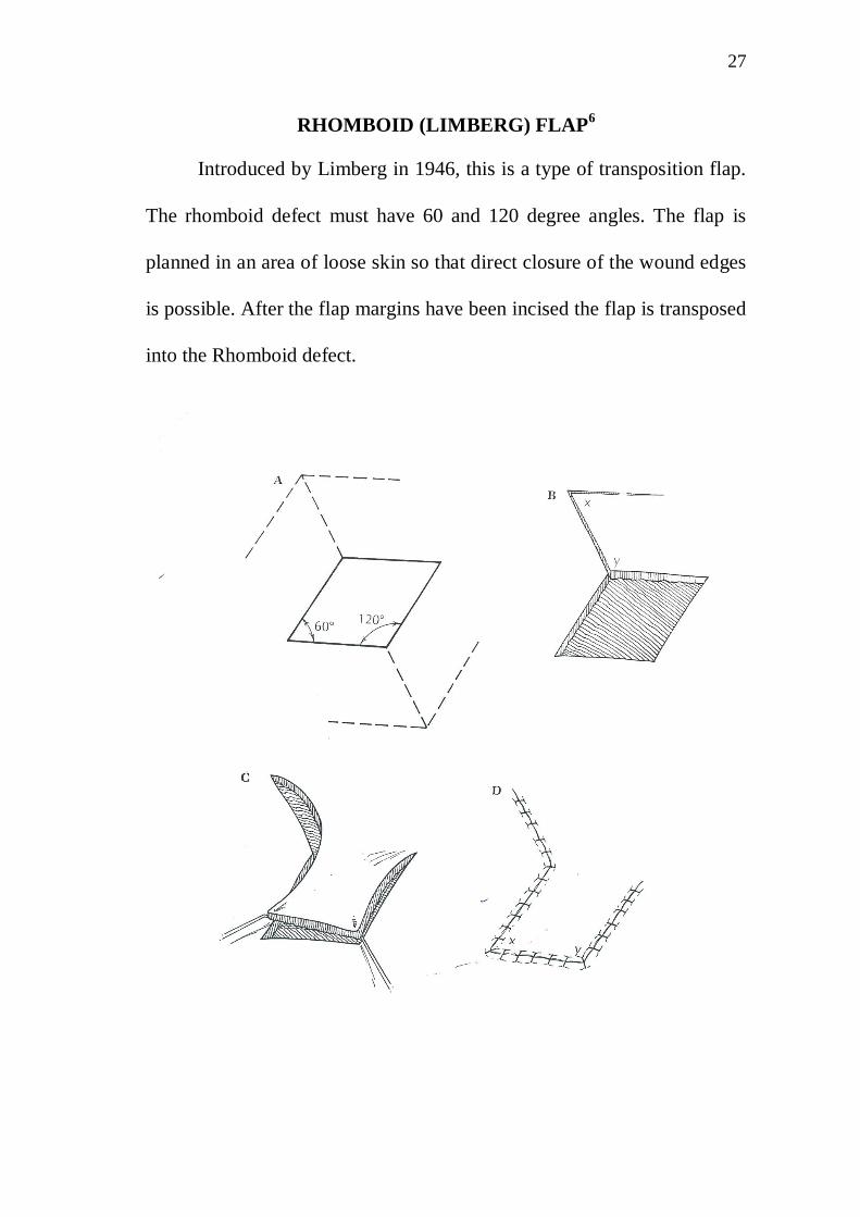
RHOMBOID (LIMBERG) FLAP6
Introduced by Limberg in 1946, this is a type of transposition flap.
The rhomboid defect must have 60 and 120 degree angles. The flap is
planned in an area of loose skin so that direct closure of the wound edges
is possible. After the flap margins have been incised the flap is transposed
into the Rhomboid defect.
28
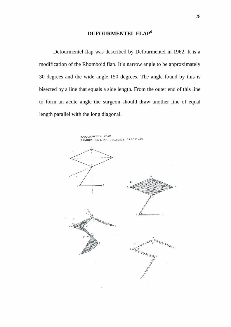
DUFOURMENTEL FLAP6
Defourmentel flap was described by Defourmentel in 1962. It is a
modification of the Rhomboid flap. It’s narrow angle to be approximately
30 degrees and the wide angle 150 degrees. The angle found by this is
bisected by a line that equals a side length. From the outer end of this line
to form an acute angle the surgeon should draw another line of equal
length parallel with the long diagonal.

29
BILOBED FLAP6
Described by Esser in 1918 in facial reconstruction it is most useful
on the nose. This is basically a rotation flap that spreads the load. The
planned defect is outlined and two flaps are drawn.
The width of flap one is slightly less than the diameter of the defect
and that of flap two is correspondingly less than flap one. Each flap may
rotate 90 degree or less. Test the tissue availability to assess the
feasibility of the flap.
GLABELLAR FLAPS6
The glabellar donor area contains an abundant source of skin for
resurfacing nasal defects. Because the skin in this area is thin, it provides
a good colour and texture match for the upper nasal area. The glabellar
flaps can be transfered in three ways; as a rotation flap, as a midline
transposition flap, and as a island flap .The last two types are more
flexible and can be enlarged and contoured as required. The classic
glabllelar flap is a rotation flap that incorporates a V-Y advancement in
the glabellar region. The lesion is resected in a triangular fashion. The
flap is designed and is raised at the level just above the glabellar
musculature. The movement of the flap is partly rotational, partly
30
transpositional. The donor defect is closed without difficulty and with
only slight distortion of the medial end of the right eyebrow
Problems
1. Eyebrow hair is present in the glabellar region area and the
rotation of this skin moves the hair bearing area down into the
medial canthus.
2. The thickness of the glabellar skin is greater than that of the skin
removed from the medial canthal area and this causes some
convexity in this region.
PARAMEDIAN FOREHEAD FLAPS10
Midforehead flaps, which include the median and paramedian flaps
and their many variations. It is based on a single supratrochlear artery, it
has replaced the median forehead flap for nasal reconstruction because it
has a more axial design, narrower base, and greater effective length .The
design enables the simultaneous use of two vertically oriented forehead
flaps. Removal of muscle and subcutaneous fat from the distal portion
can make this flap thin, pliable and easily contoured to fit any defect of
the nose. The attachment of the frontalis muscle to this flap is used when
more bulk is required to fill defects of considerable depth. Proper
thinning of the flap will ensure the shape and contour of the defect . The
31
supratrochlear artery travels superiorly in the subcutaneous or
subdermal tissue plane from a point 1 cm superior to the level of the
eyebrow. Hence no compromise of the flap’s vasculature. The ability to
modulate the thickness of the flap exactly match the thickness of the
defect, greatly enhances the aesthetic result. When this flap is used as
interpolated flaps, a second operation to separate the pedicle is required
The pedicle may safely be divided just 10 to 14 days after flap transfer.
Disadvantage
1. Donor site scar
2. Two stage procedure
KELOID11
It is a locally raised itchy scar extending over normal tissue which
may develop up to one year after injury and does not regress on it’s own.
This often results after minor trauma and can continue to spread for years.
Simple surgical excision is often followed by recurrence. There may be
genetic abnormality involved in keloid scarring. In minor keloid typical
site includes earlobes.

32
Initial management – Silicon Gel Sheeting, Intralesional steroid
injection, localized pressure therapy.
Secondary management – laser therapy, surgery with adjunctive
silicon gel sheeting, occasionally cryo therapy, radio therapy and recently
interferons.

33
MATERIALS AND METHODS
STUDY DESIGN
It is a prospective study carried out from September 2009 to
February 2012.
PLACE OF STUDY
The study was carried out at the Department of Burns and Plastic
and Reconstructive Surgery, Kilpauk Medical College, Chennai - 600010.
SELECTION OF PATIENTS
Patients with Cutaneous Lesions in head and neck regions were
selected from outpatients and inpatients.
Brief clinical history and other details were collected as per
proforma attached herewith.
INCLUSION AND EXCLUSION CRITERIA
All the patients with cutaneous lesions head and neck region who
underwent surgery in our department.
Patients with cutaneous lesions other than head and neck region
were excluded.

34
STUDY METHODS
All patients with cutaneous lesions of head and neck region were
evaluated. Detailed information was recorded in a predesigned data
collection sheet (Appendix 1). Information included was particulars of the
patient clinical features, surgery done, and it’s outcome. Permission to
carry out the study was obtained from the Ethical Committee.
STATISTICAL ANALYSIS
The data obtained were compiled and analysed using standard
statistical analysis.
35
OBSERVATION AND RESULTS
Total no of cases studied 100
TYPE OF CUTANEOUS LESIONS
36
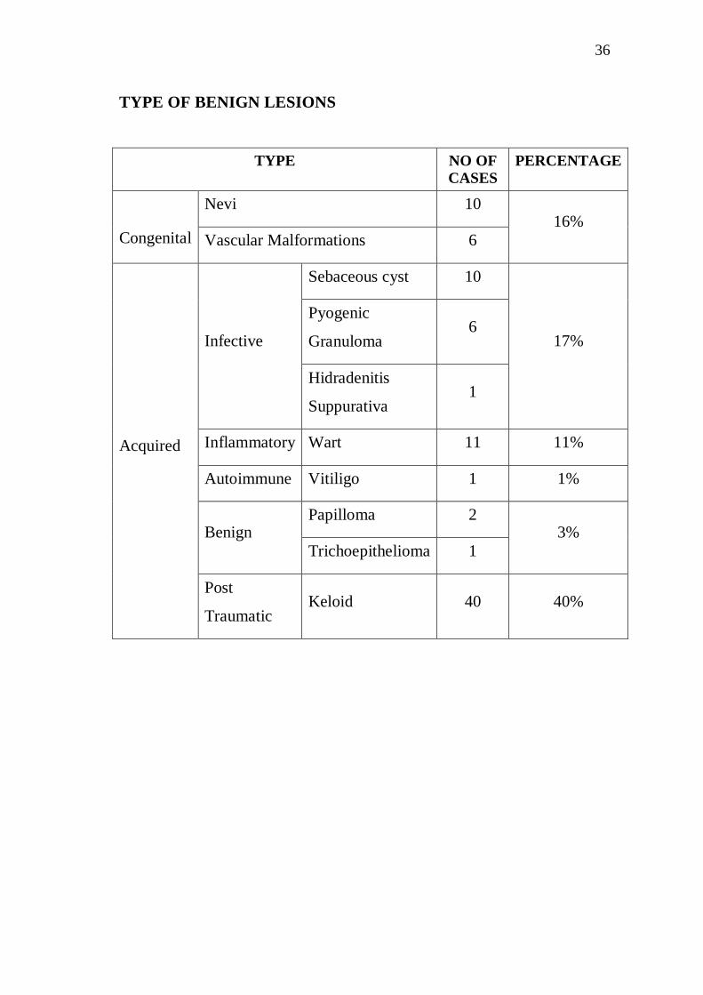
TYPE OF BENIGN LESIONS
TYPE NO OFCASES
PERCENTAGE
Nevi 10
Congenital Vascular Malformations 616%
Sebaceous cyst 10
Pyogenic
Granuloma6
Infective
Hidradenitis
Suppurativa1
17%
Inflammatory Wart 11 11%
Autoimmune Vitiligo 1 1%
Papilloma 2Benign
Trichoepithelioma 13%
Acquired
Post
TraumaticKeloid 40 40%

37
KELOID
OCCURRENCE NO OF CASES
UNILATERAL 30
BILATERAL 8
LOBULE 33
HELIX 5
SITE
MANDIBLE 2
MALE 4SEX
FEMALE 36
<10 1
11 - 20 20
AGE GROUP
20 - 40 19
38
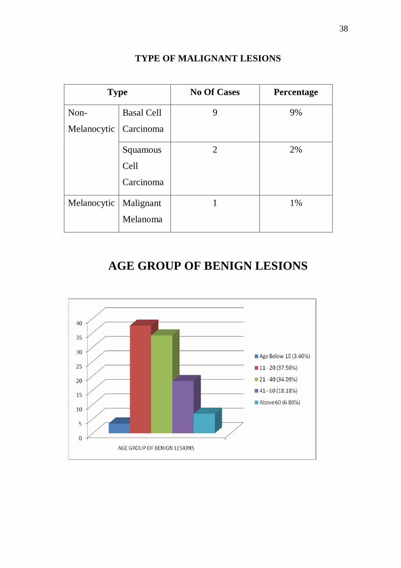
TYPE OF MALIGNANT LESIONS
Type No Of Cases Percentage
Basal Cell
Carcinoma
9 9%Non-
Melanocytic
Squamous
Cell
Carcinoma
2 2%
Melanocytic Malignant
Melanoma
1 1%
AGE GROUP OF BENIGN LESIONS

39
AGE GROUP OF BENIGH LESIONS
AGE GROUP IN
YEARS
NO OF CASES PERCENTAGE
<10 3 3.40%
11 – 20 33 37.50%
21 - 40 30 34.09%
41 - 60 16 18.18%
>60 6 6.80%
AGE GROUP OF MALIGNANT LESIONS
TYPE AGE GROUP
IN YEARS
NO OF
CASES
PERCENTAGE
40 – 50 2 22%Basal Cell
Carcinoma >50 7 78%
Squamous Cell
Carcinoma
>60 2 100%
Malignant
Melanoma
20 – 40 1
40
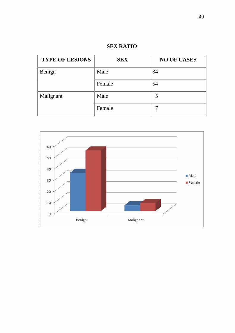
SEX RATIO
TYPE OF LESIONS SEX NO OF CASES
Male 34Benign
Female 54
Male 5Malignant
Female 7

41
BASEL CELL CARCINOMA - SITE OF OCCURENCE
SITE NO OF CASES PERCENTAGE
Cheek 4 44.4%
Forehead 2 22.2%
Nose 1 11.11%
Eye Lid 1 11.11%
Scalp (Post Auricular) 1 11.11%
BASEL CELL CARCINOMA – SITE
42
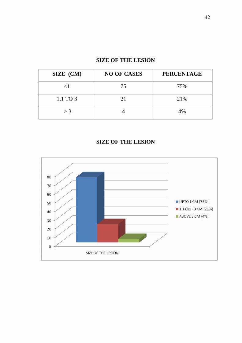
SIZE OF THE LESION
SIZE (CM) NO OF CASES PERCENTAGE
<1 75 75%
1.1 TO 3 21 21%
> 3 4 4%
SIZE OF THE LESION

43
SURGICAL PROCEDURES

44
SURGICAL PROCEDURES – BENIGN LESIONSType No
OfCases
Treatment
Nevi 10 Excision AndPrimary Closure
1 Excision AndPrimary ClosureCongenital
Vascular Malformations
5 Intra Lesional Inj.Sclerosing Agents
Sebaceous cyst 10PyogenicGranuloma 6InfectiveHidradenitisSuppurativa 1
Excision AndPrimary Closure
5 Excision AndPrimary Closure
4 Diathermic Excision1 Excision And Skin
Grafting
Inflamatory Wart
1 Excision AndLimberg Flap Cover
Autoimmune Vitiligo 1 Excision AndAdvancement Flap
Papilloma 2 Excision AndPrimary Closure
BenignTrichoepithelioma 1 Excision And Skin
Grafting
34IntralesionalExcision AndPrimary Closure
1 Excision And SkinGrafting
1 Excision AndPrimary Closure
Acquired
PostTraumatic
Keloid
4 Intra LesionalSteroids Inj.
45
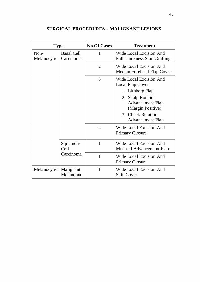
SURGICAL PROCEDURES – MALIGNANT LESIONS
Type No Of Cases Treatment1 Wide Local Excision And
Full Thickness Skin Grafting2 Wide Local Excision And
Median Forehead Flap Cover3 Wide Local Excision And
Local Flap Cover1. Limberg Flap2. Scalp Rotation
Advancement Flap(Margin Positive)
3. Cheek RotationAdvancement Flap
Basal CellCarcinoma
4 Wide Local Excision AndPrimary Closure
1 Wide Local Excision AndMucosal Advancement Flap
Non-Melanocytic
SquamousCellCarcinoma 1 Wide Local Excision And
Primary ClosureMelanocytic Malignant
Melanoma1 Wide Local Excision And
Skin Cover
46
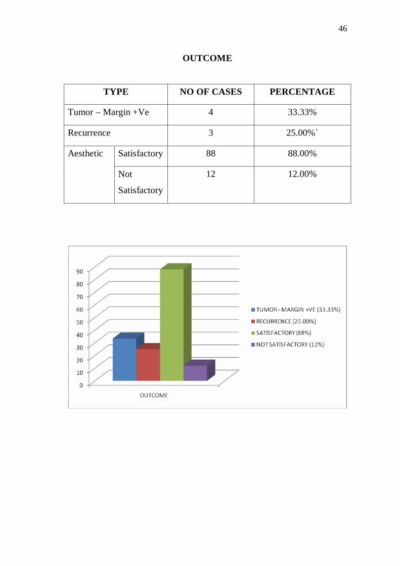
OUTCOME
TYPE NO OF CASES PERCENTAGE
Tumor – Margin +Ve 4 33.33%
Recurrence 3 25.00%`
Satisfactory 88 88.00%Aesthetic
Not
Satisfactory
12 12.00%
47
DISCUSSION
Evaluation of patients with cutaneous lesions. 100 patients have
been studied. Benign cases were 88 (88%) and maligmant ones were 12
(12%).
Patients with benign cutaneous lesions attending our department
were more because lesions in face, neck and scalp are easily visible.
Hence early medical attention is sought by them. Better aesthetic results
motivates them to attend Plastic surgery Department. Among these
cutaneous lesions female(61%) patients are more affected than
male(39%). Benign cutaneous lesions occur mostly in young individuals
(72% between 11 to 40 years). The youngest patient with benign
cutaneous lesion who attended our department was 3.5 year old girl with
Hemangioma cheek who underwent excision and primary closure. Benign
cutaneous lesions like Nevi, Wart and Sebaceous cyst occurs in old age
also (more than 40 years 25%).
In the benign cutaneous lesions 40% cases are keloids of the
earlobe. In this category the youngest female with Keloid was 6 years.
She had keloid earlobule. Studying the occurrence of Keloids, females
were 36 and males 4. Among these unilateral Keloid lesions observed in
30 cases and bilateral in 8 cases. Maximum site of occurrence was lobule
48
(33 cases), helix (5 cases) and mandible (2 cases). Keloid earlobe
attending our department were between 10 to 40 years (50% in 11 – 20
years, 49% in 21 to 40 years). Mandibular area, keloids were post
traumatic and post burn in etiology and excision with primary closure
and SSG were done respectively.
Next to keloid, congenital lesions are common. 16% (10% Nevi
and 6% hemangioma and vascular malformations). Here male and female
were equally affected. Youngest presentation was 3.5 years and at 40
years was the oldest. For Hemangiomas sclerosant therapy was advocated
in 5 patients and 1 patient had total excision and primary closure. In the
nevi group most of the lesions were less than 1 cm hence excision and
primary closure done in RSTL or wrinkle lines. One patient with nevi had
excision and slide swing flap cover.
Next to congenital patients infective lesions like sebaceous cyst
and pyogenic granuloma accounted for 16% cases which were excised
and the defect closed primarily. Here the incision was planned in such a
way that the future scar comes in the RSTL or wrinkle lines or anatomical
junction sites.
Next to infective lesions the viral inflammatory wart stands in the
line which is 10%; less than 1cm in size, being the major lesion. Excision

49
and primary closure was done in 4 patients, 3cm veruccous growth in the
scalp was excised after wedge biopsy and covered with SSG. One female
patient with Wart above the right eyebrow in the forehead was excised
and local Limberg flap cover given.
In malignant lesions, basal cell casinoma occurred in the age group
40 to 70 years that coincides with the other studies in the literature review
(95% of BCC occurs between 40 to 70 years). BCC ratio between male
and female were 1:2. Squamous cell carcinoma patients were manual
labourers with smoking habits and found to be above 60 years of age.
Patient who had melanoma of the scalp was 30 years old for whom wide
local excision and temporary skin cover given. Biopsy revealed
amelanotic melanoma with margins positive for tumor cells. Patient had
further treatment in the oncology department. Site analysis of BCC
showed, cheek involvement in 44%, forehead and periorbital area in 22%
each and periauricular area in 11% of cases.
74% cases had excision and primary closure (70 benign and
4 malignant). Because majority of the lesions presented early with size of
the lesion around 1cm, benign lesions did not necessitate marginal
clearance and in elderly patients with lax skin. Early surgery was

50
planned in such a way that the future scar was in the natural skin crease
line / RSTL and did not distort the anatomical landmarks and symmetry.
The incision and future scar were hidden by placing them at the
junction of aesthetic units.
Local flaps were used in 8% of patients to cover the post excision
defect (ie 3 benign and 5 malignant lesions). Local flaps were planned in
these patients for the following advantages of same colour, texture, and
same operative field. Secondary defects were also closed primarily. These
can be done as OP procedure and overall gave better aesthetic outcome.
Skin grafting was done in 4 patients. One patient had FTSG for the
forehead defect and three for scalp defect. Both did not give satisfying
results. Patients with scalp lesion were willing for future tissue expander
reconstruction to correct post surgical alopecia. BCC of the forehead,
reconstructed with FTSG did not show good aesthetic result and the
biopsy report showed inadequate marginal clearance. Hence wide local
excision and bilateral scalp rotation advancement flap was given to cover
the defect which gave better aesthetic results and the margins were tumor
free.
51
Regional flap cover was given to 2 patients. BCC in the medial
periorbital region was managed with wide local excision and
reconstructed with median forehead flap with advantage of same colour,
texture with a satisfied aesthetic outcome.
Intralesional sclerosants and steroids for hamangioma and keloids
were given as primary mode of treatment to reduce the size of the lesion
and secondary treatment as debulking for hamangiomas and intra lesional
excision for the keloids were done.
To analyse the overall outcome in malignant lesions biopsy showed
margin positive in 4 cases out of 12 malignant cutaneous lesions and 3
recurrence patients were in the H zone of BCC occurrence which also
coincides with the literature. Aesthetic outcome was analysed and 88%
patients were satisfied and 12% had no satisfaction.

52
CONCLUSION
In the cutaneous lesions of head and neck region seen in our study
majority were benign (88%) and malignant lesions were only 12%.
In the benign cases aetiology of the lesions in the order of
incidence were: post traumatic keloid 40%, congenital nevi and vascular
malformations 16%, infective sebaceous cyst and pyogenic granuloma
16%, inflammatory wart 11%, papilloma 2%, trichoepithelioma 1% and
vitiligo 1%.
Benign lesions occur in the age group of 10 – 40 years (72%) and
malignant lesions were above 40 years.
Females with benign lesions were a majority (54) and 34 were
males.
Majority of the malignant cutaneous lesions were basel cell
carcinoma and majority occurs between 40 – 70 years with female
preponderance.
Overall surgical procedures done for benign and malignant lesions
were as follows.

53
75% of patients had excision and primary closure. 8% of the
patients had excision and local flap cover. 2% of the patients had excision
and regional flap cover. 2% of the patients had excision and skin grafting.
4% of patients had diathermy excision for wart. 5% of the patients had
intralesional sclerosant injection for hemangiomas and 4% of the patients
had intralesional steroids for keloid of earlobule.
In the outcome analysis, 4 patients had tumor margin positive.
They underwent secondary surgical procedures. Recurrence in 3 patients
of malignant lesions had further resection and reconstruction done.
Satisfactory aesthetic outcome observed in 88% of patients and
unsatisfactory in 12 patients.
Keloid patients were advised to follow scar massage therapy,
steroid injection in suture lines or silicon gel sheeting after intralesional
excision of keloids.

BIBLIOGRAPHY
1. BORGES, A.F. ALEXANDER, J.E.- "Relaxed skin tension Lines,
Z-plasties on scars and fusiform excision of lesions". British
Journal of Plastic Surgery. Julio 1962. Vol. XV, no 3, Page 242.
2. Borges AF: Elective Incisions and Scar Revision, Boston, Little,
Brown, 1973.
3. Charles H Thorne, Grabb and Smith’s Plastic Surgery 2007 6th ed,
Lippincott Williams and wilkins, Philadelphia.
4. E Weiss and K Frese: Tumours of skin, Bull World Health
Organisation, 1974_50_(1-2).
5. Gibson, T., AND Kenedi, R.M., Biomeechanical Properties of
Skin, Surg. Clin. North am 47 : 279, 1967.
6. Jackson Ian T: Local Flaps in Head and Neck Reconstruction.
St.Louis, Mosby, 1985.
7. McGregor IA : Fundamental Techniques in Plastic Surgery and
Their Surgical Applications 7th ed, Edinburgh ,Churchill
Livingstone , 1980.
8. Mulliken JB : Vascular Anomalies. In Aston SJ, Beasley RW,
Thome CHM [eds]: Grabb and Smith’S Plastic Surgery, 5th ed
Philadelphia Lippincott-Raven, 1997.
9. RUBÍN, Leonard R.- "LANGER 's lines and facial scars". Plastic
and Reconstructive Surgery. March 1948. Vol. 3, no 2, Page 147
10. Shan R Baker: Local Flaps in Facial Reconstruction, 2nd ed,
Mosby, 2007.
11. Stephen J Mathes: Plastic Surgery, Volume I, General principles
2006, Saunders, Philadelphia.
12. T. Fattahi - An overview of facial aesthetic units. Journal of Oral
and Maxillofacial Surgery, Volume 61, Issue 10.
13. Valentina Sorvillo: Journal of Skin Cancer
Volume 2011 (2011), Article ID 476362

PROFORMA
Sl No Name Age Sex Occupation
Habits
Main complaint
Duration of illness
Previous treatment
Examination
Lesion site Size
Any other change
Surgery Done
Post Operative Period
Biopsy
Follow up
Satisfactory Or Not Satisfactory
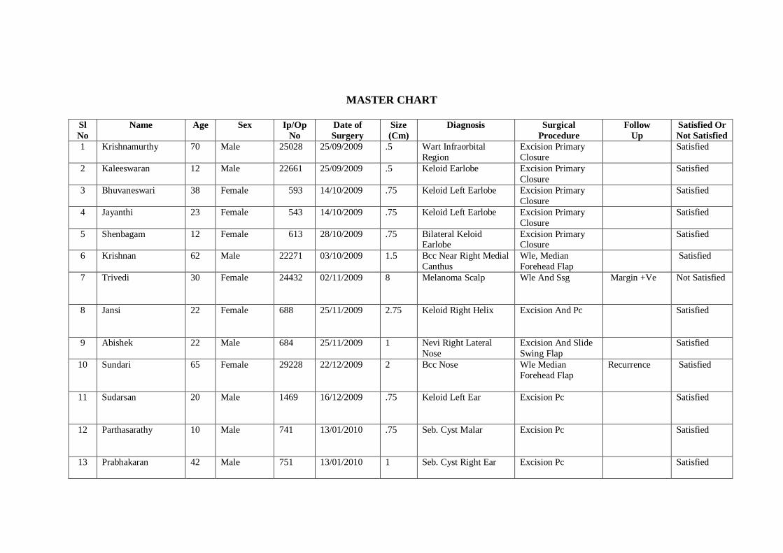
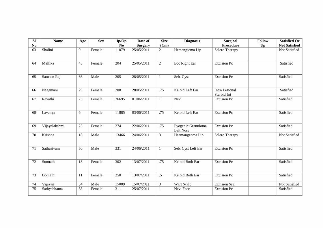
MASTER CHART
SlNo
Name Age Sex Ip/OpNo
Date ofSurgery
Size(Cm)
Diagnosis SurgicalProcedure
FollowUp
Satisfied OrNot Satisfied
1 Krishnamurthy 70 Male 25028 25/09/2009 .5 Wart InfraorbitalRegion
Excision PrimaryClosure
Satisfied
2 Kaleeswaran 12 Male 22661 25/09/2009 .5 Keloid Earlobe Excision PrimaryClosure
Satisfied
3 Bhuvaneswari 38 Female 593 14/10/2009 .75 Keloid Left Earlobe Excision PrimaryClosure
Satisfied
4 Jayanthi 23 Female 543 14/10/2009 .75 Keloid Left Earlobe Excision PrimaryClosure
Satisfied
5 Shenbagam 12 Female 613 28/10/2009 .75 Bilateral KeloidEarlobe
Excision PrimaryClosure
Satisfied
6 Krishnan 62 Male 22271 03/10/2009 1.5 Bcc Near Right MedialCanthus
Wle, MedianForehead Flap
Satisfied
7 Trivedi 30 Female 24432 02/11/2009 8 Melanoma Scalp Wle And Ssg Margin +Ve Not Satisfied
8 Jansi 22 Female 688 25/11/2009 2.75 Keloid Right Helix Excision And Pc Satisfied
9 Abishek 22 Male 684 25/11/2009 1 Nevi Right LateralNose
Excision And SlideSwing Flap
Satisfied
10 Sundari 65 Female 29228 22/12/2009 2 Bcc Nose Wle MedianForehead Flap
Recurrence Satisfied
11 Sudarsan 20 Male 1469 16/12/2009 .75 Keloid Left Ear Excision Pc Satisfied
12 Parthasarathy 10 Male 741 13/01/2010 .75 Seb. Cyst Malar Excision Pc Satisfied
13 Prabhakaran 42 Male 751 13/01/2010 1 Seb. Cyst Right Ear Excision Pc Satisfied
SlNo
Name Age Sex Ip/OpNo
Date ofSurgery
Size(Cm)
Diagnosis SurgicalProcedure
FollowUp
Satisfied OrNot Satisfied
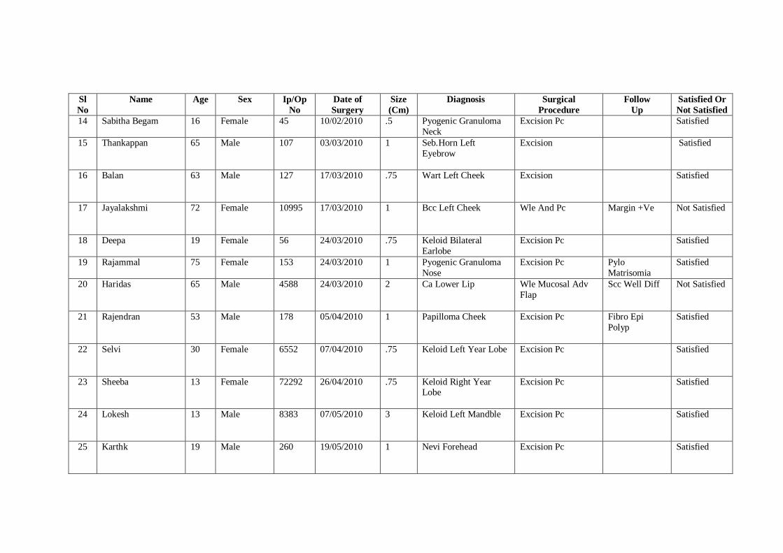
14 Sabitha Begam 16 Female 45 10/02/2010 .5 Pyogenic GranulomaNeck
Excision Pc Satisfied
15 Thankappan 65 Male 107 03/03/2010 1 Seb.Horn LeftEyebrow
Excision Satisfied
16 Balan 63 Male 127 17/03/2010 .75 Wart Left Cheek Excision Satisfied
17 Jayalakshmi 72 Female 10995 17/03/2010 1 Bcc Left Cheek Wle And Pc Margin +Ve Not Satisfied
18 Deepa 19 Female 56 24/03/2010 .75 Keloid BilateralEarlobe
Excision Pc Satisfied
19 Rajammal 75 Female 153 24/03/2010 1 Pyogenic GranulomaNose
Excision Pc PyloMatrisomia
Satisfied
20 Haridas 65 Male 4588 24/03/2010 2 Ca Lower Lip Wle Mucosal AdvFlap
Scc Well Diff Not Satisfied
21 Rajendran 53 Male 178 05/04/2010 1 Papilloma Cheek Excision Pc Fibro EpiPolyp
Satisfied
22 Selvi 30 Female 6552 07/04/2010 .75 Keloid Left Year Lobe Excision Pc Satisfied
23 Sheeba 13 Female 72292 26/04/2010 .75 Keloid Right YearLobe
Excision Pc Satisfied
24 Lokesh 13 Male 8383 07/05/2010 3 Keloid Left Mandble Excision Pc Satisfied
25 Karthk 19 Male 260 19/05/2010 1 Nevi Forehead Excision Pc Satisfied

SlNo
Name Age Sex Ip/OpNo
Date ofSurgery
Size(Cm)
Diagnosis SurgicalProcedure
FollowUp
Satisfied OrNot Satisfied
26 Chamundi 42 Female 322 09/06/2010 .75 Wart Scalp Excision Pc Satisfied
27 Radha 20 Female 373 07/07/2010 .75 Keloid Left Ear Lobe Excision Pc Satisfied
28 Kuppu 36 Female 174 14/07/2010 1 Keloid Right Ear Lobe Excision Pc Satisfied
29 Krishna 47 Male 1850 23/06/2010 3 Vasc Mal Sclerosant Satisfied
30 Revathi 20 Female 416 28/07/2010 .75 Keloid Right Ear Lobe Excision Pc Satisfied
31 Krishnamurthi 41 Male 428 04/08/2010 1.5 Bcc Right Cheek Wle And Pc MarginNegative
Satisfied
32 Nagalakshmi 25 Female 440 07/08/2010 2 Vitiligo Forehead Excision Adv Flap Satisfied
33 Jaya 20 Female 462 03/09/2010 .75 Keloid Right Ear Excision Pc Satisfied
34 Lavanya 15 Female 499 17/09/2010 .75 Keloid Left Ear Excision Pc Satisfied
35 Ponnurangan 58 Male 498 17/09/2010 .5 Wart Right Cheek Excision Pc Satisfied
36 Anandakishore 33 Male 531 24/09/2010 1 Pyogenic Granuloma Excision Pc Satisfied37 Hamesh 12 Male 843 08/10/2010 .75 Papilloma Right Cheek Excision Pc Satisfied

SlNo
Name Age Sex Ip/OpNo
Date ofSurgery
Size(Cm)
Diagnosis SurgicalProcedure
FollowUp
Satisfied OrNot Satisfied
38 Chandrasekar 61 Male 553 11/10/2010 1 Seb.Horn Left SideNeck
Excision Pc Satisfied
39 Jothi 64 Female 560 13/10/2010 .75 Wart Fore Head Excision LimbergFlap
Satisfied
40 Tamil Mani 58 Male 568 18/10/2010 1.5 Bcc Below Left Eye Wle And Pc Satisfied
41 Anthoniammal 24 Female 572 20/10/2010 .75 Nevi Left Cheek Excision Pc Satisfied
42 Mani 55 Male 580 22/10/2010 1 Seb. Cyst Excision Pc Satisfied
43 Vaishnavi 16 Female 516 25/10/2010 1 Keloid Left Ear Excision Pc Satisfied
44 Kalpana 24 Female 592 29/10/2010 .75 Keloid Left Ear Excision Pc Satisfied
45 Geetha 28 Female 451 08/11/2010 .75 Keloid Left Ear Excision Pc Satisfied
46 Renuka 28 Female 608 15/11/2010 1 Keloid Both Ear Excision Pc Satisfied
47 Harikrishnan 54 Male 30488 01/12/2010 2 Hid. Suppurativa Excision Pc Satisfied
48 Deshbhai 23 Male 341 01/12/2010 .75 Seb. Cyst Left Face Excision Pc Satisfied49 Kannan 16 Male 2465 09/12/2010 3 Hemangioma
Lower LipSclero Therapy Not Satisfied

SlNo
Name Age Sex Ip/OpNo
Date ofSurgery
Size(Cm)
Diagnosis SurgicalProcedure
FollowUp
Satisfied OrNot Satisfied
50 Ashwini 19 Female 7 07/01/2011 .75 Keloid Left Ear Excision Pc Satisfied
51 Jayanthi 25 Female 35 04/02/2011 1 Keloid Right Ear Excision Pc Satisfied
52 Padma 32 Female 53 14/02/2011 1 Keloid Right Ear Excision Pc Satisfied
53 Thangaraj 67 Male 38 18/02/2011 .75 Wart Excision Diathermy Satisfied
54 Sasi Kani 16 Female 50 23/02/2011 3 Seb.Cyst Neck Excision Pc Satisfied
55 Vennila 17 Female 648 23/02/2011 .75 Keloid Left Ear Excision Pc Satisfied
56 Filomina 42 Female 70 28/02/2011 2 Bcc Cheek Wle Pc Not Satisfied
57 Raja 29 Male 686 02/03/2011 .75 Keloid Both Ear Excision Pc Satisfied
58 Praveena 18 Female 3 13/03/2011 .75 Keloid Both Ear Excison Pc Satisfied
59 Rose 26 Female 4368 21/03/2011 10 Keloid Mandible Neck Excision Ssg Not Satisfied
60 Shanthi 30 Female 188 11/05/2011 1 Keloid Left Ear Excision Pc Satisfied61 Lavanya 15 Female 499 01/05/2011 .75 Keloid Left Helix Excision Pc62 Divya 12 Female 669 18/05/2011 .75 Keloid Both Ear Excision Pc Satisfied
SlNo
Name Age Sex Ip/OpNo
Date ofSurgery
Size(Cm)
Diagnosis SurgicalProcedure
FollowUp
Satisfied OrNot Satisfied
63 Shalini 9 Female 11079 25/05/2011 2 Hemangioma Lip Sclero Therapy Not Satisfied
64 Mallika 45 Female 204 25/05/2011 2 Bcc Right Ear Excision Pc Satisfied
65 Samson Raj 66 Male 205 28/05/2011 1 Seb. Cyst Excision Pc Satisfied
66 Nagamani 29 Female 200 28/05/2011 .75 Keloid Left Ear Intra LesionalSteroid Inj
Satisfied
67 Revathi 25 Female 26695 01/06/2011 1 Nevi Excision Pc Satisfied
68 Lavanya 6 Female 11885 03/06/2011 .75 Keloid Left Ear Excision Pc Satisfied
69 Vijayalakshmi 23 Female 274 22/06/2011 .75 Pyogenic GranulomaLeft Nose
Excision Pc Satisfied
70 Krishna 18 Male 13466 24/06/2011 3 Haemangeoma Lip Sclero Therapy Not Satisfied
71 Sathasivam 50 Male 331 24/06/2011 1 Seb. Cyst Left Ear Excision Pc Satisfied
72 Sunnath 18 Female 302 13/07/2011 .75 Keloid Both Ear Excision Pc Satisfied
73 Gomathi 11 Female 250 13/07/2011 .5 Keloid Both Ear Excision Pc Satisfied
74 Vijayan 34 Male 15089 15/07/2011 3 Wart Scalp Excision Ssg Not Satisfied75 Sathyabhama 38 Female 311 25/07/2011 1 Nevi Face Excision Pc Satisfied

SlNo
Name Age Sex Ip/OpNo
Date ofSurgery
Size(Cm)
Diagnosis SurgicalProcedure
FollowUp
Satisfied OrNot Satisfied
76 Asharaf 23 Female 214 25/07/2011 .75 Keloid Both Ear Excision Pc Satisfied
77 Sutha 27 Female 301 01/08/2011 .75 Keloid Left Helix Excision Pc Satisfied
78 Vishwanath 20 Male 338 17/08/2011 .5 Nevi Nose Excision Pc Satisfied
79 Amritha 40 Female 335 17/08/2011 .5 Nevi Upper Lip Excision Pc Satisfied
80 Sangeetha 12 Female 377 14/09/2011 1 Pyogenic Granuloma Excision Pc Satisfied
81 Gajapathi 20 Female 383 19/09/2011 .5 Nevi Nose Excision Pc Satisfied
82 Imran Khan 19 Male 375 12/10/2011 .75 Nevi Ear Excision Pc Satisfied
83 Ragavendra 55 Male 21369 19/10/2011 2 Bcc Fore Head 1.Wle Ftsg2.Scalp Rot AdvFlap
MarginPositive
Satisfied
84 Krishnan 70 Male 22789 24/10/2011 1 Ca Lower Lip Wle , PrimaryClosure
Not Satisfied
85 Poongavanam 38 Female 23749 28/10/2011 1 Bcc Right Eye Brow Wle Limberg Flap Satisfied
86 Kavitha 32 Female 429 30/10/2011 .75 Keloid Right Ear Intra LesionalSteroid Inj.
Satisfied
87 Kamachi 26 Female 2631 14/11/2011 .75 Keloid Left Ear Intra Lesional Satisfied

SlNo
Name Age Sex Ip/OpNo
Date ofSurgery
Size(Cm)
Diagnosis SurgicalProcedure
FollowUp
Satisfied OrNot Satisfied
Steroid Inj.88 Poonkodi 21 Female 469 18/11/2011 .75 Keloid Left Ear Excision Pc Satisfied
89 Vijaya 34 Female 468 18/11/2011 .75 Keloid Right Ear Intra LesionalSteroid Inj
Satisfied
90 Jaya 58 Female 25734 21/11/2011 1.5 Bcc Left Nose Wle Cheek Rot AdvFlap
MarginPositive
Not Satisfied
91 Sundar 20 Male 467 09/11/2011 2 Tricho Epithelioma Excision Ssg Satisfied
92 Kuppusamy 62 Male 503 02/12/2011 1.5 Wart Lower Lip Excision Pc Satisfied
93 Karpagam 50 Female 528 14/12/2011 .5 Hyper KerototicPapule
Excision Pc Satisfied
94 Ganapathy 46 Male 530 14/12/2011 1 Pyogenic Granuloma Excision Pc Satisfied
95 Rajendra Babu 52 Male 11 11/01/2012 1 Wart Neck Diatherny Satisfied
96 Mekala 30 Female 29 13/01/2012 .5 Nevi Nose Excision Pc Satisfied
97 Loganya 42 Female 31 20/01/2012 1 Fibroma Excision Pc Satisfied
98 Sindhu 3.5 Female 2380 03/02/2012 2 Hemangioma RightCheek
Excision Pc Satisfied
99 Dhananjayan 24 Male 421 28/10/2011 4 Venous Malf. Sclero Therapy AndDebulking
Satisfied
100 Nisha 13 Female 64 15/02/2012 .5 Keloid Right Nose Excision Pc Satisfied
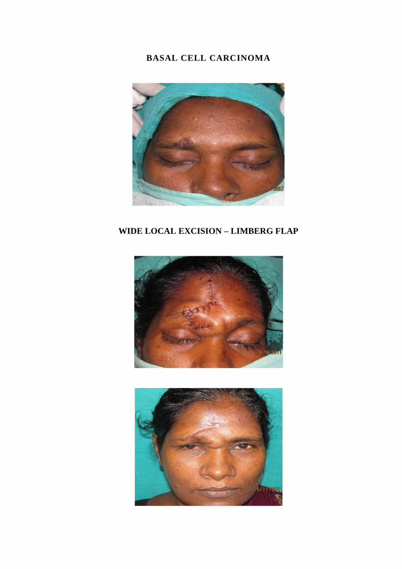
BASAL CELL CARCINOMA
WIDE LOCAL EXCISION – LIMBERG FLAP
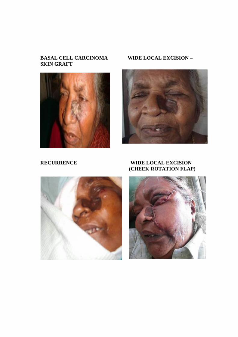
BASAL CELL CARCINOMA WIDE LOCAL EXCISION –SKIN GRAFT
RECURRENCE WIDE LOCAL EXCISION (CHEEK ROTATION FLAP)

HEMANGIOMA
EXCISION – PRIMARY CLOSURE

NEVI
EXCISION – PRIMARY CLOSURE
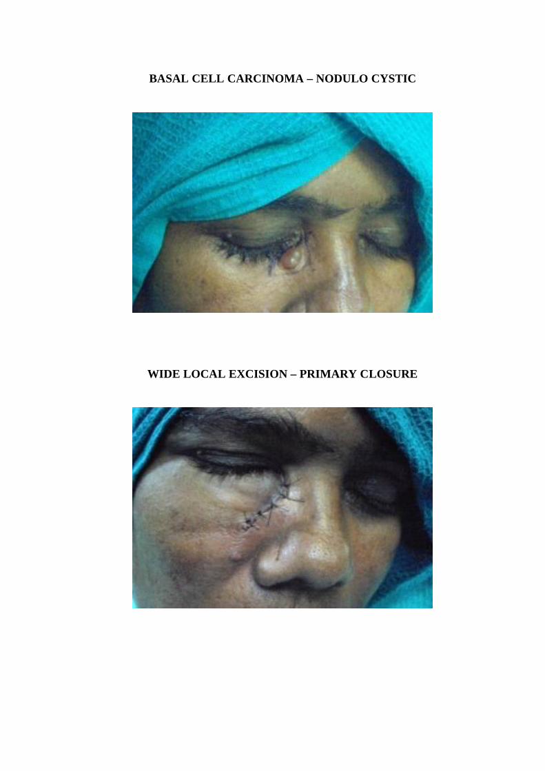
BASAL CELL CARCINOMA – NODULO CYSTIC
WIDE LOCAL EXCISION – PRIMARY CLOSURE
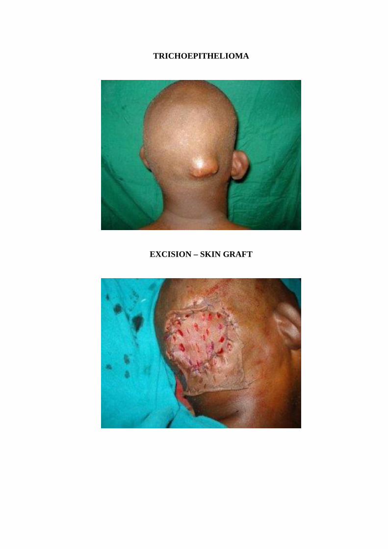
TRICHOEPITHELIOMA
EXCISION – SKIN GRAFT
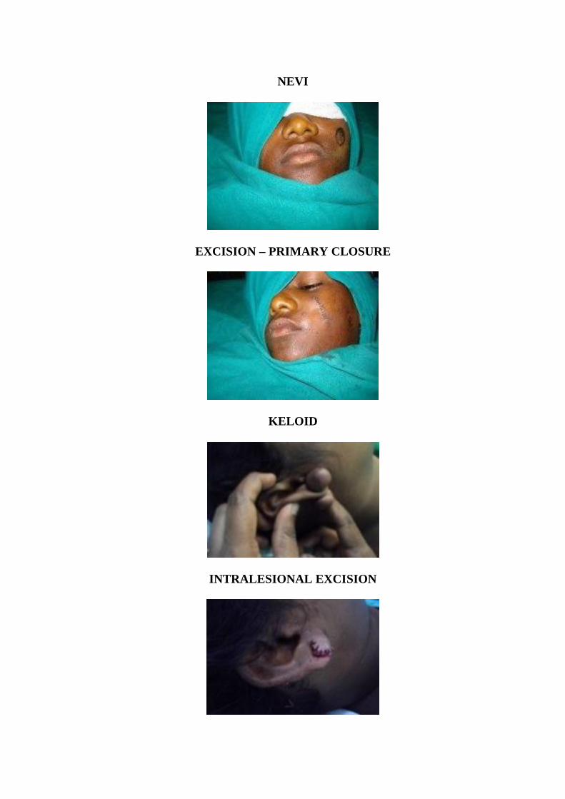
NEVI
EXCISION – PRIMARY CLOSURE
KELOID
INTRALESIONAL EXCISION
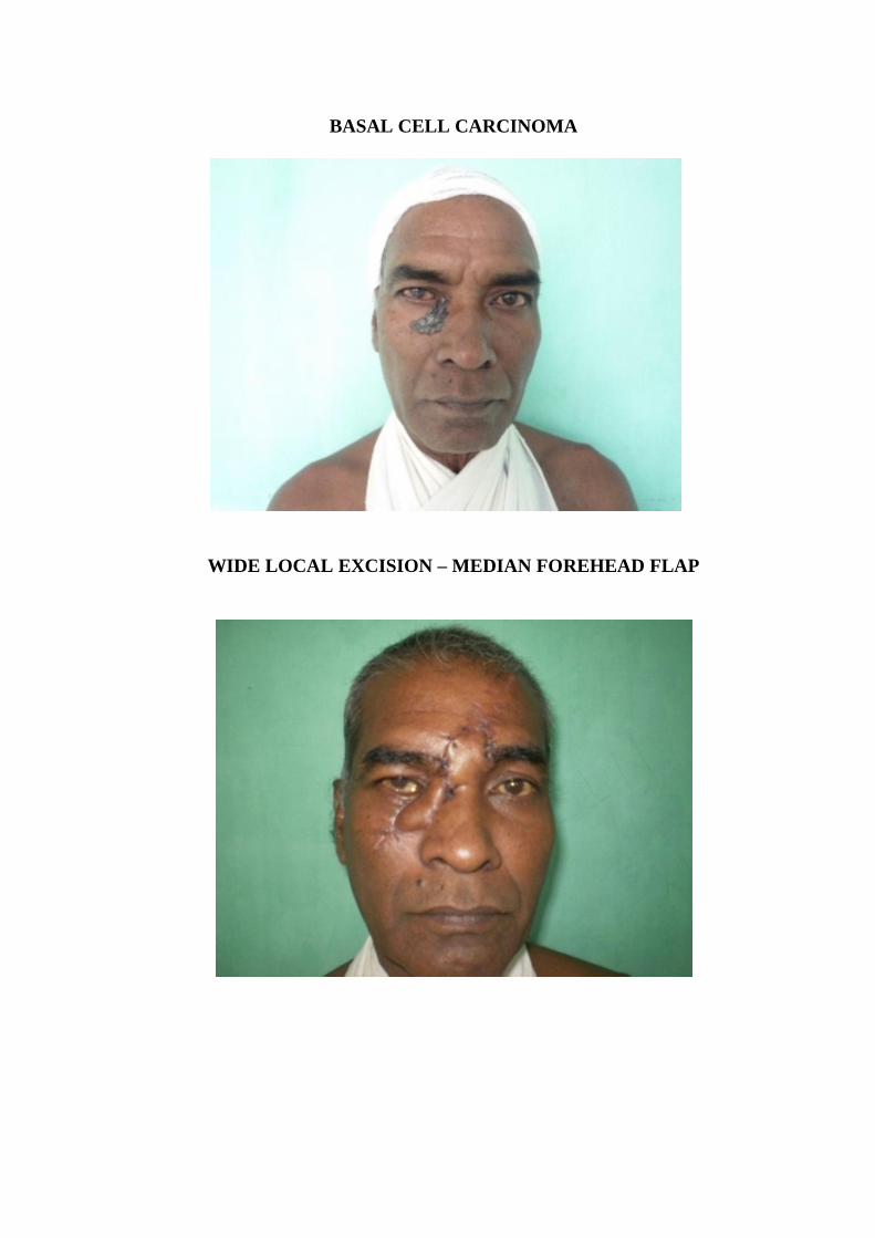
BASAL CELL CARCINOMA
WIDE LOCAL EXCISION – MEDIAN FOREHEAD FLAP
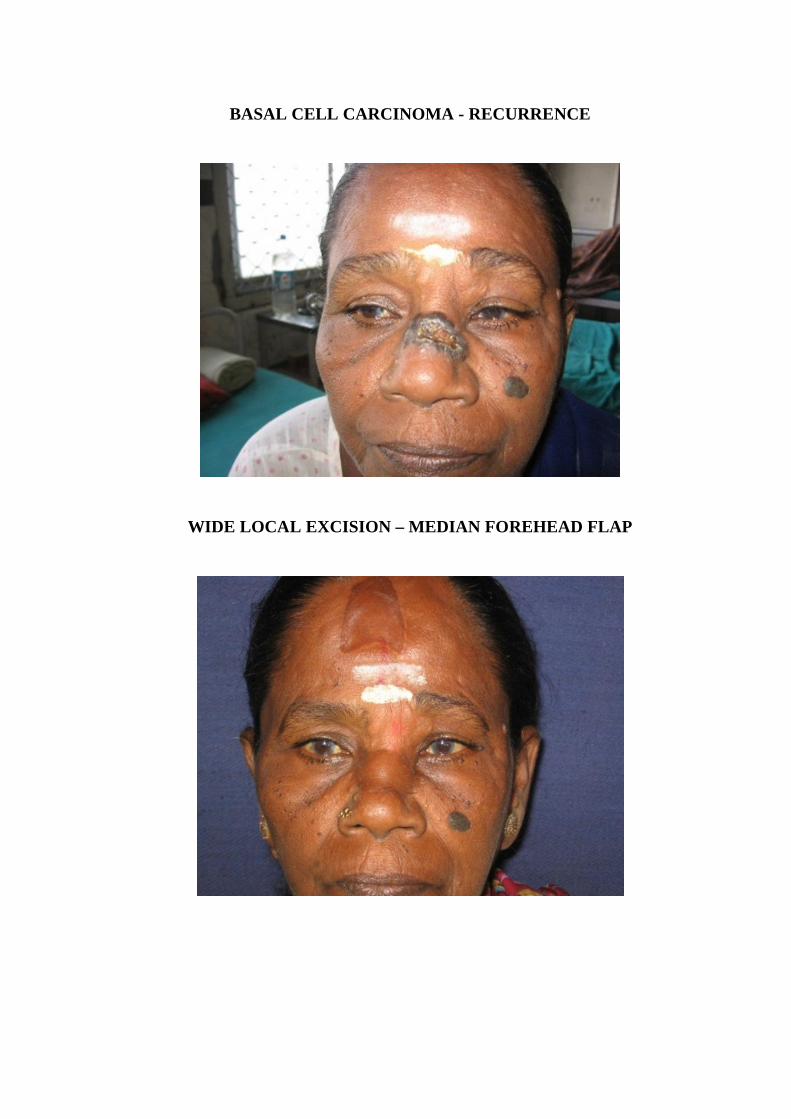
BASAL CELL CARCINOMA - RECURRENCE
WIDE LOCAL EXCISION – MEDIAN FOREHEAD FLAP
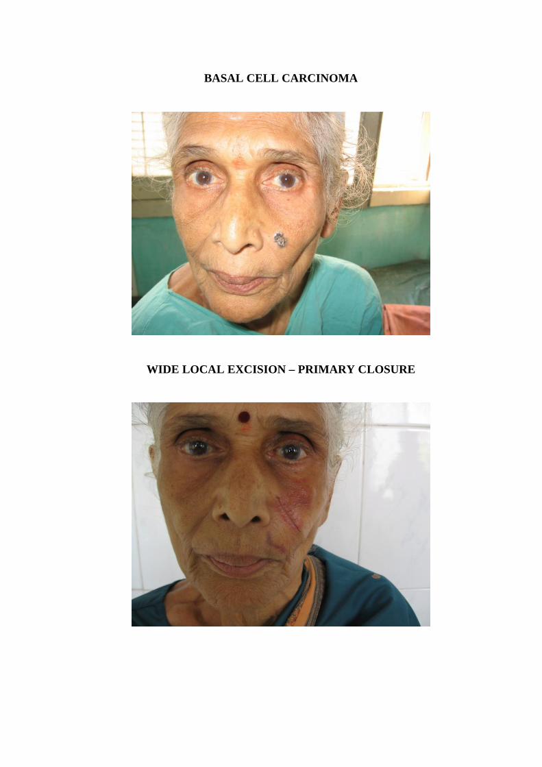
BASAL CELL CARCINOMA
WIDE LOCAL EXCISION – PRIMARY CLOSURE
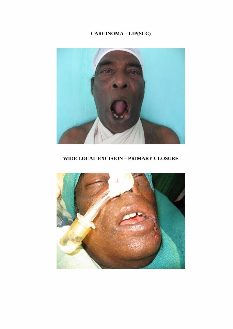
CARCINOMA – LIP(SCC)
WIDE LOCAL EXCISION – PRIMARY CLOSURE
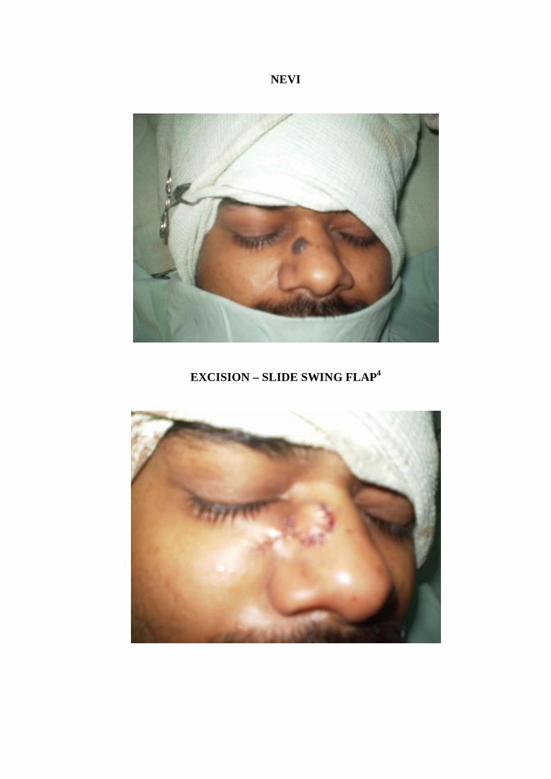
NEVI
EXCISION – SLIDE SWING FLAP4

BASAL CELL CARCINOMA
WIDE LOCAL EXCISION - FTSG

MARGIN POSITIVE
WIDE LOCAL EXCISION
BILATERAL SCALP ROTATION FLAP


![Loki N3 15 WHL P75F006, 3583, 3580 - Dell inspiron 3583 p75f... · 2019-06-18 · 3urgxfw &rpsoldqfh 'dwdvkhhw _ (19 _ $ 'hoo ,qf zzz ghoo frp / v v o h r } v ( ] v ] o , 6wdwhphqw](https://img.dokumen.tips/doc/110x75/5f75f821869d8267936ce0e2/loki-n3-15-whl-p75f006-3583-3580-dell-inspiron-3583-p75f-2019-06-18-3urgxfw.jpg)