Embed Size (px)
Citation preview

Hindawi Publishing CorporationInternational Journal of Biomedical ImagingVolume 2006, Article ID 86747, Pages 1–6DOI 10.1155/IJBI/2006/86747
Brain Structure Segmentation from MRI byGeometric Surface Flow
Greg Heckenberg,1 Yongjian Xi,1 Ye Duan,1 and Jing Hua2
1 Department of Computer Science, College of Engineering, University of Missouri-Columbia, Columbia, MO 65211-2060, USA2 Department of Computer Science, College of Liberal Arts and Sciences, Wayne State University, Detroit, MI 48202, USA
Received 1 August 2005; Revised 22 September 2005; Accepted 28 September 2005
Recommended for Publication by Ming Jiang
We present a method for semiautomatic segmentation of brain structures such as thalamus from MRI images based on the con-cept of geometric surface flow. Given an MRI image, the user can interactively initialize a seed model within region of interest.The model will then start to evolve by incorporating both boundary and region information following the principle of variationalanalysis. The deformation will stop when an equilibrium state is achieved. To overcome the low contrast of the original imagedata, a nonparametric kernel-based method is applied to simultaneously update the interior probability distribution during themodel evolution. Our experiments on both 2D and 3D image data demonstrate that the new method is robust to image noise andinhomogeneity and will not leak from spurious edge gaps.
Copyright © 2006 Greg Heckenberg et al. This is an open access article distributed under the Creative Commons AttributionLicense, which permits unrestricted use, distribution, and reproduction in any medium, provided the original work is properlycited.
1. INTRODUCTION
Thalamus is the relay center for nerve impulses in the brain.It mediates communication among sensory, motor, and as-sociative brain regions. Axons from almost every sensorysystem connect here as the last site before the informationreaches the cerebral cortex. Information received from the di-verse brain regions is passed on to the cortex through the tha-lamus. Anatomically, thalamus is the largest, most internalstructure of the diencephalon consisting of dual lobe massesof gray matter. It is located at the rostral end of the mid-brain on each side of the third ventricle. Each lobe is about4 centimeters. Motor nuclei of the thalamus receive signalsfrom the striatum and cerebellum and project into the mo-tor and premotor areas of the cerebral cortex. The thalamusplay a major role in the regulation of consciousness, alert-ness, arousal, and attention and is thus considered part ofthe limbic system.
Thalamus segmentation has become more and more es-sential for a wide range of clinical and research applica-tions. For example, thalamus changes in terms of volumeand intensity are involved in a large number of diseases, suchas schizophrenia, Parkinson’s disease and multiple sclerosis,and so forth. Manual segmentation is very labor intensiveand the result is not reproducible. On the other hand, dis-crete methods such as thresholding or region growing are not
reliable because of the low contrast and discontinuous edgesin the MRI images of thalamus.
In this paper, we present a new semiautomatic frameworkfor thalamus segmentation based on the concept of geomet-ric surface flow. Unlike previous methods with edge infor-mation only, we apply a nonparametric kernel-based methodthat can simultaneously update the interior region statis-tics along with the boundary shape evolution. By integratingboth boundary and region information, the new method isrobust to image noise and inhomogeneity and will not leakfrom spurious edge gaps.
2. ACTIVE CONTOURS AND MEDICAL IMAGESEGMENTATION
Segmenting structures from medical images and reconstruct-ing a compact geometric representation of these structuresis difficult due to the sheer size of the datasets and the com-plexity and variability of the anatomic shapes of interest. Fur-thermore, the shortcomings typical of sampled data, such assampling artifacts, spatial aliasing, and noise, may cause theboundaries of structures to be indistinct and disconnected.The challenge is to extract boundary elements belonging tothe same structure and integrate these elements into a co-herent and consistent model of the structure. Among var-ious segmentation techniques, active contours/deformable

2 International Journal of Biomedical Imaging
models have been very successful since their invention inlate 80’s. The mathematical foundation of deformable mod-els stems from the confluence of geometry, physics, and ap-proximation theory. Terzopoulos pioneered the theory ofcontinuous (multidimensional) deformable models basedon Lagrangian dynamics [1] and formulated deformationenergies for generalized splines with controlled continuity[1]. Kass et al. [2] introduced the active contour model or“snake,” a deformable model which is essentially a 2D splinethat minimizes an internal deformation energy subject to ex-ternal forces derived from images. Later, a voluminous litera-ture on deformable models appeared in computer vision, andespecially in the field of medical image analysis [3–9]. For acollection of seminal works, see [10, 11].
3. THALAMUS SEGMENTATION BY GEOMETRICSURFACE FLOW
3.1. Geometric surface flow
Our new thalamus segmentation framework is based on theconcept of geometric surface flow. The general formulationof the geometric surface flow is the following initial-value dy-namical system of nonlinear PDEs:
∂�s(�p)
∂t= F(t, k, k′, f , . . . )�n
(�p, t), �s
(�p, 0) =�s0
(�p),
(1)
where F is the speed function, t is the time variable, k andk′ are the surface curvature and its derivative at the point�p, �s0(�p) is the initial surface, �n is the surface normal vector.Equation (1) can be either directly provided by the user, ormore generally, obtained as a gradient descent flow of theEuler-Lagrange equation of certain energy functionals by thecalculus of variations.
In general, there are two approaches to numerically sim-ulate PDEs such as (1): explicit Lagrangian approach or im-plicit level-set approach. Implicit level-set methods [3, 6] arebecoming popular recently mainly because of their ease inhandling topology changes, however since all the compu-tations are conducted in a higher dimension, the compu-tational cost tends to be quite expensive. In this paper, wetake the explicit Lagrangian approach to simulate the sur-face flow. In particular, we apply the new framework we re-cently proposed [12, 13]. In our new framework, the geom-etry and topology of the model are always explicitly repre-sented throughout the simulation process. To ensure the reg-ularity of the model and the stability of the numerical in-tegration process, powerful Laplacian tangential smoothing,along with commonly used mesh optimization techniques,are employed throughout the geometric deformation andtopological variation process. In addition, a novel particle-based collision detection scheme is conducted to automati-cally handle topology changes during the deformation. Thenew framework for surface flow simulation is fast, simple,and accurate. More importantly, it allows the user to directlyinteract with the model during the deformation. For moredetails of our new framework, please refer to [12, 13].
3.2. Thalamus segmentation
When applying the above geometric surface flow for tha-lamus segmentation, the speed function F in (1) is explic-itly formulated as a linear combination of the following twoterms:
F = a1Freg + Fdata, (2)
here Freg is the regularity term to maintain the smoothnessof the model. Freg is usually a function of the curvature (e.g.,mean curvature, Gaussian curvature). In this paper, we de-fine Freg as the difference between the mean curvature Hcurr
at the current position and the average mean curvature Havg
of the whole model:
Freg = Hcurr −Havg. (3)
Comparing with other commonly used regularity terms suchas mean curvature or Gaussian curvature, (3) works muchbetter in our experiment. This is probably because of the vol-ume preserving property of (3), which can be considered asa shape prior term for the thalamus’s oval shape. A more de-tailed explanation can be found in [14], where a similar termis used.
Fdata is the term that interacts the model with the im-age data, and is often defined as a function of the edge in-formation of the image data. However, since the thalamushas low contrast, using boundary information alone is notvery reliable. It is more preferable to combine the edge in-formation along with the region information as suggested byHuang et al. [15]. Hence, in this paper, we propose to employboth the edge information as well as the region information.In particular, we define Fdata as a linear combination of thefollowing two terms:
Fdata = a2Fedge + a3Fregion, (4)
Fedge is the speed function corresponding to the edge infor-mation in the image data that will attract the contour movingtowards the edges of the image data and is defined as
Fedge = −Δ(Gσ ∗ I(s)
), (5)
where Δ is the Laplacian operator, I is the image intensityfunction, and Gσ ∗ I is the smoothed intensity function byconvoluting with a Gaussian filter with variance σ . Varianceσ can be assigned according to the image resolution of theMRI dataset. In our experiment, σ is set as 1.0.
Fregion is the speed function corresponding to the regioninformation, and is defined as
Fregion = 1
1 +∣∣∇(B(s)
)∣∣2 , (6)

Greg Heckenberg et al. 3
where ∇ is the gradient operator, B(s) is the binary imagecreated by the interior probability estimation of the currentmodel, which will be explained in more detail in the follow-ing section (Sections 3.3.2 and 3.3.3). In a nutshell, Fregion willexpand the contour outwards in regions that are consideredcompatible with the current segmented volume’s statistics.
Substituting (3), (4), (5), and (6) into (2), we obtainedthe following speed function for thalamus segmentation:
F = a1(Hcurr −Havg
)− a2Δ(Gσ ∗ I(s)
)
+ a31
1 +∣∣∇(B(s)
)∣∣2 .(7)
Here, a1, a2, a3 are the corresponding weighting coeffi-cients. In our experiments, a1, a2, a3 are set as 0.1, 1, 1, re-spectively.
3.3. Algorithm pipeline
There are four main steps in our thalamus segmentationframework: (1) seed initialization; (2) interior statistics es-timation; (3) binary image creation; (4) model evolution.
3.3.1. Seed initialization
First, the user interactively selects a pixel/voxel inside the re-gion of interest in the image data. Considering the thala-mus has an oval shape, a circular contour centered at thepixel/voxel is then automatically created and serves as the ini-tial seed model.
3.3.2. Interior statistics estimation
Then, the intensity probability density function of the in-terior regions enclosed by the seed contour is estimated.Specifically, we approximate the distribution by a Parzen-window-function-based nonparametric method [16] be-cause it is differentiable, more generic, and can representcomplex multimodal intensity distributions. We choose theGaussian kernel as the Parzen window function. Suppose themodel M is placed on an image I , the volume of the imageregion bounded by the current model M is V , then the prob-ability of a pixel’s (voxel’s) intensity value i being consistentwith the model interior intensity can be derived as
P(i |M) = 1V
∫∫∫1√2πσ
e−(i−I(y))2/2σ2, (8)
where σ is a constant that specifies the width of the Gaussiankernel and is set as 1.0 in our experiment. Since (8) is a simpleintegral, it can be calculated very efficiently as an incremen-tal update, that is, only newly added voxels are calculated toupdate the integral value at each time step.
3.3.3. Binary image creation
Next, based on the interior probability density distribution ofmodel M obtained in the previous step, the image intensity
probability map PI of every pixel’s (voxel’s) intensity is ob-tained. Then a small threshold (e.g., the mean probabilityover the entire image domain) is applied on PI to producea binary image B(s), in which pixels/voxels with probabilityhigher than the threshold have value 1, zero otherwise.
3.3.4. Model evolution
Finally, the model will start to evolve according to (1) and(7). More specifically, the surface evolution process is numer-ically approximated using a simple, explicit iterative equa-tion:
�S(�p, t + Δt
) = �S(�p, t)
+ F(�p, t)�N(�p, t)Δt. (9)
When advancing the model, we must enforce a constrainton the size of the time step Δt. In particular, the time step Δtmust satisfy the following stability condition: the velocity ofchange must be strictly restrained by the minimum detail inthe system. In our system, this condition is
Δt ≤ me
MF, (10)
where me is the unit grid cell length of the image data and MF
is the maximum magnitude of the speed F obtained by (7).Before each deformation step, we will calculate the velocity Fat each vertex point and determine the maximum magnitudeof the velocity MF . A proper time step can then be estimatedfrom (10). At each deformation cycle, the model will loopfrom step 2 to step 4 until an equilibrium state is achieved,then the deformation stops and the geometry of the thalamusis extracted from the MRI image.
3.4. Experimental results
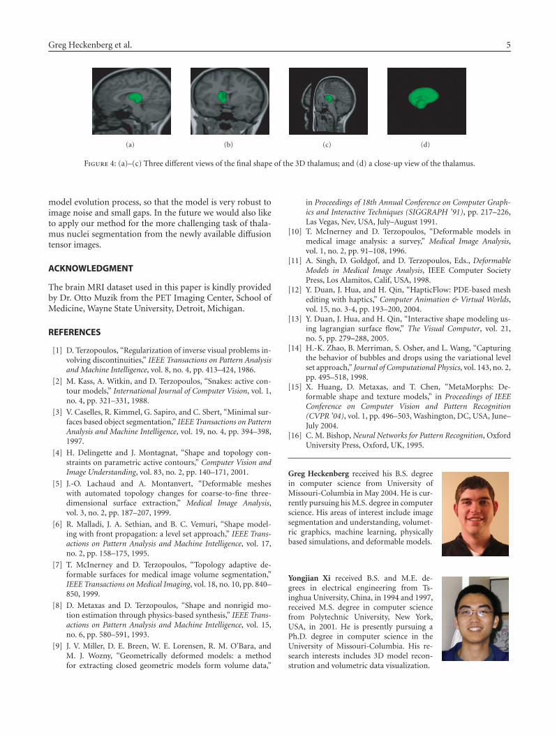
Some of the experimental results for 2D and 3D thalamussegmentations are shown in Figure 1 to Figure 4. In partic-ular, an example of our 2D thalamus segmentation result isshown in Figures 1 and 2. Each of the two figures shows fivesnapshots of the model evolution process including the ini-tial seed (a) and the final shape (e). Figure 1 shows the modelsuperimposed with the original image, while Figure 2 showsthe same five snapshots superimposed with the binary im-age created by the interior statistics estimation (Sections 3.3.2and 3.3.3). The 3D thalamus segmentation result is shown inFigures 3 and 4. Figure 3 shows the four snapshots during themodel evolution process. The four different views of the final3D shape of the thalamus are shown in Figure 4.
4. CONCLUSION
We proposed a semiautomatic thalamus segmentation meth-od based on an explicit simulation of geometric surface flow.To overcome the low contrast of the thalamus, we employ anonparametric kernel-based statistics estimation that can in-corporate both the boundary and region information in the

4 International Journal of Biomedical Imaging
(a) (b) (c)
(d) (e)
Figure 1: The model evolution process of the 2D thalamus segmentation superimposed with original image. (a) Initial seed; (b)–(d) threeintermediate stages; and (e) final extracted shape.
(a) (b) (c)
(d) (e)
Figure 2: The same five snapshots of the model evolution process of the 2D thalamus segmentation superimposed with the binary maskcreated by interior statistics estimation.
(a) (b) (c) (d)
Figure 3: The model evolution process of the 3D thalamus segmentation superimposed with original image. (a) Initial seed; (b)-(c) twointermediate stages; and (d) final extracted shape.
Greg Heckenberg et al. 5
(a) (b) (c) (d)
Figure 4: (a)–(c) Three different views of the final shape of the 3D thalamus; and (d) a close-up view of the thalamus.
model evolution process, so that the model is very robust toimage noise and small gaps. In the future we would also liketo apply our method for the more challenging task of thala-mus nuclei segmentation from the newly available diffusiontensor images.
ACKNOWLEDGMENT
The brain MRI dataset used in this paper is kindly providedby Dr. Otto Muzik from the PET Imaging Center, School ofMedicine, Wayne State University, Detroit, Michigan.
REFERENCES
[1] D. Terzopoulos, “Regularization of inverse visual problems in-volving discontinuities,” IEEE Transactions on Pattern Analysisand Machine Intelligence, vol. 8, no. 4, pp. 413–424, 1986.
[2] M. Kass, A. Witkin, and D. Terzopoulos, “Snakes: active con-tour models,” International Journal of Computer Vision, vol. 1,no. 4, pp. 321–331, 1988.
[3] V. Caselles, R. Kimmel, G. Sapiro, and C. Sbert, “Minimal sur-faces based object segmentation,” IEEE Transactions on PatternAnalysis and Machine Intelligence, vol. 19, no. 4, pp. 394–398,1997.
[4] H. Delingette and J. Montagnat, “Shape and topology con-straints on parametric active contours,” Computer Vision andImage Understanding, vol. 83, no. 2, pp. 140–171, 2001.
[5] J.-O. Lachaud and A. Montanvert, “Deformable mesheswith automated topology changes for coarse-to-fine three-dimensional surface extraction,” Medical Image Analysis,vol. 3, no. 2, pp. 187–207, 1999.
[6] R. Malladi, J. A. Sethian, and B. C. Vemuri, “Shape model-ing with front propagation: a level set approach,” IEEE Trans-actions on Pattern Analysis and Machine Intelligence, vol. 17,no. 2, pp. 158–175, 1995.
[7] T. McInerney and D. Terzopoulos, “Topology adaptive de-formable surfaces for medical image volume segmentation,”IEEE Transactions on Medical Imaging, vol. 18, no. 10, pp. 840–850, 1999.
[8] D. Metaxas and D. Terzopoulos, “Shape and nonrigid mo-tion estimation through physics-based synthesis,” IEEE Trans-actions on Pattern Analysis and Machine Intelligence, vol. 15,no. 6, pp. 580–591, 1993.
[9] J. V. Miller, D. E. Breen, W. E. Lorensen, R. M. O’Bara, andM. J. Wozny, “Geometrically deformed models: a methodfor extracting closed geometric models form volume data,”
in Proceedings of 18th Annual Conference on Computer Graph-ics and Interactive Techniques (SIGGRAPH ’91), pp. 217–226,Las Vegas, Nev, USA, July–August 1991.
[10] T. McInerney and D. Terzopoulos, “Deformable models inmedical image analysis: a survey,” Medical Image Analysis,vol. 1, no. 2, pp. 91–108, 1996.
[11] A. Singh, D. Goldgof, and D. Terzopoulos, Eds., DeformableModels in Medical Image Analysis, IEEE Computer SocietyPress, Los Alamitos, Calif, USA, 1998.
[12] Y. Duan, J. Hua, and H. Qin, “HapticFlow: PDE-based meshediting with haptics,” Computer Animation & Virtual Worlds,vol. 15, no. 3-4, pp. 193–200, 2004.
[13] Y. Duan, J. Hua, and H. Qin, “Interactive shape modeling us-ing lagrangian surface flow,” The Visual Computer, vol. 21,no. 5, pp. 279–288, 2005.
[14] H.-K. Zhao, B. Merriman, S. Osher, and L. Wang, “Capturingthe behavior of bubbles and drops using the variational levelset approach,” Journal of Computational Physics, vol. 143, no. 2,pp. 495–518, 1998.
[15] X. Huang, D. Metaxas, and T. Chen, “MetaMorphs: De-formable shape and texture models,” in Proceedings of IEEEConference on Computer Vision and Pattern Recognition(CVPR ’04), vol. 1, pp. 496–503, Washington, DC, USA, June–July 2004.
[16] C. M. Bishop, Neural Networks for Pattern Recognition, OxfordUniversity Press, Oxford, UK, 1995.
Greg Heckenberg received his B.S. degreein computer science from University ofMissouri-Columbia in May 2004. He is cur-rently pursuing his M.S. degree in computerscience. His areas of interest include imagesegmentation and understanding, volumet-ric graphics, machine learning, physicallybased simulations, and deformable models.
Yongjian Xi received B.S. and M.E. de-grees in electrical engineering from Ts-inghua University, China, in 1994 and 1997,received M.S. degree in computer sciencefrom Polytechnic University, New York,USA, in 2001. He is presently pursuing aPh.D. degree in computer science in theUniversity of Missouri-Columbia. His re-search interests includes 3D model recon-strution and volumetric data visualization.

6 International Journal of Biomedical Imaging
Ye Duan is an Assistant Professor of com-puter science at University of Missouri-Columbia. He received his Ph.D. degree incomputer science from the State Universityof New York at Stony Brook (2003). He re-ceived his M.S. degree in computer sciencefrom the State University of New York atStony Brook in 1998. In 1996, he receivedhis M.S. degree in mathematics from UtahState University. He received his B.A. degreein mathematics from Peking University in 1991. His research in-terests include biomedical imaging, computer graphics, scientificvisualization, computer vision, geometric and physics-based mod-eling, virtual reality and human-computer Interaction, and com-puter animation and simulation.
Jing Hua is an Assistant Professor in com-puter science at the Wayne State Univer-sity and he is the Director of Graphics andImaging Lab. He received his Ph.D. degreein computer science from the State Univer-sity of New York at Stony Brook in 2004.His research interests include biomedicalimage analysis, computational geometry,physics-based modeling, scientific visual-ization, human-computer interaction, andcomputer vision.