Embed Size (px)
Citation preview

8/3/2019 Brain Overview - Sub Cortical
http://slidepdf.com/reader/full/brain-overview-sub-cortical 1/49
Overview of Gross Neuroanatomy
References:
DeArmond, S.J., Fusco, M.M., Dewey M.M., (1988), Structure of the
Human Brain, 3rd Ed., Oxford, NY
Warner J.J., Atlas of Neuroanatomy with Systems Organization and
Case Correlations, 2001
University of Florida class notes on Medical Neuroscience
Carpenter,Human Neuroanatomy, 7th ed., 1976

8/3/2019 Brain Overview - Sub Cortical
http://slidepdf.com/reader/full/brain-overview-sub-cortical 2/49
Directional Terms
8/3/2019 Brain Overview - Sub Cortical
http://slidepdf.com/reader/full/brain-overview-sub-cortical 3/49
Rostral (beak) Caudal (tail)
Anterior (front) Posterior (back)
Dorsal (top) Ventral (belly)
Superior (top) Inferior (bottom)
Medial (midline) Lateral (side)
Axes on the brain

8/3/2019 Brain Overview - Sub Cortical
http://slidepdf.com/reader/full/brain-overview-sub-cortical 4/49
Planar cuts
Coronal
(parallel to face)
Axial
(parallel to ground)
Sagittal
(parallel to ears)

8/3/2019 Brain Overview - Sub Cortical
http://slidepdf.com/reader/full/brain-overview-sub-cortical 5/49
Parts of the brain

8/3/2019 Brain Overview - Sub Cortical
http://slidepdf.com/reader/full/brain-overview-sub-cortical 6/49
Interhemispheric fissure
- visible on coronals and axials
- separates R and L hemispheres
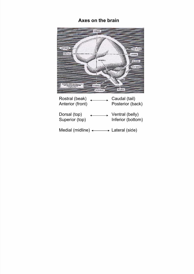
Fissures
Sylvian fissure
- present on both R and L
- separates the temporal lobe
- commonly asymmetric (L>R)
Sylvian
Interhemispheric
8/3/2019 Brain Overview - Sub Cortical
http://slidepdf.com/reader/full/brain-overview-sub-cortical 7/49
insula
Sylvian
Fissure
insula
Sylvian
Fissure

8/3/2019 Brain Overview - Sub Cortical
http://slidepdf.com/reader/full/brain-overview-sub-cortical 8/49
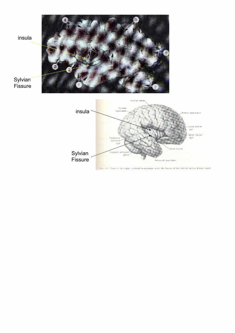
Layers inside the skull
R hemisphere
cerebellum
8/3/2019 Brain Overview - Sub Cortical
http://slidepdf.com/reader/full/brain-overview-sub-cortical 9/49
Meninges:- Dura mater: Opaque, tough, follows skull boundary and major fissures.
Consists of 2 parts: falx cerebri, falx cerebelli
- Arachnoid mater: Web-like, white, bridges over major folds
- Pia mater: Transparent, follows brain surface continuously- pial surface
dura

8/3/2019 Brain Overview - Sub Cortical
http://slidepdf.com/reader/full/brain-overview-sub-cortical 10/49
Gray Matter (GM): White Matter (WM):(functions of the brain) (connectivity of the brain)
- Neo-cortex: sulci, gyri - Association fibers
- Sub-cortical structures - Midline structures
neo-cortex sub-cortical
midline WM association fibers
neo-cortex
association fibers
sub-cortical
Tissue classes

8/3/2019 Brain Overview - Sub Cortical
http://slidepdf.com/reader/full/brain-overview-sub-cortical 11/49
Lobes

8/3/2019 Brain Overview - Sub Cortical
http://slidepdf.com/reader/full/brain-overview-sub-cortical 12/49
Lobes

8/3/2019 Brain Overview - Sub Cortical
http://slidepdf.com/reader/full/brain-overview-sub-cortical 13/49

8/3/2019 Brain Overview - Sub Cortical
http://slidepdf.com/reader/full/brain-overview-sub-cortical 14/49
Frontal Lobe: motor, executive learning, semantic processing, emotion
Boundaries: central sulcus, Sylvian fissure
Temporal Lobe: auditory processingBoundaries: Sylvian fissure
Parietal Lobe: somatosensory
Boundaries: central sulcus, parieto-occipital sulcus (imaginary projection)
Occipital Lobe: vision
parieto-occipital sulcus
Limbic Lobe: emotion, memory, learning
Boundaries: paracingulate sulcus, hippocampus, amygdala

8/3/2019 Brain Overview - Sub Cortical
http://slidepdf.com/reader/full/brain-overview-sub-cortical 15/49

8/3/2019 Brain Overview - Sub Cortical
http://slidepdf.com/reader/full/brain-overview-sub-cortical 16/49
Bottom view
Top view
Brain stem
Top
View
BottomView
Cerebellar peduncles
Superior colliculi
Inferior colliculi
Pons
Thalamus

8/3/2019 Brain Overview - Sub Cortical
http://slidepdf.com/reader/full/brain-overview-sub-cortical 17/49
Cerebellum

8/3/2019 Brain Overview - Sub Cortical
http://slidepdf.com/reader/full/brain-overview-sub-cortical 18/49
Blood and water supply

8/3/2019 Brain Overview - Sub Cortical
http://slidepdf.com/reader/full/brain-overview-sub-cortical 19/49
Blood-brain barrier:
Substances navigating inside the blood do not penetrate
into brain as in other organs. There is a selective penetration
Blood-CSF barrier:
Substances navigating inside the blood do not penetrate into
ventricles
Note:No CSF-brain barrier. When dyes are injected into ventricles,
the brain is stained.

8/3/2019 Brain Overview - Sub Cortical
http://slidepdf.com/reader/full/brain-overview-sub-cortical 20/49
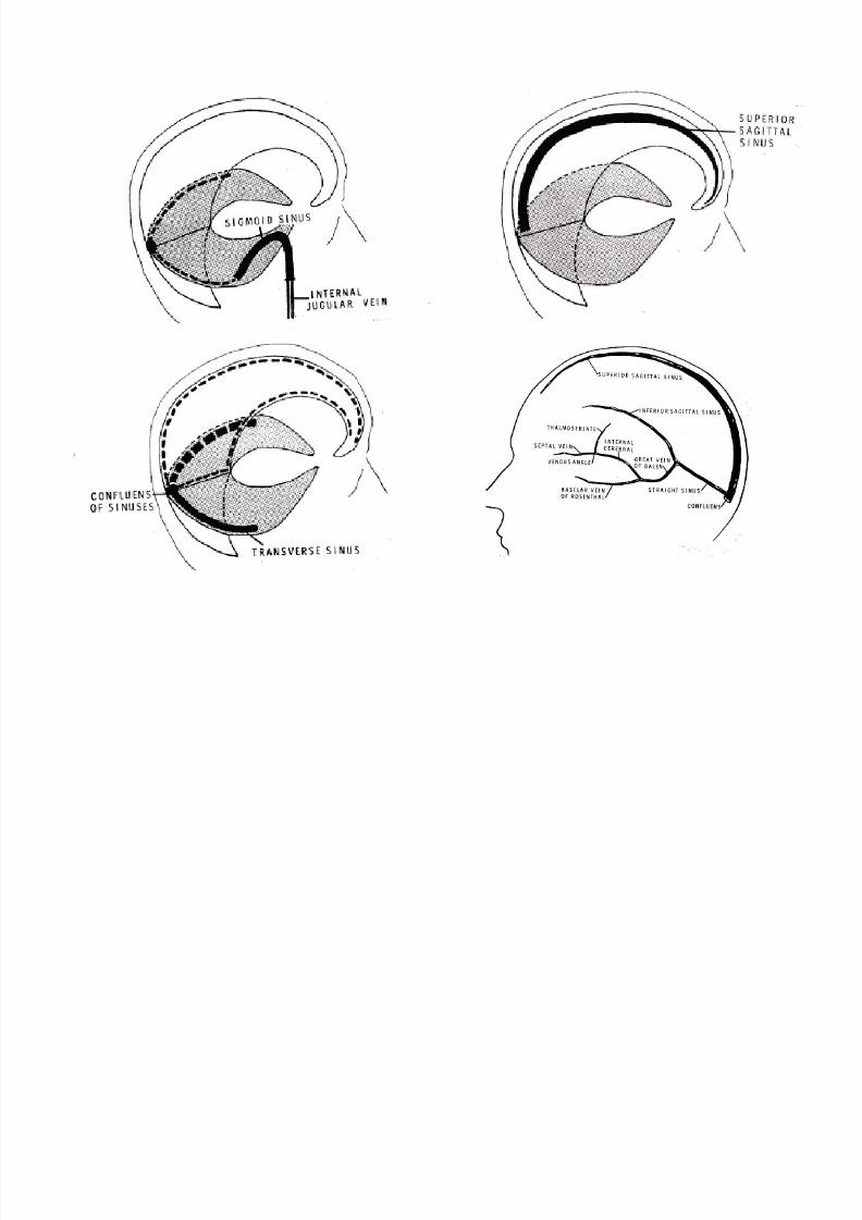
Veins
Veins run in sub dural space
8/3/2019 Brain Overview - Sub Cortical
http://slidepdf.com/reader/full/brain-overview-sub-cortical 21/49

8/3/2019 Brain Overview - Sub Cortical
http://slidepdf.com/reader/full/brain-overview-sub-cortical 22/49
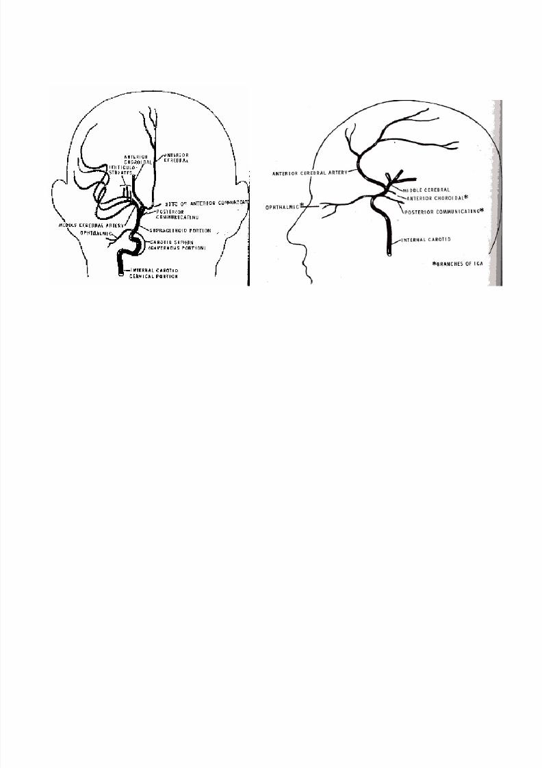
Arteries
Arteries run in sub arachnoid space
8/3/2019 Brain Overview - Sub Cortical
http://slidepdf.com/reader/full/brain-overview-sub-cortical 23/49

8/3/2019 Brain Overview - Sub Cortical
http://slidepdf.com/reader/full/brain-overview-sub-cortical 24/49
Ventricles
- CSF runs in ventricles, and subarachnoid space

8/3/2019 Brain Overview - Sub Cortical
http://slidepdf.com/reader/full/brain-overview-sub-cortical 25/49
Ventricles
Anterior Posterior

8/3/2019 Brain Overview - Sub Cortical
http://slidepdf.com/reader/full/brain-overview-sub-cortical 26/49
Choroid plexus:
Produces CSF
Septum pellucidum

8/3/2019 Brain Overview - Sub Cortical
http://slidepdf.com/reader/full/brain-overview-sub-cortical 27/49
Interhemispheric structures

8/3/2019 Brain Overview - Sub Cortical
http://slidepdf.com/reader/full/brain-overview-sub-cortical 28/49
WM: Anterior Comissure (AC) GM: Massa IntermediaPosterior Comissure (PC) (or interthalamic adhesion)
Corpus Calossum
anterior posterior

8/3/2019 Brain Overview - Sub Cortical
http://slidepdf.com/reader/full/brain-overview-sub-cortical 29/49
AC
PC
genu
splenium
body
septum pellicidummassa intermedia
rostrum

8/3/2019 Brain Overview - Sub Cortical
http://slidepdf.com/reader/full/brain-overview-sub-cortical 30/49
anterior
comissure
corpus
calossum

8/3/2019 Brain Overview - Sub Cortical
http://slidepdf.com/reader/full/brain-overview-sub-cortical 31/49
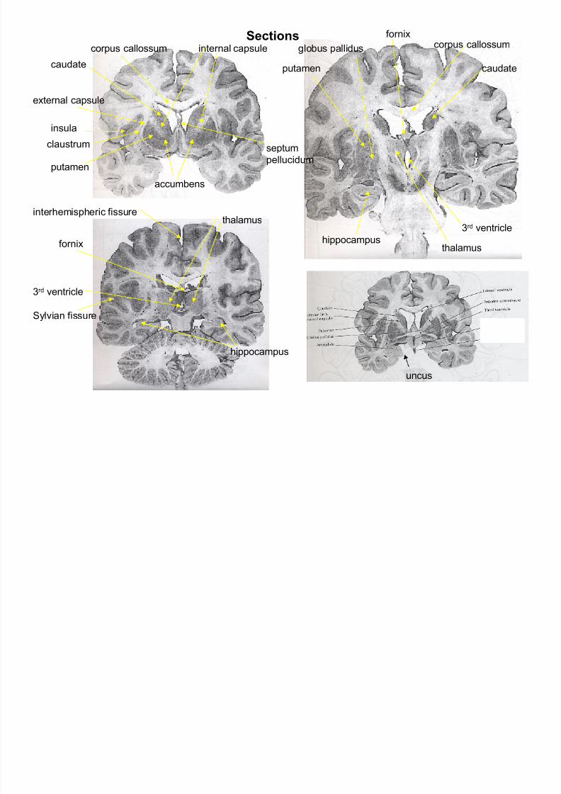
Subcortical structures

8/3/2019 Brain Overview - Sub Cortical
http://slidepdf.com/reader/full/brain-overview-sub-cortical 32/49
Subcortical GM:
Basal ganglia: caudate, putamen, globus pallidus, claustrum (initiating behavior)
Thalamus: Sensory gateway to the cerebral cortex
Hypothalamus: Autonomic nervous system (motivated behaviour)
Basal forebrain: Amygdala (emotion)
Hippocampal formation: (memory)
Adjacent WM:
Internal capsule
External capsule
Extreme capsule
Fornix
Ant
Post
Basal

8/3/2019 Brain Overview - Sub Cortical
http://slidepdf.com/reader/full/brain-overview-sub-cortical 33/49
Basal
Ganglia

8/3/2019 Brain Overview - Sub Cortical
http://slidepdf.com/reader/full/brain-overview-sub-cortical 34/49
Internal capsule

8/3/2019 Brain Overview - Sub Cortical
http://slidepdf.com/reader/full/brain-overview-sub-cortical 35/49
Internal capsule
Sk t h

8/3/2019 Brain Overview - Sub Cortical
http://slidepdf.com/reader/full/brain-overview-sub-cortical 36/49
Basal ganglia
Thalamic nuclei
Hippocampus and fornix
Temporal Lobe
Sketches
Posterior Anterior
fornixSections
8/3/2019 Brain Overview - Sub Cortical
http://slidepdf.com/reader/full/brain-overview-sub-cortical 37/49
insula
accumbens
uncus
3rd ventricle
internal capsule
external capsule
claustrum
hippocampus
globus palliduscorpus callossum
Sylvian fissure
interhemispheric fissure
fornix
hippocampus
putamen
caudate putamen caudate
thalamus
thalamus
corpus callossum
3rd ventricle
septum
pellucidum
Sections

8/3/2019 Brain Overview - Sub Cortical
http://slidepdf.com/reader/full/brain-overview-sub-cortical 38/49
Amygdala and hippocampus
amygdala
hippocampus

8/3/2019 Brain Overview - Sub Cortical
http://slidepdf.com/reader/full/brain-overview-sub-cortical 39/49
Fornix
fornixfornix
fornix
Anterior
comissureMamillary
Body
Ant
Post

8/3/2019 Brain Overview - Sub Cortical
http://slidepdf.com/reader/full/brain-overview-sub-cortical 40/49
Thalamus and Hypothalamus

8/3/2019 Brain Overview - Sub Cortical
http://slidepdf.com/reader/full/brain-overview-sub-cortical 41/49
Hints for identifying subcortical structures
Caudate: always on the walls of lateral ventricle
frontally, it is located superior to internal capsule
Putamen:runs on the inferior part of internal capsule
Globus pallidus: next to putamen seperated by wm tracts
Thalamus: on the walls of third ventricle
Hippocampus: inferior to the inferior horn of lateral ventricle
Amygdala: Inside part of uncus
Anterior Comissure: Close to the frontal horns of fornix
Claustrum: skinny GM between extreme capsule and insula

8/3/2019 Brain Overview - Sub Cortical
http://slidepdf.com/reader/full/brain-overview-sub-cortical 42/49
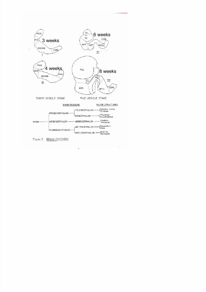
Developmental
and aging processes
8/3/2019 Brain Overview - Sub Cortical
http://slidepdf.com/reader/full/brain-overview-sub-cortical 43/49
3 weeks
4 weeks
6 weeks
8 weeks

8/3/2019 Brain Overview - Sub Cortical
http://slidepdf.com/reader/full/brain-overview-sub-cortical 44/49
12 week fetus
7 month fetus
8/3/2019 Brain Overview - Sub Cortical
http://slidepdf.com/reader/full/brain-overview-sub-cortical 45/49
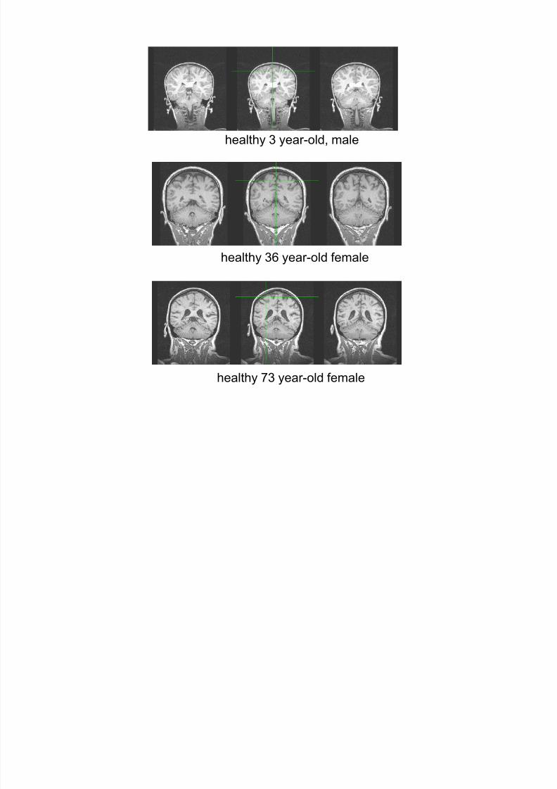
healthy 3 year-old, male
healthy 36 year-old female
healthy 73 year-old female
V l t i h

8/3/2019 Brain Overview - Sub Cortical
http://slidepdf.com/reader/full/brain-overview-sub-cortical 46/49
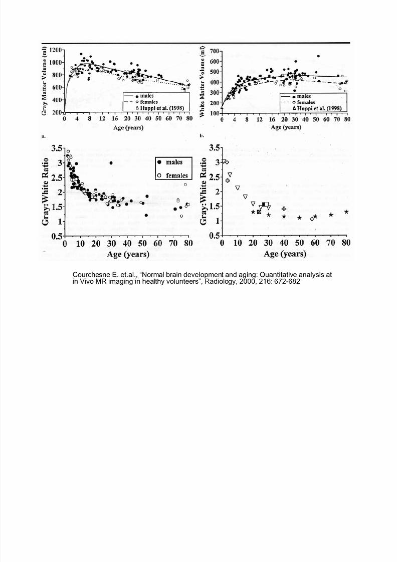
Courchesne E. et.al., “Normal brain development and aging: Quantitative analysis atin Vivo MR imaging in healthy volunteers”, Radiology, 2000, 216: 672-682
Volumetric changes across ages
8/3/2019 Brain Overview - Sub Cortical
http://slidepdf.com/reader/full/brain-overview-sub-cortical 47/49
Courchesne E. et.al., “Normal brain development and aging: Quantitative analysis atin Vivo MR imaging in healthy volunteers”, Radiology, 2000, 216: 672-682

8/3/2019 Brain Overview - Sub Cortical
http://slidepdf.com/reader/full/brain-overview-sub-cortical 48/49
Volumetric changes vs performance in aging
reference
n=73
Walhovd et.al., 2005
S
8/3/2019 Brain Overview - Sub Cortical
http://slidepdf.com/reader/full/brain-overview-sub-cortical 49/49
Summary
• Whole brain and intracranial space grows about 25% between early
childhood and adolescence
• By age 80 this growth is reversed such that whole brain of agedpeople are about the same size of young children
• Between early to late childhood, GM increases about 13%.
• After the 4th decade of life, GM decreased about 13% until age 80.
• Increase in CSF between adolescence and old age is about 100%
• GM loss is not always bad. Effective pruning occurs twice: at 8
months old and at adolescence