Embed Size (px)
Citation preview

Eur. J. Immunol. 2012. 42: 1–10 ImmunomodulationDOI: 10.1002/eji.201142341 1
Both IFN-γ and IL-17 are required for the developmentof severe autoimmune gastritis
Eric Tu1, Desmond K.Y. Ang1, Shayne A. Bellingham1, Thea V. Hogan1,Michele W. L. Teng2,3, Mark J. Smyth2,3, Andrew F. Hill1
and Ian R. van Driel1
1 Department of Biochemistry and Molecular Biology, Bio21 Molecular Science andBiotechnology Institute, The University of Melbourne, Parkville, Victoria, Australia
2 Cancer Immunology Program, Trescowthick Laboratories, Peter MacCallum Cancer Centre,St. Andrews Place, East Melbourne, Victoria, Australia
3 Department of Pathology, University of Melbourne, Parkville, Australia
IL-17, produced by a distinct lineage of CD4+ helper T (Th) cells termed Th17 cells, inducesthe production of pro-inflammatory cytokines from resident cells and it has been demon-strated that over-expression of IL-17 plays a crucial role in the onset of several auto-immune diseases. Here we examined the role of IL-17 in the pathogenesis of autoimmunegastritis, a disease that was previously believed to be mediated by IFN-γ. Significantlyhigher levels of IL-17 and IFN-γ were found in the stomachs and stomach-draining lymphnodes of mice with severe autoimmune gastritis. Unlike IL-17, which was produced solelyby CD4+ T cells in gastritic mice, the majority of IFN-γ-producing cells were CD8+ T cells.However, CD8+ T cells alone were not able to induce autoimmune gastritis. T cells thatwere deficient in IL-17 or IFN-γ production were able to induce autoimmune gastritisbut to a much lower extent compared with the disease induced by wild-type T cells.These data demonstrate that production of neither IL-17 nor IFN-γ by effector T cells isessential for the initiation of autoimmune gastritis, but suggest that both are requiredfor the disease to progress to the late pathogenic stage that includes significant tissuedisruption.
Keywords: Autoimmune disease � Cytokines � IFN-γ � IL-17 � Immunopathology � T cells
Supporting Information available online
Introduction
CD4+ helper T cells play vital roles in the immune system by estab-lishing and augmenting immune responses against pathogens.However, these cells have also been found to induce autoimmunediseases and allergic inflammation. The three most characterisedTh subsets are Th1, Th2 and Th17 cells. They can be distinguished
Correspondence: Dr. Ian R. van Driele-mail: [email protected]
based on their unique transcription factors and cytokine profiles,which confer separate effector functions [1–4].
The association of Th1 response with autoimmunity cameinto question after the discovery of IL-23, which shares thep40 subunit with IL-12, but is engaged with a different sec-ond chain, IL-23p19 instead of IL-12p35 [5]. It is now clearthat the prevention of autoimmune diseases, including exper-imental autoimmune encephalomyelitis and collagen-inducedarthritis, by treatment with neutralising antibodies to IL-12p40 orin IL-12p40-deficient mice is conferred by inhibition of IL-23, butnot IL-12 [6,7]. Therefore, the IL-23–IL-17 axis rather than the
C© 2012 WILEY-VCH Verlag GmbH & Co. KGaA, Weinheim www.eji-journal.eu

2 Eric Tu et al. Eur. J. Immunol. 2012. 42: 1–10
Figure 1. Increased levels of IL-17+CD4+
T cells in the paragastric lymph nodeof mice with severe autoimmune gastri-tis. Irradiated wild-type BALB/cCrSlc.CD90.1congenic mice received 5 × 10⁷ lymphnode cells from either wild-type (normal)or H/Kα−/− (gastritic) mice. Eight weeksafter transfer, gastric pathology was deter-mined by histological examination andthe frequency of IL-17+CD4+ T cells inlymph nodes (identified by CD4 and CD90.2expression) was assessed by intracellularflow cytometry. (A) Quantitation of gas-tric pathology. (B) Representative flow cyto-metric plots. The values indicate the per-centages of IL-17+CD4+ T cells in thedonor CD90.2+ population. (C) Frequency ofIL-17+CD4+ T cells in the donor CD4+ T-cellpopulation. (D) Representative flow cyto-metric plots. Shown are IL-17+ and IFN-γ+
cells in the donor CD4+ T-cell populationin the paragastric lymph node. Data arerepresentative of (B and D) or pooled from(A and C) three independent experimentsand each symbol represents one mouse.Mann–Whitney U test was used; bars, meanand ***p < 0.001.
IL-12–IFN-γ axis is critically involved in the development ofautoimmunity in these models, which were previously thoughtto be caused by pathogenic Th1 responses. In light of these dis-coveries, we examined the role of IL-17 in the onset and pro-gression of autoimmune gastritis. Autoimmune gastritis is a CD4+
T cell-mediated organ-specific autoimmune disease in which thegastric H+/K+ ATPase has been identified as the target antigen inboth the mouse model and human equivalent [8,9]. The diseaseis associated with an inflammatory infiltrate in the gastric mucosa,the loss of gastric parietal and zymogenic cells, and hypertrophyof gastric mucosa [10]. Several studies have also indicated that adysregulated Th1 response is linked to the pathogenesis of auto-immune gastritis; CD4+ T cells isolated from the gastric mucosaof gastritic humans and mice showed a Th1 phenotype [11,12],and neutralising-IFN-γ antibody was able to prevent the onset ofmurine autoimmune gastritis [13]. Additionally, T cells from micedeficient in IFN-γ (IFN-γ−/−) or IL-12 have a reduced pathogenic-ity in causing autoimmune gastritis. On the other hand, IL-4,a signature cytokine of Th2 cells, seems to play a protectiverole [14].
One previous report has demonstrated that Th1, Th2 and Th17cells that are differentiated in vitro from a monoclonal gastrito-genic T-cell (A23 T cells) are all able to cause severe autoimmunegastritis in athymic nude mice [15]. It is noteworthy that naiveA23 T cells and polyclonal T cells have the capacity to inducesevere gastritis in athymic mice without prior differentiation toTh effectors [16]. As a result, whether IL-17-producing T cells areactually generated during, and responsible for, the development of
autoimmune gastritis in mice with a normal polyclonal repertoireis still unclear.
Using disease models that involve polyclonal pathogenicT cells, we show here that high levels of IL-17 and IFN-γ aredetected in mice with severe autoimmune gastritis, suggestingboth cytokines may be involved in the pathogenesis of the disease.Moreover, we demonstrate that the autoimmune gastritis can beinduced in the absence of either IL-17 or IFN-γ alone, but bothcytokines are required in combination for autoreactive T cells tocause severe damage.
Results
Increased levels of IL-17-producing T cells in micewith severe autoimmune gastritis
To address the role of IL-17 in autoimmune gastritis, gastritic andcontrol mice were produced by transferring lymph node cells fromeither HKα−/− or wild-type mice into sublethally irradiated wild-type mice. As previously described [17], HKα−/− mice containedhigh numbers of HKα-specific T cells and transfer of these cellsled to the severest form (score 6) of autoimmune gastritis in allrecipient mice (“Gastritic”) whereas mice that received wild-typecells remained unaffected (“Normal”) (Fig. 1A). Donor-derivedT cells were identified by the donor- and T cell-specific markerCD90.2 (Supporting Information Fig. 1). A significant increase inthe IL-17+ population of donor-derived CD4+ T cells was detected
C© 2012 WILEY-VCH Verlag GmbH & Co. KGaA, Weinheim www.eji-journal.eu
Eur. J. Immunol. 2012. 42: 1–10 Immunomodulation 3
in the paragastric lymph node of gastritic mice compared to that ofnormal mice. Furthermore, an elevated proportion of IL-17+CD4+
T cells was only found in the paragastric lymph node but notin the inguinal lymph nodes of gastritic mice (Fig. 1B and C).In both groups of mice, IL-17+ cells were confined to the CD4+
T-cell population (Fig. 1B) and IL-17 and IFN-γ were produced bytwo distinct CD4+ T-cell populations as IL-17+IFN-γ+ cells werenot detected (Fig. 1D). This indicated that Th17 cells were theonly source of IL-17 in the paragastric lymph node of mice withautoimmune gastritis.
Increased levels of IFN-γ-producing T cells in micewith severe autoimmune gastritis
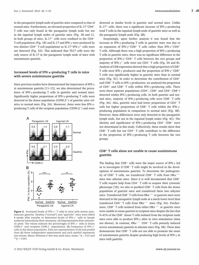
Since previous studies have demonstrated the importance of IFN-γin autoimmune gastritis [11–13], we also determined the preva-lence of IFN-γ-producing T cells in gastritic and normal mice.Significantly higher proportions of IFN-γ-producing T cells weredetected in the donor population (CD90.2+) of gastritic mice rel-ative to normal mice (Fig. 2A). Moreover, there were less IFN-γ-producing T cells of the recipient population (CD90.2−) and were
Figure 2. Increased levels of IFN-γ+ T cells in mice with severe auto-immune gastritis. Healthy (“normal”) and “gastritic” mice were killed8 weeks after transfer to determine levels of IFN-γ+ cells in lymphnodes by intracellular flow cytometry. (A) Representative flow cytomet-ric plots. The values indicate the percentages of IFN-γ+ cells in donorCD90.2+ and recipient CD90.2− populations. (B) Frequency of IFN-γ+
cells in the donor population. Data are representative of (A) and pooledfrom (B) three independent experiments and each symbol representsone mouse. Mann–Whitney U test was used; bars, mean, **p < 0.01 and***p < 0.001.
detected at similar levels in gastritic and normal mice. UnlikeIL-17+ cells, there was a significant increase of IFN-γ-producingtotal T cells in the inguinal lymph node of gastritic mice as well asthe paragastric lymph node (Fig. 2B).
Surprisingly, upon further analysis it was found that theincrease in IFN-γ-producing T cells in gastritic mice was due toan expansion of IFN-γ+CD8+ T cells rather than IFN-γ+CD4+
T cells. Although there was a high proportion of IFN-γ-producingT cells in gastritic mice, there was no significant difference in theproportion of IFN-γ+CD4+ T cells between the two groups andmajority of IFN-γ+ cells were not CD4+ T cells (Fig. 3A and B).Analysis of CD8 expression showed that a high proportion of CD8+
T cells were IFN-γ producers and the proportion of IFN-γ+CD8+
T cells was significantly higher in gastritic mice than in normalmice (Fig. 3C). In order to determine the contribution of CD4+
and CD8+ T cells to IFN-γ production, we analysed the proportionof CD4+ and CD8+ T cells within IFN-γ-producing cells. Therewere three separate populations (CD4+, CD8+ and CD4+ CD8+)detected within IFN-γ-producing cells. In both gastritic and nor-mal mice, majority of IFN-γ-producing cells were CD8+ T cells(Fig. 4A). Also, gastritic mice had lower proportions of CD4+ Tcells but higher proportions of CD8+ T cells within the IFN-γ-producing population in comparison to normal mice (Fig. 4B).However, these differences were only detected in the paragastriclymph node, but not in the inguinal lymph nodes (Fig. 4C). Theidentity and significance of IFN-γ-producing CD4+ CD8+ werenot determined in this study. Collectively, these results show thatCD8+ T cells but not CD4+ T cells contribute to the differencein the proportion of IFN-γ-producing T cells between the twogroups.
CD8+ T cells alone are unable to cause autoimmunegastritis
The finding that CD8+ cells were the major source of IFN-γ ledus to investigate if CD8+ T cells might be involved in the devel-opment of autoimmune gastritis. To determine the pathogenic-ity of CD8+ T cells, we transferred CD8+ T cells from HKα−/−
mice into athymic mice. Since it is well documented that CD8+
T cells require help from CD4+ T cells to acquire their cytotoxicphenotype [18], we also re-purified CD8+ T cells from the donorpopulation of gastritic mice and transferred them into athymicmice. Transferred CD8+ T cells from HKα−/− or gastritic mice weredetected in the paragastric lymph node at a much lower level thantransferred CD4+ T cells from HKα−/− mice (Fig. 5A). Further-more, CD8+ T cells isolated from either HKα−/− or gastritic micewere unable to cause gastritis in recipient mice despite the fact that9–41% of the CD8+ donor T cells isolated from the recipient nudemice were able to produce IFN-γ after in vitro stimulation (datanot shown). In contrast, HKα−/− CD4+ T cells potently inducedsevere autoimmune gastritis in athymic mice (Fig. 5B). These datademonstrate that CD8+ T cells are not able to promote the onsetof autoimmune gastritis despite producing high levels of IFN-γ inmice with gastritis.
C© 2012 WILEY-VCH Verlag GmbH & Co. KGaA, Weinheim www.eji-journal.eu

4 Eric Tu et al. Eur. J. Immunol. 2012. 42: 1–10
Figure 3. IFN-γ is produced by both CD4+ and CD8+ T cells in gastritic mice. Healthy (“normal”) and “gastritic” mice were killed 8 weeks aftertransfer to determine levels of IFN-γ+ cells in lymph nodes by intracellular flow cytometry. (A) Representative flow cytometric plots. The numbersindicate the percentages of IFN-γ+CD4+ and IFN-γ+CD4− T cells in the donor CD90.2+ population. (B) Frequency of IFN-γ+CD4+ T cells in the donorCD4+ T-cell population. (C) Frequency of IFN-γ+CD8+ T cells in the donor CD8+ T-cell population. T cells were identified by identified by CD4, CD8and CD90.2 expression. Data are representative of (A) and pooled from (B and C) three independent experiments and each symbol represents onemouse. Mann–Whitney U test was used; bars, mean, **p < 0.01 and ***p < 0.001.
Increased levels of IL-17 and IFN-γ in the stomachs ofgastritic mice
It was difficult to isolate sufficient number of cells fromthe stomach tissue for intracellular flow cytometric analysis,therefore, we performed Q-PCR to determine if cytokine mRNAlevels increased in the stomachs of mice with autoimmune dis-ease. Significantly higher levels of IL-17, IFN-γ and IL10 mRNAwere detected in the stomachs of gastritic mice (Fig. 6). How-ever, TGFβ, a signature cytokine produced by Foxp3+ regulatoryT (Treg) cells was found at similar levels to the normal stomach(Fig. 6). These results suggest that IL-17- and IFN-γ-producingT cells do not only reside in the stomach draining lymph nodebut also migrate to the stomach and release effector cytokinestherein.
Th17 and Th1 cells in the severity of autoimmunegastritis
Next, we investigated if there was a correlation between IL-17and IFN-γ levels and the severity of autoimmune gastritis. Sincemice that received T cells from H/Kα−/− mice uniformly developsevere autoimmune gastritis [17], this disease model was not suit-able for this purpose. Therefore, mice that had been neonatallythymectomised and which had a broad range of gastritis severitywere used. Mice with gastritis scores 1–4 have mild to substantialmononuclear cell infiltrate in the gastric mucosa and submucosalregions. On the other hand, mice with scores 5 and 6 have severedisease where there is substantial depletion of end-stage cells inthe gastric mucosa and mucosal hypertrophy [19]. Hence there isa qualitative difference in the stomachs of mice with disease scores
Figure 4. Majority of IFN-γ-producing cells in gastritic mice are CD8+ T cells. Healthy (“normal”) and “gastritic” mice were killed 8 weeks aftertransfer to determine levels of IFN-γ+ cells in lymph nodes by intracellular flow cytometry. (A) Representative flow cytometric plots. The valuesindicate the percentages of CD4+, CD8+ and CD4+CD8+ cells within IFN-γ+ cells derived from the donor CD90.2+ population. (B) Frequency ofIFN-γ+CD4+ and IFN-γ+CD8+ T cells in IFN-γ+ cells derived from the donor populations in the paragastric lymph node. (C) Frequency of CD4+ andCD8+ T cells in IFN-γ+ cells derived from the donor populations in the inguinal lymph node. Data are representative of (A) and pooled from (B andC) three independent experiments and each symbol represents one mouse. Mann–Whitney U test was used; bars, mean and ***p < 0.001.
C© 2012 WILEY-VCH Verlag GmbH & Co. KGaA, Weinheim www.eji-journal.eu

Eur. J. Immunol. 2012. 42: 1–10 Immunomodulation 5
Figure 5. CD8+ T cells are not able to cause autoimmune gastritis. Athymic mice were transferred with 1 × 10⁶ CD8+ T cells from H/Kα−/−, ordonor CD8+ T cells from “gastritic” or healthy (“normal”) mice. Athymic mice that received 1 × 10⁶ CD4+ T cells from H/Kα−/− mice were used asdiseased controls. All recipient mice were killed 8 weeks after transfer. (A) Frequency of donor T cells in the paragastric lymph node. (B) Gastritisscore, each symbol represents one mouse. Data are pooled from two independent experiments. Mann–Whitney U test was used; bars, mean, *p <
0.05.
Figure 6. Cytokine expression in stomach tissue. Healthy (“normal”)and “gastritic” mice were killed 8 weeks after transfer and total RNA wasextracted from stomach homogenates. Expression of cytokine mRNA instomach homogenates was measured by Q-PCR, normalised to Tbp andHprt. Data are shown as mean + SE of n = 12 samples in each group,pooled from three independent experiments. Mann–Whitney U test wasused, **p < 0.01, ***p < 0.001 and ns = not significant.
1–4 (mild gastritis) compared to those with disease scores 5 and6 (severe gastritis).
An increase in the IL-17+ population of CD4+ T cells was onlyobserved in mice with severe autoimmune gastritis (score 5 and 6),and the increase was significantly higher compared to normal mice(score 0 and 1), and mice with mild or moderate disease (score2∼4) (Fig. 7A). A higher proportion of IFN-γ+CD4+ T cells werealso detected in the paragastric lymph node of mice with severe
gastritis (score 5 and 6), as well as moderate disease (score 3)(Fig. 7B), albeit not as pronounced as the increase in IL-17+CD4+
T cells. Moreover, the proportion of IFN-γ+CD8+ T-cell popula-tion also increased significantly in mice with autoimmune gastritis(Fig. 7C). Treg cells were found to be at similar levels amongst allgroups (Fig. 7D), suggesting that the difference in disease severitywas not caused by the variation in Treg cell level in these mice.
CD25− T cells from IL-17−/− and IFN-γ−/− mice areunable to induce severe gastritis
To further define the roles of IL-17 and IFN-γ in pathogenesisof autoimmune gastritis, CD25− cells were purified from lymphnodes of IL-17−/−, IFN-γ−/− or wild-type mice and transferredthem into athymic mice. As previously described [20], transferof CD25− cells from wild-type mice induced severe autoimmunegastritis in athymic recipients due to the absence of Treg cells(Fig. 8A). Autoimmune gastritis was also observed in mice thatreceived CD25− cells from IL-17−/− and IFN-γ−/− mice (Fig. 8A),which demonstrated that pathogenic T cells can cause the diseasein the absence of either IL-17 or IFN-γ. Although CD4+ T cells fromIL-17−/− and IFN-γ−/− mice were able to localise to the paragastriclymph node to the same degree as CD4+ T cells from wild-typemice (Fig. 8B), significantly lower grade disease was observed inmice that received CD4+ T cells from either IL-17−/− or IFN-γ−/−
mice. Only 1 of 15 mice that received T cells from IL-17−/− orIFN-γ−/− mice developed severe gastritis (score 5 or 6) comparedto 4 of 6 mice developing severe gastritis in mice that receivedwild-type T cells (Fig. 8A). Furthermore, the lack of IL-17 did nothave any effect on the proportion of IFN-γ+CD4+ T cells (Fig. 8C)and likewise for the lack of IFN-γ on the IL-17+CD4+ population(Fig. 8D). This suggests that the lower pathogenicity of T cellsfrom IL-17−/− and IFN-γ−/− mice is the direct consequence of theabsence of T cell-derived IL-17 or IFN-γ. Also, the lower diseaseseverity in mice that received IL-17−/− and IFN-γ−/− cells wasnot caused by increased proportion of Treg cells in the absence of
C© 2012 WILEY-VCH Verlag GmbH & Co. KGaA, Weinheim www.eji-journal.eu

6 Eric Tu et al. Eur. J. Immunol. 2012. 42: 1–10
Figure 7. Proportion of IL-17+CD4+ T cellsand IFN-γ+CD4+ T cells correlate to gastri-tis severity. Thymectomy was performed onBALB/cCrSlc and BALB.B6-Gasa congenic miceat 3 days of age and mice were killed at 12weeks of age. The proportions of (A) IL-17,(B and C) IFN-γ and (D) Foxp3 in CD4+ (A, Band D) or CD8+ (C) T cells were assessed byintracellular flow cytometry. Data are pooledfrom at least 100 independent thymectomies.Mann–Whitney U test was used; bars, mean,*p < 0.05, **p < 0.01 and ***p < 0.001.
IL-17 and IFN-γ (Fig. 8E). Together, these data demonstrate thatautoimmune gastritis can develop in the absence of either IL-17or IFN-γ, however, the disease is not able to progress to the latepathogenic stages.
We also examined the infiltrating cells in the gastric mucosae ofmice that received IL-17−/− and IFN-γ−/− cells. Nearly all infiltrat-ing cells were mononuclear cells and very few polymorphonuclearneutrophils were observed (Supporting Information Fig. 2).
Discussion
Here we have re-evaluated the roles of cytokines in the onsetand progression of autoimmune gastritis, which was believed tobe mediated by IFN-γ-producing Th1 cells [11–13]. Significantly,high levels of IL-17-producing CD4+ T cells were found in theparagastric lymph node of mice with severe autoimmune gastri-tis induced by either the transfer of H/Kα−/− T cells or neonatalthymectomy. Whereas, the proportions of IL-17-producing CD4+
T cells in non-draining peripheral lymph nodes were similarbetween gastritic and normal mice. Additionally, high levels ofIL-17+CD4+ T cells were only detected in mice with severe dis-ease, but not in mild and moderate conditions. All IL-17-producingcells detected in gastritic mice were CD4+ T cells, which rulesout the involvement of IL-17-producing CD8+ T cells [21,22].
IL-17-producing NKT cells are CD4− [23], therefore it seemsunlikely that NKT cells are involved in IL-17 production either.Together, these results suggest that IL-17 produced by Th17 cellsis linked to the development of severe autoimmune gastritis. Fur-thermore, IL-17 and IFN-γ were produced by separate lineages ofCD4+ T cells in autoimmune gastritis since IL-17+IFN-γ+ double-producing Th cells that are involved in intestinal inflammation incolitis [24] were not detected in gastritic mice.
Consistent with other studies that demonstrated the essentialrole of IFN-γ in the pathogenesis of autoimmune gastritis [13,14],significantly higher levels of IFN-γ-producing cells were detectedin both draining and non-draining lymph nodes of gastritic micecompared to normal mice. However, unlike IL-17, the majorityof IFN-γ producers were non-CD4+ T cells and there was noincrease in the proportion of IFN-γ+CD4+ T cells in gastriticmice that received H/Kα−/− T cells. Although high proportionsof IFN-γ+CD4+ T cells were found in neonatally thymectomisedmice with severe disease, the increase was not as pronounced asIL-17+CD4+ T cells. A high proportion of IFN-γ+CD4+ T cells wasalso found in mice with moderate gastritis (score 3). Since massiveinfiltrate of mononuclear cells was often observed in mice at thispathogenic stage (Fig. 7), it is possible that a high level of IFN-γis produced to facilitate the recruitment of T cells into stomachtissue.
Analysis of CD8 expression revealed an elevation of IFN-γ-producing CD8+ T cells in gastritic mice, which accounted for
C© 2012 WILEY-VCH Verlag GmbH & Co. KGaA, Weinheim www.eji-journal.eu

Eur. J. Immunol. 2012. 42: 1–10 Immunomodulation 7
Figure 8. IL-17 and IFN-γ are both required for the development of severe gastritis. Athymic mice received 2 × 10⁶ CD25− lymph node cells fromIL-17−/−, IFN-γ−/− or wild-type (WT) mice and killed 8 weeks after transfer. (A) Quantitation of gastric pathology. (B) Frequency of donor T cells.(C) Frequency of IFN-γ+CD4+ T cells in the donor CD4+ T-cell population in the paragastric lymph node. (D) Frequency of IL-17+CD4+ T cells inthe donor CD4+ T-cell population in the paragastric lymph node. (E) Frequency of Foxp3+CD4+ T cells in the donor CD4+ T-cell population in theparagastric lymph node. Data are pooled from four independent experiments; each symbol represents a single mouse. Mann–Whitney U test wasused; bars, mean, *p < 0.05, **p < 0.01, ***p < 0.001 and ns = not significant.
the significant increase of total IFN-γ-producing T cells. How-ever, neither naıve CD8+ T cells from H/Kα−/− mice nor CD8+
T cells from gastritic mice were able to induce autoimmune gas-tritis, which is consistent with past reports showing that auto-immune gastritis is caused by H+/K+ ATPase-specific CD4+ Tcells, but not CD8+ T cells [14,25]. The inability of CD8+ T cellsto initiate disease may be related to their reduced ability to accu-mulate in the paragastric lymph node (Fig. 5A). This explanationis supported by recent studies that revealed that CD8+ T cellsrequire assistance from CD4+ T cells to gain entry into the targettissue [26].
High levels of IL-17 and IFN-γ gene expression were detectedin the stomach tissue of gastritic mice, which reflected the highproportion of IL-17- and IFN-γ-producing T cells in the paragas-tric lymph node. This indicates the presence of IL-17- and IFN-γ-producing effector T cells in gastritic stomach, which is in line withour previous hypothesis suggesting that the disruption of gastricmucosal cell development in autoimmune gastritis is caused bycytokines released by pathogenic T cells [27]. Although there aresignificantly more Treg cells in the stomach infiltrate of mice withautoimmune gastritis [17], high levels of TGF-β were not detectedin the stomach of gastritic mice. We found that increased levelsof IL10 mRNA in gastritic mucosae as well, although the leveland fold increase compared were not as great as those seen forIFN-γ and IL-17. IL-17, IFN-γ and other pro-inflammatorycytokines released in the gastritic environment may have served
to inhibit the regulatory functions of Treg cells, as observed in thecases of dry eye disease and NOD diabetes [28,29].
To determine if IL-17 and/or IFN-γ are required for the onsetand progression of autoimmune gastritis, CD25− T cells fromIL-17−/− or IFN-γ−/− mice were transferred to athymic mice.Autoimmune gastritis developed in the absence of either CD4+
T cell-derived IL-17 or IFN-γ but to a much lower severity com-pared to disease induced by wild-type T cells. The dispensablerole of IFN-γ in the onset of CD25− T cell-induced autoim-mune gastritis has also been reported by others using T cellsfrom IFN-γ−/− mice [14], although treatment of neutralising-IFN-γ antibody inhibits the initiation of neonatal thymectomy-induced gastritis [13]. Contradictory results obtained from usingneutralising antibodies and knockout mice have also been reportedin NOD diabetes and T cell-mediated colitis [30–33]. It is possi-ble that IFN-γ produced by cells of the innate immune system inathymic recipients aids the development of diseases.
Since it has been shown that monoclonal gastritogenic Th1 andTh17 cells can both induce autoimmune gastritis [15], it is likelythat gastritis in mice that received IL-17−/− T cells is mediated bya Th1 response and disease in mice that received IFN-γ−/− T cellsrelies on a Th17 response. It remains to be determined whetherautoimmune gastritis can develop in the absence of both IL-17 andIFN-γ.
Since there is an antagonistic cytokine network involved inthe development of Th1 and Th17 cells [34–36], IL-17 and
C© 2012 WILEY-VCH Verlag GmbH & Co. KGaA, Weinheim www.eji-journal.eu

8 Eric Tu et al. Eur. J. Immunol. 2012. 42: 1–10
IFN-γ have been shown to serve a protective role in certain mod-els of autoimmunity as the result of increased Th1 and Th17 cellnumbers in the absence of IL-17 and IFN-γ, respectively [37–39].However, we did not find a mutual regulation of IL-17- and IFN-γ-producing T cells by IFN-γ and L-17 in autoimmune gastritis, nordid we observe a more aggressive disease in mice that receivedIL-17−/− T cells or IFN-γ−/− T cells. In contrast, both IL-17−/−
and IFN-γ−/− T cells induced significantly lower grade diseasein comparison to wild-type T cells. This suggests that both IL-17and IFN-γ act together to promote the progression autoimmunegastritis to late pathogenic stages, which is consistent with thefinding of high proportions of IL-17- and IFN-γ-producing T cellsin mice with severe autoimmune gastritis. This finding is similar tothat observed in bacterial-induced T cell-dependent colitis, whereIL-17 and IFN-γ are both indispensable for the induction of maxi-mal intestinal inflammation [40].
In summary, we show that IL-17- and IFN-γ-producing Tcells are present at significant levels in mice with severe auto-immune gastritis in two different gastritis models. Our study fur-ther demonstrates that the early stages of autoimmune gastritiscan develop in the absence of either CD4+ T cell-derived IL-17 orIFN-γ, yet, both cytokines are required in combination for T cellsto cause severe damage. The ultimate goal of immunotherapy isto reverse ongoing autoimmune diseases by dampening effector Tcells. Treatments that target either IL-17 or IFN-γ alone may besufficient to abrogate the progression of autoimmunity or ame-liorate established inflammation. However treatments that targetboth IL-17 and IFN-γ may be required for a complete reversal ofautoimmune disease that is similar to autoimmune gastritis.
Materials and methods
Mice
BALB/cCrSlc, BALB/cCrSlc.CD90.1 congenic, H+/K+ ATPase α
subunit-knockout (H/Kα−/−) [41], BALB.B6-Gasa congenic [42],IL-17-deficient (IL-17−/−) [43] and IFN-γ−/− [44] mice havebeen previously described. H/Kα−/− mice were crossed toBALB/c.CD90.1 mice to generate H/Kα−/−.CD90.1 mice. AthymicBALB/cnu/nu mice (6–8 weeks) were purchased from the AnimalResource Centre (Perth, Australia). All mice and experiments wereapproved by the University of Melbourne Animal ExperimentationEthics Committee.
Antibodies and flow cytometric analysis
The following antibodies were used for the analysis of cell sur-face markers: anti-90.1 FITC (HIS51), anti-90.2 FITC (30H-12),anti-CD4 PerCP (RM4.5) and anti-CD8 APC (53–6.7) were pur-chased from BD Pharmingen; anti-CD4 Pacific Blue (RM4.5) andanti-CD8 PE Cy7 (53–6.7) were purchased from eBioscience. Intra-cellular staining of Foxp3 was performed using anti-Foxp3 allo-
phycocyanin (FJK-16s) and eBioscience Foxp3 Staining Buffer Setaccording to the manufacturer’s instruction. Intracellular stainingof cytokines was performed using anti-IL-17 PE (TC11–18H10),anti-IFN-γ allophycocyanin (XMG1.2) and BD Cytofix/CytopermPlus according to the manufacturer’s instruction.
Induction of autoimmune gastritis and assessmentof disease severity
Autoimmune gastritis was induced by transferring 5 × 10⁷ lymphnode cells from H/Kα−/− mice into irradiated wild-type miceas previously described [17]. In some experiments, the diseasewas induced in BALB/cCrSlc and BALB.B6-Gasa congenic mice byneonatal thymectomy as previously described [42]. The severity ofautoimmune gastritis was determined in each mouse by histologi-cal examination of stomach sections as previously described [19].Scoring was performed in a blind fashion and each slide was inde-pendently scored by two people.
Cytokine quantitation in stomach tissue using Q-PCR
Stomach tissue samples were homogenised in RNAlater and totalRNA was purified using TRI Reagent (Molecular Research Cen-ter, Inc.). All samples had a RNA integrity number of >9 asdetermined by an Agilent Technologies 2100 Bioanalyzer. cDNAsynthesised using M-MLV reverse transcriptase and Oligo dT(both obtained from Invitrogen). IL-17 (Il17a, Mm00439618 m1),IFN-γ (Ifng, Mm01168134 m1), IL10 (Il10, Mm00439614 m1)and TGFβ (Tgfb1, Mm01178820 m1) gene expression was deter-mined by Q-PCR using mouse-specific TaqMan Gene ExpressionAssays (Applied Biosystems). TaqMan Gene Expression Mastermix was used to setup 20 μL singleplex reactions in quadrip-licates. Internal endogenous reference genes were mouse Tbp(Mm00446971 m1) and Hprt1 (Mm00446968 m1). qPCR reac-tions were run on a StepOnePlus qPCR machine (Applied Biosys-tems) and analysed using the comparative DeltaDelta CT method.Relative quantification of mRNA levels shown use normal mice asthe normaliser and Tbp and Hprt1 as the internal reference genes.
Assessment of pathogenicity of CD8+ T cells
CD8+ T cells from lymph nodes and spleens of H/Kα−/−.CD90.1mice were purified by FACS sorting. A total of 1 × 10⁶ puri-fied CD8+ T cells were injected intravenously into athymic mice.Eight weeks after the transfer, athymic recipients were sacri-ficed and stomachs were taken for histological examination. Insome experiments, 1 × 10⁶ CD8+ T cells from the donor popu-lation in either ‘gastritic’ or ‘normal’ mice were transferred intoathymic mice. ‘Gastritic’ and ‘normal’ mice were set up by trans-ferring 5 × 10⁷ lymph node cells from either H/Kα−/−.CD90.1 orBALB/cCrSlc.CD90.1 into irradiated CD90.2 wild-type mice.
C© 2012 WILEY-VCH Verlag GmbH & Co. KGaA, Weinheim www.eji-journal.eu

Eur. J. Immunol. 2012. 42: 1–10 Immunomodulation 9
Adoptive transfer of CD25− cell populations
For isolation of CD25− cell populations, single cell suspensionsfrom lymph nodes were stained with anti-25-FITC, followed byanti-FITC-labelled microbeads (Miltenyi Biotec). Labelled-CD25+
cells were depleted using an AutoMACS separator (MiltenyiBiotec) according to the manufacturer’s instructions. A total of 2 ×10⁶ CD25− lymph node cells from IL-17−/−, IFN-γ−/− or wild-typemice were injected intravenously into athymic mice. Developmentof autoimmune gastritis in the recipient mice was determined after8 weeks.
Statistical analysis
Statistical analysis was performed using GraphPad Prism 5.0(GraphPad). Data were analysed using the Mann–Whitney U testand a p-value < 0.05 was considered significant.
Acknowledgments: This work was supported by research awards,a Biomedical Training Fellowship (SAB), an Australia fellowship(MJS) and a CDF1 (MWLT) from the National Health and Med-ical Research Council of Australia, Future Fellowship from theAustralian Research Council (AFH) and research funding from theUniversity of Melbourne. We thank Prof. Geoff Hill for the supplyof IL17 deficient mice.
Conflict of interest: The authors declare no financial or commer-cial conflict of interest.
References
1 Abbas, A. K., Murphy, K. M. and Sher, A., Functional diversity of helper
T lymphocytes. Nature 1996. 383: 787–793.
2 Murphy, K. M. and Reiner, S. L., The lineage decisions of helper T cells.
Nat. Rev. Immunol. 2002. 2: 933–944.
3 Iwakura, Y., Nakae, S., Saijo, S. and Ishigame, H., The roles of IL-17A in
inflammatory immune responses and host defense against pathogens.
Immunol. Rev. 2008. 226: 57–79.
4 Korn, T., Bettelli, E., Oukka, M. and Kuchroo, V. K., IL-17 and Th17 cells.
Annu. Rev. Immunol. 2009. 27: 485–517.
5 Oppmann, B., Lesley, R., Blom, B., Timans, J. C., Xu, Y., Hunte, B., Vega,
F. et al., Novel p19 protein engages IL-12p40 to form a cytokine, IL-23,
with biological activities similar as well as distinct from IL-12. Immunity
2000. 13: 715–725.
6 Cua, D. J., Sherlock, J., Chen, Y., Murphy, C. A., Joyce, B., Seymour, B.,
Lucian, L. et al., Interleukin-23 rather than interleukin-12 is the critical
cytokine for autoimmune inflammation of the brain. Nature 2003. 421:
744–748.
7 Murphy, C. A., Langrish, C. L., Chen, Y., Blumenschein, W., McClana-
han, T., Kastelein, R. A., Sedgwick, J. D. et al., Divergent pro- and anti-
inflammatory roles for IL-23 and IL-12 in joint autoimmune inflamma-
tion. J. Exp. Med. 2003. 198: 1951–1957.
8 Jones, C. M., Callaghan, J. M., Gleeson, P. A., Mori, Y., Masuda, T. and Toh,
B. H., The parietal cell autoantigens recognized in neonatal thymectomy-
induced murine gastritis are the alpha and beta subunits of the gastric
proton pump [corrected]. Gastroenterology 1991. 101: 287–294.
9 Karlsson, F. A., Burman, P., Loof, L. and Mardh, S., Major parietal cell
antigen in autoimmune gastritis with pernicious anemia is the acid-
producing H+, K+-adenosine triphosphatase of the stomach. J. Clin.
Invest. 1988. 81: 475–479.
10 Toh, B. H., van Driel, I. R. and Gleeson, P. A., Pernicious anemia. N. Engl.
J. Med. 1997. 337: 1441–1448.
11 D’Elios, M. M., Bergman, M. P., Azzurri, A., Amedei, A., Benagiano, M.,
De Pont, J. J., Cianchi, F. et al., H(+),K(+)-atpase (proton pump) is the
target autoantigen of Th1-type cytotoxic T cells in autoimmune gastritis.
Gastroenterology 2001. 120: 377–386.
12 Martinelli, T. M., van Driel, I. R., Alderuccio, F., Gleeson, P. A. and Toh,
B. H., Analysis of mononuclear cell infiltrate and cytokine production in
murine autoimmune gastritis. Gastroenterology 1996. 110: 1791–1802.
13 Barrett, S. P., Gleeson, P. A., de Silva, H., Toh, B. H. and van Driel, I. R.,
Interferon-gamma is required during the initiation of an organ-specific
autoimmune disease. Eur. J. Immunol. 1996. 26: 1652–1655.
14 Suri-Payer, E. and Cantor, H., Differential cytokine requirements for reg-
ulation of autoimmune gastritis and colitis by CD4(+)CD25(+) T cells. J.
Autoimmun. 2001. 16: 115–123.
15 Stummvoll, G. H., DiPaolo, R. J., Huter, E. N., Davidson, T. S., Glass, D.,
Ward, J. M. and Shevach, E. M., Th1, Th2, and Th17 effector T cell-induced
autoimmune gastritis differs in pathological pattern and in susceptibility
to suppression by regulatory T cells. J. Immunol. 2008. 181: 1908–1916.
16 DiPaolo, R. J., Glass, D. D., Bijwaard, K. E. and Shevach, E. M., CD4+CD25+T cells prevent the development of organ-specific autoimmune dis-
ease by inhibiting the differentiation of autoreactive effector T cells. J.
Immunol. 2005. 175: 7135–7142.
17 Tu, E., Ang, D. K., Hogan, T. V., Read, S., Chia, C. P. Z., Gleeso, P. A. and
van Driel, I. R., A convenient model of severe, high Incidence autoim-
mune gastritis caused by polyclonal effector T cells and without pertur-
bation of regulatory T cells. PLoS One 2011. 6: e27153.
18 Williams, M. A. and Bevan, M. J., Effector and memory CTL differentia-
tion. Annu. Rev. Immunol. 2007. 25: 171–192.
19 Read, S., Hogan, T. V., Zwar, T. D., Gleeson, P. A. and Van Driel, I. R.,
Prevention of autoimmune gastritis in mice requires extra-thymic T-cell
deletion and suppression by regulatory T cells. Gastroenterology 2007. 133:
547–558.
20 Sakaguchi, S., Sakaguchi, N., Asano, M., Itoh, M. and Toda, M., Immuno-
logic self-tolerance maintained by activated T cells expressing IL-2 recep-
tor alpha-chains (CD25). Breakdown of a single mechanism of self-
tolerance causes various autoimmune diseases. J. Immunol. 1995. 155:
1151–1164.
21 He, D., Wu, L., Kim, H. K., Li, H., Elmets, C. A. and Xu, H., CD8+ IL-17-
producing T cells are important in effector functions for the elicitation
of contact hypersensitivity responses. J. Immunol. 2006. 177: 6852–6858.
22 Tajima, M., Wakita, D., Noguchi, D., Chamoto, K., Yue, Z., Fugo, K.,
Ishigame, H. et al., IL-6-dependent spontaneous proliferation is required
for the induction of colitogenic IL-17-producing CD8+ T cells. J. Exp. Med.
2008. 205: 1019–1027.
23 Coquet, J. M., Chakravarti, S., Kyparissoudis, K., McNab, F. W., Pitt, L. A.,
McKenzie, B. S., Berzins, S. P. et al., Diverse cytokine production by NKT
cell subsets and identification of an IL-17-producing CD4-NK1.1- NKT cell
population. Proc. Natl. Acad. Sci. USA 2008. 105: 11287–11292.
C© 2012 WILEY-VCH Verlag GmbH & Co. KGaA, Weinheim www.eji-journal.eu

10 Eric Tu et al. Eur. J. Immunol. 2012. 42: 1–10
24 Ahern, P. P., Schiering, C., Buonocore, S., McGeachy, M. J., Cua, D. J.,
Maloy, K. J. and Powrie, F., Interleukin-23 drives intestinal inflammation
through direct activity on T cells. Immunity 2011. 33: 279–288.
25 De Silva, H. D., Van Driel, I. R., La Gruta, N., Toh, B. H. and Gleeson, P.
A., CD4+ T cells, but not CD8+ T cells, are required for the development
of experimental autoimmune gastritis. Immunology 1998. 93: 405–408.
26 Nakanishi, Y., Lu, B., Gerard, C. and Iwasaki, A., CD8(+) T lymphocyte
mobilization to virus-infected tissue requires CD4(+) T-cell help. Nature
2009. 462: 510–513.
27 Judd, L. M., Gleeson, P. A., Toh, B. H. and van Driel, I. R., Autoimmune
gastritis results in disruption of gastric epithelial cell development. Am.
J. Physiol. 1999. 277: G209–218.
28 Chauhan, S. K., El Annan, J., Ecoiffier, T., Goyal, S., Zhang, Q., Saban, D.
R. and Dana, R., Autoimmunity in dry eye is due to resistance of Th17 to
Treg suppression. J. Immunol. 2009. 182: 1247–1252.
29 Gregori, S., Giarratana, N., Smiroldo, S. and Adorini, L., Dynamics of
pathogenic and suppressor T cells in autoimmune diabetes development.
J. Immunol. 2003. 171: 4040–4047.
30 Hultgren, B., Huang, X., Dybdal, N. and Stewart, T. A., Genetic absence
of gamma-interferon delays but does not prevent diabetes in NOD mice.
Diabetes 1996. 45: 812–817.
31 Debray-Sachs, M., Carnaud, C., Boitard, C., Cohen, H., Gresser, I.,
Bedossa, P. and Bach, J. F., Prevention of diabetes in NOD mice treated
with antibody to murine IFN gamma. J. Autoimmun. 1991. 4: 237–248.
32 Powrie, F., Leach, M. W., Mauze, S., Menon, S., Caddle, L. B. and Coffman,
R. L., Inhibition of Th1 responses prevents inflammatory bowel disease
in SCID mice reconstituted with CD45RBhi CD4+ T cells. Immunity 1994.
1: 553–562.
33 Simpson, S. J., Shah, S., Comiskey, M., de Jong, Y. P., Wang, B., Mizoguchi,
E., Bhan, A. K. et al., T cell-mediated pathology in two models of experi-
mental colitis depends predominantly on the interleukin 12/Signal trans-
ducer and activator of transcription (Stat)-4 pathway, but is not condi-
tional on interferon gamma expression by T cells. J. Exp. Med. 1998. 187:
1225–1234.
34 Park, H., Li, Z., Yang, X. O., Chang, S. H., Nurieva, R., Wang, Y. H., Wang,
Y. et al., A distinct lineage of CD4 T cells regulates tissue inflammation
by producing interleukin 17. Nat. Immunol. 2005. 6: 1133–1141.
35 Harrington, L. E., Hatton, R. D., Mangan, P. R., Turner, H., Murphy, T.
L., Murphy, K. M. and Weaver, C. T., Interleukin 17-producing CD4+effector T cells develop via a lineage distinct from the T helper type 1
and 2 lineages. Nat. Immunol. 2005. 6: 1123–1132.
36 Li, M. O., Wan, Y. Y., Sanjabi, S., Robertson, A. K. and Flavell, R. A.,
Transforming growth factor-beta regulation of immune responses. Annu.
Rev. Immunol. 2006. 24: 99–146.
37 Ferber, I. A., Brocke, S., Taylor-Edwards, C., Ridgway, W., Dinisco,
C., Steinman, L., Dalton, D. et al., Mice with a disrupted IFN-gamma
gene are susceptible to the induction of experimental autoimmune
encephalomyelitis (EAE). J. Immunol. 1996. 156: 5–7.
38 Komiyama, Y., Nakae, S., Matsuki, T., Nambu, A., Ishigame, H., Kakuta,
S., Sudo, K. et al., IL-17 plays an important role in the development of
experimental autoimmune encephalomyelitis. J. Immunol. 2006. 177: 566–
573.
39 O’Connor, W., Jr., Kamanaka, M., Booth, C. J., Town, T., Nakae, S.,
Iwakura, Y., Kolls, J. K. et al., A protective function for interleukin
17A in T cell-mediated intestinal inflammation. Nat. Immunol. 2009. 10:
603–609.
40 Kullberg, M. C., Jankovic, D., Feng, C. G., Hue, S., Gorelick, P. L., McKenzie,
B. S., Cua, D. J. et al., IL-23 plays a key role in Helicobacter hepaticus-
induced T cell-dependent colitis. J. Exp. Med. 2006. 203: 2485–2494.
41 Spicer, Z., Miller, M. L., Andringa, A., Riddle, T. M., Duffy, J. J.,
Doetschman, T. and Shull, G. E., Stomachs of mice lacking the gastric
H,K-ATPase alpha -subunit have achlorhydria, abnormal parietal cells,
and ciliated metaplasia. J. Biol. Chem. 2000. 275: 21555–21565.
42 Ang, D. K., Brodnicki, T. C., Jordan, M. A., Wilson, W. E., Silveira, P.,
Gliddon, B. L., Baxter, A. G. et al., Two genetic loci independently confer
susceptibility to autoimmune gastritis. Int. Immunol. 2007. 19: 1135–1144.
43 Nakae, S., Komiyama, Y., Nambu, A., Sudo, K., Iwase, M., Homma, I.,
Sekikawa, K. et al., Antigen-specific T-cell sensitization is impaired in IL-
17-deficient mice, causing suppression of allergic cellular and humoral
responses. Immunity 2002. 17: 375–387.
44 Street, S. E., Cretney, E. and Smyth, M. J., Perforin and interferon-gamma
activities independently control tumor initiation, growth, and metasta-
sis. Blood 2001. 97: 192–197.
Full correspondence: Dr. Ian R. van Driel, Department of Biochemistryand Molecular Biology, Bio21 Molecular Science and BiotechnologyInstitute, The University of Melbourne, Parkville, Victoria 3010,AustraliaFax: +61-3 9348 1421e-mail: [email protected]
Current address: Dr. Thea V. Hogan, MRC National Institute for MedicalResearch, The Ridgeway, Mill Hill, London, NW7 1AA, United Kingdom
Received: 18/12/2011Revised: 6/5/2012Accepted: 12/6/2012Accepted article online: 10/7/2012
C© 2012 WILEY-VCH Verlag GmbH & Co. KGaA, Weinheim www.eji-journal.eu