Embed Size (px)
Citation preview

Book-Chapter 6: Transport and ATP synthesis in mitochondr ia (1978-1984)
IV. K+-transport: Evidence for mitochondr ial F0F1-ATPase being a K+-pump.
Reinhold Kiehl, RKI-Institute, Laboratory and Research for Mol.Medicine/Biology, Saliterweg 1,D-93437 Furth im Wald, Phone: 09973/801056, E.Mail: [email protected], Internet: www.rki-i.com
Abbreviations: NSPM, N'-[N''-n-nonyl-4-sulfamoylphenyl]-maleimide; TTFB, 4,5,6,7-tetrachloro-2-trifluoromethylbenzimidazole; ACMA, 9-amino-6-chloro-2-methoxyacridine; SMP,submitochondrial particles; o = outside mitochondria
Summary: Our experiments demonstrate that mitochondria contain an energy driven K+/H+-antiporter. The energy is derived from substrate oxidation by the respiratory chain. Thisantiporter is Mgo
2+-sensitively stimulated by NSPMo, Cdo2+, dicyclohexylcarbodiimideo and
Cao2+. Quinine prevents the Mgo
2+-sensitivity. Ruthenium red prevents Cdo2+ and Cao
2+
sensitivity.The experiments prove further that mitochondria contain an oligomycin-sensitive, ATP- drivenK+-pump, which is identical to the oligomycin-sensitive F1F0-ATPase. This K+-pump isstimulated by NSPM, picrylacetate, Cd2+(Mg2+,Ca2+) and inhibited by dicyclohexylcarbodiimide.The synthesis of ATP on the Pi/H
+-symporter, as proposed, is then highly probable, or evenproven. K+/H+-antiporter, K+-pump and Pi/H
+-symporter are linked together in versatile energy-driven K+/H+-cycling and oscillations. This system is controlled by O2, and the free Mg2+ andCa2+ concentrations in the cytosol of the cells and most likely also able to handle "abnormal"Ca2+ concentrations. A high physiological K+-gradient between cytosol (K+=high, ∼175 mMo)and matrix (K+=low,∼1mM i)is established and maintained by the system. Under normalphysiological conditions, no protons are detectable in the bulk phase; they are therefore eithermoving along the membranes via hydrogen bridges, or the proton-pumping systems are directlylinked. The results imply a direct connection (and regulatory function) between nervous system(brain) and mitochondria (body and brain): for thermoregulation, substrate oxidation, as well asO2-uptake/-reduction (anorexia/dystrophia), which can lead to arteriosclerosis, heart attack, andcancer on the one hand, and edema during anoxia/hypoxia, or shock and dilusion with coma(death) on the other hand are examples. Key words: Energy driven K+/H+-antiporter,oligomycin-sensitive ATP-driven K+ -pump, K+/H+-cycling, oscillations, hydrogen bridges,connection: brain-mitochondria.
Zusammenfassung: Unsere Ergebnisse zeigen, daß die Mitochondrien einen Energie-getriebenen K+/H+-Antiporter enthalten. Die Energie wird durch Substrat-Oxidation in derAtmungskette geliefert. Dieser Antiporter wird Mga
2+-sensitiv stimuliert durch NSPMa, Cda2+,
dicyclohexylcarbodiimida und Caa2+. Quinine verhindert die Mga
2+-Sensivität, Ruthenium Rotverhindert die Cda
2+- und Caa2+-Sensivität. Die Experimente beweisen weiterhin, daß die
Mitochondrien eine Oligomycin-sensitive ATP-getriebene K+-Pumpe, identisch mit derOligomycin-sensitiven F1F0-ATPase, besitzen. Diese K+-Pumpe wird stimuliert durch NSPM,Picrylacetat, Cd2+ (Mg2+, Ca2+) und inhibiert durch Dicyclohexylcarbodiimid. Die Synthese vonATP an dem Pi/H
+-Symporter - wie vorgeschlagen - ist damit sehr wahrscheinlich, wenn nichtbewiesen. K+/H+-Antiporter, K+-Pumpe und Pi/H
+-Symporter sind miteinander durch Energie-

getriebene zyklische K+/H+-Bewegungen und Oszillationen verbunden. Dieses System wirddurch O2 und die freien Mg2+-und Ca2+-Konzentrationen im Cytosol der Zellen kontrolliert undist höchstwahrscheinlich auch in der Lage, "abnormale" Ca2+-Konzentrationen zu beseitigen. Eingroßer physiologischer K+-Gradient zwischen Cytosol (K+ = hoch, ∼ 175 mMa) und Matrix(K+ = tief, ∼ 1 mM i) wird durch das System erstellt und erhalten. Da unter normalenphysiologischen Bedingungen keine Protonen in der wässrigen Phase zu sehen sind, bewegen siesich entweder entlang der Membran über Wasserstoffbrücken oder aber die H+-Pumpen sinddirekt miteinander verbunden. Die Resultate implizieren eine direkte Verbindung (undRegulation) zwischen Nervensystem (Gehirn) und Mitochondrien (Körper und Gehirn):Thermoregulation, Substrat-Oxidation wie auch O2-Aufnahme/-Reduktion (Anorexia/Dystrophia)mit Arteriosklerose, Herzinfarkt, Krebs auf der einen Seite und Bildung von Ödemen währendAnoxia/Hypoxia oder Schock, Wahnvorstellungen mit Koma (Tod) auf der anderen Seite sindBeispiele. Schlüsselwörter : Energie-getriebener K+/H+-Antiport, Oligomycin-sensitive ATP-getriebene K+-Pumpe, zyklische K+/H+-Bewegungen, Oszillationen, Wasserstoffbrücken,Verbindung: Gehirn-Mitochondrien.
Introduction
NSPM, picrylacetate and Cd2+ have been shown to be very potent and specific reagents forinhibition or stimulation of various membrane-associated functions, e.g. electron transfer andtransport activities [1-8].
Our hypothesis for mitochondrial ATP synthesis and the consequences of this hypothesis [4]implicate the F0F1-ATPase as a K-pump. Past literature includes numerous reports in support ofthis supposition, including our own results [4, 9]. The present manuscript focuses on K+ (Ca2+)-transport across the mitochondrial and submitochondrial membranes and its relationship to theenergy-linked functions performed by these particles; especially electron-transfer and K+- orproton-pumping activities. This relationship was investigated by using various functional group-trapping reagents in addition to the compounds NSPM, picrylacetate, and Cd2+ as mentionedabove. Preliminary results were presented [10, 11].
Mater ials and Methods
Rat liver mitochondria were prepared according to Kaschnitz et al [12] and suspended in 0.22 Mmannitol, 70 mM sucrose, 2 mM K-hepes, pH 7.4. Oxygen uptake of mitochondria was measuredwith an oxygen electrode. All mitochondrial preparations were checked for structural integrityusing the criterion of respiratory control [13]. Assay of endogenous calcium and phosphateconcentrations, as well as swelling experiments were performed as in [2, 3]. K+-transport rateswere monitored continously at 300C by means of a Beckman K+-electrode. Calibration of the K+-electrode in each experiment was done by adding a known amount of a KCl solution [2, 3, 14].The experiments were only possible at low K+-concentrations. pH-jump experiments using a pH-electrode were performed as in [15-17].
∆ϕ of mitochondria was followed using DiS-C3-(5) fluorescence changes [18]. The fluorescenceemission of DiS-C3-(5) was measured at 667 nm at an excitation wavelength of 622 nm.

Mitochondria (1.2 mg in 40 µl 0.22 M mannitol, 70 mM sucrose, 2 mM K-hepes, pH 7.4) in 2.5ml of a solution containing 4 µM DiS-C3-(5), 0.22 M mannitol, 70 mM sucrose, 2 mM K-hepes,pH 7.4, 20 mM KCl, 5 mM MgCl2, 10 mM K-phosphate, pH 7.4, 25 µM Rotenone, wereenergized with 8 mM K-succinate, pH 7.4 at 300C.
4-Methylumbelliferon (4-MU) fluorescence was used as one more indicator of ∆ pH inmitochondria [19]. The fluorescence emission of 4-MU was measured at 450 nm at an excitationwavelength of 356 nm. Mitochondria (1.2 mg in 40 µl 0.22 M mannitol, 70 mM sucrose, 2 mMK-hepes, pH 7.4) in 2.5 ml of a solution containing 0.2 µM 4-MU, 0.22 M mannitol, 70 mMsucrose, 2 mM K-hepes, pH 7.4, 20 mM KCl, 5 mM MgCl2, 10 mM K-phosphate, pH 7.4, 25 µMRotenone, were energized with 8 mM K-succinate, pH 7.4 at 300C.
Phosphorylating submitochondrial particles and submitochondrial particles were preparedaccording to published procedures [8, 20, 21].
ATP/O-ratio, ATP-Pi-exchange (without BSA and added phospholipids), ATPase activities in thepresence and absence of KCl and either in an ATP regenerating system or by the amount ofanorganic phosphate released, ATP-driven reduction of NAD by K-succinate,transhydrogenation from NADH to AcPyNADP and proton permeability in 150 mM KCl by theATP-jump method were performed as described [8, 17, 22-26].
Succinate- and ATP-dependent spectral response of oxonol VI and ACMA-fluorescence insubmitochondrial particles and complex V [8] were followed as described in [8].
The effect of Cd2+ on the respiratory chain was evaluated on phosphorylating submitochondrialparticles using a Shimadzu ultraviolet 300 dual-wavelength scanning spectrophotometer [27, 28].
O-acetyl-picric acid was prepared according to [29]. 3H-acetyl-picric acid was synthesized from3H-acetic anhydride (Amersham, 500 mCi/ mmol): 64.6 mg picric acid (0.282 mmol) werewarmed to 56oC, then a mixture of 25 µl acetic anhydride and 40 µl 3H-acetic anhydride wasadded and stirred with a glass rod. After further addition of 1 µl 70 % perchloric acid, the mixturewas kept for 5 minutes at 56oC, then cooled to 0oC and the product purified by being washedthree times with 1 ml of 4 % ice-cold acetic acid and dried over anhydrous calcium chloride. Theyield of 3H-picrylacetate was 55 mg and the specific radioactivity was 22900 cpm/0,1 nmol.
The 3H-acetyl labeling experiments were done with 10 mM in anhydrous acetone freshlydissolved 3H-acetyl-picric acid. 3H-acetylation studies of various particles, complex V, and F1
were performed identically to the picrylacetate inhibition studies on their different activities.
NSPM, DCCD and the sources of the other materials used were as described [2, 3, 8, 23], orotherwise commercially available and analytically graded. 4-Methylumbelliferon and DiS-C3-(5)were gifts from Dr. H. Kiefer, Basel Institute of Immunology.
Results
Fig. 1 shows the effects of various uncouplers and ionophores on the coupled respiration of rat

liver mitochondria. 21 nmoles/mg of the thiol reagent NSPM (A) and 4 nA/mg of the dithiolreagent Cd2+ (B) stimulate Mg2+-dependent respiration rate [2, 30]. Inhibition of the increasedrespiratory rates is almost complete by addition of 5.7 mM Mg2+ to the assay medium directlyafter adding the thiol reagents, or complete inhibition results after several minutes on addition ofMg2+ before the compounds. 1.1 mM quinine prevents the Mg2+ inhibition of the increasedrespiratory rates (tested at NSPM-stimulated respiration). The low concentrations of the thiolcompounds used completely inhibit the uncoupling normally seen with 2.4-dinitrophenol byinhibition of the phosphate-proton-symporter as discussed [2]. 1 µg of the ionophore nigericin(catalyzing K+/H+-exchange) (C) stimulate – similar to NSPM or Cd2+ - in a high K+-concentration medium Mg2+-sensitive respiration. 2.4-dinitrophenol-uncoupling is not inhibitedfrom this type of compound as expected. 1 µg of the K+-ionophore, valinomycin (D), stimulatesrespiration as nigericin does, but in contrast to the experiments with the K+/H+-exchanger, Mg2+
further stimulates the respiration rate. A similar behavior, although not as pronounced, has beenobtained using 114 µM 2.4-dinitrophenol (E).
Fig.1. Effects of various uncouplers on the coupled respiration of rat liver mitochondria. The lines represent theoutput from the oxygen electrode. The numbers on the lines are respiration rates; nanogram atoms of oxygen permin./ per mg protein. Reaction medium contained 0.22 M mannitol, 70 mM sucrose, 2 mM K-hepes, pH 7.4, 1.2 mgmitochondria, 5 µM rotenone, 10 mM Na-succinate (A, B, E) or 10 mM K-succinate (C, D) and 30 mM KCl (C).Additions: 21 nmoles NSPM/mg protein, 5.7 mM MgCl2, 114 µM DNP, 1.14 mM KCN, 5 nA CdCl2/mg protein, 1µg nigericin (Nig), 1 µg valinomycin (Val).
Fig. 2 shows active swelling of rat liver mitochondria in the presence of NSPM. 21 nmolesNSPM/mg lead to a large amplitude swelling with almost identical Mg2+ sensitivity underessentially the same conditions as described for the experiments depicted in Fig. 1. Cd2+-inducedactive swelling is about half that induced by NSPM, and is sensitive to 2 nmol ruthenium red/mgmitochondria, similarly to the increased respiratory rate caused by Cd2+, which is also inhibitedon addition of oligomycin (+ADP). The same holds for the respiratory rate increased by Ca2+/Pi.The Ca2+/Pi-induced active swelling and the increased respiratory rate are strongly inhibited by 2nmol ruthenium red/mg at concentrations also inhibiting Ca2+/Pi uptake by about 50 % [2, 3, 6].Ca2+-uptake or release results in a Pi-drain in the same direction, most probably via the Pi/H
+-

symport system and the dicarboxylate carrier.
The results presented above refer to the experiments of Connelly and Lardy [31] and should, ofcourse, be viewed with respect to the work of Li et al [32]. A rough summation of our resultsmakes a further exploration of the K+-transport symport system – which is without any doubt thecommon denominator of our results – necessary. We therefore decided to investigate the K+-transport activities of mitochondria.
A summary of the results is presented in Fig. 3. The relative rates of K+-release by K+-loaded ratliver mitochondria in the presence of increasing concentrations of NSPM can be seen in theaerobe/energized state, as well as the anaerobe/deenergized state. A typical assay may bedescribed as follows: addition of 10 µM rotenone to the mitochondria in the normal O2-saturatedassay medium leads to the release of accumulated K+ as the endogenous substrate oxidation isblocked. Upon energization with succinate, a rapid reuptake of K+ follows to an internal averageconcentration of 175 mM i, with a remaining external concentration of 1,4 mMo. This differencemay be calculated to a K+-concentration gradient of roughly 130 mV. After about four minutes,
Fig. 2. Swelling of rat liver mitochondriain the presence of NSPM. Reactionmedium contained 0.22 M mannitol, 70mM sucrose, 2 mM K-hepes, pH 7.4, 1.2mg mitochondria, 5 µM rotenone, 5 mMNa-succinate. Additions: 21 nmolesNSPM/mg protein, 10 mM MgCl2.
Fig. 3. K+ release of rat liver mitochondria in thepresence of NSPM. Reaction medium contained0.22 M mannitol, 70 mM sucrose, 2 mM K-hepes,pH 7.4, 14.4 mg mitochondria, 10 µM rotenone,energization with 2 mM tris-succinate. Additions:indicated amounts of NSPM. Anaerobiosis at thebreak in the release curves, as calculated fromoxygen-uptake measurements.

there is a break in the gradually declining K i+ concentrations at anaerobiosis of the assay medium
(as calculated from oxygen uptake measurements) to higher K+-release values. The difference inthe K+ concentrations between the energized (+O2) and deenergized (-O2) state could best beresolved by using indicated concentrations of NSPM. Fig. 3 demonstrates clearly that K+-pumping activity is linked to the substrate oxidizing respiratory chain. The results described inFigs. 1 to 3 indicate that NSPM has at least two effects on the K+-transport system ofmitochondria: at low concentrations of NSPM (below 10 nmol/mg), stimulation of K+-uptake isvisible, while at higher concentrations (above 10 nmol/mg), stimulation is counteracted byenhanced K+-release. Taken together, both effect by NSPM add up in K+-cycling, an enhancedMg2+-sensitive respiration-rate and swelling.
Former results imply the question about accompanying pH-changes. The aerobe pH-jumpexperiments, performed as described in [15], show essentially nigericin-sensitive, oppositemovements of protons to K+ (and an H+-gradient of ∼ 130 mV? which would be - together withthe K+-gradient - ∼ 260 mV), with essentially the same sensitivity to NSPM. As expected, theobtained pH is much lower in the presence of phosphate than in its absence. Most interestingly,under the conditions used pH-oscillations were evident which are normally seen allusively in theother measurements. The K+-release curve obtained under aerobic conditions shown in Fig. 3
could be explained by the counter movement of K+-ions. The same explanation holds for the H+-release curve, although in this case for protons. The opposite K+- and proton-movements areoscillating in phase.
Fig. 4 demonstrates impressively the oscillatory phenomena on proton release, membranepotential (K+-movements), and swelling as induced by 21 nmoles NSPM/mg mitochondria. These
Fig. 4. pH-gradient (4-MU), membrane potential(DiS-C3-5)) and swelling(in the presence of 10 mMK-phosphate) of rat livermitochondria in thepresence of NSPM.Reaction medium as givenin Methods and Materials.Additions: 21 nmolesNSPM/mg protein, 1 µgvalinomycin (Val), 0.8 mMKCN.

three reactions are almost parallel and oscillating in phase. There is no ∆pH visible under ourapplied conditions without the use of valinomycin or NSPM (A), but as expected, succinateinduces a rapid uptake of K+, which is sensitive to these reagents (B). The obvious result:valinomycin and NSPM change the succinate supported K+-gradients into pH-gradients,presumably due to the K+-cycling caused by the compounds.
The results described up to now demonstrate: there is a respiratory chain linked energy dependentK+/H+-antiport system, responsible for transformation of the energy obtained from substrateoxidation into the energy of an H+- and K+-gradient across the mitochondrial membrane. There isa natural limitation for the uptake of osmotically active K+-ions: the totally extended
mitochondrial membranes! Therefore, K+ is released (H+ taken up) if the osmotic pressure of H2O(as supported by the K+-ions) becomes too high. The oscillations under our applied conditions arebest described by this explanation.
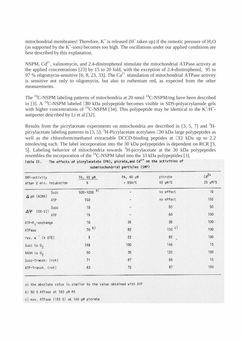
NSPM, Cd2+, valinomycin, and 2.4-dinitrophenol stimulate the mitochondrial ATPase activity atthe applied concentrations [23] by 15 to 20 fold, with the exception of 2.4-dinitrophenol, 95 to97 % oligomycin-sensitive [6, 8, 23, 33]. The Ca2+ stimulation of mitochondrial ATPase activityis sensitive not only to oligomycin, but also to ruthenium red, as expected from the othermeasurements.
The 14C-NSPM labeling patterns of mitochondria at 20 nmol 14C-NSPM/mg have been describedin [3]. A 14C-NSPM labeled ∼80 kDa polypeptide becomes visible in SDS-polyacrylamide gelswith higher concentrations of 14C-NSPM [34]. This polypeptide may be identical to the K+/H+-antiporter described by Li et al [32].
Results from the picrylacetate experiments on mitochondria are described in [3, 5, 7] and 3H-picrylacetate labeling patterns in [3, 5]. 3H-Picrylacetate acetylates ∼30 kDa large polypeptides aswell as the chloroform/methanol extractable DCCD-binding peptides at ∼12 kDa up to 2.2nmoles/mg each. The label incorporation into the 30 kDa polypeptides is dependent on RCR [3,5]. Labeling behavior of mitochondria towards 3H-picrylacetate at the 30 kDa polypeptidesresembles the incorporation of the 14C-NSPM label into the 33 kDa polypeptides [3].

Tables 1 and 2 [5] show the effects of 20 nmoles NSPM/mg submitochondria, 40 µMpicrylacetate and 25 µM Cd2+ with 2 minutes incubation time. The results may be roughlysummarized as follows:
1. Succinate- or ATP-supported ACMA response increases on NSPM or picrylacetate addition,picrate is without effect, and Cd2+ has the opposite effect: the succinate response is abolished, theATP response raises to the NSPM and picrylacetate values, while t1/2 of H+-release is notaffected by NSPM.
2. Succinate- and ATP-supported oxonol VI-response is lowered by NSPM and picrylacetate orpicrate; Cd2+ lowers the succinate-supported response, but has little effect on the ATP-inducedone.
3. ATP/O, ATP-Pi-exchange and ATPase are only slightly, or not at all influenced by NSPM orCd2+. In contrast, picrylacetate almost abolishes ATP-Pi-exchange and lowers ATPase, whilepicrate reduces ATP-Pi-exchange and stimulates ATPase.
4. ATP-driven reverse electron transfer from succinate to NAD is abolished by NSPM,picrylacetate, lowered by picrate and not much affected by Cd2+. Succinate oxidation isstimulated by NSPM and picrate, but not affected by picrylacetate, and almost abolished byCd2+.
5. NSPM and picrylacetate decrease the NADH-oxidation rate, picrate stimulates this activityand Cd2+ shows no effect.
6. NSPM, picrylacetate and picrate lower the succinate- and ATP-driven transhydrogenaseactivities, but Cd2+ effects only the succinate-driven activity. The ATP-driven transhydrogenaseis much more sensitive to NSPM in the presence of KCN instead of rotenone.
Most effects by Cd2+ can be easily explained. Measurements on submitochondrial particles [27,28] indicate Cd2+-induced changes in the cytochromes: The high potential cytochrome b562 ofcomplex III disappears almost totally on Cd2+ addition. Comparing experiments with antimycinA + Cd2+ on aerobe/anaerobe-working particles, indicates inhibition by Cd2+ of energy transferfrom the high potential cyt b to the low potential cyt b .
The results described demonstrate that proton (cation) movement (ACMA response), andmembrane potential (oxonol VI response) are not correlated with the ATP/O-ratio or the ATP-Pi-exchange activities performed by the particles. These results, therefore, encouraged us to moreexplicitly explore the energy-dependent ACMA quench and oxonolVI responses [Fig.5 to Fig.9].
The succinate dependent ACMA fluorescence quenching in the applied assay mixture is verysmall. But this quench can be increased to about 600 % by addition of 24 nmoles NSPM/mgparticles, and can be abolished by addition of 0.2 mM CdCl2. 130 nmoles N-ethyl-maleimide/mgare only marginally effective. The response remains sensitive to 2 µM of the uncoupler TTFBand to anaerobiosis under all conditions [Fig. 5]. The ATP-dependent ACMA fluorescencequenching in the same assay mixture is much larger than the succinate-dependent level, and canbe increased further to about 140 % by addition of 24 nmoles NSPM/mg or 0.2 mM CdCl2. The

Fig.9. Energy-dependentresponse of oxonol VI insubmitochondrial particles(SMP). Reaction mediumcontaining 0.25 M sucrose,50 mM Tris-sulfate, 10 mMMgSO4, pH 7.5, 0.4 mgSMP, 3.8 µM oxonol VI,energization with 5 mM Na-succinate respectively 5 mMNa-ATP. Additions (each):20 µM PA, 20 nmol/mgNSPM, 250 nmol/mgNEM;0.4 (---) and 4 mM CdCl2(___) at ATP-, 0.4 mM (-.-), 1mM (---) and 4 mM CdCl2(___) at succinateenergization; 2 µM CCCP.
Fig. 5. Energy-dependent ACMA fluorescence quenchingin the presence of NSPM, NEM and CdCl2. Reactionmedium contained 0.25 M sucrose, 50 mM Tris-sulfate,10 mM MgSO4, pH 7.5, 0.8 mg submitochondrialparticles, 0.5 µM ACMA, energization with 5 mM Na-succinate or 5 mM Na-ATP, respectively. Additions(each): 24 nmol/mg NSPM, 43 nmol/mg NEM, 0.2 mMCdCl2, 2 µM TTFB.
Fig. 6. Energy-dependent ACMA fluorescencequenching in the presence of NSPM, CdCl2 and PA.Conditions as described in Fig. 4. Additions (each):24 nmol/mg NSPM, 0.2 mM CdCl2, 11 µM PA, 2µM TTFB.

response is fully sensitive to TTFB. The increased ATP- or succinate-dependent ACMAfluorescence quench becomes equal. 33 µM picrylacetate behave similarly to 24 nmolesNSPM/mg [Fig. 6]. 36 (48) mM KCl result in nigericin-sensitive, but valinomycin-insensitivefull responses of either ATP- or succinate-supported ACMA fluorescence quench. The quenchinduced by 18 mM CaCl2 reaches a level of about 60 to 70 % of the KCl values [Fig. 7].
10 nmol dicyclohexylcarbodiimide/mg have almost no effect on the succinate/KCl-supportedquench, but are highly effective in abolishing or preventing the ATP-(ATP/KCl) supportedquench [Fig. 8]. All fluorescence quenches described remain fully sensitive to O2 depletionand/or TTFB additions.
The effects of the described compounds on the energy-dependent absorbance increase of oxonolVI have been summarized in Fig. 9. With essentially the same conditions as described for theexperiments on the ACMA fluorescence quench (Fig. 5 to Fig. 8) 60 to 80 µM picrylacetate, 20nmoles NSPM/mg SMP, 8 to 12 mM or 0.4 to 1 mM CdCl2, respectively, abolish the ATP- orsuccinate-supported absorbance increase of oxonol VI. The absorbance increases are therefore 2to 4 times less sensitive to picrylacetate or CdCl2, but equally sensitive to NSPM if compared
Fig. 7. Energy-dependent ACMA fluorescencequenching in the presence of KCl and CaCl2.Conditions as described in Fig. 4. Additions (each):12 mM KCl, 6 mM CaCl2, 1 µg nigericin (Nig), 1 µgvalinomycin (Val), 2 µM TTFB.
Fig. 8. Energy-dependent ACMA fluorescence quenching inthe presence of KCl and DCCD. Conditions as described in Fig.4. Additions (each): 2 nmol DCCD/mg (10 nmol/mgbefore ATP), 12 mM KCl, 2 µM TTFB. M Tris-sulfate, 10 mMMgSO4, pH 7.5, 0.4 mg SMP, 3.8 µM oxonol VI, energizationwith 5 mM Na-succinate or 5 mM Na-ATP, respectively.

with ACMA fluorescence quenches. N-ethylmaleimide (500 nmoles/mg SMP) has almost noeffect on the energized oxonol VI response, which is similar to the results obtained for ACMAfluorescence quenches. All oxonol VI responses are, or remain, fully sensitive to 2 µM CCCPand/or O2 depletion.
Fig. 10. ATP-dependent responseof oxonol VI in complex V.Conditions as described [8].Additions: 2 mM phosphoenol-pyruvate (PEP), 20 µg pyruvatekinase (PK), 10 µM CCCP.
Fig. 11. Mg-ATP-dependent response of oxonol VI incomplex V. Conditions as described [8], but withoutMg2+. Additions: 2 mM phosphoenolpyruvate (PEP),20 µg pyruvate kinase (PK), Mg as MgSO4, 10 µMCCCP. Controls show: PK is active, although notmaximal at the applied starting conditions.

Figs. 10 and 11 show K+- and Mg2+-modulations of the ATP-dependent absorbance increases ofoxonol VI on complex V. In the ATP regenerating system at low steady-state ATP, the responsecan be modulated by K+ (Fig. 10), Mg2+ (Fig. 11) or Ca2+-ions (not shown). K+ (Ca2+) lowers,Mg2+, in contrast, enhances dye response [9].
Labeling patterns of 14C-NSPM-treatedsubmitochondrial particles, complex V andsoluble F1-ATPase are described andshown [1-3, 35], plus 3H-picrylacetate-treated submitochondrial particles and F1
[2, 3, 5]. 40 µM 3H-picrylacetate acetylateabout 0.4 to 0.6 nmoles 30 kDapolypeptides, as well as 0.2 to 0.3 nmolesDCCD-binding peptide. At 120 µM 3H-picrylacetate there is label incorporation ofabout 1 to 1.5 nmoles/mg into the 30 kDapolypeptides, and 0.6 to 0.8 nmoles/mginto the proteolipids. This labelincorporation raises to about 2 nmoles/mgor 1.2 nmoles/mg into the 30 kDa –respectively 12 kDa peptides on treatmentof SMP in the normal assay medium with200 µM 3H-picrylacetate. Labelincorporation into the proteolipids in SMPis dependent on pH and reducing agents(Fig. 12). As may also be seen in Fig. 12,the proteolipids obviously appear inaggregated multiple forms under theapplied conditions. The labeling behaviorof SMP towards 3H-picrylacetate at the 30kDa polypeptides resembles the 14C-NSPMlabel incorporation into the 33/30 kDa
.. polypeptides. 3H-picrylacetate acetylates in
. F1 the α-, β- and γ-subunits [5].
Discussion
We do not intend to review all literature data presented over the last 30 years. Instead, we willbriefly summarize the results described above and the information related to the aim of ourstudy (i.e. the K+/Ca2+-transport activities of mitochondria). Figures will be presented showingK+ (Ca2+)-fluxes obtained, which demonstrate the effects of our "aids" in evaluating transportactivities. Then we will discuss some of the most important literature data, in our opinion, inrelation to our flux schemes, and will finally describe some newly-discovered mitochondrialfunctions.
Fig. 12. Distribution of radioactivity due to 40 µM 3H-picrylacetate-induced acetylation of SMP electrophoresed on pH 5.0acetate NaDodSO4-acrylamide gels (-DTE, continuous curve),and on 12.5 % pH 7.0 Weber-Osborn NaDodSO4-acrylamidegels (+ 5 mM DTE, interrupted line).

The results of our experiments may be rationalized as shown in Fig 13. It is obvious that we aredealing with the K+/H+-antiport system of mitochondria [32]. This K+/H+-antiporter is drivenwith the energy derived from substrate oxydation by the respiratory chain. It is sensitive to Cd2+,NSPM, Ca2+ or DCCD (all stimulate) from the outside and is regulated by Mg2+ and quinine.Cd2+ and Ca2+ sensitivity are prevented by ruthenium red. As expected, K+/H+ exchange isstimulated, Mg2+ sensitively, by nigericin-supported K+/H+-cycling. It is promoted byvalinomycin/+ Mg, presumably through direct interaction with the F0F1-ATPase/ATPsynthase[4], and by 2.4-dinitrophenol at the µM0 or mM i concentrations applied, respectively, and itsdirect interaction with the F0F1-ATPase/ATPsynthase [2-4].
Fig. 13. Summation of various mitochondrial energy-linked functions. Location and effects of energy transferinhibitors and uncouplers on K+/H+-antiporter, K+-pump and Pi/H
+-symporter. Mitochondrial coupling to thenervous system. Glu = glutathione, TH = transhydrogenase, Oli = oligomycin, PA = picrylacetate, Val =valinomycin, RR = ruthenium red, Nig = nigericin, SR/ER = sarcoplasmic/endoplasmic reticulum.
The coupling modes between e--transfer and the K+/H+-antiport system are unclear to us. How isit possible to link a K+/H+-antiporter to a respiratory chain just pumping protons as described byLi et al [32], assuming we are dealing with this antiporter (which seems to be the case based onthe presented results, although Li et al used sonicated uncoupled mitochondria for their studyresulting in different DCCD effects compared to our study)? Green [36] demonstrated activeand opposite H+- and K+-transport activities by cytochrome oxidase (complex IV) reconstitutedin phospholipid vesicles. His results and the model derived from his studies agree much morewith our data than the current descriptions of electron transfer coupling, and should thus beconsidered when explaining our results.

The modulations of the K+/H+-antiporter activity described above lead to corresponding swellingor contraction of the mitochondrial matrix space by the osmotically active K+-ions and inmodified oligomycin-sensitive (valinomycin, NSPM, Cd2+, Ca2+) or ruthenium red-sensitive(Cd2+, Ca2+) ATPase activities. NSPM and Cd2+ - at the low concentrations applied - act from theoutside, since β-hydroxybutyrate or NADH-dehydrogenase respectively, and complex IIIactivities were not modified by the compounds. Swelling/contraction and ATPase activity weretherefore indirectly influenced via osmotically-active K+-ions. The oligomycin sensitivity of thechanged ATPase activities suggests mitochondrial F0F1-ATPase involvement, most probably bythe release of K+-ions. The osmotic back-pressure [37] at high K+-load (see description of theoscillatory phenomena in results) may also open up the ATPase for K+-release, in which theATPase inhibitor IF1 could be involved [38]. Ruthenium red prevents Cd2+-, Ca2+-induced K+-uptake and accompanies ATPase modulations together with K+-release.
Cd2+ and Ca2+ under 10 µM bind (Mg2+-sensitive) at high affinity binding sites ( ∼ 1 nA/mg) [2,3, 6, 39], thereby inducing Mg2+-sensitive K+-uptake, but without being accumulated itself. Thefree active cytosolic Mg2+ concentration should be low in contrast to our applied concentrations( ∼ 6 to 10 mM). In fact, free concentrations of cellular cytosolic Mg2+ have been estimated to be< 1 mM [40]. Respiratory activity, K+-cycling, swelling/contraction phenomena and ATPaseactivity are thus controlled by available O2, and most importantly by the free Ca2+ and Mg2+
concentrations!
A high noradrenaline/adrenaline-ratio in plasma leads in low glycolysis rates and as consequencelowered ATP concentrations in the cell. ADP-, Pi- and free Mg concentrations in the cytosol, aswell as ATP synthesis [41, 42] and oxidized glutathione concentrations in mitochondria, areconcomitantly raised, but the reduced glutathione concentrations in the cells lowered. Thechanged ATP/ADP or Ca/Mg ratio, respectively, determines, in this case, the respiratory rates(Table III). Our results and conclusions are based on extrapolations from summarized results onmitochondrial oscillations [44] and are, furthermore, one way to explain the molecularmechanism leading to these phenomena. Longer noradrenaline exposure of cells leads toconstantly lowered cAMP-concentrations and the consequences described [43].
Fig. 14. Summation of varioussubmitochondrial energy-linkedfunctions. Location and effects ofenergy transfer inhibitors anduncouplers on K+/H+-antiporter, K+-pump and Pi/H
+-symporter.Val = valinomycin, Nig = nigericin,PA = picrylacetate, TH =transhydrogenase.

A rise in cytosolic Ca by extracellular signals leads to muscle contractions, myofibril mobility,stimulation of respiration, etc. with liberation of ADP, Pi and Mg, resynthesis of ATP by creatinephosphate [42] and rebinding of Mg. As a consequence, there is a constantly changing Ca/Mg-ratio with oscillating mitochondria. The changing Cacyt determines the Ca/Mg or ATP/ADP ratio,respectively (Table III).
At Cd2+ or Ca2+ concentrations above 10 µM, both ions were taken up. But Cd2+, at < 10 µM,prevents a Ca2+ (Pi)-accumulation by binding to the Pi/H
+- and Ca2+-binding sites. Takentogether, Ca2+-ions behave similarly in many ways to K+-ions (uptake, swelling/contraction,ATPase, etc.). This is not surprising since the Ca2+ concentrations in the cytosol of our cell isonly about 1 to 10 µM under normal physiological conditions (Table III) and its K+-cyclingsystem may, therefore, also be able to handle high "abnormal" Cao
2+ concentrations (as well asCai
2+/Pi) (Fig. 13).

Submitochondrial particles can be compared to mitochondria: in both systems we are dealingwith energy-driven K+/H+-exchange activities, driven either by substrate-supported electrontransfer or by ATP-hydrolysis (Fig. 14). As may be seen in the summation of our results, thesuccinate-driven K+/H+-antiporter works opposite to mitochondria in SMP under the appliedconditions. The arising nigericin-sensitive (valinomycin-insensitive) K+ and H+ gradientsbecome maximal and stimulated by succinate oxidation at low NSPM and picrylacetate, and athigh K+ or Ca+ concentrations, but are abolished at moderate Cd2+ concentrations, atanaerobiosis, or on TTFB addition.
The abolishment by Cd2+ can be explained in detail by the action of this heavy metal ion oncomplex III activity, and thereby the inhibition of the energy transfer from the high potential tothe low potential cyt b (resulting in inhibition of the forward, succ → O2, but not the backreaction, reverse e--transfer, Table II). However, the question of why Cd2+ inhibits succ → O2,but not NADH → O2, remains to be answered. We think that a possible answer may be found inthe ubiquinone pool.
The direction of the succinate-driven K+/Ca2+(and H+)-gradients are obviously dependent on theforces the K+/H+-antiport system is exposed to in the SMP-membranes (opposite bending to themitochondrial membrane). Otherwise, we do not have a plausible explanation for the differentdirections of the gradients in SMP or mitochondria. There is definitely a relationship to theforces effective on the membrane proteins during the membrane transitions (oscillations) inmitochondria.
ATP-hydrolysis (similarly to succinate oxidation) produces large nigericin-sensitive/valinomycin-insensitive (site-directed binding to F0 [4]) K+/H+-gradients, which becomemaximal and almost identical to the succinate-supported gradients at low levels of NSPM,picrylacetate and Cd2+, and at high levels of K+, Ca2+, but which disappear at low concentrationsof dicyclohexylcarbodiimide or TTFB, in contrast to mitochondria and isolated complex V [8].In fact, the ATP-supported K+- or H+-gradients in our SMP particles are highly DCCD sensitive.
This kind of DCCD sensitivity has not been observed so far for the other activities performed bySMP [8], mitochondria [32] or even complex V-preparations [8, 23, 45]. DCCD abolishes ATPsynthetic activity (RCR)/Pi-transport > K+-transport >>> ATPase in mitochondria, presumablyby first modifying the phospholipids [32] before trapping any proteolipid or Pi-carrier. In SMP,the sequence of the DCCD-sensitive reactions is K+-transport > ATP-Pi-exchange or Pi-transport>>> ATPase and proves 1) side-specific reaction of DCCD and 2) separation of the variousreactions by DCCD. Modification of the ATPase leads to abolishment of the K+-gradients,although not of their establishing force, the Mg-ATPase. Our complex V-preparations show noATP-dependent ACMA-responses because they are not vesicularized [8].
Before discussing data on oxonol response, some points about valinomycin have to be added.Valinomycin is able to abolish K+-gradients in mitochondria, which leads to K+-cycling and ahigh proton gradient (by stimulation of the K+/H+-exchange and the ATPase), together withstimulated respiration (uncoupling, as shown). This is not the case with submitochondria, whichcould be demonstrated with ACMA. If one considers all the results for the explanation of thisfact, including the binding of valinomycin to the F0-part of the ATPase, one reaches the onlypossible conclusion: Valinomycin stimulates the ATPase activity (K+-pump) of mitochondria

(oligomycin-sensitive) from the outside. For these measurements, all the ions present (includingTris+) are important. It should be added that the movement of valinomycin through purephospholipid membranes is already very difficult, and should be almost impossible throughmitochondrial membranes. Pressman's final conclusions [46] are mainly derived fromphospholipid bilayer measurements and are different from his original ones [47, 48].
Why should all the different microorganisms produce these highly complex "ionophores" just forphospholipid bilayer membranes, when their antibiotic activity is directed against systemscontaining only minimal or no phospholipid bilayers? A synthesis directed directly against theion-transporting units is much easier to imagine.
The cyclic decapeptide antamanide for instance has been described by Pressman as neutral“ ionophore” because he found stimulation of K+-uptake by mitochondria and complexation ofNa+ > K+ by this compound [46]. However, studies by Ovchinnikov [49] demonstrate that thebiological action of antamanide is not connected to ionophoric action, and in accordance withthis finding, photoaffinity labelling studies [50] demonstrate specific binding of this compoundto membrane polypeptides. Our results on "protonophoric" uncoupling agents [2] match perfectlythe experimental findings on "ionophoric" compounds.
Normally a model system has to be proven in every aspect and dismissed if not valid in allpoints. In the case described above, it is obviously easier to keep a wrong model rather than toperform difficult new experiments.
The oxonal VI response of SMP is due to the membrane potential and has been extensivelydiscussed [8]. The absorbance increase of oxonol VI is less sensitive to picrylacetate or CdCl2,but equally sensitive to NSPM if compared with the sensitivity of the ACMA fluorescencequench to these compounds. The difference between the oxonol VI responses described [8], andthis paper is due to the use of a Tris+-buffer, which replace K+ or H+, respectively, in theexperiments. The oxonol response in the isolated ATPase complex reflects a more localized,ATP-dependent and energy-related process in contrast to SMP [4, 8, 9], showing, most likely,K+ (Ca2+)-induced conformational changes of the ATPase [9]. The oxonal VI response in SMPthus might reflect K+-pumping activities.
The K+-pumping activity cannot be located on the Pi/H+-symporter, which should be inhibited
for Pi-translocation at the high ADP/ATP and/or low NSPM concentrations applied during theexperiments. The protons cannot be pumped by F0F1 because of the direction in which they areforced. They are most likely delivered by ATP hydrolysis, or Tris+ (used as a buffer), possibly byinvolvement of the Pi/H
+-symporter. Only the F0F1-ATPase is then able to operate as a K+-pump.
Furthermore, we add four additional points to those listed in [4], which imply that the F0F1-ATPase acts as a K-pump:
1. Mg2+ is not required for ATP synthesis [51], but for ATPase activity. Our proposedmechanism for ATP synthesis does not require Mg2+ [4].
2. Concentrations of our "aids" (NSPM, etc.) leading to maximal K+- and H+-gradients andmoderately-stimulated, oligomycin-sensitive ATPase acitivities, are not very effective on theATP/O-ratio or ATP-Pi-exchange.

3. Sandri et al [52] required monovalent cations, including K+, besides F6 for membrane bindingof F1; a point possibly important for a K-pumping activity of F0F1.
4. Schneider et al [53] found K+-translocation on EF0 by performing conductance studies onblack lipid membranes.
The summation of all described points concerning K+-translocation – other than by the electrontransfer linked K+/H+-exchange (Fig.13, 14) proves that mitochondria contain a valinomycin-and oligomycin-sensitive, ATP-driven K+-pump as originally suggested [47, 48], which isidentical to the well-known oligomycin-sensitive F0F1-ATPase, but not with the ATP synthase(Fig. 13, 14). Also, the results obtained on reconstituted CF0F1liposomes [54-56] are inagreement with the ones presented here. ATP-Pi
33-exchange/ATP synthesis may be due toavidin-sensitive propionyl-CoA-carboxylase [8, 57], to (coupled) AP5A-sensitive adenylatekinase [8, 54], or should be compared to the sarcoplasmic reticulum Ca2+-pump ATP synthesis[58]. Our hypothesis for ATP synthesis [4] is therefore not only probable, but indirectly provenand requires only a few more experiments to be directly proven.
The results of Lardy et al [4, 31, 33] become clearer in light of our results: the anti-swellingactivity of ATP and the contraction of swollen mitochondria by ATP are due to the describedATP-driven K-pump or F0F1-ATPase, respectively. Under normal physiological conditions, thispump is responsible for the establishment and maintenance of a high K+-gradient, which meansthe K+ concentration is very low inside (matrix), but high outside (cytosol). The physiological K+
concentrations in the mitochondrial matrix space are normally in the range of about 1 mM i , inthe cytosol about 175 mMo (the maximal K+ concentrations for the matrix space underdeenergized/uncoupled conditions) (Table III). Under normal physiological conditions, noprotons are either moving from the K+/H+-exchanger to the Pi/H
+-symporter (ATP synthase)along the membrane via hydrogen bridges [59], or the K+/H+-exchanger and the Pi/H
+-symporter(ATP synthase) are directly linked [61] (Fig. 13). The conclusion for a delocalized, protonmotiveforce as presented in [43] is thus, in light of our results, incorrect.
The results presented above imply a direct connection (and regulatory function) between thenervous system (brain) and mitochondria (body and brain). Our body temperature may then beregulated by mitochondria via nervous signals from the brain. The normal substrate oxidationand O2-uptake/-reduction in our bodies (anorexia/dystrophia) is directly linked to our nerveimpulses, and thus essentially to the brain. Substrate overload leads to a rise in acetyl-CoAconcentrations, and consequently also to a rise in cholesterol, hormone and Vitamin (D3)concentrations, which as a result, can in turn lead to such diseases as arteriosclerosis [60], heartattack and cancer.
Oxygen deficiency, or even depletion in our cells, results in an imbalance of the Na+/K+
concentrations across the cellular and mitochondrial membranes, and to osmotic imbalancesresulting in H2O decreases with damage to mitochondria and cells. As a consequence, the build-up of edema during anoxia/hypoxia, or shock and dilusion with coma (death) can occur.
The above examples may be just some of the possible interactions between nervous system andmitochondria. Mitochondria are in the true sense of the word the “hearts” of our cells.

A technical realization of bioenergy production (via molecular biotechnology) is made possibleby our results. Mitochondria are ideal oscillators and predestined, through mutual coupling withthe plasma membranes, to function in the brain for memory and thinking. Such new materialscould thus lead to the development of thinking computers.
Acknowledgements
I thank Prof. E. Neumann, in whose laboratory I learned more about plasma membrane receptorsand also about some of the possible problems which may arise during interdisciplinary researchefforts, and Prof. K. Altendorf for allowing us to use his fluorescence spectrophotometer. Wealso thank Prof. Th. Wieland, Mrs. E. Dirscherl and Mrs. C. Cavanna for their help in preparingthis manuscript.
References
1 Kiehl R (1980)1st Eur.Bioenerg.Conference, Urbino, p.113-114, IUB-IUPAB BioenergeticGroups.2 Kiehl R (1995) part I.3 Kiehl R (1995) part II.4 Kiehl R (1995) part III.5 Kiehl R, Akinpelu O, Hoffmann-Posorske E, Kordt S (1982) 2nd Eur. Bioenerg. Conference,Lyon, p. 91-92, IUB-IUPAB Bioenergetic Groups.6 Kiehl R (1982) 12. Internat. Congr. of Biochem., Perth, Abstr. 004-56.7 Kiehl R (1982) RACI-Meeting, Canberra, Joint Meeting to the 12. Int. Congr. of Biochem,Perth.8 Kiehl R and Hanstein WG (1984) Biochim. Biophys. Acta 766, 375-385.9 Kiehl R and Hanstein WG (1984) 3rd Eur. Bioenerg. Conference, Hannover, p. 323-324, IUB-IUPAB Bioenergetic Groups.12 Kaschnitz RM, Hatefi Y, Morris HP (1976) Biochim. Biophys. Acta 449, 224-235.13 Chance B (1959) in: Ciba Symposium Regulation Cell Metabolism (Wolstenholme GE andO'Connor, Eds.) 91-129, Little Brown, Boston, Mass.14 Rossi C, Scarpa A and Azzone GP (1967) Biochemistry 6, 3902-3910.15 Mitchell P (1968) Eur. J. Biochem. 4, 530-539.16 Archbold GPR, Farrington CL, Lappin SA, Mc Kay AM and Malpress FH (1979) Biochem.J. 180, 161-174.17 Hanstein WG and Hatefi Y (1974) Proc. Nat. Acad. Sci USA 71, 288-292.18 Waggoner A (1976) J Membrane Biol 27, 317-334.19 Grünhagen HH and Witt HT (1970) Z Naturforscher 25 b, 373-386.20 Hansen M, Smith AL (1964) Biochim.Biophys.Acta 81, 214-222.21 Löw H and Vallin L (1963) Biochim. Biophys. Acta 69, 361- 374.22 Ragan CI and Racker E (1973) J. Biol. Chem. 248, 6876-6884.23 Kiehl R and Hatefi Y (1980) Biochemistry 19, 541-548.24 Le Bel D, Poirier GG, Beaudoin AR (1978) Anal. Biochem. 85, 86-89.25 Lam KW, Warshaw JB and Sanadi DR (1967) Arch. Biochem.Biophys. 199, 477-484.26 Hatefi Y and Hanstein WG (1973) Biochemistry 12, 3515-3522.27 Becker WG, von Jagow G, Anke T and Steglich W (1981) FEBS Letters 132, 329-333.28 Surkow SA and Konstantinov A (1979) FEBS Letters 109, 283-288.29 Leader JE and Whitehouse MW (1966) Biochem. Pharmacology 15, 1379-1387.

30 Kiehl R and Bäuerlein E (1976) FEBS Letters 72, 24-28.31 Connelly JL and Lardy HA (1964) Biochemistry 3, 1969-1973.32 Li X, Hegazy MG, Mahdi F, Jezek P, Lane RD, Garlid KD (1990) J. Biol. Chem. 265,15316-15322.33 Lardy HA, Connaly JL, Johnson D (1964) Biochemisry 3, 1961-1968.34 Kiehl R, Dissertation (1976) Universität Heidelberg und Max-Planck-Institut für Med.Forschung, Heidelberg, FRG.35 Kiehl R (1980) FEBS Letters 109, 280-282.36 Green DE (1977) Proc. Nat. Acad. Sci USA 74, 3662-3666; Seminar at Scripps Clinic andRes. Foundation, 13 of April, 1979.37 Brdicka R (1969) Grundlagen der phys. Chemie, UEB Deutscher Verlag der Wissenschaften,Berlin, 330-340 and 637-638.38 Pullman ME and Monroy GC (1963) J. Biol. Chem. 238, 3762-3765.39 Mitchell P (1977) FEBS Letters 77, 136-140.40 Veloso D, Guynn RW, Oskarsson M and Veech RL (1973) J. Biol.Chem. 248, 4811-4819.41 Soboll S and Bünger R (1981) Hoppe-Seyler's Z. Physiol. Chem. 362, 125-132.42 Klingenberg M and Heldt HW (1982) in: Metabolic Compartmentation (Sies H, Ed.) 101-122, Academic Press, London.43 Kiehl R (1993) Dear Colleague 2(2):4-6.44 Hess B and Boiteux A (1971) Ann. Rev. Biochem. 40, 237-258.45 Hatefi Y and Matsumi-Yagi A (1992) in: Adenine Nucleotides in Cellular Energy Transferand Signal Transduction (Papa S, Ed) 33-46, Dirkhäuser-Verlag, Basel.46 Pressman BC (1976) Ann Rev of Biochem 45, 501-530.47 Pressman BC (1965) Fed Proc 24, 425-428.48 Pressman BC (1965) Proc Natl Acad Sci 53, 1076-1081.49 Ovchinnikov YA, Ivanov VT, Barsukov LI, Melnik EI, Oreshnikova ND, Bogolyubova ND,Ryabova JD, Miroshnikov and Rimskaya VA (1972) Experientia 28/4, 399-401.50 Wieland Th, Nassal M, Kramer W, Fricker G, Bickel U, Kurz G (1984) Proc Natl Acad Sci81, 5232-5236.51 Chan TL, Greenawalt JW and Pedersen PL (1970) J. Cell. Biol. 45, 291-305.52 Sandri G, Wojtczak L and Ernster L (1981) Int. Symp. On Vect. Reactions in Electron- andIon-trransport in Mit. And Bact., Fasano, 14.53 Schneider E, Schmid R, Deckers G, Steffens K and Altendorf K (1981) Int. Symp. on Vect.Reactions in Electron- and Ion-transport in Mit. and Bact., Fasano, 48.54 Pick U and Racker E (1979) J Biol Chem 254, 2793-2799.55 Schmidt G and Gräber P (1985) Biochim Biophys Acta 808, 46-51.56 Richard P, Rigaud JL and Gräber P (1990) Eur J Biochem 193, 9211-925.57 Stigall DL, Galante Y and Hatefi Y (1978) J Biol Chem 253, 956-964.58 Hasselbach W (1978) Biochim Biophys Acta 515, 23-53.59 Christen HR (1969) Grundlagen der allgemeinen und anorganischen Chemie, VerlagSauerländer, Aaran, 151-178.60 Murray M, Schrodt GR and Berg FH (1968) Biochem Med 2, 118-135.61 Kiehl R, Schuermann M and Hanstein WG (1988) Biochem-life Sci Adv 7, 71-75.