Embed Size (px)
Citation preview
Acta Orthop Scand 2002; 73 (3): 359–368 359
Bone stress injuries of the lower extremityA review
Jan Lassus1, Ilkka Tulikoura1, Yrjö T Konttinen2,3,4, Jari Salo1 and Seppo Santavirta1
Departments of 1Orthopaedics and Traumatology, 2Internal Medicine, Helsinki University Hospital, 3ORTON, Research Institute, Invalid Foundation, Helsinki, 4Anatomy, University of Helsinki, Finland. Correspondence: Dr. Seppo Santavirta. E-mail: seppo.santavirta@hus.� Submitted 01-02-03. Accepted 01-09-19
Copyright © Taylor & Francis 2002. ISSN 0001–6470. Printed in Sweden – all rights reserved.
ABSTRACT – Bone stress injuries can cause long-last-ing damage, especially in young athletes and military conscripts, if not diagnosed and treated properly.
Diagnosis has been traditionally based on clinical, radiographic and scintigraphic examinations, but MRI has become increasingly important. High resolution MRI is particularly valuable for the grading of bone stress injuries. The clinician should be aware of the wide range of bone stress injuries and available diagnostic methods. Early diagnosis is the prerequisite for avoiding long-lasting complications.
Most bone stress injuries heal with closed treatment, but surgery is necessary in some cases. They heal well if the diagnosis is not delayed and the treatment ade-quate.
n
Bone stress injuries are mostly seen among long-distance runners, soldiers, dancers, athletes and other sportsmen. The patients are usually young or middle-aged (Table 1).
About 400–500 sportsmen are affected by a bone stress injury annually in Finland (population 5 million; Hulkko and Orava 1991). The global incidence of bone stress injuries in sports has been estimated to be 2–4%: 2% in men and 7% in women (Johnson et al. 1994). Female athletes run an up to four times higher risk of a bone stress injury (Brunet et al. 1990, Zernicke et al. 1993). Among athletes, one � fth of all musculoskeletal stress injuries are located in bones (Bennell et al. 1996). Bone stress injuries of the lower extremities are commonest in the shin of long-distance runners (Table 1).
The recurrence rate of bone stress injuries among athletes during a 4-year follow-up varied between 2% and 13% (Sullivan et al. 1984, Mathe-son et al. 1987), and 11–20% in military conscripts
(Milgrom et al. 1985). Multiple stress injuries are seen in 10% of runners (Sullivan et al. 1984) and in over 20% of female athletes (Bennell et al. 1996).
Etiology
The main factor predisposing to bone stress inju-ries is repeated mechanical load. The amount of load seems to correlate directly to the extent of injury (Marti et al. 1988).
The ratio of exercise to rest seems to be impor-tant, according to the authors’ own experience and some reports (Nordin and Frankel 1989). In sports, the quality of running shoes and road or track surfaces are important mechanical factors for bone stress injuries. If bone is given enough time to recover from strain, it will become stronger with exercise (Conroy et al. 1993). The quality and
Table 1. Location (%) of bone stress injuries according to various kinds of sports according to Bennell and Brukner 1997
Sports Shin Foot Fibular Navicular bone bone bone
Athletics 39 21 17 73Long-distance running 42 12 27 4Dance 3 43 23 0Basket ball 0 2 0 4Racket games 3 2 7 4Aerobic 0 7 3 0Other sports 14 12 23 16
Act
a O
rtho
p D
ownl
oade
d fr
om in
form
ahea
lthca
re.c
om b
y 11
1.68
.111
.42
on 1
0/27
/14
For
pers
onal
use
onl
y.

360 Acta Orthop Scand 2002; 73 (3): 359–368
amount of the bone’s reaction depend on the type of stress (Haapasalo et al. 1994): strength exercises to improve strength increase bone mass more than running (Robinson et al. 1995).
The effect of load on bone changes when the type of training is modi� ed. We have found that shock absorbers or different shoe models had no effect on the occurrence of bone stress injuries, which agrees with other reports (Jones et al. 1989). Height and weight do not seem to affect the risk of developing bone stress injuries, but the percentage of body fat in women shows a negative correlation
(Jones et al. 1989, Bennell et al. 1996). Some con-troversy exists about the effect of age in that one report found no in� uence on the incidence of stress injuries (Shwayhat et al. 1994).
We have noted that when bone stress injuries recur often and are bilateral or multi-focal, some important risk factor probably exists. Anteversion of the femur, varus or valgus knees, tibia vara, varus- or valgus-rotated calcaneus, hyperprona-tion of the foot, plain or curved foot or a foot with an intermetatarsal neurinoma are regarded as anomalies that increase the risk of stress injuries (Matheson et al. 1987, Krivickas 1997). Flat foot
(Sullivan et al. 1984) and leg-length discrepancy (Brunet et al. 1990) also seem to increase the risk for stress injuries. Currently there is hardly any scienti� c evidence to prove that certain anatomical features—e.g., the anatomical shape of the middle foot—really affects the development of stress inju-ries (Krivickas 1997).
A lower bone density increases the risk of stress injuries in women more than in men (Bennell et al. 1996). However, men with bone stress injuries do not usually have less bone density (Bennell et al. 1996). The relevance of bone density to stress injuries therefore remains controversial (Bennell et al. 1995).
Menstrual disturbances and delayed menarche seem to increase the risk of sustaining a bone stress injury 2–4-fold (Bennell et al. 1996). In some stud-ies, contraceptives seemed to reduce the risk of stress injuries by 50% (Myburgh et al. 1990), but in others had no effect (Kadel et al. 1992). Estro-gen replacement therapy may reduce the incidence of stress injuries (Bennell and Brukner 1997). Spontaneously normal menstrual periods seem to prevent bone stress injuries and enhance density of
bone matrix more effectively than any substitutes
(Jonnavithula et al. 1993).No correlation has been found between the
development of bone stress injuries and nutrition in men, but anorectic women run a higher risk of stress injuries (Bennell et al. 1996).
Pathogenesis
Bone is a dynamic tissue, which needs a constant load for normal remodeling (Sterling et al. 1992), according to Wolff’s law (Chamay and Tschants 1972). Repeated traumatic strain induces remodel-ing, starting with osteoclastic activation and bone resorption (Chamay and Tschants 1972, Carter et al. 1981, Burr 1993). This process reaches its maximum in 3 weeks (Jones et al. 1989, Sallis and Jones 1991, Sterling et al. 1992). Thereafter, the new bone created � lls resorption tunnels and pits. This usually takes about 3 months (Frost 1991, Sallis and Jones 1991). Macroscopically, apart from a periosteal reaction, stress-induced endos-teal bone growth can often be seen even when no fractures are present (Uhthoff and Jaworski 1986). Later, the new bone matures and forms normal lamellar layers (Uhthoff and Jaworski 1986).
In optimally-directed loading, with enough time for the remodeling process, bone mass remains unchanged and no stress injury will result, but the bone becomes stronger (Uhthoff and Jaworski 1986, Frost 1991, Sallis and Jones 1991, Frost 1994). However, if the load is excessive, prolonged or recurrent, the resorption process dominates and formation of new bone does not proceed properly. This weakens the bone and a stress injury occurs
(Burr et al. 1985, Jones et al. 1989, Frost 1994, Maitra and Johnson 1997). Repeated micro-trauma of a previously stress-injured bone may eventually result in a stress fracture, which can be seen on plain radiographs (Carter and Caler 1983, Frost 1994). We have measured high intraosseous hydro-static pressures—i.e., 46–58 cm H2O (normal 21– 23 cm H2O) in patients having symptoms related to bone stress injuries, but no detectable fracture (unpublished observations) and found that a stress-injured bone with an obviously increased uptake in scintigraphy may have focal necroses on histologi-cal examination. This is probably due to intraosse-ous compartment syndrome-like conditions which, because of poor local blood circulation, result in
Act
a O
rtho
p D
ownl
oade
d fr
om in
form
ahea
lthca
re.c
om b
y 11
1.68
.111
.42
on 1
0/27
/14
For
pers
onal
use
onl
y.
Acta Orthop Scand 2002; 73 (3): 359–368 361
bone necrosis. Ischemia may also explain the local pain. Poor blood circulation and ischemia as well as a low pH contribute to enhanced resorption and reduce the formation of new bone.
Diagnoses
Load-related pain is typical of bone stress injuries. The onset of pain usually occurs during the � rst 2 weeks after the training becomes more strenuous, but it may be delayed by several months (Greaney et al. 1983, Jones et al. 1989, Maitra and Johnson 1997). The onset of symptoms is gradual. At � rst, pain is present only during stress and it depends partly on the amount of the load. As the process goes on, it starts to occur also at rest and even during the night. Surrounding soft tissues may become swollen. Early suspicion of a possible stress injury is essential for adequate treatment. Local tenderness can be detected by simply tap-ping the affected bone.
Conventional radiography remains the � rst routine examination for suspected stress injuries of bone (Daffner 1978). Radiographic changes become visible 2–12 weeks after the onset of symptoms, if ever (Sullivan et al. 1984, Mathe-son et al. 1987). The sensitivity of radiography is 15–35% in the early stage (Daffner 1978, Greaney et al. 1983). Findings indicating stress fracture are seen in 30–70% of cases only during the follow-up (Daffner 1978, Matheson et al. 1987). The � rst sign, on the surface of long bones, is the so-called “grey cortex”. A grey-looking hypodensic area seen in compact bone is related to the resorption phase of remodeling (Daffner 1984, Mulligan 1995). After this phase, periosteal new bone forms and endosteal bone thickens (Daffner 1978, Martin and Burr 1982, Greaney et al. 1983). A fracture, if present, can be seen as a radiolucent line in com-pact cortical bone and as a sclerotic line in cancel-lous bone (Daffner 1978, Mulligan 1995).
In most cases CT is less sensitive than conven-tional radiography for the detection of stress inju-ries of bone. However, certain fracture lines, such as longitudinal and spiral ones, can be seen more clearly with CT (Daffner 1978, Torg et al. 1982, Matheson et al. 1987, Allen 1988).
Scintigraphy has long been considered to be the best diagnostic method for bone stress injuries
(Daffner 1978) with a sensitivity close to 100%
(Stafford et al. 1986, Matheson et al. 1987). How-ever, false negative diagnoses have been reported (Sterling et al. 1993), possibly because necrotic bone tissue is not labeled (Kanstrup 1997). In these injuries, an increase in uptake can be detected as early as 6–72 hours after the onset of symptoms
(Greaney et al. 1983, Matheson et al. 1987, Kan-strup 1997). An oval signal, the intensity of which correlates to the severity of the bone stress injury, can be seen with scintigraphy (Floyd et al. 1987). Asymptomatic mild stress injuries can also be seen with scintigraphy as clusters of increased uptake
(Daffner 1978, Matheson et al. 1987). Follow-ups with scintigraphy, however, are not entirely satis-factory (Kanstrup 1997).
MRI provides important information in studies of bone stress injuries. It is as sensitive, but more speci� c than scintigraphy (Daffner 1978, Stafford et al. 1986, Lee and Yao 1988, Meyers and Weiner 1991). In addition to changes in bone, it gives information about the surrounding soft tissues in all three dimensions. T2- and STIR-techniques are useful for detecting edema of cancellous bone, periosteum and bone marrow (Daffner 1978, Staf-ford et al. 1986, Resnick 1995). Edema, however, is a nonspeci� c � nding. A fracture can be seen as a lower signal intensity in the middle of the edema
(Daffner 1978, Stafford et al. 1986, Resnick 1995), but it is better seen as a low-density signal line on T1-images. Edema disappears before the signal indicating new formation of bone appears. There-fore, MRI-analysis is most accurate during the � rst 3 weeks (Daffner 1978, Stafford et al. 1986, Martin et al. 1993). The diagnosis is clear if a fracture line can be seen. If only a swelling without signs of a fracture is visible, an accurate diagnosis cannot be made (Daffner 1978, Stafford et al. 1986, Martin et al. 1993). Although bone tissue is seen more ef� ciently on CT than on MRI, the latter is better for early detection and a differential diagnosis
(Daffner 1978, Stafford et al. 1986, Martin et al. 1993, Uhmans and Pavlov 1994). If radiographs show normal bone structure despite pathologi-cal � ndings on scintigraphy, MRI usually gives additional information. In case of uncertainty one can repeat the radiographic and MRI examinations after 3–4 weeks.
Jones et al. (1989) suggested a practical clas-si� cation of bone stress injuries (Table 2). The
Act
a O
rtho
p D
ownl
oade
d fr
om in
form
ahea
lthca
re.c
om b
y 11
1.68
.111
.42
on 1
0/27
/14
For
pers
onal
use
onl
y.

362 Acta Orthop Scand 2002; 73 (3): 359–368
grade of a stress injury is known to have a positive correlation with the intensity of the scintigraphic signal (Zwas et al. 1987, Matin 1988). We mainly use both MRI- and scintigraphic-based classi� ca-tions (Table 3; Fredricson et al. 1995) to get more accurate and useful information for diagnosis, classi� cation and follow-up. In conclusion, the MRI-based classi� cation gives more detailed information about the bone stress injury, but scin-tigraphy is good for screening and measuring the activity of the lesion.
Differential diagnosis. A negative scintigraphy � nding rules out bone stress injuries with great certainty. In medial tibial stress syndrome, an increase in the uptake is seen in a large area on the surface of the shin bone. The signal is seen only in the collecting phase, but not with angiography or bone-pole phases in connection with bone stress injuries. MRI shows swelling only in the periosteal area (Daffner 1978, Matheson et al. 1987, Kan-strup 1997).
Chronic sclerotic osteomyelitis causes bone changes that usually extend throughout the bone circumference and affect a larger area than those associated with stress injuries. Variable cortical thickening, uneven sclerosis and swelling of the bone are seen. In chronic sclerotic osteomyelitis,
radiographs do not change during a few weeks of follow-up (Daffner 1978, Daffner and Pavlov 1992).
Ewing’s sarcoma, which usually occurs in young patients, may be the most important differential diagnostic problem. New bone growth is more even and lamellar-like in stress injuries (Uhmans and Pavlov 1994). Ewing’s sarcoma causes more severe periosteal changes, such as uneven surfaces and spine-like spiculum, and a soft tissue com-ponent is always present. Osteolysis is usually severer in malignant diseases than in bone stress injuries (Daffner 1978, Martin and Burr 1982, Daffner and Pavlov 1992). A biopsy is sometimes necessary for the diagnosis.
Treatment
We consider an early diagnosis is necessary for optimal treatment. To diagnose and classify bone stress injuries correctly, one must combine the clinical, MRI, radiographic and scintigraphic � nd-ings. It is also important to understand the speci� c characteristics and natural healing ability of inju-ries in various bones.
Closed treatment is suf� cient in most bone stress injuries of the lower extremities (Sallis and Jones 1991). The main principle is to treat the injury and prevent recurrences and complications. To achieve this goal, one has to understand the natural history of the injury (Reeder et al. 1996).
It is essential to reduce or eliminate the bone load from the beginning (Sallis and Jones 1991). The time the patient should avoid weight bearing varies according to which bone is injured and how severely it has been affected (McBryde 1982, Matin 1988, Fredricson et al. 1995). The treatment of the shin bone, based on the MRI classi� cation, is shown in Table 4 (McBryde 1982).
Table 2. Classi� cation of bone stress injuries according to Jones et al. 1989
Grade Scinti- Stress Local Radio- Frac- graphy pain tenderness graphy ture
0 + – – – –1 + + – – –2 + + + – –3 + + + + –4 + + + + +
Table 3. Classi� cation of bone stress injuries by MRI according to Fredricson et al. 1995
Grade T1 T2 and STIR
0 normal normal1 -------- mild periosteal and no bone marrow swelling2 -------- evident periosteal and bone marrow swelling3 bone marrow swelling severe periosteal and bone marrow swelling4 bone marrow swelling severe periosteal and bone marrow swelling “low-signal“ -fracture severe bone marrow swelling
Act
a O
rtho
p D
ownl
oade
d fr
om in
form
ahea
lthca
re.c
om b
y 11
1.68
.111
.42
on 1
0/27
/14
For
pers
onal
use
onl
y.

Acta Orthop Scand 2002; 73 (3): 359–368 363
Non-steroidal anti-in� ammatory drugs (NSAID) have been recommended for treatment of the early phase of stress injury (Sallis and Jones 1991, Reeder et al. 1996), but no evidence shows that they affect healing. Theoretically, the NSAIDs may have an osteoblast-inhibiting effect. A variety of physiotherapeutic methods have been recom-mended, such as cold, phonophoresis, electric and electromagnetic treatments (Sallis and Jones 1991, Fredricson et al. 1995, Reeder et al. 1996). Several uncontrolled studies have been published concern-ing electric/electromagnetic methods in the treat-ment of delayed fracture healing (Brighton and Pollack 1985, Behari 1991). Such treatments may have a positive effect on healing of stress injuries
(Matheson et al. 1987, Benazzo et al. 1995).Pulsed low intensity ultrasound is a new promis-
ing non-operative treatment for bone stress inju-ries. The energy given is extremely low and the treatment protocol includes daily treatment ses-sions for several weeks (Brand et al. 1999). This method reduces pain and permits patients to return to their sports activities earlier (Brand et al. 1999).
The use of long air-casts has shortened the recovery time of fractures (Dale et al. 1993) and minor stress injuries (Whitelaw et al. 1991, Swen-son et al. 1997).
The second phase of the treatment—i.e., a gradual increase in load which should be started 14 days after painless walking (Sallis and Jones 1991, Reeder et al. 1996) and done in an orderly fashion. Initially practice is started with a stationary bike and exercise in a pool, and only then continued with running and jumping exercises. Stress-related pain is a valuable way to assess the ability to exercise.
The incidence of stress injuries has been reduced from 5% to 2% by changing the training methods
(Scully and Besterman 1982). Proper studies are still needed concerning the value of supporting soles in shoes (Reeder et al. 1996). The following rules are important in prophylaxis: avoid sudden major changes in running track surface and shoes, ensure adequate nutrition (anorexia is common in young female endurance athletes), avoid over-training (natural steroid balance can be disturbed) and shorten workouts when � rst signs of overuse occur.
Surgical treatment is indicated if a fracture dislo-cates or does not heal with closed treatment (Sallis and Jones 1991). Many methods are available. Drilling the bone improves healing in our experi-ence and that of others (Hulkko and Orava 1991). It lowers excessive hydrostatic pressure in the bone which, due to lowered perfusion resistance, improves the blood circulation in local bone. As the bone pressure falls, pain subsides. We have used drilling, especially in patients who suffer from a symptomatic bone stress injury with no detectable fracture. The value of drilling, however, remains controversial.
For fractures resection or autograft transplanta-tion, alone or combined with osteosynthesis, has also been used (Monteleone 1995, Reeder et al. 1996, Egol et al. 1998). A bone transplantation improves healing of the fracture. Tension wires, screws, sliding screws, plates and intramedullary nails have been used for osteosynthesis.
Various bones in the lower extremities have very different mechanical and biological individual properties that affect their healing and treatment. The physician should know the principles men-tioned below to be able to handle different kinds of stress fractures.
In most cases, sesamoid bone stress fractures of the big toe do not unite with closed treatment, especially if the fracture is dislocated (Hulkko and Orava 1991). Cases with persistent pain have been treated with bone transplantation (Bergfeld and Khoury 1994) and partial or total excision of the sesamoid bone (Bergfeld and Khoury 1994). In our experience, total excision of this bone does not give good results—e.g., in runners.
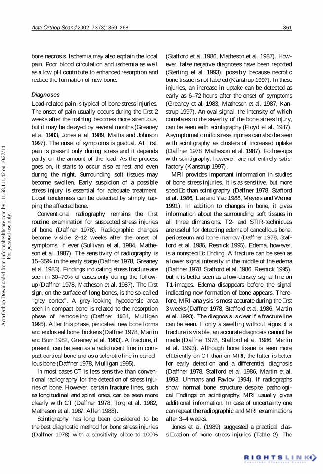
Metatarsal bone (MT) stress fractures, espe-cially when located in the diaphyseal middle and proximal � fth areas are common in � eld and track athletes (Figure 1). Stress injuries of the diaphyseal
Table 4. Conservative treatment protocols for various grades of stress injuries of the shin bone, according to Fredricson et al. 1995 and present authors
Grade Time without loading Gradual increase in weight bearing
1 2–3 weeks according to pain2 4–6 weeks according to pain3 6–9 weeks according to pain4 cast for 6 weeks without cast for 6 weeks according to pain
Act
a O
rtho
p D
ownl
oade
d fr
om in
form
ahea
lthca
re.c
om b
y 11
1.68
.111
.42
on 1
0/27
/14
For
pers
onal
use
onl
y.
364 Acta Orthop Scand 2002; 73 (3): 359–368
area of MT II in dancers heal almost always during 4–6 weeks after closed treatment, with the excep-tion of the proximal injury of the MT V bone. In case of delayed healing, drilling seems to help in our experience. If a patient has a proximal fracture of the � fth MT with a wide lateral fracture line, sclerotic fracture edges and a high activity level, one should consider surgery with compression osteosynthesis by an intramedullary screw (Berg-feld and Khoury 1994, Quill 1995) or tension wire osteosynthesis (Hulkko and Orava 1991). We have had good results with the intramedullary screw.
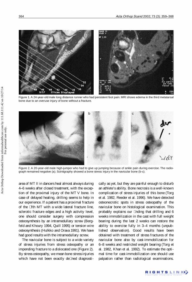
The navicular bone is subject to a wide variety of stress injuries from stress osteopathy or an impending fracture to a dislocated one (Figure 2). By stress osteopathy, we mean bone stress injuries which have not been exactly de� ned diagnosti-
cally as yet, but they are painful enough to disturb an athlete’s ability. Bone necrosis is a well-known complication of stress injuries of this bone (Torg et al. 1982, Reeder et al. 1996). We have detected osteonecrotic spots in stress osteopathy of the navicular bone on histological examination. This probably explains our � nding that drilling and 6 weeks immobilization in the cast with full weight bearing during the last 2 weeks can restore the ability to exercise fully in 3–4 months (unpub-lished observation). Good results have been obtained with treatment of stress fractures of the navicular bone also by cast-immobilization for 6–8 weeks and restricted weight bearing (Torg et al. 1982, Khan et al. 1992). To estimate the opti-mal time for cast-immobilization one should use palpation rather than radiological examinations.
Figure 1. A 24-year-old male long distance runner who had persistent foot pain. MRI shows edema in the third metatarsal bone due to an overuse injury of bone without a fracture.
Figure 2. A 20-year-old male high-jumper who had to give up jumping because of ankle pain during exercise. The radio-graph remained negative (a). Scintigraphy showed a bone stress injury in the navicular bone (b-c).
a b c
Act
a O
rtho
p D
ownl
oade
d fr
om in
form
ahea
lthca
re.c
om b
y 11
1.68
.111
.42
on 1
0/27
/14
For
pers
onal
use
onl
y.
Acta Orthop Scand 2002; 73 (3): 359–368 365
It takes in average of 4–5 months for the patient to return to sports (Monteleone 1995). Combined bone transplantation and screw-� xation help although both methods have also been used alone with 6–8 weeks of immobilizationin a cast after surgery (Khan et al. 1992, Bergfeld and Khoury 1994). We advocate surgery for stress injuries of the navicular bone.
Stress fracture of the talus is rare and affects the collum or corpus areas (Greaney et al. 1983, Uhmans and Pavlov 1994, Brandshaw et al. 1996). The recommended treatment for stress osteopathies of the collum area is drilling followed by 6 weeks immobilization in a cast and restricted weight bear-ing. Stress fractures in the collum can be treated at � rst with immobilization for 6–8 weeks in a cast. However, screw-� xation is recommended if the fracture tends to dislocate and healing is delayed. The results are poor with closed treatment of bone stress injuries of the corpus area. The risk of bone necrosis has to be remembered and weight bearing should be delayed (Black and Ehlert 1994). Drill-ing may also be useful.
Dislocated stress fractures of the calcaneus are rare. According to a few reports and our clinical experience, stress injury of this bone heals in most cases after 6 weeks of reduced activity and even without immobilization in a cast (Reeder et al. 1996, Marcelli 1997).
Fibular stress injuries mostly affect the upper or lower third of the bone (Monteleone 1995). Most of them heal without surgery in 6 weeks (Montele-one 1995). If healing is delayed, drilling seems to be helpful in our experience. Closed treatment of fractures located in the upper part of the malleolus can cause complications. Dislocation leading to malunion easily follows total fractures (Guille et al. 1997).
Medial malleolar stress fractures usually have a relatively vertical course from the corner of the joint. Osteopathies or partial and total fractures occur in the medial malleolus. Total stress fractures tend to dislocate (Schils et al. 1992) and we prefer screw-� xation in these cases. We have treated some osteopathy cases, where healing was delayed for 2 years although the patient only walked during that time. Healing improved with drilling. In other cases with delayed healing for 8–12 months, drill-ing also enhanced recovery (Orava et al. 1995). Early surgery may be indicated for athletes who are very active.
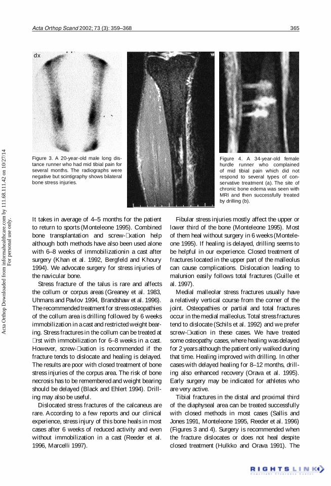
Tibial fractures in the distal and proximal third of the diaphyseal area can be treated successfully with closed methods in most cases (Sallis and Jones 1991, Monteleone 1995, Reeder et al. 1996) (Figures 3 and 4). Surgery is recommended when the fracture dislocates or does not heal despite closed treatment (Hulkko and Orava 1991). The
Figure 3. A 20-year-old male long dis-tance runner who had mid tibial pain for several months. The radiographs were negative but scintigraphy shows bilateral bone stress injuries.
dx
Figure 4. A 34-year-old female hurdle runner who complained of mid tibial pain which did not respond to several types of con-servative treatment (a). The site of chronic bone edema was seen with MRI and then successfully treated by drilling (b).
a b
Act
a O
rtho
p D
ownl
oade
d fr
om in
form
ahea
lthca
re.c
om b
y 11
1.68
.111
.42
on 1
0/27
/14
For
pers
onal
use
onl
y.

366 Acta Orthop Scand 2002; 73 (3): 359–368
usual therapy for tibial fracture is used in such cases. Stress fractures on the tension side of the anterior aspect of the middle third of the tibia tend to heal poorly. Closed treatment has been recom-mended for the � rst 3–6 months (Taube and Wad-sworth 1993). In many cases, early surgery can be better: drilling or bone transplantation alone or together with plate � xation (Reeder et al. 1996). Even immediate intramedullary nailing has been suggested for athletes (Plasschaert et al. 1995). Healing of ordinary stress injuries of the shin bone can take between 3 weeks and 18 months (Sullivan et al. 1984, Hulkko and Orava 1991).
Patellar stress fracture rarely occurs. It is usu-ally a partial fracture in the anterior cortical area
(Rockett and Freeman 1990) or a horizontal one in the lower pole (Rockett and Freeman 1990, Orava et al. 1996). Such fractures can heal in 3 months with closed treatment (Mata et al. 1996). A total stress fracture, however, may dislocate (Jerosch et al. 1989) and require reduction combined with osteosynthesis.
Femoral stress injury may affect any part of the bone, but the commonest sites are the shaft and femoral neck. Most fractures located in the shaft or condylar areas can be treated without surgery in two phases, as mentioned above, if the fracture is undetectable or partial. Healing usually takes 2–3 months (Clement et al. 1993). Total fractures are treated with IM-nailing. Compression side fractures of the femoral neck are mostly treated without surgery, but require long follow-ups; in most cases tension side stress fractures result in total fractures and run a risk of avascular necro-sis. It is therefore advisable to operate on them by using several parallel screws or a dynamic hip screw (Egol et al. 1998). Avascular necrosis of the femoral caput is a serious complication after a stress injury of the femoral neck.
The EVO/Helsinki University Central Hospital and the Foundation for Sports Research in Finland supported this study.
Allen G J. Longitudinal stress fractures of the tibia: diagno-sis with CT. Radiology 1988; 167: 799-801.
Behari J. Electrostimulation and bone fracture healing. Biomed Eng 1991; 18: 4.
Benazzo F, Mosconi M, Beccarisi G et al. Use of capacitive coupled electric � elds in stress fracture in athletes. Clin Orthop 1995; 310: 145-9.
Bennell K L, Brukner P D. Epidemiology and site speci� city of stress fractures. Clin Sports Med 1997; 16: 179-96.
Bennell K L, Malcolm S A, Thomas S A et al. The incidence and distribution of stress fractures in competitive track and � eld athletes. A twelve-month prospective study. Am J Sports Med 1996; 24: 211-7.
Bergfeld J A, Khoury M. Stress fractures in sports. Sports Medicine Symposium: 1994 Sept 14-16, Burlington (VT), University of Vermont 1994; 144-65.
Black K P, Ehlert K J. A stress fracture of the lateral process of the talus in a runner. J Bone Joint Surg (Am) 1994; 76: 441-3.
Brand J C Jr, Brindle T, Nyland J, Caborn D N, Johnson D L. Does pulsed low intensity ultrasound allow early return to normal activities when treating stress fractures? A review of one tarsal navicular and eight tibial stress fractures. Iowa Orthop J 1999; 19: 26-30.
Brandshaw C, Khan K, Brukner P. Stress fracture of the body of the talus in athletes demonstrated with computer tomography. Clin J Sports Med 1996; 6: 48-51.
Brighton C T, Pollack S R. Treatment of recalcitrant non-union with a capacitively-coupled electrical � eld. J Bone Joint Surg (Am) 1985; 67: 577-85.
Brunet M E, Cook S D, Brinker M R, Dickinson J A. A survey of running injuries in 1505 competitive and recreational runners. J Sports Med Phys Fitness 1990; 30: 307-15.
Burr D B. Remodelling and the repair of fatigue damage. Calcif Tissue Int (Suppl 1) 1993; 53: S75-S80; discussion S80-81.
Burr D B, Martin R B, Schaf� er M B et al. Bone remodelling in response to in vivo fatigue microdamage. J Biomech 1985; 18: 189-200.
Carter D R, Caler W E. Cycle-dependent and time-depen-dent bone fracture with repeated loading. J Biomech Eng 1983; 105: 166-70.
Carter D R, Caler W E, Spengler D M et al. Uniaxial fatigue of human cortical bone. The in� uence of tissue physical characteristics. J Biomech 1981; 14: 461-70.
Chamay A, Tschants P. Mechanical in� uence in bone remod-elling: experimental research on Wolff’s law. J Biomech 1972; 2: 173-80.
Clement D B, Amman W, Taunton J E et al. Exercise-induced stress injuries to the femur. Int J Sports Med 1993; 14: 347-52.
Conroy B P, Kraemer W J, Maresh C M et al. Bone mineral density in elite junior Olympic weightlifters. Med Sci Sports Exerc 1993; 25: 1103-9.
Daffner R H. Stress fractures: current concepts. Skeletal Radiol 1978; 2: 221-9.
Daffner R H. Anterior tibial striations. Am J Roentgenol 1984; 143: 651-3.
Daffner R H, Pavlov H. Stress fractures: current concepts. Am J Roentgenol 1992; 159: 245-52.
Act
a O
rtho
p D
ownl
oade
d fr
om in
form
ahea
lthca
re.c
om b
y 11
1.68
.111
.42
on 1
0/27
/14
For
pers
onal
use
onl
y.

Acta Orthop Scand 2002; 73 (3): 359–368 367
Dale P A, Bronk J T, O’Sullivan M E et al. A new concept in fracture immobilisation: the application of a pressurized brace. Clin Orthop 1993; 295: 264-9.
Egol K A, Koval K J, Kummer F, Frankel V H. Stress frac-tures of the femoral neck. Clin Orthop 1998; 348: 72-8.
Floyd W N, Butler J E, Clanton T et al. Roentgenologic diagnosis of stress fractures and stress reactions. South Med J 1987; 80: 433-9.
Fredricson M, Bergman A G, Hoffman K L et al. Tibial stress reaction in runners. Correlation of clinical symptoms and scintigraphy with a new magnetic resonance imaging grading system. Am J Sports Med 1995; 23: 472-81.
Frost H M. Some ABC’s of skeletal patho-physiology vs. microdamage physiology. Calcif Tissue Int 1991; 49: 229-31.
Frost H M. Wolff’s law and bone’s structural adaptation to mechanical usage: an overview for clinicians. Angle Orthod 1994; 64: 175-8.
Greaney R B, Gerber F H, Laughlin R L et al. Distribu-tion and natural history of stress fractures in US Marine recruits. Radiology 1983; 146: 339-46.
Guille J T, Lipton G E, Bowen J R et al. Delayed union following stress fracture of the distal � bula secondary to rotational malunion of lateral malleolar fracture. Am J Orthop 1997; 26: 442-5.
Haapasalo H, Kannus P, Sievanen H et al. Long-term uni-lateral loading and bone mineral density and content in female squash players. Calcif Tissue Int 1994; 54: 249-55.
Haramati N, Staron R B, Barax C et al. MR imaging of occult fractures of the proximal femur. Skeletal Radiol 1994; 23: 19-22.
Holder L E, Schwarz C, Wernicke P G et al. Radionuclide bone imaging in the early detection of fractures of the proximal femur. A multifactorial analysis. Radiology 1990; 174: 509-15.
Hulkko A, Orava S. Diagnosis and treatment of delayed and non-union stress fractures in athletes. Ann Chir Gynecol 1991; 80: 177-84.
Jerosch J G, Castro W H, Jantea C. Stress fracture of the patella. Am J Sports Med 1989; 17: 579-80.
Johnson A W, Weiss C B, Wheeler D L. Stress fractures of the femoral shaft in athletes-more common than expected. Am J Sports Med 1994; 22: 248-56.
Jones B H, Harris J, Vinh T N, Rubin C. Exercise-induced stress fractures and reactions of bone. Epidemiology, etiol ogy and classi� cation. Exerc Sports Sci Rev 1989; 17: 379-422.
Jonnavithula S, Warren M P, Fox R P et al. Bone density is compromised in amenorrheic women despite return of menses: a 2-year study. Obstet Gynecol 1993; 81: 669-74.
Kadel N J, Teitz C C, Kronmal R A et al. Stress fractures in ballet dancers. Am J Sports Med 1992; 20: 445-9.
Kanstrup I L. Bone scintigraphy in sports medicine: a review. Scand J Med Sci Sports 1997; 7: 322-30.
Khan K, Fuller P J, Brukner P D et al. Outcome of conserva-tive and surgical management of navicular stress fracture in athletes. Eighty-six cases proven with computerized tomography. Am J Sports Med 1992; 20: 657-66.
Krivickas L S. Anatomical factors associated with overuse sports injuries. Sports Med 1997; 24: 406-7.
Lee J K, Yao L. Stress fractures: MR imaging. Radiology 1988; 169: 217-20.
Maitra R S, Johnson D L. Stress fractures. Clinical history and physical examination. Clin Sports Med 1997; 16: 259-74.
Marcelli C. Fatigue fractures of the foot. Rev Prat 1997; 47: 50-5.
Marti B, Vader J P, Minder C E et al. On the epidemiology of running injuries. The 1984 Berne Grand Prix study. Am J Sports Med 1988; 16: 285-94.
Martin R B, Burr D B. A hypothetical mechanism for the stimulation of osteonal remodelling by fatigue damage. J Biomech 1982; 15; 137-9.
Martin S D, Healey J H, Horowitz S. Stress fracture MRI. Orthopedics 1993; 16: 75-8.
Mata S G, Grande M M, Ovejero A H. Transverse stress fracture of the patella: a case report. Clin J Sports Med 1996; 6: 259-61.
Matheson G O, Clement D B, McKenzie D C et al. Stress fractures in athletes. A study of 320 cases. Am J Sports Med 1987; 15: 46-58.
Matin P. Basic principles of nuclear medicine techniques for detection and evaluation of trauma and sports medicine injuries. Semin Nucl Med 1988; 18: 90-112.
McBryde A M Jr. Stress fractures in runners. In: Prevention and treatment of running injuries (Eds. D’Ambrosia R, Drez D Jr.). Thorofare, NJ: Slack, 1982: 21-42.
Meyers S P, Weiner S N. Magnetic resonance imaging fea-tures of fractures using the short tau inversion recovery (STIR) sequence: correlation with radiographic � ndings. Skeletal Radiol 1991; 20: 499-507.
Milgrom C, Giladi M, Stein M et al. Stress fractures in mili-tary recruits. A prospective study showing an unusually high incidence. J Bone Joint Surg (Br) 1985; 67: 732-5.
Monteleone G P Jr. Stress fractures in the athlete. Sports Med 1995; 26: 423-32.
Mulligan M E. The “gray cortex“: an early sign of stress fracture. Skeletal Radiol 1995; 24: 201-3.
Myburgh K H, Hutchins J, Fataar A B et al. Low bone den-sity is an etiologic factor for stress fractures in athletes. Ann Intern Med 1990; 113: 754-9.
Nordin M, Frankel V H. Biomechanics of the hip. In: Basic biomechanics of the musculoskeletal system. 2nd edition (Eds. Nordin M, Frankel V H) .Malvern, PA: Lea and Febiger 1989: 135-51.
Orava S, Karpakka J, Taimela S et al. Stress fracture of the medial malleolus. J Bone Joint Surg (Am) 1995; 77; 362-5.
Orava S, Taimela S, Kvist M et al. Diagnosis and treatment of stress fracture of the patella in athletes. Knee Surg Sports Traumatol Arthrosc 1996; 4: 206-11.
Act
a O
rtho
p D
ownl
oade
d fr
om in
form
ahea
lthca
re.c
om b
y 11
1.68
.111
.42
on 1
0/27
/14
For
pers
onal
use
onl
y.

368 Acta Orthop Scand 2002; 73 (3): 359–368
Plasschaert V F P, Johansson C G, Micheli L J. Anterior tibial stress fracture treated with intramedullary nailing: a case report. Clin J Sports Med 1995; 5: 58-62.
Quill G E Jr. Fractures of the proximal � fth metatarsal. Orthop Clin North Am 1995; 26: 353-61.
Reeder M T, Dick B H, Atkins J K et al. Stress fractures. Current concepts of diagnosis and treatment. Sports Med 1996; 22: 198-212.
Resnick D. Diagnosis of bone and joint disorders. 3rd ed. Philadelphia, PA: Saunders 1995: 2580-603 .
Robinson T L, Snow-Harter C, Taaffe D R et al. Gymnasts exhibit higher bone mass than runners despite similar prevalence of amenorrhea and oligomenorrhea. J Bone Miner Res 1995; 10: 26-35.
Rockett J F, Freeman B L. Stress fracture of the patella. Con� rmation by triple-phase bone imaging. Clin Nucl Med 1990; 15: 873-5.
Sallis R E, Jones K. Stress fractures in athlete: How to spot this underdiagnosed injury. Post Grad Med 1991; 89: 185-92.
Schils J P, Andrish J T, Piraino D W et al. Medial malleolar stress fractures in seven patients: review of the clinical and imaging features. Radiology 1992; 185: 219-21.
Scully T J, Besterman G. Stress fracture-a preventable train-ing injury. Mil Med 1982; 147: 285-7.
Shwayhat A F, Linenger J M, Hofherr L K, Slymen D J, Johnson C W. Pro� les of exercise history and overuse injuries among United States Navy Sea, Air, and Land (SEAL) recruits. Am J Sports Med 1994; 22: 835-40.
Stafford S A, Rosenthal D I, Gebhardt M C et al. MRI in stress fractures. AJR Am J Roentgenol 1986; 147: 553-6.
Sterling J C, Edelstein D W, Calvo R D et al. Stress fractures in the athlete: diagnosis and management. Sports Med 1992; 14: 336-46.
Sterling J C, Webb R F, Meyers M C et al. False negative bone scan in a female runner. Med Sci Sports Exerc 1993; 25: 179-85.
Sullivan D, Warren R F, Pavlov H et al. Stress fractures in 51 runners. Clin Orthop 1984; 187: 188-92.
Swenson E J, DeHaven K E, Sebastianelli W J et al. The effect of a pneumatic leg brace on return to play in ath-letes with tibial stress fracture. Am J Sports Med 1997; 25: 322-8.
Taube R R, Wadsworth L T. Managing tibial stress fractures. Physician Sports Med 1993; 21: 123-8.
Torg J S, Pavlov H, Cooley L H et al. Stress fractures of the tarsal navicular. J Bone Joint Surg (Am) 1982; 64: 700-12.
Uhmans H, Pavlov H. Stress fractures of the lower extrem-ity. Semin Roentgenol 1994; 24: 176-93.
Uhthoff H K, Jaworski Z F G. Periosteal stress-induced reac-tions resembling stress fractures: a radiologic and histo-logic study in dogs. Clin Orthop 1986; 199: 284-91.
Whitelaw G P, Wetzler M J, Levy A S et al. A pneumatic leg brace for the treatment of tibial stress fractures. Clin Orthop 1991; 270: 301-5.
Zernicke R, McNitt-Gray J, Otis et al. Stress fracture risk assessment among elite collegiate women runners. Inter-national Society of Biomechanics XIVth Congress 1993: 1506-7.
Zwas S T, Elkanovitch R, Frank G. Interpretation and classi-� cation of bone scintigraphic � ndings in stress fractures. J Nucl Med 1987; 28: 452-7.
Act
a O
rtho
p D
ownl
oade
d fr
om in
form
ahea
lthca
re.c
om b
y 11
1.68
.111
.42
on 1
0/27
/14
For
pers
onal
use
onl
y.