Embed Size (px)
Citation preview
Accepted Manuscript
Bone health evaluation one year after aromatase inhibitorscompletion
Marta Pineda-Moncusí, Sonia Servitja, Guillem Casamayor,Maria Lourdes Cos, Abora Rial, Jaime Rodriguez-Morera, IgnasiTusquets, Adolfo Diez-Perez, Natalia Garcia-Giralt, XavierNogués
PII: S8756-3282(18)30348-XDOI: doi:10.1016/j.bone.2018.09.010Reference: BON 11752
To appear in: Bone
Received date: 25 June 2018Revised date: 13 September 2018Accepted date: 13 September 2018
Please cite this article as: Marta Pineda-Moncusí, Sonia Servitja, Guillem Casamayor,Maria Lourdes Cos, Abora Rial, Jaime Rodriguez-Morera, Ignasi Tusquets, Adolfo Diez-Perez, Natalia Garcia-Giralt, Xavier Nogués , Bone health evaluation one year afteraromatase inhibitors completion. Bon (2018), doi:10.1016/j.bone.2018.09.010
This is a PDF file of an unedited manuscript that has been accepted for publication. Asa service to our customers we are providing this early version of the manuscript. Themanuscript will undergo copyediting, typesetting, and review of the resulting proof beforeit is published in its final form. Please note that during the production process errors maybe discovered which could affect the content, and all legal disclaimers that apply to thejournal pertain.
ACC
EPTE
D M
ANU
SCR
IPT
1
Bone health evaluation one year after aromatase inhibitors
completion
Marta Pineda-Moncusí1, Sonia Servitja
2, Guillem Casamayor
3, Maria Lourdes Cos
3, Abora
Rial3, Jaime Rodriguez-Morera
3, Ignasi Tusquets
2, Adolfo Diez-Perez
1,3, Natalia Garcia-Giralt
1,
Xavier Nogués1,3
1. IMIM (Hospital del Mar Research Institute), Centro de Investigación Biomédica en Red de
Fragilidad y Envejecimiento Saludable (CIBERFES), Barcelona, Spain.
2. Cancer Research Program, IMIM (Hospital del Mar Research Institute), Barcelona, Spain.
3. Internal Medicine Department, Hospital del Mar, Universitat Autònoma de Barcelona,
Barcelona, Spain.
* Corresponding author at: Natalia Garcia-Giralt
Centro de Investigación Biomédica en Red de Fragilidad y Envejecimiento Saludable
(CIBERFES)
IMIM (Hospital del Mar Research Institute),
88 Doctor Aiguader Street, 08003, Barcelona, Spain.
ORCID:0000-0001-6507-0147
E-mail address: [email protected]
ACCEPTED MANUSCRIPT
ACC
EPTE
D M
ANU
SCR
IPT
2
Abstract
Introduction
Breast cancer patients treated with aromatase inhibitors (AIs) experience increased bone loss
during their treatment. However, there is little information about bone mineral density (BMD)
after completing AI-treatment. The present study aimed to assess BMD changes one year after
AI-therapy completion.
Methods
Data were collected from 864 postmenopausal women treated with AI during 5 years (5y-AI
group), or during 2-3 years after taking tamoxifen therapy (pTAM-AI group). Participants with
osteoporosis were treated with oral bisphosphonates (BP). BMD changes in lumbar spine (LS),
femoral neck (FN) and total hip (TH) between baseline, end of treatment, and at one year post-
treatment were assessed using repeated-measures ANOVA.
Results
At the end of AI-treatment, 382 patients had available BMD values and 316 also had post-
treatment BMD values. As expected, BMD levels were decreased at AI-completion in non-BP
treated patients. After one year, LS BMD increased in both groups (5y-AI: +2.11% [95%CI:
1.55 to 2.68], p<0.001; pTAM-AI: +1.00% [95%CI: 0.49 to 1.51], p<0.001) compared with the
end of AI-therapy, while values at FN and TH remained stable. On the other hand, BMD values
of BP-treated patients were increased or maintained at the end of AI-treatment and also at post-
treatment.
Conclusions
At one year after AI-completion, FN and TH BMD remained reduced in non-BP treated women,
while LS BMD was recovered in the 5y-AI group and partially recovered in the pTAM-AI
group. BP treatment increased or maintained BMD values at the end of therapy and at one year
post-treatment.
Keywords: breast cancer, aromatase inhibitors, bone mineral density, bisphosphonates, B-ABLE
cohort.
ACCEPTED MANUSCRIPT

ACC
EPTE
D M
ANU
SCR
IPT
3
Introduction
Aromatase inhibitor (AI) is recommended by the American Society of Clinical Oncology as the
adjuvant endocrine therapy to treat estrogen receptor positive (ER+) early breast cancer in
postmenopausal women. Despite its great efficiency, compared to tamoxifen (TAM) as the
alternative [1-3], AI has been associated with side effects that could affect the patient’s quality
of life and its adherence to treatment, being arthralgia and bone loss induction among the most
common [4, 5].
Previous studies have described an accelerated decrease in bone mineral density (BMD)
associated with AI therapy, leading to osteopenic or osteoporotic bone status, both of which are
related to osteoporotic fracture [1, 4]. Clinical guidelines for the management of AI-related bone
loss strongly recommend a close monitoring of BMD and other risk factors to reduce the
fracture risk by means of antiresorptive therapies [6].Treatment with bisphosphonates (BPs) is
the current recommendation to avoid this bone loss [7-9].
Even though bone parameters have been monitored during AI treatment in many studies [10,
11], there is scarce information about bone status after completion of AI treatment. A small sub-
analysis in the ATAC trial, with 23 evaluated patients, showed an increase of bone mass at
lumbar spine after one year of AI-completion [12]. In the MA.17R trial [13], an increase in
lumbar spine (LS) and total hip (TH) BMD was reported 5-7 years post-treatment in women
mainly treated with TAM followed by AI; however, half of the patients were treated with BP,
concealing the results. Despite the insights on bone behavior related to AI treatment gained
from these previous randomized control trials (RCTs), bone health after AI cessation has not
been explored in actual clinical conditions.
In the present study, we analyzed BMD changes at the end of treatment and at one year after AI-
completion in an observational prospective cohort (B-ABLE). In this study, the effect of
previous tamoxifen and/or BP treatment was taken into account.
Materials and Methods
Study design and participants
Caucasian postmenopausal woman diagnosed with ER+ early breast cancer and candidates for
AI-treatment (letrozole, exemestane, or anastrozole) were consecutively recruited from January
2006 to January 2018 in B-ABLE cohort – a prospective, non-selected, observational, clinical
cohort study – in Hospital del Mar (Barcelona, Spain). The study protocol was approved by the
ethics committee of Parc de Salut Mar (2016/6803/I) and it was carried out in accordance with
the Declaration of Helsinki. A written informed consent was obtained from all participants after
ACCEPTED MANUSCRIPT
ACC
EPTE
D M
ANU
SCR
IPT
4
they had read the study information sheet and any questions had been answered. The privacy
rights of human subjects must always be observed.
Participants were enrolled at point of starting AI therapy, either six weeks post-surgery or one
month after the last cycle of chemotherapy (5y-AI group) or, alternatively, once starting
menopause after taking TAM for two to three years (pTAM-AI group). End of treatment was
considered a total of five years of hormonal adjuvant therapy, according to classic American
Society of Clinical Oncology recommendations [14]. Follow-up was from the first day of AI
intake to one year after AI-completion. Postmenopausal status was defined as patients >55 years
old with amenorrhea for >12 months, or those ≤55 with levels of luteinizing hormone >30
mIU/mL or follicle-stimulating hormone values >40 mIU/mL. Eligible participants were
excluded for previous history of any bone, metabolic or endocrine diseases, as well as
alcoholism, rheumatoid arthritis, and concurrent or prior treatment with BP, oral corticosteroids,
or any other bone-active drug except tamoxifen.
At the outset of the study, patients were stratified by the corresponding therapeutic regimen: 1)
those with osteoporosis [T score < −2.5] or with a T score ≤ −2.0 at any site plus 1 major risk
factor (i.e. family history of femoral fracture, or early menopause) or prevalent fragility
fractures were treated with weekly oral risedronate or alendronate therapy (BP-treated patients)
2) all other patients were allocated to no active antiresorptive therapy (non-BP-treated patients).
BMD was assessed every 12 months until one year after the end of AI therapy (post-treatment).
Those who developed osteoporosis during the treatment were immediately offered oral BP
treatment and were censored from the study at that point.
Additionally, all participants received supplements of calcium and 25(OH)vitD3 tablets (1000
mg and 800 IU daily, respectively), and those with baseline 25(OH)vitD deficiency (<30
ng/mL) received an additional dose of 16,000 IU of oral calcifediol (HIDROFEROL® FAES
FARMA) every 2 weeks.
Variables
Bone Mineral Density
The main outcomes of the study are the absolute and cumulative percentage change in lumbar
spine (LS), femoral neck (FN) and total hip (TH) BMD from baseline to the end of treatment
and at one year post-treatment.
BMD measures were obtained using a DXA densitometer QDR 4500 SL® (Hologic, Waltham,
MA, USA), according to manufacturer recommendations. In our department, in vivo
coefficients of variation of these techniques are 1.0% at LS, 1.60 at TH, and 1.65% at FN.
As a secondary analysis, non-BP-treated patients were categorized according to its LS-BMD
shift, and their distribution was plotted.
ACCEPTED MANUSCRIPT

ACC
EPTE
D M
ANU
SCR
IPT
5
Other variables
At the time of recruitment, data from large clinical variables were registered, including: age,
body mass index (BMI), age of menarche and menopause, number of children, months of
breastfeeding, and prior chemotherapy, among others.
Statistical methods
Significant differences between variables in the groups of the study were analyzed with One-
way ANOVA, Kruskal-Wallis and Chi-square tests, according to variables’ nature. In each
group, BMD changes between baseline, end of treatment, and post-treatment were evaluated by
repeated-measures ANOVA.
Statistical analysis was done with R for Windows version 3.3.3 (foreign, compareGroups,
pgirmess, fifter, boot, ggplot2 and scales packages) and SPSS Statistics version 22.0. All
statistical contrasts were corrected by Bonferroni test per multiple comparisons and P values
lower than 0.05 were considered significant.
Results
Participants
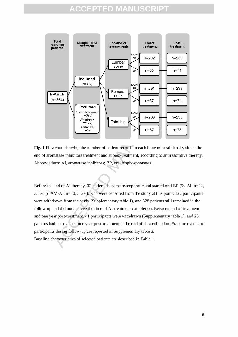
From 864 participants included in the B-ABLE cohort, 382 patients completed AI treatment and
had BMD values registered at this point, and 316 of those patients had BMD values at one year
after AI-treatment completion (Fig. 1).
ACCEPTED MANUSCRIPT
ACC
EPTE
D M
ANU
SCR
IPT
6
Fig. 1 Flowchart showing the number of patient records in each bone mineral density site at the
end of aromatase inhibitors treatment and at post-treatment, according to antiresorptive therapy.
Abbreviations: AI, aromatase inhibitors; BP, oral bisphosphonates.
Before the end of AI therapy, 32 patients became osteoporotic and started oral BP (5y-AI: n=22,
3.8%; pTAM-AI: n=10, 3.6%), who were censored from the study at this point; 122 participants
were withdrawn from the study (Supplementary table 1), and 328 patients still remained in the
follow-up and did not achieve the time of AI-treatment completion. Between end of treatment
and one year post-treatment, 41 participants were withdrawn (Supplementary table 1), and 25
patients had not reached one year post-treatment at the end of data collection. Fracture events in
participants during follow-up are reported in Supplementary table 2.
Baseline characteristics of selected patients are described in Table 1.
ACCEPTED MANUSCRIPT
ACC
EPTE
D M
ANU
SCR
IPT
7
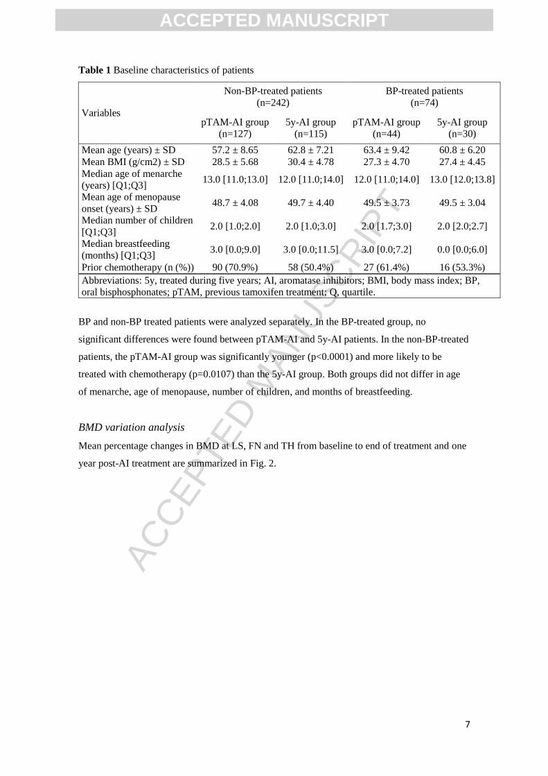
Table 1 Baseline characteristics of patients
Variables
Non-BP-treated patients
(n=242)
BP-treated patients
(n=74)
pTAM-AI group
(n=127)
5y-AI group
(n=115)
pTAM-AI group
(n=44)
5y-AI group
(n=30)
Mean age (years) ± SD 57.2 ± 8.65 62.8 ± 7.21 63.4 ± 9.42 60.8 ± 6.20
Mean BMI (g/cm2) ± SD 28.5 ± 5.68 30.4 ± 4.78 27.3 ± 4.70 27.4 ± 4.45
Median age of menarche
(years) [Q1;Q3] 13.0 [11.0;13.0] 12.0 [11.0;14.0] 12.0 [11.0;14.0] 13.0 [12.0;13.8]
Mean age of menopause
onset (years) ± SD 48.7 ± 4.08 49.7 ± 4.40 49.5 ± 3.73 49.5 ± 3.04
Median number of children
[Q1;Q3] 2.0 [1.0;2.0] 2.0 [1.0;3.0] 2.0 [1.7;3.0] 2.0 [2.0;2.7]
Median breastfeeding
(months) [Q1;Q3] 3.0 [0.0;9.0] 3.0 [0.0;11.5] 3.0 [0.0;7.2] 0.0 [0.0;6.0]
Prior chemotherapy (n (%)) 90 (70.9%) 58 (50.4%) 27 (61.4%) 16 (53.3%)
Abbreviations: 5y, treated during five years; AI, aromatase inhibitors; BMI, body mass index; BP,
oral bisphosphonates; pTAM, previous tamoxifen treatment; Q, quartile.
BP and non-BP treated patients were analyzed separately. In the BP-treated group, no
significant differences were found between pTAM-AI and 5y-AI patients. In the non-BP-treated
patients, the pTAM-AI group was significantly younger (p<0.0001) and more likely to be
treated with chemotherapy (p=0.0107) than the 5y-AI group. Both groups did not differ in age
of menarche, age of menopause, number of children, and months of breastfeeding.
BMD variation analysis
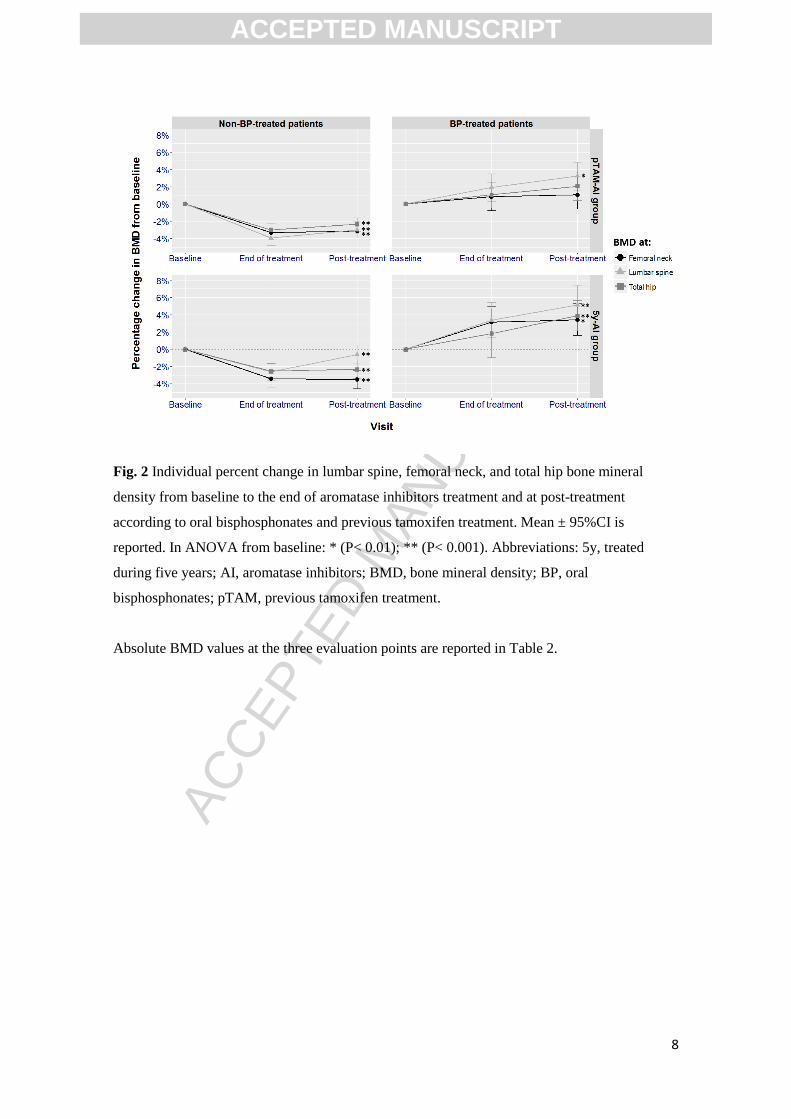
Mean percentage changes in BMD at LS, FN and TH from baseline to end of treatment and one
year post-AI treatment are summarized in Fig. 2.
ACCEPTED MANUSCRIPT
ACC
EPTE
D M
ANU
SCR
IPT
8
Fig. 2 Individual percent change in lumbar spine, femoral neck, and total hip bone mineral
density from baseline to the end of aromatase inhibitors treatment and at post-treatment
according to oral bisphosphonates and previous tamoxifen treatment. Mean ± 95%CI is
reported. In ANOVA from baseline: * (P< 0.01); ** (P< 0.001). Abbreviations: 5y, treated
during five years; AI, aromatase inhibitors; BMD, bone mineral density; BP, oral
bisphosphonates; pTAM, previous tamoxifen treatment.
Absolute BMD values at the three evaluation points are reported in Table 2.
ACCEPTED MANUSCRIPT
ACC
EPTE
D M
ANU
SCR
IPT
9
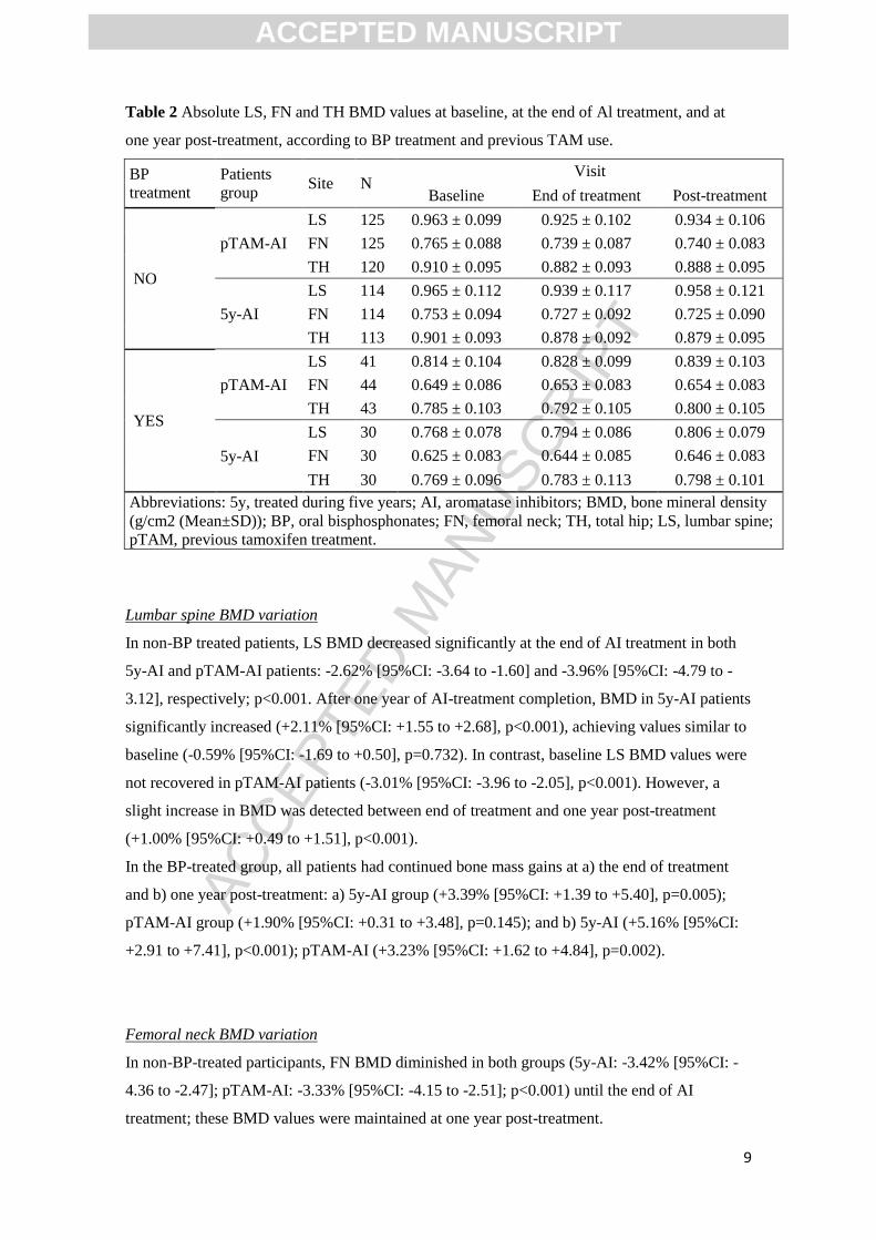
Table 2 Absolute LS, FN and TH BMD values at baseline, at the end of Al treatment, and at
one year post-treatment, according to BP treatment and previous TAM use.
BP
treatment
Patients
group Site N
Visit
Baseline End of treatment Post-treatment
NO
pTAM-AI
LS 125 0.963 ± 0.099 0.925 ± 0.102 0.934 ± 0.106
FN 125 0.765 ± 0.088 0.739 ± 0.087 0.740 ± 0.083
TH 120 0.910 ± 0.095 0.882 ± 0.093 0.888 ± 0.095
5y-AI
LS 114 0.965 ± 0.112 0.939 ± 0.117 0.958 ± 0.121
FN 114 0.753 ± 0.094 0.727 ± 0.092 0.725 ± 0.090
TH 113 0.901 ± 0.093 0.878 ± 0.092 0.879 ± 0.095
YES
pTAM-AI
LS 41 0.814 ± 0.104 0.828 ± 0.099 0.839 ± 0.103
FN 44 0.649 ± 0.086 0.653 ± 0.083 0.654 ± 0.083
TH 43 0.785 ± 0.103 0.792 ± 0.105 0.800 ± 0.105
5y-AI
LS 30 0.768 ± 0.078 0.794 ± 0.086 0.806 ± 0.079
FN 30 0.625 ± 0.083 0.644 ± 0.085 0.646 ± 0.083
TH 30 0.769 ± 0.096 0.783 ± 0.113 0.798 ± 0.101
Abbreviations: 5y, treated during five years; AI, aromatase inhibitors; BMD, bone mineral density
(g/cm2 (Mean±SD)); BP, oral bisphosphonates; FN, femoral neck; TH, total hip; LS, lumbar spine;
pTAM, previous tamoxifen treatment.
Lumbar spine BMD variation
In non-BP treated patients, LS BMD decreased significantly at the end of AI treatment in both
5y-AI and pTAM-AI patients: -2.62% [95%CI: -3.64 to -1.60] and -3.96% [95%CI: -4.79 to -
3.12], respectively; p<0.001. After one year of AI-treatment completion, BMD in 5y-AI patients
significantly increased (+2.11% [95%CI: +1.55 to +2.68], p<0.001), achieving values similar to
baseline (-0.59% [95%CI: -1.69 to +0.50], p=0.732). In contrast, baseline LS BMD values were
not recovered in pTAM-AI patients (-3.01% [95%CI: -3.96 to -2.05], p<0.001). However, a
slight increase in BMD was detected between end of treatment and one year post-treatment
(+1.00% [95%CI: +0.49 to +1.51], p<0.001).
In the BP-treated group, all patients had continued bone mass gains at a) the end of treatment
and b) one year post-treatment: a) 5y-AI group (+3.39% [95%CI: +1.39 to +5.40], p=0.005);
pTAM-AI group (+1.90% [95%CI: +0.31 to +3.48], p=0.145); and b) 5y-AI (+5.16% [95%CI:
+2.91 to +7.41], p<0.001); pTAM-AI (+3.23% [95%CI: +1.62 to +4.84], p=0.002).
Femoral neck BMD variation
In non-BP-treated participants, FN BMD diminished in both groups (5y-AI: -3.42% [95%CI: -
4.36 to -2.47]; pTAM-AI: -3.33% [95%CI: -4.15 to -2.51]; p<0.001) until the end of AI
treatment; these BMD values were maintained at one year post-treatment.
ACCEPTED MANUSCRIPT
ACC
EPTE
D M
ANU
SCR
IPT
10
In contrast, BMD improved with BP therapy (5y-AI: +3.17% [95%CI: +1.37 to +4.98],
p<0.003; and pTAM-AI: +0.85% [95%CI: -0.73 to +2.44], p=0.145) up to the end of treatment;
no significant changes were detected between AI-completion and post-treatment.
Total hip BMD variation
Similar to FN BMD behavior, a significant decrease in TH BMD was detected after AI-
treatment completion in non-BP-treated patients (-2.53% [95%CI: -3.40 to -1.65], p<0.001 in
5y-AI group; and -3.01% [95%CI: -3.80 to -2.22], p<0.001 in pTAM-AI group). The decreased
TH BMD levels remained stable at one year post-treatment.
In the BP-treated patients, BMD increases were detected only in the 5y-AI group at one year
post treatment (+3.89% [95%CI: +2.14 to +5.64], p<0.001).
Patient distribution by LS BMD categories
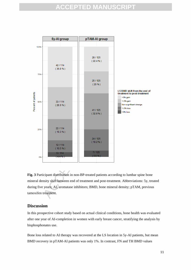
As the major BMD variations were detected at the LS location in patients without BP between
end of AI treatment and one year post-treatment, patient distribution according to LS BMD
changes was explored (Fig. 2). A total of 65.8% of 5y-AI and 42.4% of pTAM-AI patients
experienced an intra-individual BMD gain equal to or greater than 1%. In 19.3% and 32.8% of
patients, respectively, BMD values remained constant (Fig. 3). However, in 14.9% and 24.8%,
respectively, bone mass had continued to decrease, by 1% or more, at one-year follow-up.
ACCEPTED MANUSCRIPT
ACC
EPTE
D M
ANU
SCR
IPT
11
Fig. 3 Participant distribution in non-BP-treated patients according to lumbar spine bone
mineral density shift between end of treatment and post-treatment. Abbreviations: 5y, treated
during five years; AI, aromatase inhibitors; BMD, bone mineral density; pTAM, previous
tamoxifen treatment.
Discussion
In this prospective cohort study based on actual clinical conditions, bone health was evaluated
after one year of AI-completion in women with early breast cancer, stratifying the analysis by
bisphosphonates use.
Bone loss related to AI therapy was recovered at the LS location in 5y-AI patients, but mean
BMD recovery in pTAM-AI patients was only 1%. In contrast, FN and TH BMD values
ACCEPTED MANUSCRIPT
ACC
EPTE
D M
ANU
SCR
IPT
12
remained reduced at one year post-treatment even though the bone loss was stopped. In BP-
treated patients, LS, FN and TH BMD levels were maintained or continued to gain at the end of
follow-up.
About half of non-BP treated patients experienced a clinically significant gain (greater than or
equal to 1%) in LS-BMD at one year after AI cessation, and half of this subgroup had gained
more than 3%. Only 15% of 5y-AI and 25% of pTAM-AI patients continued to lose bone mass
at the end of follow-up. Hence, in most patients the deleterious effect of AI in LS bone mass
stopped, with a trend towards recuperation of baseline BMD after completing AI treatment. On
the other hand, a lack of FN and TH BMD recovery was observed in the first year post-
treatment. This could be due to the lower capacity for change at these locations [11].
Similar findings were reported in the bone sub-study of the ATAC trial [12], in which 65.2% of
participants had increased their LS BMD one year after AI cessation. Similar to our study, LS
BMD values increased (+2.35% [interquartile range: -5.34 to 8.19, p=0.04]) and remained stable
in TH BMD (+0.71% [interquartile range: -9.42 to 4.63, p=0.3]). Their results, obtained from a
small sample (n=21 in LS; n=23 in TH), were confirmed in our cohort.
In the Ma.17R trial [13], patients presented a mean gain in BMD of +4.5% in the spine and
+22.4% in the hip at 5 to 7 years post-treatment. The higher increase observed in that trial could
be explained by differences in length of follow-up and a lack of stratification by BP use and
previous TAM treatment. In fact, one of the strengths of the present study is that we separate
participants in different groups according to previous tamoxifen treatment and current BP use,
which revealed differences in BMD behavior between treatment groups. In this regard, BP-
treated patients improved their BMD values, as would be expected.
Bone mass loss during AI treatment is one of the most important adverse effects experienced by
breast cancer patients on adjuvant endocrine therapy. In fact, the decrease of BMD has been
described as the major factor of fragility fractures [15]. Moreover, some clinical trials with AI
have reported an increase of fractures in both osteopenic and osteoporotic patients [13, 16].
Even though bone mass seems to be recovered after AI cessation, bone health should be
evaluated in all patients since BMD in LS remained decreasing almost in 1/4 of patients after AI
treatment was ended. In these cases, BPs could be a recommended option since BP treated
patients in our study showed better BMD values.
In this line, patients from our cohort are subjected to strict monitoring of 25(OH)vitD levels and
calcium diet intake. All patients receive a high 25(OH)vitD supplementation from baseline.
Hence, 25(OH)vitD levels improved significantly in our cohort with the proposed repletion
regimen raised until the normal range reaching an average >30 ng/ mL, and persisted by the
ACCEPTED MANUSCRIPT
ACC
EPTE
D M
ANU
SCR
IPT
13
follow-up [17]. Data from previous studies strongly recommend that individuals in AI
treatment, including those who are at low risk for fractures and not candidates for BP treatment,
should receive calcium and 25(OH)vitD supplements [18], especially in cases where calcium
intake is not enough. These supplementations could contribute to the recovery of BMD values
observed after AI-completion in our study.
One limitation of our study is the limited tracking time after treatment completion, which
precludes any predictions about how BMD will evolve during a longer-term follow-up, and in
particular, whether FN and TH BMD will be recovered. Further research is needed to explain
why patients previously treated with TAM experienced lower LS BMD recovery than patients
receiving AI monotherapy. We hypothesize that the accelerated bone loss during AI treatment in
pTAM-AI patients, as previously described in B-ABLE cohort [19], might delay LS BMD
recovery. Likewise, we cannot know if this BMD could return to baseline levels at long term.
It is worth mentioning that although BMD measured by DXA is the gold standard surrogate for
the diagnosis of osteoporosis, it is well-known that the increased risk of non-traumatic fractures
is determined not only by the mineral content but also by bone quality and material properties,
such as trabecular microarchitecture [10], the accumulation of microfractures, a disordered bone
remodeling, bone affecting drugs, toxic habits or the influence of extra-skeletal risk factors [20].
In summary, AI-related bone loss stopped at one year after AI-completion and, in the lumbar
location, BMD values were totally recovered in most patients who had received AI
monotherapy and partially recovered in patients who were previously treated with TAM.
However, monitoring of bone health and calcium and 25(OH)vitD supplementation is essential
for the clinical management of patients after finalizing AI adjuvant therapy. Larger studies are
needed to determine whether the observed BMD behavior persists beyond the one year post-
treatment.
Conflicts of interest
Marta Pineda-Moncusí, Sonia Servitja, Guillem Casamayor, Maria Lourdes Cos, Abora Rial,
Jaime Rodriguez-Morera, Ignasi Tusquets, Adolfo Diez-Perez, Natalia Garcia-Giralt, Xavier
Nogués declare that they have no conflict of interest.
Acknowledgments
The authors thank Elaine M. Lilly, Ph.D, for providing language assistance, helpful advice and
critical reading of the manuscript. This work was supported by the Centro de Investigación
Biomédica en Red de Fragilidad y Envejecimiento Saludable (CIBERFES, grant number
ACCEPTED MANUSCRIPT

ACC
EPTE
D M
ANU
SCR
IPT
14
CB16/10/00245) and FIS Project (grant number PI16/00818); Instituto de Salud Carlos III
(ISCIII), and FEDER funds.
ACCEPTED MANUSCRIPT
ACC
EPTE
D M
ANU
SCR
IPT
15
References
[1] L. Ryden, M. Heibert Arnlind, S. Vitols, M. Hoistad, J. Ahlgren, Aromatase inhibitors alone or
sequentially combined with tamoxifen in postmenopausal early breast cancer compared with tamoxifen or
placebo - Meta-analyses on efficacy and adverse events based on randomized clinical trials, Breast 26
(2016) 106-14. doi: https://doi.org/10.1016/j.breast.2016.01.006
[2] A.T.A.C. Group, J.F. Forbes, J. Cuzick, A. Buzdar, A. Howell, J.S. Tobias, M. Baum, Effect of
anastrozole and tamoxifen as adjuvant treatment for early-stage breast cancer: 100-month analysis of the
ATAC trial, Lancet Oncol 9(1) (2008) 45-53. doi: https://doi.org/10.1016/S1470-2045(07)70385-6
[3] E.B.C.T.C. Group, Aromatase inhibitors versus tamoxifen in early breast cancer: patient-level meta-
analysis of the randomised trials, Lancet 386(10001) (2015) 1341-1352. doi:
https://doi.org/10.1016/S0140-6736(15)61074-1
[4] R. Eastell, J.E. Adams, R.E. Coleman, A. Howell, R.A. Hannon, J. Cuzick, J.R. Mackey, M.W.
Beckmann, G. Clack, Effect of anastrozole on bone mineral density: 5-year results from the anastrozole,
tamoxifen, alone or in combination trial 18233230, J Clin Oncol 26(7) (2008) 1051-7. doi:
https://doi.org/10.1200/JCO.2007.11.0726.
[5] F. Laroche, J. Coste, T. Medkour, P.H. Cottu, J.Y. Pierga, J.P. Lotz, K. Beerblock, C. Tournigand, X.
Decleves, P. de Cremoux, D. Bouhassira, S. Perrot, Classification of and risk factors for estrogen
deprivation pain syndromes related to aromatase inhibitor treatments in women with breast cancer: a
prospective multicenter cohort study, J Pain 15(3) (2014) 293-303. doi:
https://doi.org/10.1016/j.jpain.2013.11.004.
[6] P. Hadji, J.J. Body, M.S. Aapro, A. Brufsky, R.E. Coleman, T. Guise, A. Lipton, M. Tubiana-Hulin,
Practical guidance for the management of aromatase inhibitor-associated bone loss, Ann Oncol 19(8)
(2008) 1407-16. doi: https://doi.org/10.1093/annonc/mdn164
[7] P. Hadji, R.E. Coleman, C. Wilson, T.J. Powles, P. Clezardin, M. Aapro, L. Costa, J.J. Body, C.
Markopoulos, D. Santini, I. Diel, A. Di Leo, D. Cameron, D. Dodwell, I. Smith, M. Gnant, R. Gray, N.
Harbeck, B. Thurlimann, M. Untch, J. Cortes, M. Martin, U.S. Albert, P.F. Conte, B. Ejlertsen, J. Bergh,
M. Kaufmann, I. Holen, Adjuvant bisphosphonates in early breast cancer: consensus guidance for clinical
practice from a European Panel, Ann Oncol 27(3) (2016) 379-90. doi:
https://doi.org/10.1093/annonc/mdv617
[8] F.A. Tremollieres, I. Ceausu, H. Depypere, I. Lambrinoudaki, A. Mueck, F.R. Perez-Lopez, Y.T. van
der Schouw, L.M. Senturk, T. Simoncini, J.C. Stevenson, P. Stute, M. Rees, Osteoporosis management in
patients with breast cancer: EMAS position statement, Maturitas 95 (2017) 65-71. doi:
https://doi.org/10.1016/j.maturitas.2016.10.007
[9] P. Hadji, M.S. Aapro, J.J. Body, M. Gnant, M.L. Brandi, J.Y. Reginster, M.C. Zillikens, C.C. Gluer,
T. de Villiers, R. Baber, G.D. Roodman, C. Cooper, B. Langdahl, S. Palacios, J. Kanis, N. Al-Daghri, X.
Nogues, E.F. Eriksen, A. Kurth, R. Rizzoli, R.E. Coleman, Management of Aromatase Inhibitor-
Associated Bone Loss (AIBL) in postmenopausal women with hormone sensitive breast cancer: Joint
position statement of the IOF, CABS, ECTS, IEG, ESCEO IMS, and SIOG, J Bone Oncol 7 (2017) 1-12.
doi: https://doi.org/10.1016/j.jbo.2017.03.001
[10] R.-S. María, P.-M. Marta, S. Sonia, G.-G. Natalia, M. Tamara, T. Ignasi, M.-G. Maria, R.-M. Jaime,
D.-P. Adolfo, A. Joan, N. Xavier, TBS and BMD at the end of AI-therapy: A prospective study of the B-
ABLE cohort, Bone 92 (2016) 1-8. doi: https://doi.org/10.1016/j.bone.2016.08.008
[11] A.R. Hong, J.H. Kim, K.H. Lee, T.Y. Kim, S.A. Im, H.G. Moon, W.S. Han, D.Y. Noh, S.W. Kim,
C.S. Shin, Long-term effect of aromatase inhibitors on bone microarchitecture and macroarchitecture in
non-osteoporotic postmenopausal women with breast cancer, Osteoporos Int 28(4) (2017) 1413-1422.
doi: https://doi.org/10.1007/s00198-016-3899-6
ACCEPTED MANUSCRIPT

ACC
EPTE
D M
ANU
SCR
IPT
16
[12] R. Eastell, J. Adams, G. Clack, A. Howell, J. Cuzick, J. Mackey, M.W. Beckmann, R.E. Coleman,
Long-term effects of anastrozole on bone mineral density: 7-year results from the ATAC trial, Ann Oncol
22(4) (2011) 857-62. doi: https://doi.org/10.1093/annonc/mdq541
[13] P.E. Goss, J.N. Ingle, K.I. Pritchard, N.J. Robert, H. Muss, J. Gralow, K. Gelmon, T. Whelan, K.
Strasser-Weippl, S. Rubin, K. Sturtz, A.C. Wolff, E. Winer, C. Hudis, A. Stopeck, J.T. Beck, J.S. Kaur,
K. Whelan, D. Tu, W.R. Parulekar, Extending Aromatase-Inhibitor Adjuvant Therapy to 10 Years, N
Engl J Med 375(3) (2016) 209-19. doi: https://doi.org/10.1056/NEJMoa1604700
[14] E.P. Winer, C. Hudis, H.J. Burstein, A.C. Wolff, K.I. Pritchard, J.N. Ingle, R.T. Chlebowski, R.
Gelber, S.B. Edge, J. Gralow, M.A. Cobleigh, E.P. Mamounas, L.J. Goldstein, T.J. Whelan, T.J. Powles,
J. Bryant, C. Perkins, J. Perotti, S. Braun, A.S. Langer, G.P. Browman, M.R. Somerfield, American
Society of Clinical Oncology technology assessment on the use of aromatase inhibitors as adjuvant
therapy for postmenopausal women with hormone receptor-positive breast cancer: status report 2004, J
Clin Oncol 23(3) (2005) 619-29. doi: https://doi.org/10.1200/JCO.2005.09.121
[15] N. O'Flynn, Risk assessment of fragility fracture: NICE guideline, Br J Gen Pract 62(605) (2012)
667-8. doi: https://doi.org/10.3399/bjgp12X659475
[16] M. Colleoni, A. Giobbie-Hurder, M.M. Regan, B. Thurlimann, H. Mouridsen, L. Mauriac, J.F.
Forbes, R. Paridaens, I. Lang, I. Smith, J. Chirgwin, T. Pienkowski, A. Wardley, K.N. Price, R.D. Gelber,
A.S. Coates, A. Goldhirsch, Analyses adjusting for selective crossover show improved overall survival
with adjuvant letrozole compared with tamoxifen in the BIG 1-98 study, J Clin Oncol 29(9) (2011) 1117-
24. doi: https://doi.org/10.1200/JCO.2010.31.6455
[17] D. Prieto-Alhambra, S. Servitja, M.K. Javaid, L. Garrigos, N.K. Arden, C. Cooper, J. Albanell, I.
Tusquets, A. Diez-Perez, X. Nogues, Vitamin D threshold to prevent aromatase inhibitor-related bone
loss: the B-ABLE prospective cohort study, Breast Cancer Res Treat 133(3) (2012) 1159-67. doi:
https://doi.org/10.1007/s10549-012-2013-9
[18] C. Markopoulos, E. Tzoracoleftherakis, A. Polychronis, B. Venizelos, U. Dafni, G. Xepapadakis, J.
Papadiamantis, V. Zobolas, J. Misitzis, K. Kalogerakos, A. Sarantopoulou, N. Siasos, D. Koukouras, Z.
Antonopoulou, S. Lazarou, H. Gogas, Management of anastrozole-induced bone loss in breast cancer
patients with oral risedronate: results from the ARBI prospective clinical trial, Breast Cancer Res 12(2)
(2010) R24. doi: https://doi.org/10.1186/bcr2565
[19] M. Rodriguez-Sanz, D. Prieto-Alhambra, S. Servitja, N. Garcia-Giralt, L. Garrigos, J. Rodriguez-
Morera, J. Albanell, M. Martinez-Garcia, I. Gonzalez, A. Diez-Perez, I. Tusquets, X. Nogues, AI-related
BMD variation in actual practice conditions: A prospective cohort study, Endocr Relat Cancer 23(4)
(2016) 303-12. doi: https://doi.org/10.1530/ERC-16-0025
[20] J.A. Kanis, A. Oden, H. Johansson, F. Borgstrom, O. Strom, E. McCloskey, FRAX and its
applications to clinical practice, Bone 44(5) (2009) 734-43. doi:
https://doi.org/10.1016/j.bone.2009.01.373
ACCEPTED MANUSCRIPT
ACC
EPTE
D M
ANU
SCR
IPT
17
Highlights
Bone mineral density (BMD) levels decrease during aromatase inhibitor (AI) therapy.
At one year after AI-completion, femoral neck and total hip BMD remained reduced.
At one year after AI-completion, lumbar spine BMD was recovered in patients treated
with 5 years of AI and partially recovered in AI-patients previously treated with
tamoxifen.
Bisphosphonate treatment increased or maintained BMD values at the end of AI therapy
and at one year post-treatment.
ACCEPTED MANUSCRIPT


![Aromataseinhibitorsfortreatmentofadvancedbreastcancer ...researchonline.lshtm.ac.uk/4668/1/Gibson_et_al... · [Intervention Review] Aromatase inhibitors for treatment of advanced](https://img.dokumen.tips/doc/110x75/606bd20ad618f10fde1d84e4/aromataseinhibitorsfortreatmentofadvancedbreastcancer-intervention-review.jpg)


![Index [researchonline.jcu.edu.au] · aldose reductase inhibitors 157 aldosterone antagonists 147, 57 ALLHAT ... aromatase inhibitors. use in polycystic ovarian syndrome 100 arterial](https://img.dokumen.tips/doc/110x75/5fd9b9529bcf935e482a7b5a/index-aldose-reductase-inhibitors-157-aldosterone-antagonists-147-57-allhat.jpg)