Embed Size (px)
Citation preview

Bone and Joint Infections
July, 2010

Case 1
• A 12 year old female soccer player sustained a nasty bruise below her R knee during a particularly physical game. Two weeks later she complained of increased pain over the area accompanied by a low grade fever and sweats. She didn’t tell her parents. Her coach told her to quit complaining. However, her symptoms persisted and 2 weeks later she went to her pediatrician. Physical exam revealed a temperature of 38 C and a slightly swollen and warm left proximal tibia.
Case 1
• What tests would you order?
Plain film, blood culture, ESR

Case 1
www.imc.gsm.com

Case 1
• What tests might have been positive 2 weeks earlier?
Bone scan, WBC scan, AB-CD15 scan, Gallium scan, MRI

Case 1
www.imc.gsm.com

Case 1
• What is the most likely organism?
• Do you need to perform a needle biopsy for diagnosis?
• How would you treat this patient? Does she need debridement? Which antibiotics and for how long?
S. aureus > streptococci
BC+: no. Needle biopsy culture sensitivity ~ 80%, Histopathology increases yield
Antibiotics, probably not, many choices; nafcillin,ceftriaxone. Empiric treatment for MRSA?
Case 1
• Would oral antibiotics be acceptable?
YES: Zaoutis T, et.al, 2009, Pediatrics;123:636-42Retrospective cohort of children 2-17 years old, cared for at 29 childrens’ hospitals
N=1969, IV 1021, oral 948Failure: IV 5%, oral 4%
AEs: 3.4% of children on IV therapy admitted with catheter complications
Case 2
• A 26 year old thrill-seeker suffered an open fracture of his right tibia and fibula while roller-blading behind a motorcycle driven by his ex-girlfriend. The fracture was reduced and fixed with the placement of screws, plates and rods. He did remarkably well until 4 months later when he noted a pimple followed by a little drainage from one of wounds. Four days later he was chasing his ex-girlfriend up some stairs and heard a loud crack and looked down to find hardware and bone protruding through his right leg.
Case 2
• Why did his leg break (the second time)?
• What is the most likely bug?
• What specimens do you want sent to the lab?
• Can you rely on cultures taken from the sinus tract?
Pathologic fracture
S. aureus, CoNS > GNR
Bone cultures
Generally noIf S. aureus or single organism - some + predictive value

Diagnosis - Culture
• Gold standard is open bone biopsy for histopathology and culture.
• Needle biopsy has a sensitivity of 87% and a specificity of 93%. However, in the post-operative or post-trauma setting its performance is compromised.
• Histopathology of needle biopsy yields diagnosis even if a specific organism is not identified
Diagnosis - Culture
• Superficial or sinus tract cultures correlate poorly with bone cultures in most studies (< 50%).
• Perry (1991) found a 62% correlation between wound swab and operative cultures and a 55% correlation between needle biopsy and operative cultures. Better correlation demonstrated for mono-microbial infections (80 and 76%) and S. aureus infections (69% and 74%).
• Bottom line: don't trust sinus cultures unless the results yields a single organism or S. aureus

Case 2
• Should all the hardware be removed or can the leg be set and he be treated with antibiotics alone?
• What antibiotics would you recommend, by what route and how long would you treat him?
Best: 2 or 3 step procedure: remove hardware,antibiotics, new hardware laterSome success without removing hardware if infection detected early, sensitive bug
Vancomycin/rifampin/quinoloneA long time
Case 2
• Euba, AAC, 2009;53:2672-2676
• Prosepctive, randomized trial of S. aureus, non-axial osteomyelitis
• Randomized to 1) IV cloxacillin for 6 weeks followed by oral cloxacillin for 2 weeks or 2) oral TMPSMX (3 DS bid) + rifampin 600 mg/d
• Results:
• Overall cure rate 89.6% (on protocol 92.9%) – no difference b/n the groups
• Median follow up was 10 years!!
• Relapses: at median of 9 months, associated with FB retention and not following the protocol
Would oral antibiotics be acceptable?
• Cochrane Review: 2009, Issue 3: Antibiotics for treating chronic osteomyelitis in adults
• 8 studies (257 patients), 5 studies compared IV to oral
• No significant difference in outcome at 12 months
• AEs: moderated-severe: IV 15.5%, oral 4.8%
Clinical Presentation
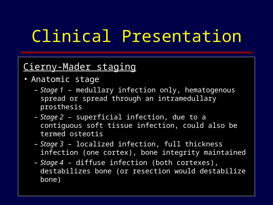
Cierny-Mader staging• Anatomic stage
– Stage 1 – medullary infection only, hematogenous spread or spread through an intramedullary prosthesis
– Stage 2 – superficial infection, due to a contiguous soft tissue infection, could also be termed osteotis
– Stage 3 – localized infection, full thickness infection (one cortex), bone integrity maintained
– Stage 4 – diffuse infection (both cortexes), destabilizes bone (or resection would destabilize bone)
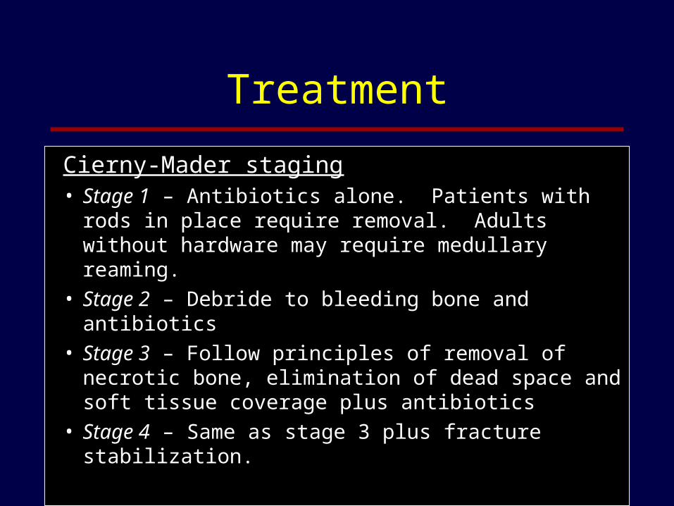
Treatment
Cierny-Mader staging• Stage 1 – Antibiotics alone. Patients with rods in
place require removal. Adults without hardware may require medullary reaming.
• Stage 2 – Debride to bleeding bone and antibiotics• Stage 3 – Follow principles of removal of necrotic
bone, elimination of dead space and soft tissue coverage plus antibiotics
• Stage 4 – Same as stage 3 plus fracture stabilization.
Case 3
• A 72 yo male who underwent a right THR 6 months ago, then developed an enterococcal UTI 3 months ago and now presents with low grade fevers and pain in the right hip that prevents ambulation.
Case 3
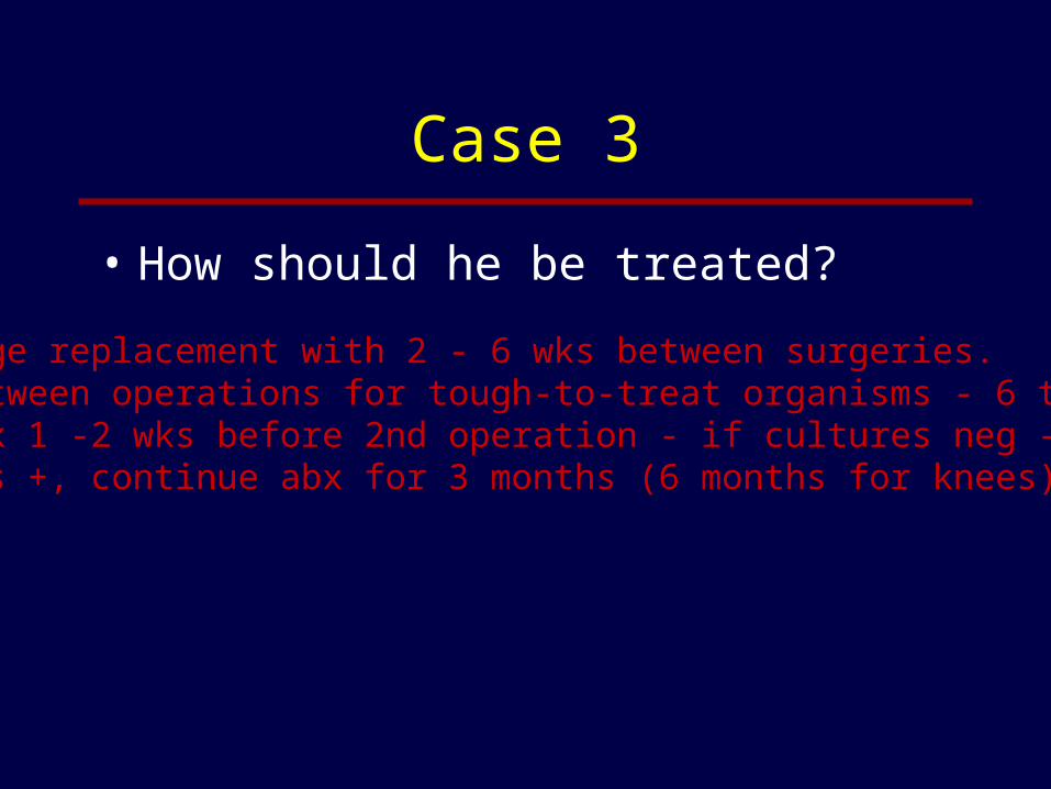
• How should he be treated?
Two stage replacement with 2 - 6 wks between surgeries.Time between operations for tough-to-treat organisms - 6 to 8 wks.Stop abx 1 -2 wks before 2nd operation - if cultures neg - stop, ifcultures +, continue abx for 3 months (6 months for knees).

Case 3
• Imaging reveals a peri-prosthetic fluid collection
• Culture of this fluid grows MRSA and enterococcus
(Lew, Lancet, 2004)
Case 3
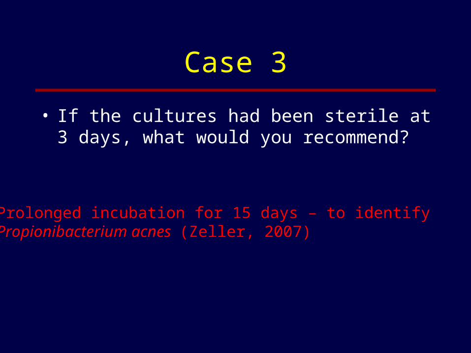
• If the cultures had been sterile at 3 days, what would you recommend?
Prolonged incubation for 15 days – to identify Propionibacterium acnes (Zeller, 2007)
Case 3
• Are there situations when the prosthesis can be retained after debridement?
Symptoms < 3 weeksStable implantEasy to treat organism
Success rates 82-100%
Case 3
• Are there indications for single stage replacement?
Symptoms >3 weeksSoft tissue in good shapeNo co-morbiditiesEasy to treat organism
Success rates 86-100%

Treatment
• Ciprofloxacin/rifampin for Osteomyelitis (Zimmerli, 1998)
• N=33, stable implants• Staphylococcus• All treated with debridement and 2
weeks of rifampin + vancomycin or flucloxacin
• Then either cipro/rifampin or cipro/placebo
• Prostheses retained• Median duration of symptoms 5d
0102030405060708090
100
Cipro Cipro/rif
Treatment
0
20
40
60
80
100
Hips Knees Bone plates Total
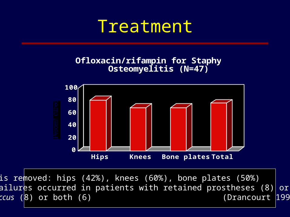
Ofloxacin/rifampin for Staphylococcal Osteomyelitis (N=47)
Prosthesis removed: hips (42%), knees (60%), bone plates (50%)All 11 failures occurred in patients with retained prostheses (8) or resistant staphylococcus (8) or both (6) (Drancourt 1993)
Case 4
• A 39 year old IVDU reports to the ER with fever and back pain. He mixes his drugs with dirty tap water and does not prep his skin before injecting. On exam his temperature is 39 C, he has a 3/6 holo-systolic murmur and tenderness over his thoracic spine on percussion. Neurological exam is initially normal.
Case 4
• Diagnoses?
• Likely organisms?
• Initial antibiotics?
• Imaging studies?
Endocarditis, vertebral OM, epidural abscess
Staphylococcus > streptococci > GNR > fungi
Nafcillin and gentamicin or vancomycin and gentamicin
MRI
Case 4
• The lab reports that 3/4 blood cultures have turned positive in 4 hours and are growing a GPC, the following day the lab reports that 2 blood cultures are also growing GNR.
• Likely organisms?
• The patient starts complaining of mid-thoracic radicular pain. What does this represent?
S. aureus > streptococci; P. aeruginosa > other GNR
Spinal ache - first sign of epidural abscess

Case 4
www.xray.2000
Tomogram CT MRI

Case 4
• What do you recommend?
• What are indications for debridement of vertebral osteomyelitis?
MRI, decompression (laminectomy or aspiration)
InstabilityAbscessCord compressionCervical infectionMedical failureNeurological signs or symptoms

Case 4
• By what route and for how long should abx be administered?
• What about follow up imaging?
No advantage of IV over oral abx (usually quinolones)Duration at least 4 weeks –Longer if hardware in place or abscesses are not drained
MR less than 4 weeks into Rx often look worse even in patients improving – don’t order!MR later – don’t follow bone changes – often progress. Focus on epidural and soft tissue changes – if these are equivocal or progress – suggests failure (Kowalski, CID, 2006)
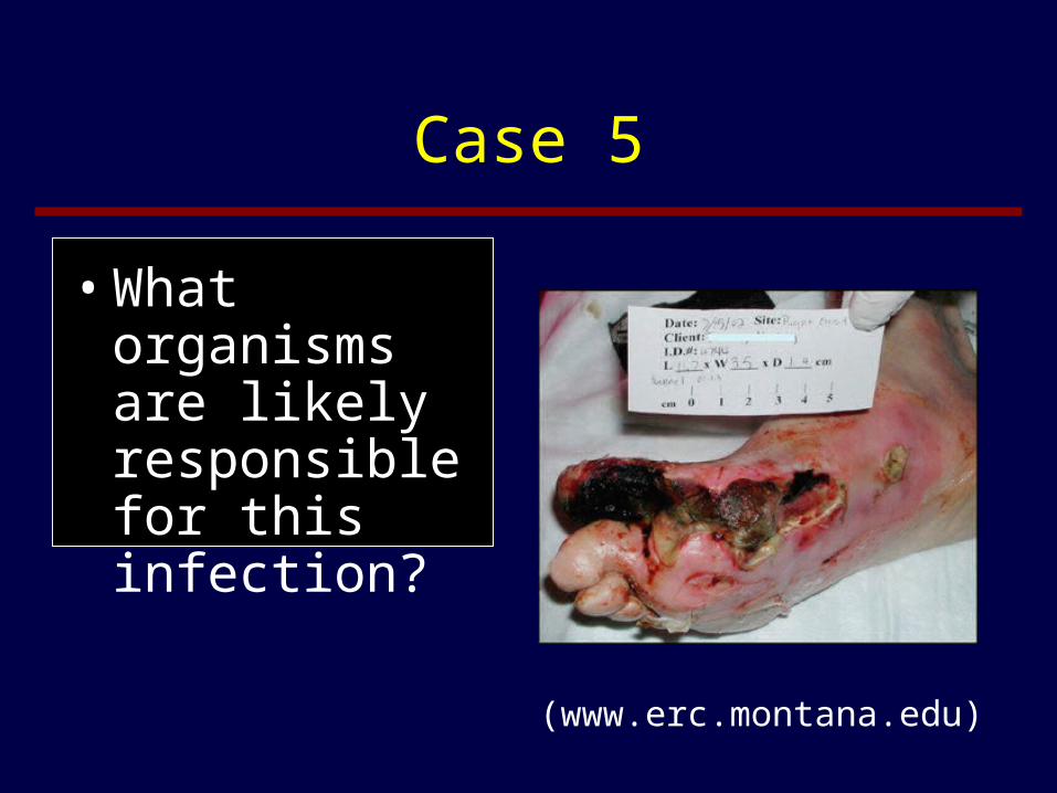
Case 5
• A 56 year old diabetic man visits his PCP for a routine visit. He is noted to have a 2.5 cm ulcer on the plantar surface of his foot at the first metatarsal head, extending up to the great toe. He was unaware of the ulcer although, in retrospect, he recalls that his socks have been stained and foul smelling lately. He has not noted fevers or chills. His physician notes a hard, gritty surface at the base of the ulcer.

Case 5
• Recommended work-up
In this case, plain films, ESR sufficientOf all imaging modalities - MR is most accurate
(sensitivity > 90%, specificity > 80%) Combination of WBC scan or ABscan with MRI can improve specificity
Diagnosis
• The gold standard is histopathologic evidence for osteomyelitis with supporting microbiologic data
• However, in many cases the diagnosis rests on clinical, laboratory and radiographic data
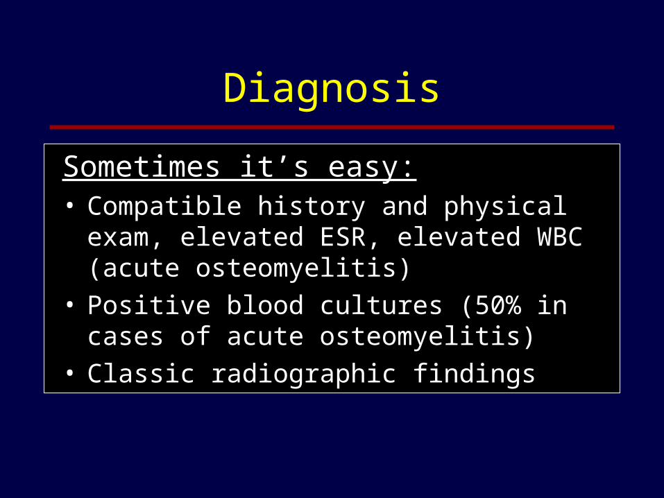
Diagnosis
Sometimes it’s easy: • Compatible history and physical exam, elevated
ESR, elevated WBC (acute osteomyelitis)• Positive blood cultures (50% in cases of acute
osteomyelitis)• Classic radiographic findings
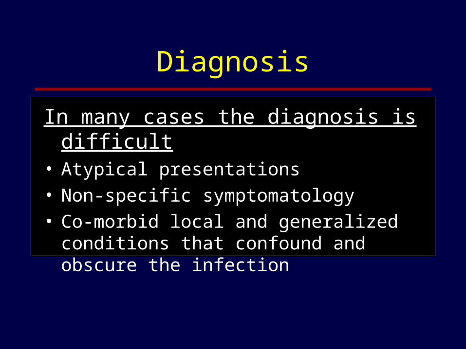
Diagnosis
In many cases the diagnosis is difficult• Atypical presentations• Non-specific symptomatology• Co-morbid local and generalized conditions that
confound and obscure the infection

Diabetic Foot Infections
What are exam findings that predict bone involvement?
– Larger (> 2cm, 92% specificity) and deeper ( > 3mm) associated with osteomyelitis
– Probe to bone – 66% sensitivity and 85% specificity, PPV around 55%, NPV 98%
– ESR > 70: 100% specificity (only 28% sensitivity)
(Grayson, JAMA,1995;273:721-3)(Newman, JAMA, 1991;266:1246-51)(Kaleta, J Am Pod Med Assoc, 2001;91:445-50)(Dinh, CID, 2008;47:519-27)
Diabetic Foot Infections
What are the best imaging modalities?• Plain film• CT scan• MRI scan• Nuclear medicine studies

Diabetic Foot Infections
Plain films • Need 30 to 50% mineral loss for x-ray
changes to be evident - takes at least 14 days
• Sensitivity 43-75%, specificity 75-83%
• Insensitive with acute osteomyelitis
• In chronic infection - sclerosis, periosteal elevation and sequestra.
(www.podiatry.files.wordpress.com)(Lipsky, CID, 1997;25:1318-26)

Diabetic Foot Infections
CT• Best method for
detecting small areas of necrosis, gas, foreign bodies
• Metallic foreign bodies compromise the image
(www.xray.2000)

Diabetic Foot Infections
MRI• Sensitivity 82-100%
• Specificity 53-94% (tumors, fractures, post surgery, sympathetic edema, infarction – all can look the same; light up on T2 weighted image)
• BEST SINGLE TEST
• Location important - – Heel and malleoli with ulcer = osteo
– Midfoot, joint-centered, no ulcer - Charcot
• Combine with Ind-111 WBC scans or gallium scans to increase specificity (www.med.harvard.edu)
(Eckman, JAMA, 1995;273:712-20)(Croll, J Vasc Surg,1996:24:266-70)(Craig, Radiology, 1997;203:849-55)(Enderle, Diabetes Care, 1999;22:294-9)

Diabetic Foot Infections
Bone scan (TC-99 labeled phosphorus)
• Soft tissue infection will be positive in the immediate (blood flow) and 15 minute (blood pool) phases while osteomyelitis will be positive in these 2 plus the delayed (> 4 hour) images.
• Sensitivity 69-100% (> 95% in acute osteomyelitis), specificity 38-82% (tumors, fractures, post-surgery, septic arthritis, Paget’s disease, Charcot foot)
(www.postgradmed.com)(Eckman, JAMA, 1995;273:712-20)(Enderle, Diabetes Care, 1999;22:294-9)
Diabetic Foot Infections
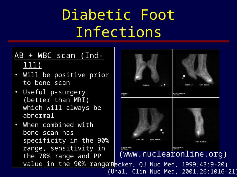
AB + WBC scan (Ind-111)• Will be positive prior to bone
scan
• Useful p-surgery (better than MRI) which will always be abnormal
• When combined with bone scan has specificity in the 90% range, sensitivity in the 70% range and PP value in the 90% range
(www.nuclearonline.org)(Becker, QJ Nuc Med, 1999;43:9-20)(Unal, Clin Nuc Med, 2001;26:1016-21)
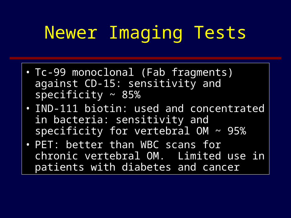
Newer Imaging Tests
• Tc-99 monoclonal (Fab fragments) against CD-15: sensitivity and specificity ~ 85%
• IND-111 biotin: used and concentrated in bacteria: sensitivity and specificity for vertebral OM ~ 95%
• PET: better than WBC scans for chronic vertebral OM. Limited use in patients with diabetes and cancer
Case 5
• What organisms are likely responsible for this infection?
(www.erc.montana.edu)
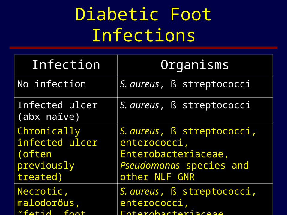
Diabetic Foot Infections
Infection Organisms
No infection S. aureus, ß streptococci
Infected ulcer (abx naïve)
S. aureus, ß streptococci
Chronically infected ulcer (often previously treated)
S. aureus, ß streptococci, enterococci, Enterobacteriaceae, Pseudomonas species and other NLF GNR
Necrotic, malodorous, “fetid” foot
S. aureus, ß streptococci, enterococci, Enterobacteriaceae, Pseudomonas species and other NLF GNR + anaerobes

Case 5
• Recommended treatment
Surgical debridement (with bone cultures)Re-vascularization if neededLong-term abx
Recent retrospective studies suggest abx alone may be sufficient treatment in many cases (Jeffcoate, 04)

Diabetic Foot Infections
• Which antibiotics should I prescribe and for how long?
(www.erc.montana.edu)

Diabetic Foot Infections
• Basic principles for choosing antibiotics:– Should always include coverage for Gram-positive
cocci, especially S. aureus– Add Gram-negative coverage for chronic wounds, for
patients previously treated with abx and for wounds classified as moderate to severe
– Provide anaerobic coverage for obviously necrotic wounds or those with a feculent odor
– Narrow coverage based on culture results
(Lipsky, Clin Micro Infect, 2007;13:351-53)

Diabetic Foot Infections
• Basic principles for choosing antibiotics:– Consider risk factors for MRSA when choosing Gram-positive
coverage– Coverage for enterococci usually not necessary unless it is the
only organism isolated– Coverage for Pseudomonas may also not be necessary unless the
wound had been treated with hydrotherapy or Pseudomonas is present and the patient is not improving without anti-Pseudomonal treatment
– Avirulent organisms (e.g. coagulase negative staphylococci, Corynebacterium species) may become real pathogens in immunocompromised hosts with significant tissue necrosis
(Lipsky, Clin Micro Infect, 2007;13:351-53)
Diabetic Foot Infections
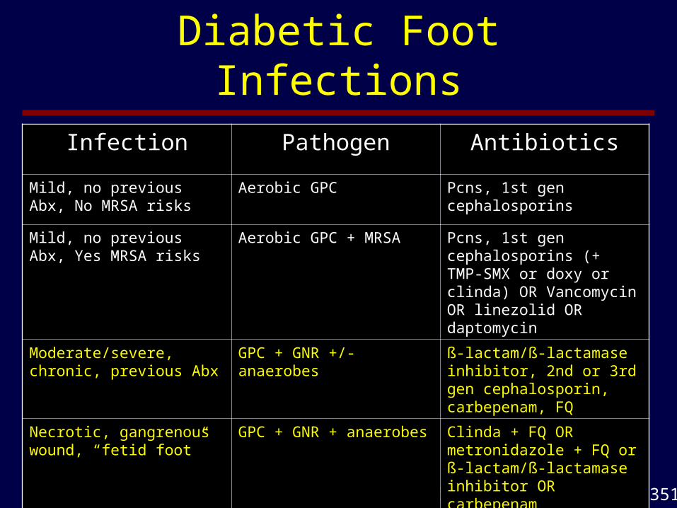
(Lipsky, Clin Micro Infect, 2007;13:351-53)
Infection Pathogen Antibiotics
Mild, no previous Abx, No MRSA risks
Aerobic GPC Pcns, 1st gen cephalosporins
Mild, no previous Abx, Yes MRSA risks
Aerobic GPC + MRSA Pcns, 1st gen cephalosporins (+ TMP-SMX or doxy or clinda) OR Vancomycin OR linezolid OR daptomycin
Moderate/severe, chronic, previous Abx
GPC + GNR +/- anaerobes ß-lactam/ß-lactamase inhibitor, 2nd or 3rd gen cephalosporin, carbepenam, FQ
Necrotic, gangrenous wound, “fetid foot”
GPC + GNR + anaerobes Clinda + FQ OR metronidazole + FQ or ß-lactam/ß-lactamase inhibitor OR carbepenam
Hydrotherapy Pseudomonas Anti-Pseudomonal FQ, Pcn or Cephalosporin or Carbepenam
Diabetic Foot Infections
• Duration of therapy– Mild infections 1-2 weeks– Moderate to severe infections: 2-4 weeks– Osteomyelitis: 4-6 weeks (or longer)

Diabetic Foot Infections
Adjuvant Therapies• G-CSF
– Cruciani, Diabetes Care, 2005;28:454460• Meta-analysis of 5 studies including 167 pateints• No effect on wound healing• Did reduce the risk for amputation (RR 0.41) and for any type of surgery
(major debridement, revascularization, angioplasty and amputation) (RR 0.38)
• Hyperbaric Oxygen– Roeckl-Wiedman, Br J Surg, 2005;92:24-32
• Meta-analysis of 6 studies, including 5 on patients with DFI (118 patients)• No effect on ulcer healing or minor amputation• Did reduce the risk of major amputation: RR 0.31

Case 6
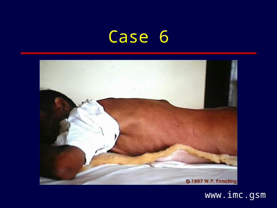
• A 43 year old male immigrant from Pakistan reports to urgent care complaining of back pain for the last 12 months. He has lost ~15 pounds. During the last 2 weeks he noticed some mild weakness in his right leg. Examination reveals a thin, stooped, muscular male with normal vital signs. His back has a tender deformity at T6. His right knee is tender and swollen. Plain films of his T-spine show anterior wedge-shaped collapse of T6.
Case 6
www.imc.gsm

Case 6
• Differential
• Diagnostic tests:
TB > Staphylococcus > other
MRI spinePPD and CXRBlood culturesBiopsyHIVLeg films

Case 6
• MRI of his spine reveals complete destruction of T6, a 20 anterior acute angle deformity and a large para-spinal fluid collection. Biopsy reveals granulomas, no AFB.
• Does he need anti-tuberculous therapy?
• Does he need surgery?
Yes
Yes

Case 6
• What are indications for surgery in Pott’s disease?
• What about his knee?
Neurological deficitsInstabilityCervical diseaseMedical failure including non-adherence
Needs evaluationSkeletal TB more common in young people with Pott’sMedical treatment alone usually sufficientIf severe destruction with abscess - debride
Skeletal Tuberculosis
Pathogenesis• In developed countries skeletal TB is a disease of
adults and represents reactivation of an old focus of infection.
• In the developing world most cases of skeletal TB occur in patients who recently acquired TB. Therefore, most skeletal TB occurs in childhood. Many patients give a history of recent trauma to the involved area.

Skeletal Tuberculosis
Clinically • Accounts for 35% of cases of extra-pulmonary TB
and 2% of all cases of TB• Indolent course, average duration of symptoms
prior to diagnosis: 16 to 19 months.• Local swelling, pain, fluctuance; systemic
symptoms (fever, sweats, etc) often absent.• Pulmonary disease present in 30%. PPD+ in >
85%
Skeletal Tuberculosis
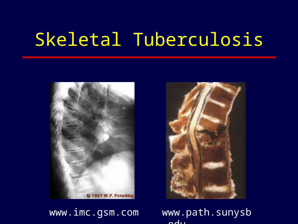
Clinically • Pott’s disease (tuberculous spondylitis)
– Responsible for 1/3 of cases of skeletal TB.
– Infection begins in the anterior aspect of the vertebral body leading to anterior collapse and spread of the infection along the anterior ligament
– Most cases involve the lumber and lower thoracic spine
– 50% of cases have associated abscesses (if calcified is diagnostic for TB)
Skeletal Tuberculosis
www.imc.gsm.com www.path.sunysb.edu
Skeletal Tuberculosis
Clinically • Pott’s disease
– 50% have weakness or paralysis at the time of presentation or during Rx
– 50% associated with disc involvement
– 50% without disc involvement are younger and more likely to have other skeletal lesions
– 77% have epidural involvement by MRI (Pertuiset, 1999)

Skeletal Tuberculosis
Clinically • Other bones: any bone; weight bearing, flat, ribs -
relatively unique to TB
Diagnosis• AFB stain and culture of biopsy specimen
(sensitivity ~85%)
Skeletal Tuberculosis
Treatment• Chemotherapy: Duration - 9 to 18 months.
Although recent studies suggest that 6 months of treatment, when combined with surgery, is as effective as longer course of antibiotics.
• Debridement of abscesses will lead to faster resolution and less kyphosis in those with severe disease at presentation.
Skeletal Tuberculosis
Treatment• Criteria for surgical intervention in Pott’s
– Neurological deficit
– Spinal instability
– Cervical spine disease
– Failure of medical therapy
– Non-adherence to medical therapy.

Case 7
• A 43 year old female with a long history of rheumatoid arthritis requiring multiple joint replacements complains to her rheumatologist of a flare of her disease with pain and swelling in one of her IP joints and her right wrist. Her temperature is 37.5C, her right wrist is warm, swollen and red as is one of her IP joints on the same hand.
• Why isn't this just a flare of her RA?
• How would you differentiate infected from non-infected joint fluid?
Too few joints
Aspiration: Gram stain 50-75%, Culture 90%, BC 50%

Case 7
• What is the bug?
• Which antibiotics would you use and for how long?
• Do the joints need to be drained? How?
S. aureus - 80% in RA
Anti-staph (anti-MRSA?), 4 to 6 weeks
YesSerial aspiration or open procedure
Case 7
• What are indications for open drainage?
Hips, shoulders, prosthetic jointsOsteomyelitis with arthritisGNRWhen aspiration fails (thick pus)

Case 8
• A 23 year old female reports to the ER with 2 days of diffuse arthralgias, low grade fever and then the development of swelling and increased pain in her right knee and wrist. She has a new boyfriend.
• Diagnosis?
• What do you find on exam?
• Is the patient likely to be menstruating?
GC > reactive arthritis
Skin lesions (< 30), tenosynovitis, additive oligoarthritis
Yes; risks for DGI: certain strains of GC, F > M, menses, complement deficiency
Case 8
• Is she likely to have genital symptoms?
• Will BC grow the organism? Joint fluid? Cervical culture?
• Should her joints be drained? Open drainage?
• Antibiotic therapy: drugs, route, duration?
• Should anyone else be treated?
No (strains that don’t fix complement - less inflammation)
< 20% ~50% > 80%
Yes, Usually not necessary
Ceftriaxone for 7-10 days
Partner

Case 9
• The ex-boyfriend of the last patient is treated as a contact. Two weeks later he reports to urgent care with pain in his toes, right knee and left hip area. He also complains of a little dysuria. Exam reveals 2 sausage digits on his right foot, a swollen, warm right knee and pain and decreased range of motion of his left hip.
• Differential diagnosis?
• Would you order any imaging studies? GC or reactive arthritis
MRI of hip
Case 9
• Would you tap his knee? His hip? His hip and knee?
• Antibiotics? Which ones? Until when?
• Would you culture anything else?
• What makes you think this is not a bacterial infection?
Yes Yes Yes
Anti-staph, strep, GC (e,g., ceftriaxone) - until cultures neg
Urethral and oral cultures
Sausage digitsMultiple sites (RA or GC > strep or staph)Negative cultures

Case 9
• Two days later he is no better and all the cultures are negative.
• How would you treat him now?
• How likely is he to HLA-B27 positive?
• Is he likely to relapse?
• Are antibiotics of any use in this disease?
• What are other risk factors for this syndrome?
Anti-inflammatories (sulfasalazine, NSAIDS, steroids)
Post CT: > 90%, post enteric infections: 50-80%
Sure, more common post CT
Doxy or macrolides for RA post CT - maybe, otherwise - No
After any enteric infection, IBD
Case 10• A 37 year old male roofer sustained a T12-S1
fracture/dislocation due to a fall. His spine was initially stabilized with rods, plates and screws. Eight weeks post-operation he was diagnosed with osteomyelitis due to S. aureus and CNS. This infection was treated by debridement of necrotic tissue and bone, removal of almost all the original hardware and immediate replacement with new hardware and a tibial allograft. The patient also received 3 months of appropriate antibiotics (vancomycin, ciprofloxacin and rifampin).
• He did well for 8 months, joined a wheelchair basketball team and then began noticing pain in his back, made worse by a rigorous game of hoops. His surgeon thinks (hopes) his pain is due to his recent increased activity but orders an ESR (32 mm/hr) and a CT (lots of post-operative changes but no obvious osteomyelitis).
Case 10
• What diagnostic tests might you order now?
• Was it a mistake to replace the infected hardware with new hardware at the same operation?
• What about the placement of the tibial allograft?
• What treatment strategy would you recommend at this point?
MRI-CT with WBC scan: Specificity 90%, PPV ~90%
Best to avoid this but in some cases not avoidable due to stabilization issues (spine)
Theoretically a bad idea (adding sequestrum!)Nevertheless, some support in the surgical literature (Shuster)
Remove hardware, antibiotics

Case 10
• The patient refuses any further treatment or work-up. A month later a draining sinus develops at the site of the original injury. The patient takes some antibiotics he had stashed at home and the sinus dries up. The infection intermittently flares over the next 15 years and each time it does the patient takes a short course of antibiotics that temporarily solves the problem. However, the most recent episode of drainage has not responded to his usual remedy and he comes back to see you. Your examination of his spine reveals an 8 cm area of tough, indurated skin with a necrotic, bleeding center draining green, purulent material.
• What are you worried about?
• What do you recommend?
Recurrent, resistant infection, squamous cell cancer
Image, debride, biopsy