Embed Size (px)
Citation preview

DECEDENT First-Middle-Last Names (Please avoid use of initials)
JAMIE ROSE BOLIN
Age
10
Birth Date
08/07/1995
Race
WHITE
Sex
F
HOME ADDRESS - No. - Street, City, State
1000 N. 8TH STREET, APT. #213, PURCELL, OK
DRIVER PASSENGER PEDESTRIANIF MOTOR VEHICLE ACCIDENT:
AUTOMOBILE LIGHT TRUCK HEAVY TRUCK BICYCLE MOTORCYCLETYPE OF VEHICLE:
EXAMINER NOTIFIED BY - NAME - TITLE (AGENCY, INSTITUTION, OR ADDRESS)
ROBERT LEE - OSBI
DATE
04/14/2006
TIME
19:34
INJURED OR BECAME ILL AT (ADDRESS)
1000 N. 8TH STREET, APT. #115
CITY
PURCELL
COUNTY
MCCLAIN
TYPE OF PREMISES
RESIDENCE
DATE
04/12/2006TIME
LOCATION OF DEATH
1000 N. 8TH STREET, APT. #115
CITY
PURCELL
COUNTY
MCCLAIN
TYPE OF PREMISES
RESIDENCE
DATE
04/14/2006
TIME
16:50
BODY VIEWED BY MEDICAL EXAMINER
901 N. STONEWALL
DESCRIPTION OF BODY RIGOR
EXTERNALPHYSICALEXAMINATION
Jaw
Neck
Arms
Legs
Complete
Absent
Passing
Passed
Decomposed
Lateral
Posterior
Anterior
Color
Regional
LIVOR EXTERNAL OBSERVATION
Beard Hair
Eyes: Color Mustache
LR
Body Length Body Weight
Opacities
NOSE MOUTH EARS
BLOOD
Significant observations and injury documentations - (Please use space below)
SEE AUTOPSY PROTOCOL
Natural
Manner of Death:
Suicide
Unknown
Accident
Homicide
Pending
Autopsy
Case disposition:
NoYes
Authorized by
Pathologist
Not a medical examiner case
MEDICAL EXAMINER
INAS YACOUB M.D.
CITY
OKLAHOMA CITY
COUNTY
OKLAHOMA
TYPE OF PREMISES
MORGUE
DATE
04/15/2006
TIME
08:15
Name, Address and Telephone No.
I hereby state that, after receiving notice of the death described herein, Iconducted an investigation as to the cause and manner of death, as required bylaw, and that the facts contained herein regarding such death are true and correctto the best of my knowledge.
Signature of Medical Examiner Date
OTHER
OFFICE OF THE CHIEF MEDICAL EXAMINER
BOARD OF MEDICOLEGAL INVESTIGATIONS
REPORT OF INVESTIGATION BY MEDICAL EXAMINER
Central Office901 N. Stonewall
Oklahoma City, Oklahoma 73117
(405) 239-7141 Fax (405) 239-2430
Eastern Division
1115 West 17thTulsa, Oklahoma 74107
(918) 582-0985 Fax (918) 585-1549
OFFICE USE ONLY
Re Co
I hereby certify that this is a trueand correct copy of the original
document. Valid only when copy
bears imprint of the office seal.
By
Date
Unknown
FOUND FOUND
MEDICAL EXAMINER:
0600829CME-1 (REV 7-98)
Computer generated report
Probable Cause of Death:
Pupils:
INAS YACOUB M.D.
ASPHYXIA
Other Significant Medical Conditions:
OTHER:
04/15/2006
INAS YACOUB M.D.
901 N. STONEWALL
OKLAHOMA CITY, OK 73117

Board of Medicolegal Investigations
Office of the Chief Medical Examiner 901 N. Stonewall
Oklahoma City, Oklahoma 73117
(405) 239-7141 Voice
(405) 239-2430 Fax
REPORT OF AUTOPSY
Decedent Age Birth Date Race Sex Autopsy No Case No
JAMIE ROSE BOLIN 10 8/7/1995 WH F 326-06 0600829
Type of Death Means ID By Authority for Autopsy
Violent, unusual or unnatural ASSAULT TOE TAG INAS YACOUB, M.D.
Present at Autopsy
PATRICK MARCOTTE / KEVIN ROWLAND
PATHOLOGICAL DIAGNOSES
I. Asphyxia evident by multiple petechiae on the face, petechiae in the eyes and curvilinear abrasions on the nose
associated with brain swelling
II. Blunt force trauma to the top of the head, right upper aspect of the back, right arm, front of the right thigh, left thigh
and left ankle, patterned appearing contusion on the left upper aspect of the chest
III. 12 cm horizontally oriented incised wound to the front and sides of the neck with resultant incision of the skin,
subcutaneous tissue, muscles, jugular veins, carotid arteries, vagus nerves, trachea, thyroid gland and esophagus;
the wound extends to the front aspect of the lower cervical vertebrae (C5-C6) with no bleeding in the airways,
aspirated food present in the airway, apparent air embolism in the brain
IV. 1.2 cm and 0.3 cm tears in the posterior aspect of the vagina / vestibular fossa associated with 0.3 cm area of
apparent hemorrhage / contusion on the cervix at the 9 o’clock location
V. Early decomposition change
VI. Generalized organ pallor
Continued on Pathological Diagnoses Page 2
CAUSE OF DEATH:
ASPHYXIA
The facts stated herein are true and correct to the best of my knowledge and belief.
OCME Central Division 4/15/2006 8:15 AM
INAS YACOUB, M.D. Pathologist Location of Autopsy Date and Time of Autopsy
CME-2 Page 1
CERTIFICATION I hereby certify that this document is a
true and correct copy of the original
document. Valid only when copy
bears imprint of the office seal.
By____________________________
Date__________________________

.
PATHOLOGICAL DIAGNOSES
(Continued)
AUTOPSY NO. ML 326-06 CASE NO. 0600829
VII. The body is covered by clear plastic, and a black plastic bag over the head / upper body and another
black plastic bag over the lower extremities and placed over a pink towel in a gray tub covered by
a lid with a piece of duct tape on each of the short sides of the tub
VIII. Atrophy of the left lobe of the liver, left kidney and left adrenal gland, incidental, old
Comment: This 10 year old was reported missing on Wednesday, April 12
th, 2006. On Friday, April 14
th,
2006 she was found dead in a closet in her neighbor’s residence. She was found there nude, wrapped in
plastic, placed in a storage tub that had duct tape on its lid. A complete autopsy was performed and
revealed the above findings. Microscopic sections confirmed the recent contusions on the right side of the
scalp, left side of the scalp, recent contusion in the right thigh, left thigh, left ankle and right side of the
back. The injury to the genital area was associated with recent hemorrhage, but no inflammatory reaction.
The injury to the genital area grossly and microscopically is due to blunt force trauma, but the
postmortem changes render its interpretation as a premortem versus a postmortem injury difficult.
Autolysis / early decomposition change was noted microscopically. No sperm is seen in the slides
prepared from the oral, vaginal or rectal swabs. Toxicology revealed 0.03% w/v ethyl alcohol in her
cavity blood. No ethyl alcohol was detected in her vitreous. It is my opinion that the probable cause of
death is asphyxia. The manner of death is homicide.
May 23, 2006
IZY/ns INAS YACOUB, M.D.

EXTERNAL EXAMINATION
AUTOPSY NO. ML 326-06 CASE NO. 0600829
DESCRIPTION
Height Weight Eyes Pupils Opacities, Etc. Hair Beard Mustache Circumcised
55 in. 46 kg. BLUE R 4 mm L 4 mm RED
RIGOR (jaw, neck, back, legs, arm, chest, abd., complete) LIVOR (color, anterior, posterior, lateral, regional) Body Heat
PASSING POSTERIOR, LEFT COOL
DESCRIPTION OF CLOTHING:
The decedent is received unclothed, however the body was covered with clear plastic and a black plastic
garbage-like bag was on the head and upper aspect of the body and another similar appearing black
garbage-like plastic bag is on the lower part of the body. The body is lying on a pink towel and is in a
gray tub. Gray duct tape is on the upper short sides of the lid. The body was received in an evidence
sealed black body bag.
EXTERNAL EXAMINATION:
The body is that of a well developed female child who appears consistent with the reported age of 10
years. Examination of the decedent’s head reveals multiple petechiae on the face and in the eyes. No
blood is observed in the nose, mouth or ear canals. Examination of the nose reveals several curvilinear
abrasions that appear red and appear consistent with fingernail marks. They range from less than 0.2 cm
to up to 0.4 cm. The location and appearance of these injuries are documented by diagrams and
photographs. Examination of the mouth reveals natural teeth, intact frenula, and no injury to the lips or
gums. Apparent stomach contents are noted in the mouth. Examination of the ears reveals apparent ear
pierce sites in each ear lobe. Examination of the head does not otherwise reveal remarkable findings
externally.
Examination of the neck reveals a 12 cm horizontally incised wound on the front and sides of the neck.
This wound is located 5 cm below the left ear lobe and approximately 6 cm below the right ear lobe. It
involves the front and sides of the neck and has at least three small cuts on the right side of its lower edge.
This incised wound has cut through the skin, subcutaneous tissue, muscles of the front and sides of the
neck, the jugular veins, the carotid arteries, the vagus nerves, the trachea, the thyroid gland, and the
esophagus, and reached the anterior aspect of the lower cervical vertebrae (C5-C6). This wound was not
associated with bleeding inside the airways, however it was associated with air emboli in the brain.
Examination of the neck does not otherwise reveal remarkable findings.
Examination of the chest and abdomen reveals a patterned contusion measuring 4.5 x 3 cm on the left
upper aspect of the chest. Examination of the genitalia reveals a 1.2 cm tear in the posterior aspect of the
vestibular fossa / vagina, at approximately 5 to 6 o’clock location, and a 0.3 cm tear at the 7 o’clock
location.
Examination of the lower extremities reveals a 2.5 x 1 cm poorly demarcated area of contusion on the
lower outer aspect of the left thigh, a 2 x 3 cm area of contusion on the lower outer aspect of the left leg, a
1 x 0.5 cm oval shaped non-contused area on the outer aspect of the left ankle and a pale green area of
discoloration on the top of the left ankle. A 2 x 2 cm poorly demarcated area of contusion is observed on
the upper aspect of the right thigh.
Continued on External Exam Page 2

External Exam Page 2 Case No. 0600829
Examination of the upper extremities reveals a 5 mm area of blue contusion on the right arm. The
fingernails appear to have been previously polished by a golden colored nail polish that is peeling. No
broken fingernails are seen.
Examination of the back reveals a 3 x 1.5 cm area of contusion on the right upper aspect of the back.

GROSS EXAMINATION
AUTOPSY NO. ML 326-06 CASE NO. 0600829
The body is examined through the customary “Y” shaped incision. No contusions are observed in the skin
and panniculus of the anterior and lateral aspect of the chest and abdomen. The 4 cm subcutaneous fat is
normally distributed, moist, and cream yellow. The musculature through the chest and abdomen is
rubbery, pink-brown, and is grossly unremarkable. The sternum is examined in the usual fashion. The
organs of the chest and abdomen appear pale and are in the normal position and relationship. The liver
edge extends 2 cm below the right costal margin at the midclavicular line. The diaphragm is intact
bilaterally. The lining of the pericardium, parietal pleura, and peritoneum is smooth and glistening. No
adhesions or abnormal accumulations of fluid are noted in the pericardial, pleural or peritoneal cavities.
NECK ORGANS:
The incised wound across the lower aspect of the front and sides of the neck have been previously
described. The skin and the panniculus of the anterior and lateral aspects of the neck are examined after
the heart is grossly examined. No contusions of these areas are noted. The pink-brown rubbery muscles of
the anterior and lateral aspects of the neck are examined. No contusions are observed in these muscles.
The neck structures have been previously described and are otherwise unremarkable. The tongue is intact,
normally papillated, and without evidence of tumor or contusion or bite marks. The hyoid bone is
fractured on the right side however this is not associated with recent hemorrhage and is interrupted as a
postmortem fracture. The cricoid and thyroid cartilages are intact and without abnormality. The epiglottis
is plate-like with no evidence of edema, trauma, or other gross pathology. The 12 gm thyroid gland is
pink-brown and has been transected by the incised wound of the neck. It is otherwise symmetrical and has
no other gross lesions. The vocal cords, folds, and respiratory lining in the larynx are unremarkable
except for the presence of aspirated food material. No other material is observed in the airways. Notably,
no blood is observed in the airways. There are no petechiae of the epiglottis, laryngeal mucosa, or thyroid
capsule.
THYMUS:
Weighs 46 gm. It is dusky pink and has a few petechiae.
CARDIOVASCULAR SYSTEM:
The heart weighs 179 gm. The epicardial surfaces are smooth and glistening. The heart has the normal
configuration and location. The coronary vessels arise and distribute normally. The coronary ostia are
normally located and widely patent. The chambers and atrial appendages are unremarkable. The valves
are normally formed and measure as follows: tricuspid = 8.0 cm, pulmonary = 6.0 cm, mitral = 8.0 cm,
and aortic = 5.0 cm. The endocardium is smooth, gray and glistening. The myocardium is maroon with
no areas of hemorrhage, masses or discoloration. The right ventricle measures 0.5 cm; the left ventricle
measures 1.5 cm; the interventricular septum measures 1.5 cm. The papillary muscles and chordae
tendineae are intact and unremarkable. The major vessels arising from the heart arise in the usual fashion.
No thromboemboli are observed in the main pulmonary artery. The major vessels arising from the aorta
arise in the usual fashion and their orifices are not narrowed. The aorta (arch, thoracic and abdominal) is
unremarkable except for the previously described incised wound of the neck that has completely
transected both right and left carotid arteries. The inferior vena cava is unremarkable.

Gross - 2 Case No. 0600829
PULMONARY SYSTEM:
The right lung weighs 163 gm, and the left weighs 197 gm. The visceral pleurae are smooth, glistening,
and intact with a few petechiae and no anthracosis or bleb formation. A 2.5 x 1.5 x 1 cm area of contusion
is observed on the upper posterior aspect of the lower lobe of the right lung deep to the previously
described 3 x 1.5 cm area of contusion on the right side of the back. The trachea, bronchi and bronchioles
have a dusky pink lining that appears smooth and has aspirated food material, but otherwise no gross
lesions. The pulmonary arterial tree is free of emboli or thrombi. The parenchyma is uniformly spongy,
pale pink, except for the area of contusion previously described, and is otherwise unremarkable. There is
no other evidence of trauma, granulomatous, or neoplastic disease. The hilar lymph nodes are
unremarkable in size, color, and consistency.
GASTROINTESTINAL SYSTEM:
The esophagus has been completely transected at the area of the incised wound to the neck, as previously
described. The esophagus has an otherwise smooth mucosa and no other gross lesions. The
gastroesophageal junction is unremarkable. The stomach is of normal configuration, is lined by an intact
mucosa, has an unremarkable wall and serosa, and contains approximately 180 cc of light brown to cream
colored viscid fluid that has some granular material and apparent cream colored food like potatoes, and
green food that appears like pickles. The duodenum is patent, shows an unremarkable mucosa and no
evidence of acute or chronic ulceration. The jejunum and ileum are unremarkable and contain green-
brown fecal material. There is no Meckel’s diverticulum. The ileocecal valve is intact and unremarkable.
The appendix is unremarkable. The colon is examined segmentally and shows no evidence of
diverticulitis, neoplasm or trauma. The large intestine contains green-brown semi-formed stools. The anus
and rectum are unremarkable.
LIVER AND GALLBLADDER:
The 830 gm liver has an intact capsule and a pale pink-brown parenchyma with no gross lesions except
for apparent atrophy of the left lobe of the liver. This is associated with atrophy of the left adrenal gland
and left kidney, to be described. The gallbladder has a smooth serosa, velvet green mucosa and no stones
or gross lesions.
SPLEEN AND LYMPH NODES:
The 126 gm spleen has an intact capsule and a dark red soft parenchyma with otherwise no gross lesions.
The lymph nodes do not appear enlarged.
PANCREAS:
The 58 gm pancreas has a lobulated tan-pink parenchyma with no gross lesions except for decomposition
change. No areas of hemorrhage, masses or obstruction to the pancreatic duct are noted.
ADRENAL GLANDS:
The left adrenal gland appears markedly atrophic with only remnants of the yellow cortex seen. The right
adrenal gland weighs 8 gm and has a yellow cortex and tan to gray medulla with no gross lesions.
GENITOURINARY SYSTEM:
The right and the left kidney weigh 93 gm and 7 gm, respectively. The left kidney appears markedly
atrophic and measures 4.3 x 2.5 x 0.7 cm. The right kidney has a 3 x 2.5 cm area of scarring on the lower
front aspect. The cortices, medulla, calyces, pelves, ureters and empty urinary bladder are otherwise
unremarkable. The ovaries, fallopian tubes, uterus, cervix and vagina are otherwise unremarkable, except

Gross - 3 Case No. 0600829
for the presence of an apparent area of contusion on the left side of the cervix at the 9 o’clock location
and the previously described tears in the vagina / vestibular fossa at 5 to 6 o’clock and 7 o’clock. The
endometrium is red-brown.
BRAIN AND MENINGES:
The scalp is reflected through the customary intermastoid incision and shows an area of recent contusion
on the right top aspect of the head measuring 4.5 x 4.5 cm and adjacent area of recent contusion
measuring 3 x 4 cm on the left upper aspect of the scalp. These areas are associated with poorly
demarcated overlying contusion on the scalp itself. These areas are not associated with skull fractures or
bleeding inside the cranial cavity. The calvarium is removed through the use of an oscillating saw and is
intact without evidence of fractures or osseous disease. No areas of epidural or subdural hemorrhage are
present. The leptomeninges are smooth and glistening. The brain weighs 1350 gm. The gyri appear
markedly swollen and there is a tendency toward obliteration of the sulci. The brain appears dusky,
swollen and soft. Apparent air embolism is noted in the cerebral vessels. The cranial nerves and circle of
Willis are otherwise unremarkable. Multiple sections of the cerebral hemispheres, midbrain, pons,
medulla, and cerebellum do not otherwise reveal remarkable findings. The ventricular system is
symmetric and unremarkable. The dura is examined. No base of the skull fractures is present.
RIBS:
Intact.
PELVIS:
Intact.
VERTEBRAE:
Apart from the incised wound, previously described, the vertebrae are otherwise unremarkable.
BONE MARROW:
Moist and dark red. Unremarkable.

.
MICROSCOPIC EXAMINATION
AUTOPSY NO. ML 326-06 CASE NO. 0600829
Microscopic sections of multiple organs like the pancreas, kidney, spleen, liver, and thyroid
gland show autolysis / early decomposition change evident by cellular damage and the presence
of microorganisms in some tissues like the heart. These are not associated with an inflammatory
reaction.
Edema is noted in the lung sections.
The thymus section shows an indistinct cortical medullary junction.
No acute inflammation is noted in the brain, meninges, heart or lungs.
The section obtained from the tear in the vulva / vagina (A) shows mucosal tear with recent
hemorrhage and no apparent inflammatory reaction. The section obtained from the red area in
the cervix at 9 o’clock also shows an area of recent contusion.
The microscopic section (B) from the right scalp contusion shows recent hemorrhage in the soft
tissue.
The microscopic section (C) from the left scalp contusion shows recent hemorrhage in the soft
tissue.
Section from the right thigh contusion (D) shows recent hemorrhage in the soft tissue.
The section from the left thigh contusion (E) shows a recent hemorrhage in the soft tissue.
The section from the left ankle contusion (F) shows recent hemorrhage in the soft tissue.
The section from the right back contusion (G) shows recent hemorrhage in the soft tissue.
No sperm is seen in the slides prepared from the oral, vaginal or rectal swabs.
May 23, 2006
IZY/ns INAS YACOUB, M.D.
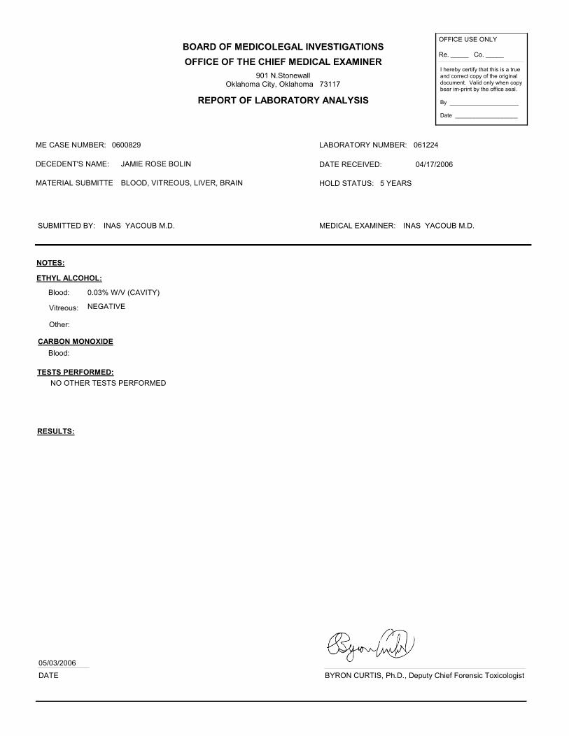
BOARD OF MEDICOLEGAL INVESTIGATIONS
OFFICE OF THE CHIEF MEDICAL EXAMINER
REPORT OF LABORATORY ANALYSIS
901 N.StonewallOklahoma City, Oklahoma 73117
OFFICE USE ONLY
Re. _____ Co. _____
I hereby certify that this is a true
and correct copy of the original
document. Valid only when copy bear im-print by the office seal.
By ______________________
Date ____________________
ME CASE NUMBER: 0600829 LABORATORY NUMBER: 061224
MATERIAL SUBMITTE BLOOD, VITREOUS, LIVER, BRAIN
SUBMITTED BY: INAS YACOUB M.D.
HOLD STATUS: 5 YEARS
DATE RECEIVED: 04/17/2006DECEDENT'S NAME: JAMIE ROSE BOLIN
NOTES:
Blood: 0.03% W/V (CAVITY)
Vitreous: NEGATIVE
TESTS PERFORMED:
NO OTHER TESTS PERFORMED
RESULTS:
ETHYL ALCOHOL:
DATE
05/03/2006
MEDICAL EXAMINER: INAS YACOUB M.D.
Other:
CARBON MONOXIDE
Blood:
BYRON CURTIS, Ph.D., Deputy Chief Forensic Toxicologist