Embed Size (px)
Citation preview
PhD program in Translational and Molecular Medicine
DIMET
Virus-host interactions in hepatitis C
virus infection: implications for
pathogenesis and therapy
Coordinator: Prof. Andrea Biondi
Tutor: Dr. Raffaele De Francesco
Discussant: Prof. Massimo Levrero
Dr. Annalisa Bianco
Matr. No. 725213
XXIV CYCLE
ACADEMIC YEAR 2010-2011


Table of Contents
CHAPTER 1. GENERAL INTRODUCTION .............................................. 1
A. Clinical aspects of Hepatitis C Virus infection .......................... 2
1. The discovery of the Hepatitis C Virus ...................................... 2
2. Epidemiology and geographic distribution of Hepatitis C ........ 3
3. Clinical characteristics of viral Hepatitis C and natural history
of the pathology ....................................................................... 6
4. Current management of Hepatitis C: diagnostic tools, clinical
decisions and therapies .......................................................... 10
5. Recently licensed HCV inhibitors ............................................ 28
6. IL28B genotype and response to therapy .............................. 33
7. Still unmet clinical needs in Hepatitis C: diagnostic tools,
biomarkers of disease progression and response to therapies,
new therapies ........................................................................ 35
B. Molecular aspects of Hepatitis C Virus infection .................... 39
1. HCV description ...................................................................... 39
2. Genomic heterogeneity and classification systems of HCV ... 51
3. HCV lifecycle ........................................................................... 52
3.1 HCV entry ........................................................................... 53
3.2 Translation and replication ................................................ 56
3.3 Assembly and release ........................................................ 57
4. The discovery of HCV defective particles ............................... 59
5. Host-pathogen interactions .................................................... 66
5.1 Lipid metabolism ................................................................ 67
5.2 Cellular stress, apoptosis, cell cycle ................................... 70
5.3 Other host factors important for HCV lifecycle: results from
siRNA screenings and other studies – The emerging role of
PI4KIIIα ............................................................................... 72
5.4 Inhibition of HCV by compounds that target host factors 76
6. Experimental models to study HCV ........................................ 82
6.1 HCV subgenomic replicon .................................................. 83
6.2 HCV cell cultured infectious model (HCVcc) ..................... 84
6.3 Additional models for HCV ................................................ 85
C. Scope of the thesis .................................................................. 88
D. Chapter 1 - References ............................................................. 90
CHAPTER 2. METABOLISM OF PHOSPHATIDYLINOSITOL 4-KINASE
IIIΑ-DEPENDENT PI4P IS SUBVERTED BY HCV AND IS TARGETED BY A
4-AMINO QUINAZOLINE WITH ANTIVIRAL ACTIVITY .................... 113
A. Abstract ................................................................................. 114
B. Introduction ............................................................................ 115
C. Results .................................................................................... 119
1. Compound AL-9 inhibits HCV replication in vitro ................. 119
2. AL-9 is an inhibitor of PI4KIIIα .............................................. 120
3. HCV alters the intracellular and plasma membrane
distribution of PI4P ............................................................... 129
4. AL-9 inhibits PI4KIIIα in HCV-replicating cells ....................... 134
5. PI4KIIIα inhibition by AL-9 alters sub-cellular distribution of
NS5A ..................................................................................... 137
D. Discussion ............................................................................... 140
E. Materials and Methods .......................................................... 147

1. Reagents and plasmids ......................................................... 147
2. Cell lines and culture conditions........................................... 147
3. Replication and infection assays .......................................... 148
4. Expression and purification of the catalytic domain of PI4KIIIα
.............................................................................................. 149
5. In vitro kinase assay.............................................................. 150
6. Indirect Immunofluorescence .............................................. 151
7. siRNA silencing ..................................................................... 153
8. T7-driven HCV polyprotein expression................................. 154
F. Supporting Information ......................................................... 154
G. Acknowledgments .................................................................. 163
H. Chapter 2 - References ........................................................... 165
CHAPTER 3. THE PRESENCE OF HEPATITIS C VIRUS DELETION
MUTANTS IS ASSOCIATED WITH NECRO-INFLAMMATORY ACTIVITY
AND PATTERN OF RESPONSE TO THERAPY IN HCV1 PATIENTS ..... 171
A. Abstract ................................................................................. 172
B. Introduction ............................................................................ 173
C. Results .................................................................................... 176
1. HCV particles with defective genomes are found in the serum
of a large fraction of chronic hepatitis C genotype 1 patients
.............................................................................................. 176
2. The presence of defective genomes is associated with patient
older age, higher viral load and increased necro-inflammatory
activity .................................................................................. 181
3. IL28B genotype profoundly influences viral kinetics during
treatment and is associated with SVR ................................. 182
4. The association between HCV defective particles and higher
histological grading is independent from IL28B genotype .. 183
5. Defective HCV particles and IL28B genotype are independent
predictors of relapse in ETR-positive patients ..................... 185
D. Discussion ............................................................................... 186
E. Materials and Methods .......................................................... 191
1. Patients ................................................................................. 191
2. Treatment ............................................................................. 192
3. Viral RNA extraction and amplification of HCV genomes .... 193
4. Genomic DNA extraction and determination of IL28B
genotype ............................................................................... 194
5. Statistical analysis ................................................................. 195
F. Acknowledgments .................................................................. 195
G. Chapter 2 - References ........................................................... 196
CHAPTER 4. THE EXPRESSION OF HCV GENOMES RESEMBLING
NATURAL SUBGENOMIC DELETIONS IS CYTOPATHIC AND ALTERS
THE EXPRESSION OF CHOLESTEROL BIOSYNTHESIS AND STRESS
RESPONSES GENES ..................................................................... 201
A. Abstract .................................................................................. 202
B. Introduction ............................................................................ 204
C. Results .................................................................................... 207
1. Expression of HCV defective RNA genomes in Huh7.5 cells 207

2. Replication of HCV defective RNA genomes into Huh7.5
impairs cell viability .............................................................. 209
3. The expression of HCV defective genomes induces a typical
reorganization of lipid droplets that co-localize with
intracellular Core .................................................................. 211
4. Global transcriptional response to the replication of HCV
defective RNA genomes in cultured hepatoma cells ........... 213
5. Genes of Cholesterol Biosynthesis pathway are
downregulated in cells expressing either J6/JFH-Δ212-886 or
J6/JFH-Δ284-736 ................................................................... 216
6. The cellular stress sensor gene ATF3 is strongly induced by the
HCV defective genomes ....................................................... 218
D. Discussion ............................................................................... 220
E. Materials and Methods .......................................................... 225
1. Reagents and plasmids ......................................................... 225
2. Cell culture, transfection and measure of cell viability ........ 225
3. Indirect immunofluorescence .............................................. 226
4. Isolation of cellular RNA, expression microarray format and
data analysis ......................................................................... 227
5. Quantitative Real-Time PCR ................................................. 228
F. Chapter 4 – References .......................................................... 229
CHAPTER 5. SUMMARY, CONCLUSIONS AND TRANSLATIONAL
IMPACT OF THE PROJECTS ......................................................... 233
A. Discovery of HCV inhibitors targeting host factors ............... 234
B. HCV-defective RNA genomes as biomarkers of disease severity
and response to anti-HCV therapy ........................................ 236
C. Chapter 5 – References .......................................................... 241
PUBLICATIONS ........................................................................... 243

1
CHAPTER 1
GENERAL INTRODUCTION
2
A. Clinical aspects of Hepatitis C Virus infection
1. The discovery of the Hepatitis C Virus
In the 1950s and ‘60s the field of viral hepatitis was initiated by the
differentiation between “infectious” and “serum” hepatitis. Later on
these two types of hepatitis were shown to be caused by hepatitis A
virus and hepatitis B virus infection, respectively. Surprisingly, when
serological tests became available in the mid-1970s, the majority of
parentally-transmitted hepatitis infections could not be assigned to
either virus. Despite international research efforts, the agent
responsible for this so-called non-A non-B hepatitis (NANBH) remained
unidentified for the next decade1, 2. In 1989, the causative agent of
NANBH was identified by molecular cloning and was denominated
hepatitis C virus (HCV). The virus was identified and characterized by
molecular cloning techniques using serum from a NANB hepatitis virus
infected chimpanzee1 and based on the similarity of the genome
organization and hydropathy profiles of several precursor proteins
classified as a member of the Flaviviridae family. However, the low
sequence homology compared to other flaviviruses eventually lead to
its classification into a new genus hepacivirus, distinct from the other
flavivirus members3.
The discovery of HCV was an important milestone in the field of
viral hepatitis. It allowed screening of blood products and the
installment of an antiviral treatment.
3
2. Epidemiology and geographic distribution of Hepatitis C
Since its discovery, HCV has been recognized as a major cause of
chronic liver disease worldwide. Currently, the World Health
Organization (WHO) estimates that 2.2-3% of the world population is
chronically infected with HCV, representing 130 to 170 million people4
and more than one million new infection cases are reported annually5,
6. In the United States alone, nearly four million persons are infected
and 30,000 acute new infections are estimated to occur each year7. In
Europe and Japan, the disease is already more important numerically
than is either hepatitis B virus (HBV) or human immunodeficiency virus
(HIV) infection and due to the availability of the HBV vaccine the
impact of hepatitis C infections will increase further. HCV infection
causes a substantial portion of chronic liver disease mortality due to
the induction of chronic hepatitis, liver cirrhosis, and hepatocellular
carcinoma (HCC)8. About 4 to 20% of patients with chronic hepatitis C
will develop liver cirrhosis within 20 years and HCC may develop after
about 20 to 35 years. There is a significant rise in the incidence of HCC
in many developed countries including Japan, Spain, France, and Italy,
where the proportion of HCC attributable to HCV ranges from 50% to
70%9. In Japan, HCV-related HCC incidence has more than tripled over
the past four decades and accounts in the 60–70 year age group for as
much as 90%10. Apart from HCC, co-infections with other viruses,
especially HIV-1 and other hepatitis viruses, have gained more
attention. These are of clinical importance, since the course of HCV
4
infection is accelerated by co-infection with HIV-111 12, hepatitis A virus
(HAV) or HBV13, 14.
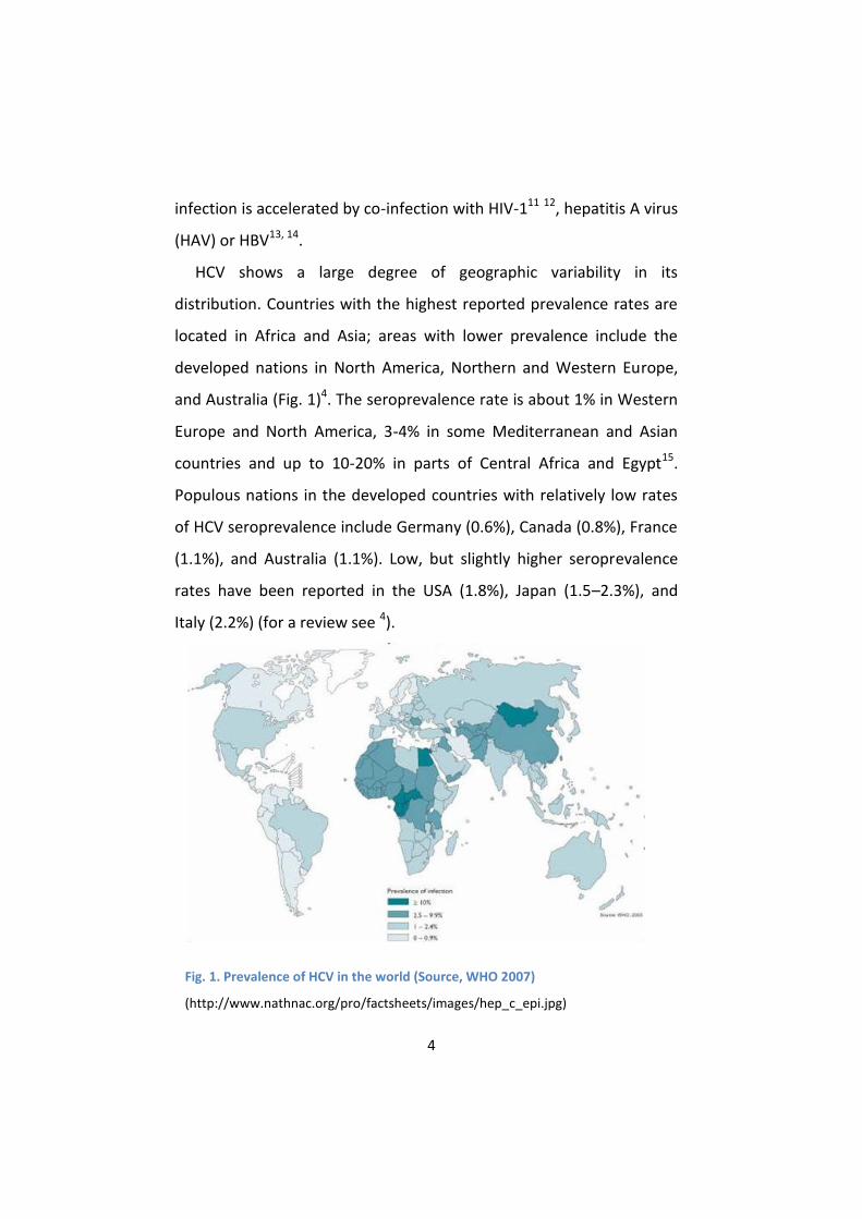
HCV shows a large degree of geographic variability in its
distribution. Countries with the highest reported prevalence rates are
located in Africa and Asia; areas with lower prevalence include the
developed nations in North America, Northern and Western Europe,
and Australia (Fig. 1)4. The seroprevalence rate is about 1% in Western
Europe and North America, 3-4% in some Mediterranean and Asian
countries and up to 10-20% in parts of Central Africa and Egypt15.
Populous nations in the developed countries with relatively low rates
of HCV seroprevalence include Germany (0.6%), Canada (0.8%), France
(1.1%), and Australia (1.1%). Low, but slightly higher seroprevalence
rates have been reported in the USA (1.8%), Japan (1.5–2.3%), and
Italy (2.2%) (for a review see 4).
Fig. 1. Prevalence of HCV in the world (Source, WHO 2007)
(http://www.nathnac.org/pro/factsheets/images/hep_c_epi.jpg)

5
There is a wide range of prevalence estimates from developing
countries, and generally less data are available to validate assumptions
about the burden of disease compared with the developed world.
China, whose citizens account for one fifth of the world’s population,
has a reported seroprevalence of 2.5-4.9%16. In other countries like
India, Indonesia and Pakistan, this may range from 0.9 % to 6.5%17-19.
Egypt, with an estimated population of 73 million, has the highest
reported seroprevalence rate at 22% due to the use of parenteral
antischistosomal therapy contaminated with HCV20.
HCV genotypes have a geographic distribution with genotype 1
being more common in the Americas and Europe, followed by
genotypes 2, 3 and 4. Subtype 1b accounts for most infections in China
and Japan, genotype 4 is more prevalent in the Middle East and North
Africa, genotype 5a is seen almost exclusively in South Africa, and
genotype 6 is common in Hong Kong and South-East Asia21, 22.
Through a phylogenetic analysis of nucleotide sequences of the HCV
genome, such as the NS5B region, it is possible to demonstrate
clustering of HCV isolates that are more or less closely related, even
within a single subtype. Using these techniques spread of HCV
genotype 1b through infected blood products and subtype 1a and 3a in
intravenous drug users, was demonstrated23, 24. Together with the
coalescent theory of population genetics, phylogenetic analysis has
been used to estimate the historical age and distribution rate of
different HCV genotypes. As a result, these models indicate that
genotype 1 originated within the last 100 years, whereas types 4 and 6
are several times older. Subtype diversity within genotypes 1, 2 and 4
6
from Western Africa and genotypes 3 and 6 in South-East Asia
implicates long-term presence of HCV in these populations25, 26. Based
on the genotype distributions, it was suggested that HCV has been
endemic in sub-Saharan Africa and South-East Asia for a considerable
time, and that the occurrence of infection in Western and other non-
tropical countries represents a relatively recent emergence in new risk
groups27, 28. In the 20th century, widespread use of blood transfusion,
unsterilized needles for injections and vaccinations caused HCV to
spread. These new transmission routes account for the epidemic
spread of HCV over the past 50 years in Europe, Egypt and elsewhere26,
27, 29.
3. Clinical characteristics of viral Hepatitis C and natural
history of the pathology
The term natural history refers to the description of the course of
the disease after infection, including clinical characteristics and factors
that influence the progression of the disease. The accurate assessment
of natural history of HCV infection has been very demanding, because
generally the acute phase of the infection is silent, although the
determination of the onset is critical to follow the full course of the
disease. Furthermore, the disease progression is modified by factors
and treatments that in principles may raise possible controversies on
the definition of the long‐term natural history of chronic HCV infection.
HCV is most efficiently transmitted by parental exposure to blood
and blood products30. The main causes of its spread are blood
transfusions and the use of contaminated needles by intravenous drug
7
users. Nowadays, transfusion-related HCV infection is almost
eliminated in countries where routine screening of blood is
obligatory31. At present, the most common risk factor in the
development world is injection drug use. Also organ transplantations,
inadequately or improperly sterilized medical or dental equipment,
tattooing, body-piercing and acupuncture can be risk factors for HCV
infection. Other routes of HCV transmission are less efficient and
include perinatal, sexual and occupational transmission. Perinatal or
“mother-to-child” transmission refers to the transmission of infection
from an infected mother to her child during the birth process
(respectively 2.7-8.4%). A higher proportion of infants are born with
HCV infection when the mother is co-infected with HIV32. Sexual
transmission of HCV has been observed, but is far less efficient than
the transmission of other sexually-transmitted viruses33. Occupational
transmission is largely confined to health-care workers who become
HCV infected by contaminated needle stick injuries34.
Acute HCV infection is in most cases asymptomatic or associated
with mild clinical illness. HCV RNA becomes detectable within 1 to 2
weeks after exposure and reaches high levels of up to 105 to 107
genome equivalents/ml in a few days35. The acute phase is mainly
characterized by mild and non-specific symptoms such as malaise,
nausea, anorexia, fatigue and abdominal discomfort; 20-30% of
patients may have jaundice. Severe acute hepatitis however is rare and
occurs in less than <1% of patients. Therefore, HCV is not often
diagnosed in the early stages of infection. The first indication of
hepatic injury is an elevated alanine aminotransferase (ALT) level,

8
which can occur 4 to 12 weeks after viral exposure. Altogether the mild
clinical manifestation of acute hepatitis generally lasts for 3 to 12
weeks. Fifteen to 20% of patients with acute hepatitis C spontaneously
clear the virus, primarily during the first 3 months following clinical
onset of infection36. Spontaneous clearance occurs more often in the
presence of symptomatic disease. Several viral and host factors appear
to affect the clinical course of infection (HCV genotype, race, gender,
HLA, co-infection with HIV, advanced age); however, none of these
factors can accurately predict spontaneous clearance. The majority of
HCV infected patients (80-85%) will develop chronic disease, defined
as the presence of HCV RNA in the blood for more than six months
following infection37. Chronic HCV infection usually remains
asymptomatic for decades, until the patient develops to a more
advanced stage of liver disease. Therefore, HCV is often designed as a
“silent disease”, since the majority of infected patients is not aware of
their disease for years after infection. Elevated ALT levels are only
found in two-thirds of patients and do not correlate with disease
severity35. Overall, 25% of chronically infected patients will develop
progressive liver fibrosis and cirrhosis. Both virus and host factors may
influence the severity and onset of liver fibrosis such as alcohol use,
immune status, sex, race etc. Each year, 4 to 5% of patients with
chronic hepatitis C develop hepatocellular carcinoma (HCC)38. The time
between infection and HCC onset varies between 10 to 30 years.
To date, liver disease related to chronic HCV infection is the most
common reason for liver transplantantion in Western countries. Being
9
a silent disease, the contribution of chronic hepatitis C to global
morbidity and mortality is generally underestimated.
However, the reported rates of cirrhosis development have been
shown to vary among studies, from 2‐8% in studies of young subjects
to 20‐30% in older patients39. These differences account for the fact
that many host factors modulate the risk of liver disease progression,
as further explained below. The pathogenesis of the liver disease is
mainly immune‐mediated. Chronic infection is associated with portal
inflammation, periportal necrosis, fibrosis and often steatosis40.
Destruction of hepatocytes by the chronic inflammation is
accompanied by liver regeneration. For mechanisms not completely
understood, in a sizable fraction of cases, liver destruction is followed
by scar formation and deposition of fibrotic tissue instead of the
normal tissue. Hepatic fibrogenesis represents a wound‐healing
response characterized by a net accumulation of extracellular matrix
(ECM) resulting from increased synthesis and decreased degradation41.
Hepatic Stellate Cells (HSCs) represent the primary source of ECM42. In
normal liver, HSCs are described as being in a quiescent state. A
distinguished feature of quiescent HSC is the presence in their
cytoplasm of multiple lipid droplets containing high amounts of
vitamin A. In response to inflammatory stimuli and Reactive Oxygen
Species (ROS), HSCs become activated, proliferate and transform into
myofibroblasts expressing α‐ smooth muscle actin (α‐SMA), an actin
isoform found in smooth muscle cells42. An increased production of
collagen type I is characteristic of this phase, a process regulated both
transcriptionally and post‐transcriptionally43, 44. Hepatocytes, Kupffer
10
cells, platelets and endothelial cells contribute to the activation of HSC.
Kupffer cells are an important source of TGF‐β1, which is a potent
stimulus for the production of ECM. TGF‐β1 also acts in an autocrine
loop, because HSCs are themselves a source of this cytokine. In
addition, TGF‐β1 inhibits cell proliferation and promotes
differentiation or apoptosis45, 46. Nevertheless, a fraction of people
with chronic HCV infection will never progress to cirrhosis, because the
disease progression may be particularly slow, depending on both
nonmodifiable and modifiable cofactors. The risk of progression is
increased by many host factors, including older age at acquisition of
the infection, male gender, alcohol consumption, coinfection with
Hepatitis B Virus (HBV) and Human Immunodeficiency Virus (HIV), iron
overload and other metabolic factors (insulin resistance, obesity).
4. Current management of Hepatitis C: diagnostic tools,
clinical decisions and therapies
The optimal approach to detecting HCV infection is to screen
persons for a history of risk of exposure to the virus, and to test
selected individuals who have an identifiable risk factor47. Currently,
injection drug use is the primary mode of HCV transmission in the U.S;
thus, all persons who use or have used illicit injection drugs in the
present or past, even if only once, as well as intranasal drug users who
share paraphernalia, should be tested for HCV infection. Individuals
who have received a blood or blood component transfusion or an
organ transplant before 1992 should also be tested. With the
introduction of sensitive tests to screen blood donors for HCV
11
antibodies in 1992, transfusion-transmission of HCV has become
rare48. Individuals with unexplained elevations of the aminotransferase
levels (alanine and/or aspartate aminotransferase; ALT/AST), those
ever on hemodialysis, children born to HCV-infected mothers, or those
with human immunodeficiency virus (HIV) infection should be tested
for the presence of HCV infection.
Other potential sources of HCV transmission include exposure to an
infected sexual partner or multiple sexual partners, exposure among
health care workers to HCV contaminated blood and blood products,
and tattooing33, 49. The prevalence of HCV infection is consistently
higher among persons with multiple sexual partners, whereas sexual
transmission of HCV between monogamous partners is uncommon33.
Nevertheless, between 1% and 5% of monogamous sexual partners of
index HCV cases test positive for anti-HCV. The hepatitis C virus is not
transmitted by hugging, kissing, sharing of eating utensils or
breastfeeding.
Folk medicine practices, including acupuncture and ritual
scarification, as well as body piercing, tattooing and commercial
barbering are potential modes for transmission of HCV infection when
performed without appropriate infection control measures50.
Transmission of HCV infection by body piercing is, however, rare.
Because symptoms are generally absent in individuals with chronic
HCV infection, recognition of infection requires risk factor screening,
which should be done whenever it is possible to link with appropriate
HCV testing and counseling47.
12
Laboratory Testing. Two classes of assays are used in the diagnosis
and management of HCV infection: serologic assays that detect specific
antibody to hepatitis C virus (anti-HCV) and molecular assays that
detect viral nucleic acid.
Tests that detect anti-HCV are used both to screen for and to
diagnose HCV infection. Anti-HCV can be detected in the serum or
plasma using a number of immunoassays. Two enzyme immunoassays
(EIAs) are approved by the U.S. Food and Drug Administration (FDA)
for clinical use, Abbott HCV EIA 2.0 (Abbott Laboratories, Abbott Park,
IL) and ORTHO® HCV Version 3.0 ELISA (Ortho-Clinical Diagnostics,
Raritan, NJ), as well as one enhanced chemiluminescence
immunoassay (CIA) VITROS® Anti-HCV assay, (Ortho-Clinical
Diagnostics, Raritan, NJ). Many commercial assays for the detection
(qualitative assays) or quantification (quantitative assays) of HCV RNA
are available. Historically, qualitative assays have been more sensitive
than quantitative assays. With the recent availability of real time
polymerase chain reaction (PCR)-based assays and transcription-
mediated amplification (TMA) assays, with sensitivities of 10-50 IU/mL,
there is no longer need for qualitative assays51. A highly sensitive assay
with this lower limit of detection is considered appropriate for
monitoring during therapy. All currently available assays have excellent
specificity, in the range of 98% to 99%. In 1997, the World Health
Organization established the first International standard for HCV RNA
nucleic acid technology, and the IU rather than viral copies is now the
preferred unit to report test results52.
13
For monitoring purposes, it is important to use the same laboratory
test before and during therapy.
Genotyping Assays. Genotyping is useful in epidemiological studies
and in clinical management for predicting the likelihood of response
and determining the optimal duration of therapy. The hepatitis C virus
can be classified into at least 6 major genotypes (genotypes 1 to 6)
based on a sequence divergence of 30% among isolates22. Genotype 1
(subtypes 1a and 1b) is the most common in the U.S., followed by
genotypes 2 and 3. Less common genotypes (genotypes 4-6) are
beginning to be observed more frequently because of the growing
cultural diversity within the United States53. Several commercial assays
are available to determine HCV genotypes using direct sequence
analysis of the 5’ non-coding region, that include Trugene 5’NC HCV
Genotyping kit (Siemens Healthcare Diagnostics Division, Tarrytown,
NY), reverse hybridization analysis using genotype specific
oligonucleotide probes located in the 5’ non-coding region, INNO-LiPa
HCV II, (Innogenetics, Ghent, Belgium), and Versant HCV Genotyping
Assay 2.0 (Siemens Healthcare Diagnostics Division, Tarrytown, NY).
Incorrect typing among the major genotypes is rare (<3%) and mixed
genotypes occur but are uncommon. Occasionally (<5%), tested
samples cannot be genotyped. This usually results from low viral
levels, issues with the PCR amplification step of the assay, or extreme
nucleotide variability within the HCV genome54.
Diagnosis of Acute and Chronic HCV Infection and Interpretation
of Assays. The diagnosis of acute or chronic HCV infection generally
requires testing of serum for both antibody to HCV (anti-HCV) and for

14
HCV RNA. Patients suspected of having acute or chronic HCV infection
should first be tested for anti-HCV.
HCV RNA testing should be also performed in patients with
unexplained liver disease whose anti-HCV test is negative and who are
immunocompromised or suspected of having acute HCV infection. A
sensitive quantitative HCV RNA assay is recommended for diagnosis
because it also provides information on the level of virus which is
helpful in management.
HCV genotyping should be performed in all HCV-infected persons
prior to interferon-based treatment in order to plan for the dose and
duration of therapy and to estimate the likelihood of response55.
One pattern is the identification of both anti-HCV and HCV RNA in a
person with recent elevation of the ALT value. This scenario is
consistent with either acute HCV infection when there is a recent
known risk exposure, with exacerbation of chronic HCV infection, or
with an acute hepatitis of another etiology in a patient with chronic
HCV infection. Another pattern is the detection of anti-HCV but with a
negative test for HCV RNA. This may represent acute HCV infection
during a period of transient clearance of HCV RNA, a false positive or
negative result or, more commonly, recovery from HCV infection.
Utility of the Liver Biopsy and Noninvasive Tests of Fibrosis. There
are three primary reasons for performing a liver biopsy: it provides
helpful information on the current status of the liver injury, it identifies
features useful in the decision to embark on therapy, and it may reveal
advanced fibrosis or cirrhosis that necessitates surveillance for
hepatocellular carcinoma (HCC). The biopsy is assessed for grade and

15
stage of the liver injury, but also provides information on other
histological features that might have a bearing on liver disease
progression56. The grade defines the extent of necroinflammatory
activity, while the stage establishes the extent of fibrosis or the
presence of cirrhosis. Several scoring systems have been conceived,
the most common being the French METAVIR, the Batts-Ludwig, the
International Association for the Study of the Liver (IASL) and the Ishak
Scoring systems57-59. The two more common non-HCV conditions that
might affect disease progression and possibly impede treatment
response are steatosis56, 60, 61 and excess of hepatocellular iron62.
Identifying either of these two features does not preclude initiating
treatment, but their presence provides additional information
regarding the likelihood of response to treatment63, 64.
The liver biopsy has been widely regarded as the “gold standard”
for defining the liver disease status, but it has drawbacks that have
prompted questions about its value. The procedure is not without risks
(including pain, bleeding and perforation of other organs), it is subject
to sampling error, it requires special expertise for interpreting the
histopathology, it adds cost to medical care, and it is anxiety-provoking
for the implicated person65. Thus, efforts are underway to seek
alternative means of establishing information on the extent of fibrosis
by focusing on noninvasive blood marker panels. These markers are
useful for establishing the two ends of the fibrosis spectrum (minimal
fibrosis and cirrhosis) but are less helpful in assessing the mid-ranges
of fibrosis or for tracking fibrosis progression66.
16
A liver biopsy may be unnecessary in persons with genotypes 2 and
3 HCV infection, since more than 80% of them achieve a sustained
virlogical response (SVR) to standard-of-care treatment. There is,
however, an ongoing debate about whether a biopsy is warranted for
persons infected with HCV, genotype 1, whose response to such
treatment approximates 50% among Caucasians and 30% among
African Americans67, 68. Even more uncertain is whether there is need
for a liver biopsy in persons infected with the other less common
genotypes (4 through 6). Thus, although the liver biopsy was
previously regarded as routine for defining the fibrosis stage in persons
with genotype 1 infection65, the issue is now in a state of flux and
possible transition. Supporters of a biopsy cite the difficult nature and
high cost of current antiviral therapy and are therefore willing to
withhold or delay treatment if liver histology displays minimal to
moderate fibrosis stage ≤2, especially if the infection is known to have
been long-standing. However, treatment is advised for those with
more advanced fibrosis stage ≥3. Therefore, the decision to perform a
liver biopsy should be based on whether treatment is being
considered, taking into account the estimated duration of infection
and other indices of advancing liver disease (e.g., the platelet count),
the viral genotype, and the patient’s willingness to undergo a liver
biopsy and motivation to be treated.
In conclusion, a liver biopsy should be considered in patients with
chronic hepatitis C infection if the patient and health care provider
wish information regarding fibrosis stage for prognostic purposes or to
make a decision regarding treatment.

17
Treatment of HCV Infection. Natural history studies indicate that
55% to 85% of individuals who develop acute hepatitis C will remain
HCV-infected37. Spontaneous resolution is more common among
infected infants and young women than among persons who are older
when they develop acute hepatitis. The risk of developing cirrhosis
ranges from 5% to 25% over periods of 25 to 30 years39. Progression to
cirrhosis may be accelerated in persons who are of older age, who are
obese, who are immunosuppressed (e.g., HIV co-infected69), and who
consume more than 50g of alcohol per day, although the precise
quantity of alcohol associated with fibrosis progression is unknown70.
Persons with HCV-related cirrhosis are at risk for the development of
hepatic decompensation (30% over 10 years) as well as hepatocellular
carcinoma (1% to 3% per year)71. Infection with HCV can also cause
extrahepatic diseases including mixed cryoglobulinemia, types II and
III. Indeed, symptomatic cryoglobulinemia is an indication for HCV
antiviral therapy regardless of the stage of liver disease.
The goal of the therapy is to prevent complications and death from
HCV infection. Because of the slow evolution of chronic HCV infection
over several decades, it has been difficult to demonstrate that therapy
prevents complications of liver disease. Accordingly, treatment
responses are defined by a surrogate virological parameter rather than
a clinical endpoint.
Short-term outcomes can be measured biochemically
(normalization of serum ALT levels), virologically (absence of HCV RNA
from serum by a sensitive PCR-based assay), and histologically (>2

18
point improvement in necroinflammatory score with no worsening in
fibrosis score)67.
Several types of virological responses may occur, labeled according
to their timing relative to treatment. The most important is the
sustained virological response (SVR), defined as the absence of HCV
RNA from serum by a sensitive PCR assay 24 weeks following
discontinuation of therapy (Fig. 2). This is generally regarded as a
“virological cure”, although liver cancer has been identified years later,
especially if cirrhosis existed at the time of achieving an SVR72.
Undetectable virus at the end of either a 24-week or 48-week
course of therapy is referred to as an end-of-treatment response (ETR).
An ETR does not accurately predict that an SVR will be achieved but is
necessary for it to occur.
A rapid virological response (RVR), defined as undetectable HCV
RNA at week 4 of treatment, using a sensitive test with a lower limit of
detection of 50 IU/mL, predicts a high likelihood of achieving an SVR73.
Only 15% to 20% of persons with HCV genotype 1 infection and 66%
with HCV genotype 2 and 3 infections achieve an RVR74, 75. Because of
the rapid clearance of virus from serum, patients who achieve an RVR
may be able to shorten the duration of treatment75.
An early virological response (EVR) is defined as a ≥2 log reduction
or complete absence of serum HCV RNA at week 12 of therapy
compared with the baseline level. Failure to achieve an EVR is the most
accurate predictor of not achieving an SVR and identifying non-
responders67, 75. Ninety-seven to 100% of treatment-naive patients
with HCV genotype 1 infection who did not reach an EVR failed to

19
achieve an SVR. Monitoring viral kinetics is thus useful for predicting
whether or not an SVR is likely to develop.
Virological breakthrough refers to the reappearance of HCV RNA
while still on therapy, while virological relapse is the reappearance of
HCV RNA in serum after treatment is discontinued and an ETR was
documented.
Persons who fail to suppress serum HCV RNA by at least 2 logs after
24 weeks of therapy are null responders, while those whose HCV RNA
levels decrease by ≤2 logs IU/mL but never become undetectable are
referred to as partial nonresponders.
Measuring the rate of viral clearance from serum is helpful in
predicting the likelihood of a response to therapy, for determining the
optimal duration of therapy and as a stopping rule for patients with
chronic HCV infection. This approach may have the benefit of limiting
exposure to PEG-IFNα and ribavirin, thus potentially leading to
reduced toxicity and a cost savings.

20
Fig. 2. Graphic display of virological responses. RVR, rapid virological response (clearance of HCV from serum by week 4 using a sensitive PCR-based assay); EVR, early virological response (≥2 log reduction in HCV RNA level compared to baseline HCV RNA level or HCV RNA negative at treatment week 12); SVR, sustained virological response (HCV RNA negative 24 weeks after cessation of treatment); relapse, reappearance of HCV RNA in serum after therapy is discontinued; non responder, failure to clear HCV RNA from serum after 24 weeks of therapy; partial non responder, 2 log decrease in HCV RNA but still HCV RNA positive at week 24; null non responder, failure to decrease HCV RNA by <2 logs after 24 week of therapy
55.
The Optimal Treatment of Chronic HCV: PEG-Interferon Alfa and
Ribavirin. The currently recommended therapy of chronic HCV
infection is the combination of a pegylated interferon alpha and
ribavirin.
There are two licensed pegylated interferons in the United States,
PEG-IFNα -2b (PEG-Intron, Schering Plough Corp., Kenilworth, NJ), with
a 12-kd linear polyethylene glycol (PEG) covalently linked to the
standard interferon alfa-2b molecule, and PEG-IFNα -2a (Pegasys,
Hoffmann-La Roche, Nutley, NJ) with a 40-kd branched PEG covalently
linked to the standard interferon alfa-2a molecule76. The doses of
these two forms of pegylated interferons differ.
21
The optimal dose of PEG-IFNα-2b, based on the original registration
trial, is 1.5 µg/kg/week dosed according to body weight, together with
ribavirin 800 to 1400 mg daily.
PEG-IFNα-2a is administered at a fixed dose of 180 µg/week given
subcutaneously together with ribavirin 1000 to 1200 mg daily.
Ribavirin has two beneficial effects: an improvement in the ETR but,
more importantly, a significant decrease in the relapse rate as
compared to PEG-IFNα monotherapy treatment.
The optimal duration of treatment should be based on the viral
genotype. The study established that patients with genotype 1 should
be treated for 48 weeks with PEG-IFNα-2a plus standard weight-based
ribavirin, whereas patients with genotypes 2 and 3 could be treated
with PEG-IFNα-2a plus low dose ribavirin (800 mg) for 24 weeks68.
For patients with HCV genotype 4 infection, combination treatment
with pegylated interferon plus weight-based ribavirin administered for
48 weeks appears to be the optimal regimen77. Patients with
genotypes 5 and 6 are underrepresented in trials of PEG-IFNα and
ribavirin due to their limited worldwide frequency.
Currently, the major challenge with regard to therapy is what new
approaches are needed to increase the SVR rates in (1) patients with
genotype 1 infection and a high viral load; (2) persons who fail to
achieve an SVR using the currently approved treatment regimens.
Multivariate analyses have identified two major predictors of an
SVR among all populations studied: the viral genotype and
pretreatment viral load67, 68. Sustained virological response rates were
higher in patients infected with genotype non-1 infection (mostly

22
genotype 2 and 3) and in those with a viral load of less than 600,000
IU/mL68. Other less consistently reported baseline characteristics
associated with a favorable response include the doses of PEG-IFNα
(1.5 µg/kg/week versus 0.5 µg/kg/week) and ribavirin (>10.6 mg/kg),
female gender, age less than 40 years, non–African-American race,
lower body weight (≤75 kg), the absence of insulin resistance, elevated
ALT levels (three-fold higher than the upper limit of normal), and the
absence of bridging fibrosis or cirrhosis on liver biopsy67.
High dose interferon induction regimens have generally been
unsuccessful. High dose ribavirin given together with standard dose
PEG-IFNα was also evaluated. Ninety percent of patients achieved an
SVR, but safety issues are the major concern for this approach since
significant anemia developed in all patients, requiring the use of
growth factors in all and blood transfusions in two patients.
Adverse Events. Almost all patients treated with PEG-IFNα and
ribavirin experience one or more adverse events during the course of
therapy. Adverse events are a major reason that patients decline or
stop therapy altogether. In the registration trials of PEG-IFNα alfa-2a
and 2b plus ribavirin, 10% to 14% of patients had to discontinue
therapy due to an adverse event67. The most common adverse events
in these trials were influenza-like side effects such as fatigue,
headache, fever and rigors, which occurred in more than half of the
patients, and psychiatric side effects (depression, irritability, and
insomnia), which occurred in 22% to 31% of patients.
Laboratory abnormalities are the most common reasons for dose
reduction. Among these, neutropenia was a frequent laboratory

23
abnormality. Despite the decline in the neutrophil count, serious
infections are uncommon and granulocyte colony stimulating factor is
rarely necessary except in patients with advanced cirrhosis. Anemia
was observed in approximately one-third of patients.
Neuropsychiatric side effects include depression, insomnia,
emotional lability, mood disorders, frank psychosis, suicidal ideation,
actual suicide, and homicide. Interferon-induced depression appears
to be composed of two overlapping syndromes — a depression-
specific syndrome characterized by anxiety and cognitive complaints,
and neurovegetative symptoms, characterized by fatigue, anorexia,
pain and psychomotor slowing78.
The most consistent risk factors for developing depression are the
presence of mood and anxiety symptoms prior to therapy. Mental or
psychiatric disease represents a significant barrier to treatment in
patients with chronic HCV infection. Significant depressive symptoms
occur in 21% to 58% of interferon-treated patients. These patients may
be successfully treated with a multidisciplinary approach to
management of adherence and neuropsychiatric side effects. Using
this approach, they can achieve SVR rates that are similar to patients
without psychiatric disorders. Most psychotropic agents are thought to
be safe for use in the management of patients with chronic HCV
infection and psychiatric disease. However, consideration should be
given to drug–drug interactions and dose modification in patients with
advanced liver disease. A past history of depression and of receiving
higher doses of interferon, as well as being female, have been
24
identified as risk factors for neuropsychiatric side effects of PEG-IFNα,
but are less reliable ones79.
Pegylated interferon may induce autoimmune disorders, such as
autoimmune thyroiditis, or may worsen preexisting autoimmune
disorders. Therefore, the presence of autoimmune conditions prior to
treatment is a relative contraindication to therapy.
With regard to ribavirin, the most common side effect is hemolytic
anemia. Since ribavirin is cleared by the kidney, the drug should be
used with extreme caution in patients with renal disease and renal
failure. Other side effects associated with ribavirin include mild
lymphopenia, hyperuricemia, itching, rash, cough and nasal stuffiness.
Ribavirin is reported to cause fetal death and fetal abnormalities in
animals and thus it is imperative for persons who receive the drug to
use strict contraceptive methods both during treatment and for a
period of 6 months thereafter. The education of patients and
caregivers about side effects and their management is an integral
component of treatment and is important for a successful outcome.
Retreatment of Persons Who Failed to Respond to Previous
Treatment. The approach to patients who fail therapy depends on the
nature of the initial response, on the potency of initial treatment and
on host–viral factors. Twenty to fifty percent of patients treated with
pegylated interferon and ribavirin will not achieve an SVR. Failure to
achieve an SVR with a course of pegylated interferon and ribavirin can
be a consequence of non-response, virological breakthrough, or
relapse. Poor adherence to the prescribed treatment and
inappropriate dose reductions can contribute to poor response rates.
25
The induction of antibodies to PEG-IFNα accounts for only a minority
of cases.
Non-responders to PEG-IFNα and ribavirin with advanced fibrosis
should follow AASLD guidelines for screening for HCC and varices and
be evaluated for liver transplantation if they are appropriate
candidates. Patients with mild fibrosis should be monitored without
treatment.
In the majority of instances, virological relapse occurs within the
first 12 weeks and late relapse, beyond 24 weeks, is extremely
uncommon. Patients with virological relapse are likely to respond to
the same regimen given a second time but will still experience an
unacceptable rate of relapse55.
Diagnosis and Treatment of HCV-Infected Children. The risk of
perinatal HCV transmission is 4% to 6%, and is 2- to 3-fold higher for
mothers with HIV/HCV co-infection. Although HCV has been identified
in breast milk of infected mothers, there are no data to show that HCV
is transmitted in breast milk; therefore breastfeeding is not prohibited
in HCV-infected mothers80.
Children who are acutely infected with HCV, like adults, are
generally asymptomatic, but they are more likely than infected adults
to spontaneously clear the virus and are more likely to have normal
ALT levels. Children with chronic HCV infection, irrespective of mode of
acquisition (vertical versus transfusion), have been shown to have
minimal progression of their disease over 5 to 20 years. Biopsy studies
in children generally have demonstrated minimal fibrosis and rare
cirrhosis 15 to 20 years after infection81.

26
Treatment of Persons with Compensated and Decompensated
Cirrhosis. Patients with HCV-related compensated cirrhosis can be
treated with the standard regimen of pegylated interferon and
ribavirin but will require close monitoring for adverse events.
Treatment of patients with decompensated cirrhosis, defined as one or
more of the clinical complications of chronic liver disease — ascites,
encephalopathy, variceal bleeding, and/or impaired hepatic synthetic
function — is more problematic. They should be referred for
consideration of liver transplantation. For those undergoing liver
transplantation, reinfection of the allograft with HCV is the rule and
progressive post-transplantation disease of the grafted liver is
common. Accordingly, since eradication of HCV pre-transplantation is
associated with a lower likelihood of post-transplantation infection,
there is a strong incentive to treat the HCV infection before
transplantation, provided the risks of treatment are acceptable. Graft
re-infection is almost universal and graft loss due to recurrent HCV
occurs in approximately 25% to 30% of patients82.
Treatment of Persons with Acute Hepatitis C. The response rate to
treatment is higher in persons with acute than with chronic HCV
infection. There is consistent evidence that treatment reduces the risk
that acute hepatitis C will evolve to chronic infection. Studies using
high doses of interferon (5-10 million units per day) for at least 12
weeks, or until serum enzymes normalized, report sustained viral
response rates of 83% to 100%, which are much higher than any
estimates of spontaneous clearance, or of response rates in persons
with chronic HCV infection. No recommendation can be made for or
27
against the addition of ribavirin and the decision will therefore need to
be considered on a case-by-case basis. It is reasonable to start
treatment within 8-12 weeks after identified acute hepatitis C, and
thus patients should be monitored monthly for this purpose. Although
HCV RNA in patients with acute infection generally is cleared from the
blood by 8 to 16 weeks in most persons who recover spontaneously,
viremia has been observed as late as 48 weeks after acute infection in
injection drug users who ultimately clear35.
Other Management Issues. An important adjunct to the therapy of
HCV is to advise chronically affected persons of measures that might
be helpful in reducing or even preventing further fibrosis progression,
independent of treatment. Most important is the issue of the potential
deleterious effect of alcohol. Excess alcohol intake may increase HCV
RNA replication and interfere with response to treatment83.
Controversy exists, however, about the level of alcohol intake that is
clearly harmful to the HCV-infected person. It is widely believed that
the daily consumption of more than 50 grams of alcohol has a high
likelihood of worsening the fibrosis, but there are reports of levels of
alcohol intake of less than that amount having a deleterious effect on
the liver disease84. It is reasonable to recommend either the complete
suspension of alcohol intake while on treatment or restricting its use
to an occasional drink during the course of the treatment.
Obesity and its associated nonalcoholic fatty liver disease are
believed to play a role in the progression of fibrosis in HCV-infected
individuals and response to treatment85. Weight reduction and
28
improvement in insulin resistance may improve the response to PEG-
IFNα plus ribavirin therapy.
Furthermore, it is recommended that persons with chronic HCV
infection who lack evidence of preexisting antibody to hepatitis A
and/or B receive the hepatitis A and B vaccines86.
Other metabolic factors contribute to the modification of the
natural course of HCV liver disease. These include: iron overload,
steatosis, insulin resistance and obesity. All these factors have been
shown to accelerate disease progression60. With regard to steatosis, a
complex relationship exists with the virus and, additionally, the
separation of the relative contributions of steatosis, obesity and insulin
resistance is challenging, because of the underlying relationships
between them. Moreover, accelerated rates of fibrosis progression
associated with steatosis are observed in a genotype‐specific way.
Both in vitro and in vivo, HCV has been shown to modulate lipid
metabolism, mainly through gene expression regulation or direct
induction of intracellular lipid droplets accumulation87, 88. Moreover,
HCV replicates within a compartment derived from intracellular
membrane alterations induced by nonstructural proteins, thus
providing another direct link between lipids and HCV life cycle.
5. Recently licensed HCV inhibitors
No direct acting antiviral drugs to treat infection with HCV have
been licensed in the 20 years since its identification. Excitingly, recent
publications herald several small revolutions in antiviral treatment of
HCV that have considerable relevance for prospective HCV therapies.
29
After a decade in which PEG-IFNα–ribavirin therapy was the only
available option, triple therapy with HCV nonstructural protein (NS)
3/4A protease inhibitors (HCV PI) in combination with PEG-IFNα–
ribavirin will become the new standard of care. In the first half of 2010,
important phase II studies of orally available inhibitors of HCV NS3/4A
protease were published.
First, McHutchison and colleagues reported on the efficacy and
safety of telaprevir in combination with PEG-IFNα–ribavirin in 465
patients with chronic hepatitis C who had not responded to at least
one course of PEG-IFNα–ribavirin therapy89. The addition of telaprevir
to standard therapy markedly increased SVR rates in these difficult to
treat patients (up to 53% compared with 14% in the control arm).
Remarkably, in the group treated for 24 weeks with this triple therapy
followed by another 24 weeks of PEG-IFNα-2b plus ribavirin the SVR
rates for patients who had previously relapsed was 76%. Even for
patients who had not responded to standard therapy the SVR rate was
up to 39% with the novel triple therapy.
In the second publication of a phase II study, Kwo et al. reported the
results of treatment with another HCV PI, boceprevir, showing that its
addition to the standard treatment leads to a notably increased SVR
(75% versus 38%) in treatment-naïve patients who are infected with
HCV genotype 190.
Importantly, all of the published studies confirm that the
combination of three agents (PEG-IFNα–ribavirin plus an HCV PI) is
needed to substantially enhance SVR rates. The potency and safety of
these two first generation HCV PIs have now been confirmed in large
30
phase III studies. In treatment-naive patients infected with HCV
genotype 1, the SVR rates were 75% with the addition of telaprevir
versus 44% with standard therapy89, and 68% versus 40% for
boceprevir91.
Although the upcoming triple therapy protocols for telaprevir and
boceprevir will be different, both will involve “response-guided
therapy”. This strategy will mean that in treatment-naïve patients,
treatment duration will be reduced to 24 weeks or 28 weeks for
patients with a rapid viral response—those with a negative result for
serum HCV RNA after 4 weeks of exposure to an HCV PI.
Although the addition of HCV PIs to HCV therapy promises to
markedly improve outcomes, IFN and ribavirin are still part of all the
treatment regimens tested in the above mentioned trials; thus
important adverse effects and several contraindications remain major
problems in HCV therapy. An IFN-free method of achieving SVR is,
therefore, the ultimate goal for HCV therapy.
In October 2010, the Lancet reported on 88 patients infected with
HCV genotype 1 who were treated with two direct acting antiviral
drugs (DAAs): a combination of the HCV PI, danoprevir, and a
nucleoside NS5B polymerase inhibitor, RG712892. Treatment with a
combination of the highest doses of both drugs led to an average 5.1
log reduction of HCV RNA plasma levels within 14 days. Overall, this
treatment was well tolerated and some patients even had HCV RNA
concentrations below the limit of detection. Notably, no viral
breakthroughs were observed during 4 weeks of treatment, which

31
might reflect the high genetic barrier to resistance associated with
nucleoside polymerase inhibitors.
The combination of two DAAs, as investigated in the INFORM-1
study, is an important step towards an IFN-free regimen that is
administered orally—hopefully with an efficacy comparable to that of
regimens that contain IFN. However, because of the protocol design of
this study all patients received a full course of PEG-IFNα–ribavirin
therapy following the 12 weeks of combination danoprevir and
RG7128 therapy. Thus, proof of the concept that SVR can be achieved
with a DAA approach administered orally is still missing.
At the moment, several companies are exploring different anti-HCV
oral therapies in ongoing studies that incorporate different
combinations of linear and cyclic HCV PIs, nucleoside as well as non-
nucleoside NS5B polymerase inhibitors, NS5A inhibitors and cyclophilin
A inhibitors.
Even in view of these imminent improvements in the standard of
care of patients with hepatitis C, there is still an urgent need for other
antiviral drugs that act via different molecular mechanisms, as the
development of viral resistance and toxic effects will certainly be a
major challenge to any new therapy. Given that HCV has a very high
mutation rate, therapies that target the host factors essential for HCV
replication might be a greater barrier to resistance than drugs that
target viral enzymes.
In the search for novel targets, Lanford and colleagues have taken a
highly original approach by antagonizing microRNA-122, the most
abundant miRNA in the liver and an essential cofactor for viral RNA
32
replication that binds to the 5'-noncoding region of the HCV genome93.
They showed that using a locked nucleic-acid-modified
phosphorothioate oligonucleotide (SPC3649) to inhibit miRNA-122
causes marked and prolonged reduction of HCV viremia in chronically
infected chimpanzees94. Except for a decrease in serum levels of
cholesterol, they did not observe any important adverse effects in the
treated animals. These data indicate the feasibility and safety of
SPC3649 as a treatment for patients with hepatitis C. A drug that
targets a specific human miRNA in a clinically common disease and
demonstrates clinical efficacy represents an extremely innovate
approach that might also be interesting for other disease areas as our
understanding of the biological functions of miRNAs expands.
How useful the novel agents will be in the most difficult to treat
patients, such as those with advanced or decompensated liver disease
or after liver transplantation, is still unclear.
Furthermore, DAAs also have to be developed for the other HCV
genotypes. Some progress has been made in meeting the next
challenges, including the management of viral resistance, tailoring
optimal treatment approaches for individual patients and eventually
the replacement of treatments that contain IFN with regimens that do
not contain IFN and are administered orally. Such therapies have to be
developed to achieve an SVR and thus cure patients infected with HCV,
even in so-called null responders to IFN-based therapies. Reported
cases of an SVR in single patients who refused to continue standard
treatment after a course of therapy with novel anti-HCV agents, such
as cyclophilin inhibitors, are promising95. Finally, with the expansion of

33
treatment options and an increasing understanding of how host
factors (such as polymorphisms in the gene that encodes interleukin-
28) affect treatment outcomes, regimens will increasingly have to be
individualized to each patient’s characteristics and preferences.
6. IL28B genotype and response to therapy
An outstanding advance in understanding the interplay between
host immunogenetics and viral clearance was the discovery of the
association between the IL28B locus on chromosome 19 and the
spontaneous or treatment‐induced viral clearance. IL28B encodes for
intereferon‐λ3 (IFN‐λ3), a member of the type‐3 interferon group. This
cytokine is structurally related to the IL10 family, but is functionally
related to type‐1 IFN96. Actually, it is an antiviral cytokine involved in
innate immune responses that functions via the JAK/STAT signaling
pathway in regulating the expression of IFN‐stimulated genes (ISGs)
and thus suppressing viral infections. Indeed, recent evidence showed
that IFN‐λ suppresses HCV replication97-99 and early clinical trials
reported a successful antiviral effect of pegylated‐IFN‐λ1 plus ribavirin
in treatment‐naive patients100. The identification of IL28B as a
significant predictor of treatment‐induced viral clearance resulted
from various GWASs performed in population of different ethnicity101-
103. These findings were further validated and the association with
spontaneous viral clearance was also demonstrated104-106. The first
GWAS performed by Ge et al.101 reported the association between
rs12979860 a Sustained Virological Response (SVR) in patients with
genotype 1 and treated with PEG-IFN‐α2a or PEG-IFN‐α2b. An

34
approximate twofold change in response to treatment was observed in
patients with the SNP rs12979860 CC genotype compared with the TT
genotype. The reported Odds Ratio (OR) was 7.3 in
European‐Americans, 7.1 in African‐Americans and 5.6 in Hispanics.
The SNP rs12979860 is located ~3 kbp upstream the IL28B gene and
resulted the strongest predictor of SVR, compared to baseline fibrosis
or baseline viral load.
Indeed, the actual causative variants have not been yet identified,
although targeted sequencing of the IL28B region revealed two
possible candidates: a non‐synonymous substitution (rs8103142,
K70A) and a variant in the promoter region of IL28B (rs28416813).
Recently, gene expression has been linked to IL28B genotypes, thus
providing a direct link between genotype and function. Indeed, lower
pre‐treatment intrahepatic expression levels of ISGs have been
reported for the genotypes of rs8099917 and rs12979860 associated
with SVR107, 108. Conversely, no association was reported between the
IL28B mRNA expression and different genotypes107. Hence, the
emerging picture is the following: a) subjects with a IL28B genotype
associated to SVR have a lower pre‐treatment activation of ISGs; b)
this reduced expression of ISGs in treatment‐naive patients may
account for the higher viral loads observed in those carrying the IL28
genotype associated with SVR101; c) the IL28B variants associated with
a poor response correspond to higher pre‐treatment levels of ISGs, a
condition that in principle might impair a strong response to
exogenous IFN during therapy; d) the therapy outcome seems not to

35
be influenced by differential expression of IL28B gene in groups with
different IL28B genotype.
Finally, the exact interplay between type‐1 and type‐3 IFNs remains
to be fully elucidated and a mechanistic model to accurately explain
this scenario is currently unavailable.
A short‐term translational value of these finding could be the
individualization of the therapy in accordance to the host genotype. A
significant number of patients fail to respond to PEG-IFN plus ribavirin
or have adverse effects, including influenza‐like symptoms,
haematologic abnormalities and neuropsychiatric symptoms.
Therefore, it is of outstanding interest to predict the therapeutic
outcome. A laboratory test for IL28B genotype is already available in
the United States and this could in principle be used together with viral
load and genotype plus other host factor to predict SVR, since a 100%
predictive power is currently not achievable with the genotype data
alone.
7. Still unmet clinical needs in Hepatitis C: diagnostic tools,
biomarkers of disease progression and response to
therapies, new therapies
Liver cirrhosis and its sequelae are the most unwanted
consequences of chronic HCV infection. Apart from virus eradication, a
very major clinical goal for HCV is to prevent or to follow closely the
transition from fibrosis to cirrhosis. The progression rate from fibrosis
to cirrhosis, varies widely among HCV patients going from few years to
several decades and it is often unpredictable109. While in some cases
36
the rate of HCV disease progression is clearly influenced by a number
of external factors such as age at infection, gender, immune status and
alcohol consumption, obesity, and liver co-infections, in most cases,
there are no clinical markers available to assess the risk of developing
progressive liver disease and liver cancer in individual subjects110.
Moreover, the liver is an organ that can often compensate very well
for the lost of a sizeable fraction of hepatic tissue. Indeed, it is not
infrequent that cirrhosis is diagnosed only at very advanced stages as
patients live acceptably well with a liver that has only 20-30% of its
functionality. Understanding what are the patients at risk of
developing a faster progressive liver disease would have great
advantages. From the clinical point of view, antiviral therapy would be
the most beneficial in patients at higher risk of developing progressive
liver disease and would therefore be given as soon as possible.
Furthermore, the gold standard for assessing hepatic fibrosis, that is
liver biopsy, is invasive, subject to sampling errors, and has rare but
occurring potentially life threatening complications, thus limiting its
acceptability in patients with mild or moderate disease. There is
therefore the medical need to develop non-invasive and reliable serum
markers that accurately reflect hepatic fibrotic and cirrhotic disease.
Moreover, effective surveillance of patients at high risk of developing
HCC (i.e., patients with cirrhosis) could potentially decrease HCC-
related mortality rate, and the availability of early and reliable
serological markers of HCC remains a huge unmet medical need.
Finally, from the basic research point of view, the study of how host
and viral factors could affect disease progression may shed light on

37
fundamental pathogenic mechanisms in the path from fibrosis to
cirrhosis to cancer, which still remain poorly defined.
Another important need in the Hepatitis C field regards the
prediction of the rate of response to therapy for each patient. Antiviral
therapy based on long-term administration of Pegylated Interferon
alpha (PEG-IFNα) and ribavirin (RBV) permits to achieve viral clearance
only in about 40-60% of cases. In addition, almost all patients treated
with PEG-IFNα and RBV experience one or more adverse events during
the course of therapy. Adverse events are a major reason that patients
decline or stop therapy altogether. The most common adverse events
in the registration trials of PEG-IFNα alfa-2a and 2b plus ribavirin were
influenza-like side effects such as fatigue, headache, fever and rigors,
which occurred in more than half of the patients, and psychiatric side
effects (depression, irritability, and insomnia), which occurred in 22%
to 31% of patients55. Both viral and host genetic variability are thought
to influence treatment success rates. Recently, whole-genome
association studies identified host single nucleotide polymorphisms
(SNPs) near the genomic region encoding IL28B as strongly associated
with both spontaneous and treatment-induced viral clearance101-103:
under PEG-IFNα/RBV therapy, HCV1 patients having the rs12979860
CT or TT genotype display reduced viral load decline and lower SVR
rates. Regarding viral genetics, HCV genotypes 1 and 4 are considered
more difficult to treat than genotypes 2 and 3111. Although IL28B
genotype is a significant pretreatment predictor of response to
therapy, there are insufficient data to determine whether IL28B testing
can be used to recommend addition of a direct antiviral agent, such as
38
one of the recently approved NS3/NS4A protease inhibitors, to the
“standard” combination of pegylated interferon-alpha and ribavirin112.
Moreover, while the inclusion of a NS3/4A protease inhibitor in a triple
combination therapy promises to markedly improve outcomes, IFN and
ribavirin are still part of all the approved treatment regimens. There is
therefore the medical need for additional predictors of response to
IFN-based therapy.
In consideration of what discussed above, the ultimate goal for HCV
therapy is the development of an IFN-free method of achieving SVR. At
the moment, several companies are exploring different anti-HCV oral
therapies in ongoing studies that incorporate different combinations of
linear and cyclic protease inhibitors, nucleoside as well as non-
nucleoside NS5B polymerase inhibitors, NS5A inhibitors and cyclophilin
A inhibitors. Even in view of these imminent improvements in the
standard of care of patients with hepatitis C, there is still an urgent
need for other antiviral drugs that act via different molecular
mechanisms, as the development of viral resistance and toxic effects
will certainly be a major challenge to any new therapy. Given that HCV
has a very high mutation rate, therapies that target the host factors
essential for HCV replication might be a greater barrier to resistance
than drugs that target viral enzymes113. The identification and the
functional characterization of host determinants essential for HCV
lifecycle will likely allow the development of new antiviral therapies
targeting host factors.

39
B. Molecular aspects of Hepatitis C Virus infection
1. HCV description
HCV is a positive single-stranded RNA virus and the only member of
the Hepacivirus genus within the Flaviviridae family. The RNA genome
of HCV is an uncapped linear molecule encoding a single open reading
frame (ORF) of approximately 9.6 kb. The ORF is flanked by 5’ and 3’
untranslated regions (UTRs) which are highly conserved RNA structures
important for genome replication and protein translation. The genetic
organization of the HCV genome is depicted in Fig. 3.
Fig. 3. Genome organization of hepatitis C virus. Solid diamonds denote cleavage sites of the HCV polyprotein precursor by the endoplasmic reticulum signal peptidase. The open diamond indicates further C-terminal processing of the Core protein by signal peptide peptidase. Arrows indicate cleavages by the HCV NS2/3 and NS3/4A proteases. Adapted from
114.
The 5’ UTR is highly conserved among different HCV isolates and is
composed of four highly ordered domains (I-IV). It contains an internal
ribosome entry site (IRES) which is required for cap-independent RNA
translation. The IRES is formed by domains II, III and IV of the 5’ UTR
and the first 24-40 nucleotides of the Core coding region. Recently it
40
was reported that an abundant liver-specific miRNA, miR-122, binds to
the 5’ UTR and that this interaction is required for efficient HCV
replication but not for translation93. The 3’ UTR structure consists of
three parts: (i) a short, highly variable region, (ii) a polypyrimidine
(poly(U/UC)) tract and (iii) a highly conserved X-tail. The X-tail consists
of three stem-loop structures which direct synthesis of the negative-
strand RNA and also increase IRES-mediated translation. Besides the 5’
and 3’ UTRs, an additional cis-acting replication element (CRE) was
identified in the C-terminal region of NS5B, namely 5BSL3.2. This stem-
loop was found to interact with a stem-loop in the 3’ X-tail via a long-
range RNA-RNA or “kissing” interaction115.
Translation of the ORF leads to the synthesis of a polyprotein
precursor of approximately 3000 aminoacids. The HCV polyprotein is
co- and post-translationally processed by viral and cellular proteases
into mature HCV proteins and can be divided into a structural (Core,
E1, E2) and non-structural region (p7, NS2, NS3, NS4A, NS4B, NS5A and
NS5B)114. The structural region is processed by host cell signal
peptidases at the endoplasmic reticulum (ER) membrane. Core protein
is additionally cleaved by signal peptide peptidase to remove the C-
terminal E1 signal sequence. All non-structural proteins, except p7, are
cleaved by the NS2/3 autoprotease and the NS3/4A protease.
41
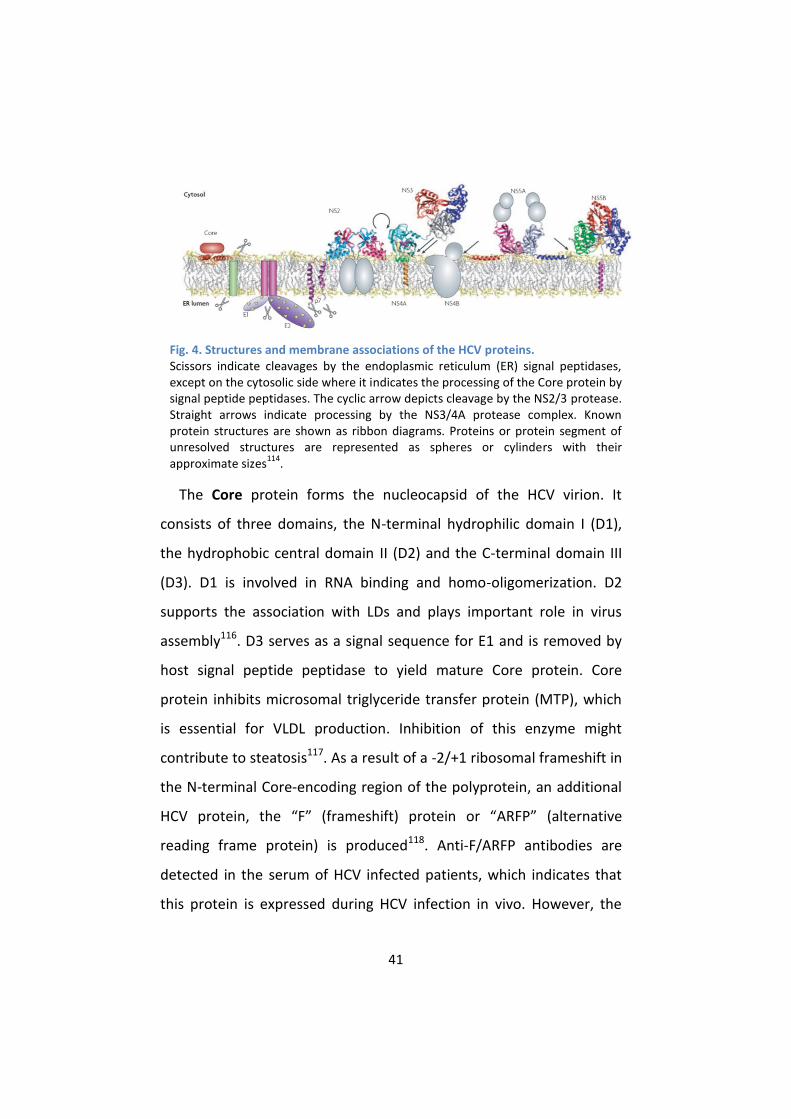
Fig. 4. Structures and membrane associations of the HCV proteins. Scissors indicate cleavages by the endoplasmic reticulum (ER) signal peptidases, except on the cytosolic side where it indicates the processing of the Core protein by signal peptide peptidases. The cyclic arrow depicts cleavage by the NS2/3 protease. Straight arrows indicate processing by the NS3/4A protease complex. Known protein structures are shown as ribbon diagrams. Proteins or protein segment of unresolved structures are represented as spheres or cylinders with their approximate sizes
114.
The Core protein forms the nucleocapsid of the HCV virion. It
consists of three domains, the N-terminal hydrophilic domain I (D1),
the hydrophobic central domain II (D2) and the C-terminal domain III
(D3). D1 is involved in RNA binding and homo-oligomerization. D2
supports the association with LDs and plays important role in virus
assembly116. D3 serves as a signal sequence for E1 and is removed by
host signal peptide peptidase to yield mature Core protein. Core
protein inhibits microsomal triglyceride transfer protein (MTP), which
is essential for VLDL production. Inhibition of this enzyme might
contribute to steatosis117. As a result of a -2/+1 ribosomal frameshift in
the N-terminal Core-encoding region of the polyprotein, an additional
HCV protein, the “F” (frameshift) protein or “ARFP” (alternative
reading frame protein) is produced118. Anti-F/ARFP antibodies are
detected in the serum of HCV infected patients, which indicates that
this protein is expressed during HCV infection in vivo. However, the
42
biological role of this protein remains to be elucidated. The Core
protein has been indicated to be involved in liver steatosis, fibrosis and
carcinogenesis in various mouse model systems119. HCV Core protein is
normally localized on the surface of cytoplasmic lipid droplets, which
are believed to represent sites of virus assembly and production of
nascent virions120. Overexpression of intracellular Core protein has
been reported cause abnormal accumulation and redistribution of lipid
droplets121, 122, as well to induce the production of free radicals and
apoptotic cell death123, all phenomena that could result in hepatocyte
injury.
The structural proteins E1 and E2 are highly N-glycosylated
transmembrane type I proteins located at the virus envelope and are
involved in the receptor-binding and membrane fusion of the virion124.
E1 and E2 contain a large N-terminal ectodomain and a C-terminal
hydrophobic region harboring a transmembrane domain. During
synthesis, the N-terminal domain is targeted towards the ER lumen
and modified by N-glycosylation, while the transmembrane domain
anchors the glycoproteins to the ER membranes. E1 and E2 contain up
to respectively 6 and 11 potential glycosylation sites. The glycans on
the envelope proteins are involved in the proper folding of these
proteins either directly or through interaction with calnexin, an ER
chaperone, and some of these glycans have been shown to be
essential for the entry function of E1 and E2125. Besides membrane
anchoring, the transmembrane domains also function as a signal
sequence, an ER retention signal and as a major regulator of the
assembly of the non-covalent E1-E2 heterodimer. This heterodimer is

43
the functional unit for productive HCV infection126. E2 plays an
important role in the interaction between HCV and its major cellular
receptors CD81 and SR-BI. Since E1 and E2 are located at the surface of
the HCV virion, they are targeted by neutralizing antibodies. These
neutralizing antibodies often inhibit the interaction(s) between E2 and
the CD81 receptor.
The HCV p7 protein belongs to the family of viroporins. Viroporins
are small, highly hydrophobic, virally-encoded proteins that
oligomerize to form channels or pores that increase the permeability
of the cell membrane to ions and other small molecules. Other known
viroporins include influenza A virus M2 protein and HIV-1 VPu protein.
P7 is a small transmembrane protein that forms hexameric
channels127. The N- and C-termini are oriented towards the ER lumen,
while the connecting loop is facing the cytoplasm128. It behaves as an
ion channel when reconstituted into artificial lipid membranes but its
function in infected cells is not well characterized. P7 is not required
for HCV replication in vitro but is essential for productive infection in
vitro and in vivo. It has been reported that p7 is involved in late steps
of virion assembly129. However, recent studies suggest that p7
increased the proton conductance in vesicles thereby preventing
acidification in otherwise acidic intracellular compartments130. This
loss of acidification is required for productive HCV infection.
The NS2/3 protease is required for intramolecular cleavage
between NS2 and NS3. The NS2 protein that is derived from the
cleavage of NS2/3 is an integral membrane protein that is targeted to
ER by its N-terminal hydrophobic domain. The C-terminal domain is a
44
cysteine protease domain and is capable to form dimers creating a
composite active site131. The function of mature NS2 is not yet fully
understood. It is not required for HCV replication, but it is essential for
the production of infectious virions132. Interestingly, the protease
activity is not necessary for virion assembly. Recently it was reported
that the NS2 protein serves as a key organizer of virus assembly by
connecting essential viral components133. It recruits the envelope
proteins to the assembly site and it crosstalks with non-structural
proteins for virus assembly134. The colocalization of NS2 with non-
structural proteins is influenced by the phosphorylation state of NS5A.
The HCV NS3 is a multifunctional protein135. The N-terminal domain of
NS3 forms, together with NS4A, a heterodimeric serine protease that
cleaves the downstream region of the HCV polyprotein into four
functional non-structural proteins (NS4A, NS4B, NS5A and NS5B). The
C-terminal domain exerts a helicase/NTPase activity that unwinds the
viral RNA. The NS3/4A protease is a serine protease and contains a
catalytic triad composed of His-57, Asp-81 and Ser-139. The minimal
NS3 domain required for full protease activity is mapped to 180
aminoacids at the N-terminus. In addition, the NS3/4A protease
activity is also responsible for an escape mechanism of innate immune
response. The intracellular presence of HCV RNA can be sensed by the
toll-like receptor 3 (TLR3) and by the retinoic acid-inducible gene I
(RIG-I). Activation of TLR3 of RIG-I leads to the activation of interferon
regulatory factor 3 (IRF3) which will transactivate the IFN-β receptor in
the nucleus. This signal pathway between activated TLR-3 or RIG-I and
activated IRF3 requires adaptor proteins TRIF (also known as TICAM-1)

45
or MAVS (also known as Cardif, IPS-1 or VISA). Both TRIF and MAVS are
substrates for the NS3/4A protease136. Cleavage of TRIF or MAVS
thereby blocks IRF3 activation. Furthermore, NS3 interacts directly
with NS5B and also with NS4B and NS5A via NS4A within the
replication complex. The NS3 helicase is a member of the superfamily
2 helicases and consists of three domains, of which domain 1 and 2 are
RecA-like domains shared with other motor proteins137. It can travel
along RNA or ssDNA in a 3’-5’ direction and is essential for viral RNA
replication. Various activities are attributed to NS3 helicase including
unwinding of duplex substrates, displacement of proteins bound to
nucleic acids, translocation along single-stranded nucleic acids, and
packaging of the RNA genome to form infectious viral particles138. The
unwinding of double- stranded RNA or single-stranded RNA with
secondary structures is coupled to ATP hydrolysis139. ATP binds the
helicase between domain 1 and 2, causing a conformational change
that leads to a decreased affinity for nucleic acids. This conformational
change allows the helicase to move like a motor. The NS3 helicase
unwinds RNA in an “inchworm” or “ratchet-like” fashion140. Genetic
variations in the HCV helicase influence its activity, which is shown by
the fact that adaptive mutations in HCV replicons frequently arise in
the helicase region. Interestingly, the enzymatic activities of protease
and helicase domains are functionally interdependent. The protease
domain promotes the binding of RNA substrate and it improves the
translocation efficiency of the helicase.
NS4A is an essential cofactor for the NS3 serine protease141. NS4A
activates the protease by inducing conformational changes and by
46
increasing binding affinity to the substrate. Furthermore, its
hydrophobic N-terminal part forms a transmembrane region that is
required for the targeting and anchoring of NS3 to the ER
membrane142. Recently, it was shown that the C-terminal acidic motif
of NS4A is essential for regulating the ATPase activity of the NS3
helicase143. Furthermore, at least for some HCV isolates, NS4A is
required for hyperphosphorylation of NS5A144.
NS4B is a highly hydrophobic protein consisting of an N-terminal
part, a central part harboring four transmembrane segments and a C-
terminal part145. It is an integral membrane protein with the N- and C-
terminal parts located on the cytosolic side of the ER membrane. The
N-terminal region contains two amphipathic α-helices of which the
second helix can traverse the membrane bilayer. Based on sequence
homology, it was proposed that the N-terminal part contains a leucine
zipper motif146. Very recently, the role of this leucine zipper domain in
the efficiency of HCV replication was demonstrated. The C-terminal
part of NS4B contains two palmitoylation sites. The importance of C-
terminal palmitoylation of NS4B remains to be clarified. NS4B plays a
central role in HCV RNA replication and assembly. It induces the
formation of the membranous web; this is the alteration of
intracellular membranes which contains the HCV replication complexes
and is essential for replication147. However, the mechanisms by which
NS4B induces web formation are unknown. NS4B also interacts with
other non-structural HCV proteins and with negative-strand HCV RNA.
Furthermore, NS4B was found to possess NTPase activity, suggested by
the presence of a nucleotide-binding motif similar to the P-loop of a
47
number of NTPases. However, this motif is not absolutely conserved
and a mutation within this motif was identified as a cell-culture
adaptive change in Con1 HCV replicons. Therefore, the enzymatic
activity of NS4B remains controversial145.
The NS5A protein is a pleiotropic protein that plays essential role in
the HCV viral lifecycle both by affecting the viral RNA replication as
well as by modulating the physiology of the host cell to favor viral
replication. It has no intrinsic enzymatic activity, but likely exerts its
functions through interactions with viral and cellular factors. NS5A
consists of three domains from which the structure of domain I has
been resolved148. Domain I contains an N-terminal amphipathic helix
that anchors NS5A to the membranes and lipid droplets and a RNA
binding domain. Crystal structure determination of domain I revealed
that a “claw-like” dimer is formed149. The groove generated by
multiple dimers is believed to serve as a railway for viral RNA,
tethering the RNA onto intracellular membranes. On the other hand,
an alternative arrangement of the domain I homodimer was reported
in which the proposed RNA binding cleft is absent150. It is conceivable
that the two dimer conformations observed thus far in crystallized
domain I reflect two different functional states of membrane-tethered
NS5A and that other configurations (even monomeric) could exist.
Domain II is poorly conserved, but is required for RNA replication151. It
is able to bind NS5B and protein kinase R (PKR), which is an IFN-
induced gene product. It also contains the so-called “interferon
sensitivity determining region” (ISDR). The ISDR has been correlated
with the efficacy of IFN treatment, but this finding remains

48
controversial152. Domain III is dispensable for replication, but is a key
factor for the assembly of HCV virions153. NS5A occurs in a basally
(p56) and hyperphosphorylated form (p58), with different putative
functions during HCV replication. It is speculated that
hyperphosphorylation serves as a molecular switch between
replication and assembly154. Mutations in NS5A that enhance viral
replication reduce NS5A hyperphosphorylation155. NS5A exerts
multiple functions by interacting with viral and cellular proteins. It was
shown that NS5A interacts with NS5B and that this interaction is
essential for viral replication. Furthermore, NS5A was found to interact
with Core protein onto lipid droplets156. This interaction seems to be
required for viral assembly. NS5A also interacts with several cellular
signal cascades157. The best studied interaction is that of NS5A and the
IFN signaling pathway. This interaction includes the binding and
inactivation of PKR. PKR is activated by binding to dsRNA, an
intermediate formed during viral RNA replication. When PKR is
activated, it shuts down protein translation. Furthermore, NS5A is also
thought to interact with pathways of cell proliferation, apoptosis,
stress-response and others.
Very recently, many groups has demonstrated the physical and
functional interaction between NS5A and the phosphatidylinositol 4-
kinase PI4KIIIα. The interaction between NS5A and PI4KIIIα was
observed for the first time in a Yeast two-hybrid screening158. Then, a
number of groups have used small interfering RNA (siRNA) screens to
identify cellular cofactors of HCV infection. Although there has been
notable divergence in the array of host cofactors of HCV infection
49
identified in these screens, a remarkable constant has been the
identification of phosphatidylinositol (PI) 4-kinase III alpha (PI4KIIIα) as
an essential cofactor of HCV RNA replication159-162. This finding is
further supported by data showing that the kinase inhibitors
wortmannin or LY294002, used at doses known to favor inhibition of
type III PI4Ks, significantly decreased HCV replication159, 161. PI kinases
and the lipids they phosphorylate are essential regulators of
membrane trafficking and protein sorting. PI4KIIIα is one of four
cellular PI 4-kinases that all function to phosphorylate
phosphatidylinositol at the 4 position of an inositol head group, which
then serves as a beacon to recruit proteins containing lipid-binding
motifs with affinity for PI4P (reviewed in references 163, 164 and 165).
The PI 4-kinases differ with respect to their subcellular localization,
thus generating pools of PI4P at specific membrane compartments.
PI4KIIIα is the predominant endoplasmic reticulum (ER) resident PI 4-
kinase163. In HCV-infected cells, PI4KIIIα colocalizes with HCV NS5A and
viral double-stranded RNA, the HCV replication intermediate,
suggesting a role in replication complex formation or function159.
All positive-stranded RNA viruses reorganize cellular membranes to
create their sites of replication. In the case of HCV, infection produces
an accumulation of heterogeneous, ER-associated vesicular structures
that has been termed the membranous web147, 166, 167. This is thought
to be the site of HCV replication, since de novo-synthesized viral RNA
colocalizes with these structures166. The expression of NS4B in the
absence of other viral proteins produces structures reminiscent of the
membranous web147, suggesting that it is a primary mediator of

50
membrane reorganization. PI4KIIIα is also implicated in HCV replication
complex formation. siRNAs that reduce PI4KIIIα accumulation appear
to perturb membranous web formation. In cell lines expressing the
HCV polyprotein, PI4KIIIα siRNAs alter the localization of the HCV NS5A
protein161, while membranous webs are difficult to detect in HCV-
infected cells pretreated with PI4KIIIα siRNAs. This led to the
hypothesis that HCV may hijack PI4KIIIα to generate PI4P, which would
serve to nucleate viral and cellular proteins to ER membranes for HCV
replication complex formation159.
The NS5B protein is a RNA-dependent RNA polymerase (RdRp),
which catalyzes the synthesis of a complementary minus-strand RNA,
using the viral RNA genome as a template, and the subsequent
synthesis of genomic plus-strand RNA form this minus-strand RNA
template. The HCV RdRp can initiate the synthesis of viral RNA de
novo, without the need of a primer, which is also thought to be the
mode of action in vivo168. It has the typical polymerase structure which
is frequently compared with a right hand, where the palm domain
contains the active site of the enzyme and where the fingers and the
thumb are responsible for the interaction with the RNA. Specific
characteristics of the viral RdRps include the fingertips; these are two
loops that extend from the finger domain and that make contact with
the thumb domain169. These fingertips completely encircle the active
site, ensuring a closed conformation. The catalytic site in the palm
domain is the most conserved region in the RdRp. In contrast, the
thumb domain is the most diverse domain among viral RdRps170. The
HCV thumb domain contains protruding extension that folds back into
51
the molecule171. These features make primer-dependent initiation
impossible. A conformational change to an “open” form is essential to
switch between initiation and elongation. The C-terminal part of the
RdRp anchors the protein to the membrane172. This C-terminal part is
dispensable for RdRp activity in vitro, but indispensable for RNA
replication in cells.
2. Genomic heterogeneity and classification systems of HCV
The HCV genome shows remarkable sequence variation generated
by the lack of proofreading activity by the RNA polymerase and a high
in vivo productive rate of an estimated 1012 virions per day173. To
encompass this diversity, HCV has been classified in genotypes and
subtypes. Initially, genotypes were considered to differ more than 20%
at the nucleotide level and more than 15% at the amino acid level.
However, phylogenetic analyses indicated that genotype 10 is closely
related to genotype 3 and genotype 7, 8, 9 and 11 to genotype 6.
Therefore, a new consensus nomenclature system was proposed to be
used for HCV classification174. According to this system, HCV is
classified into genotypes on the basis of < 70% similarity of nucleotide
sequence and phylogenetic relationship. The more closely related HCV
strains (75-80% sequence similarity) within some genotypes are
designated subtypes. Genotypes are numbered in order of discovery
and subtypes are assigned lowercase letters (Table 1). Nowadays, HCV
is classified into 6 major genotypes and more than 90 subtypes.
Sequence variation between the HCV genomes was on average
0.9%, indicative of the presence of HCV quasispecies in hepatitis C
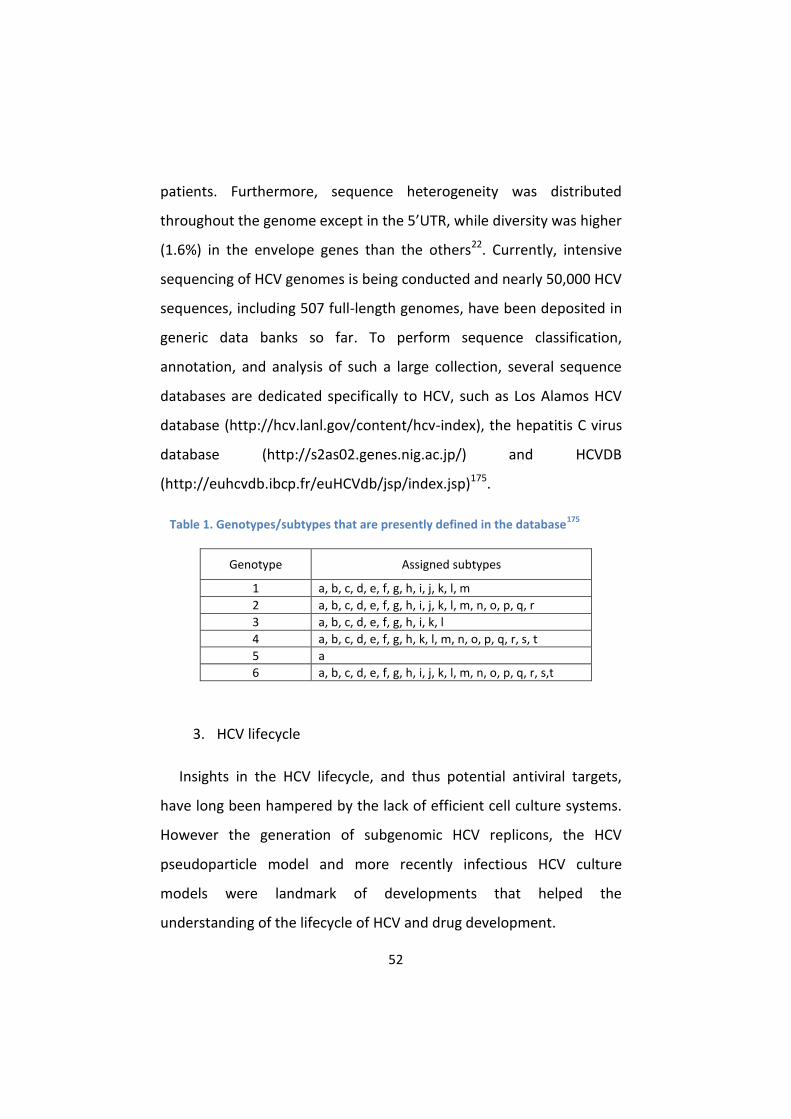
52
patients. Furthermore, sequence heterogeneity was distributed
throughout the genome except in the 5’UTR, while diversity was higher
(1.6%) in the envelope genes than the others22. Currently, intensive
sequencing of HCV genomes is being conducted and nearly 50,000 HCV
sequences, including 507 full-length genomes, have been deposited in
generic data banks so far. To perform sequence classification,
annotation, and analysis of such a large collection, several sequence
databases are dedicated specifically to HCV, such as Los Alamos HCV
database (http://hcv.lanl.gov/content/hcv-index), the hepatitis C virus
database (http://s2as02.genes.nig.ac.jp/) and HCVDB
(http://euhcvdb.ibcp.fr/euHCVdb/jsp/index.jsp)175.
Table 1. Genotypes/subtypes that are presently defined in the database175
Genotype Assigned subtypes
1 a, b, c, d, e, f, g, h, i, j, k, l, m
2 a, b, c, d, e, f, g, h, i, j, k, l, m, n, o, p, q, r
3 a, b, c, d, e, f, g, h, i, k, l
4 a, b, c, d, e, f, g, h, k, l, m, n, o, p, q, r, s, t
5 a
6 a, b, c, d, e, f, g, h, i, j, k, l, m, n, o, p, q, r, s,t
3. HCV lifecycle
Insights in the HCV lifecycle, and thus potential antiviral targets,
have long been hampered by the lack of efficient cell culture systems.
However the generation of subgenomic HCV replicons, the HCV
pseudoparticle model and more recently infectious HCV culture
models were landmark of developments that helped the
understanding of the lifecycle of HCV and drug development.

53
Fig. 5. Schematic representation of the lifecycle of hepatitis C virus.
Virus binding and internalization (a); cytoplasmic release and uncoating (b); IRES-mediated translation and polyprotein processing (c); RNA replication (d); packaging and assembly (e); virion maturation and release (f). The topology of HCV structural and non-structural proteins at the endoplasmic reticulum membrane is shown schematically. HCV RNA replication occurs in a specific membrane alteration, the membranous web. Note that IRES-mediated translation and polyprotein processing, as well as membranous web formation and RNA replication, which are illustrated here as separate steps for simplicity, might occur in a tightly coupled fashion. IRES, internal ribosome entry site. Adapted from
114.
3.1. HCV entry
HCV entry is a complex and multi-step process requiring a set of
entry proteins176. In the early stage of HCV entry, HCV virions are
concentrated at the cell surface by non-specific attachment factors.
Glycosaminoglycans seem to be the initial attachment sites for HCV.
Also the LDL receptor is thought to be involved in early attachment of
HCV, although this finding remains somehow controversial. Following
initial attachment, HCV virions bind virus-specific receptors. Currently,
a number of receptors essential for HCV entry are identified: the
54
scavenger receptor class B type I (SR-BI)177, the tetraspanin CD81178,
and tight junction proteins claudin-1 (CLDN1)179 and Occludin180.Very
recently, new players in HCV entry were uncovered: receptor tyrosine
kinases (RTKs)181.
HCV associates with very-low-density and low-density lipoproteins
(LPs) in the bloodstream. It binds the basolateral surface of
hepatocytes through glycosaminoglycans (GAGs) and the low-density
lipoprotein receptor (LDLR). Both SR-BI and CD81 bind HCV
glycoprotein E2 and are necessary but not sufficient for HCV entry. SR-
BI is a lipoprotein receptor that is responsible for the selective uptake
of cholesteryl esters from HDL (high density lipoprotein) particles. In
addition, SR-BI plays an important role in VLDL catabolism. SR-BI acts
as a post-binding receptor, since SR-BI antibodies inhibit HCV infection
when added 60 min following virus binding. CD81 belongs to the family
of tetraspanins which are ubiquitously expressed. Similar to SR-BI, it
acts as a post-binding entry factor, since anti-CD81 antibodies inhibit
HCV infection only after virus attachment. Furthermore, CD81 actively
promotes infection upon HCV binding by triggering signaling cascades
that are important for virus entry176. Very recently, RTKs were
identified as cofactors required for HCV entry, and it was shown how
pharmacological blockage of these molecules can prevent viral
infection in vitro and in vivo. Clinically approved inhibitors exist for two
of these kinases: epidermal growth factor receptor (EGFR) and ephrin
receptor A2 (EphA2). EGFR activation promotes CD81–CLDN1
interactions and membrane fusion, thereby enhancing productive HCV
uptake. RTKs, upon dimerization and autophosphorylation, provide

55
signals inducing proliferation, migration, morphogenesis and other
fundamental cellular processes181. CLDN1 is predominantly expressed
in the liver, where it forms networks at tight junctions. Silencing of
CLDN1 inhibits HCV infection in susceptible cells179. Nevertheless,
there is still no evidence for a direct interaction between the virus and
CLDN1. CLDN1 plays a role in the post-binding phase subsequent to
HCV binding to CD81 and SR-BI. Analogous to other viruses like
coxsackievirus B, it is speculated that the interaction with CLDN1 takes
place after migration of the virus-CD81/SR-BI complex to tight
junctions182. Occludin is expressed in tight junctions of polarized cells
and is also involved in the post-binding phase of HCV entry180.
Engagement by the virus of SR-BI, the tetraspanin CD81 and the
tight junction proteins claudin-1 and occludin leads to clathrin-
mediated endocytosis of virions183, delivering HCV-receptor complexes
to early endosomes. The low pH in the endosome triggers fusion of the
virion envelope with the membrane of the early endosome particle.
Membrane fusion is catalyzed by fusion peptides embedded in the
viral glycoproteins. For related viruses like flaviviruses, the structural
basis for low pH-induced membrane fusion has been elucidated184. The
envelope proteins of these viruses contain an internal fusion peptide,
which is exposed during membrane reorganization mediated by low
pH (class II fusion). The scaffolds of class II fusion proteins are
remarkably similar. Therefore it has been suggested that the entry of
all Flaviviridae, including HCV, may include a class II fusion step. The
nature of the fusion protein of HCV is not known yet. There is some
evidence that E2 is the fusion protein; however, also E1 has been
56
suggested as possible fusion protein. Following membrane fusion, the
viral RNA is released into the cyotosol185. How the viral RNA is exactly
released from the nucleocapsid is not known. It was shown for other
positive strand RNA viruses (alphaviruses) that binding of ribosomes to
the viral genome supports RNA release186.
3.2. Translation and replication
The released positive RNA genome is translated by a cap-
independent mechanism at the rough ER by the host machinery into
one polyprotein. This translation process is mediated by the IRES
located in the 5’ UTR. The translated polyprotein is cleaved co- and
post-translationally by cellular and viral proteases into ten different
proteins. Similar to related viruses, expression of HCV proteins triggers
intracellular membrane alterations166. This specific virus-induced
membrane structure is referred to as the membranous web. The
formation of the membranous web can be induced by solely
expressing HCV NS4B147. The membranous web serves as a scaffold for
the assembly of the viral replication machinery. Based on analogy of
the replication of related viruses like Dengue virus, it is hypothesized
that the viral replication complexes are located in invaginations of ER-
derived membranes187, 188. Viral RNA is thereby protected from
degradation and recognition by intracellular sensors such as RIG-I is
avoided. However, this hypothesis remains to be confirmed for HCV.
Newly synthesized positive HCV RNA can either be used for translation,
replication or encapsidation.
57
3.3. Assembly and release
Newly synthesized HCV RNA genomes can be assembled into new
virions189. Viral proteins involved in this assembly process are the core
protein, the envelope proteins E1 and E2, viroporin p7, and non-
structural proteins NS2 and NS5A153, 156, 190. Lipid droplets and the VLDL
assembly pathway are major contributors of the host cell to virion
assembly. The mature HCV virion consists of a nucleocapsid, composed
of core proteins and the viral genome, and an outer envelope,
containing a lipid membrane and the envelope proteins. The early
stage of assembly, i.e. nucleocapsid formation, involves
oligomerization of the core and encapsidation of the viral genome.
Currently it is not clear where the nucleocapsid formation initiates. In a
first model, nucleocapsid formation is initiated at the surface of
cytosolic LDs. Viral RNA is delivered to the core protein via NS5A that is
also mobilized onto LDs or remains bound to the ER. In a second
model, assembly initiates at the ER membrane. Core protein is initially
transferred to LDs but is re-recruited to ER membranes at the
assembly sites where it interacts with NS5A.
Very recently, it was demonstrated that a concerted action of HCV
p7 and NS2 regulates Core localization at the ER and virus assembly.
The JFH1 HCV strain replicates and assembles in association with LD-
associated membranes, around which viral core protein is
predominantly detected. In contrast, despite its intrinsic capacity to
localize to LDs when expressed individually, they found that the Core
protein of the high-titer Jc1 recombinant virus was hardly detected on
58
LDs of cell culture-grown HCV (HCVcc)-infected cells, but was mainly
localized at ER membranes where it colocalized with the HCV envelope
glycoproteins. Furthermore, high-titer cell culture-adapted JFH-1 virus,
obtained after long-term culture in Huh7.5 cells, exhibited an ER-
localized core in contrast to non-adapted JFH-1 virus, strengthening
the hypothesis that ER localization of core is required for efficient HCV
assembly. They also demonstrated that p7 and NS2 are HCV strain-
specific factors that govern the recruitment of core protein from LDs to
ER assembly sites. Indeed, using expression constructs and HCVcc
recombinant genomes, they found that p7 is sufficient to induce Core
localization at the ER, independently of its ion-channel activity.
Importantly, the combined expression of JFH1 or Jc1 p7 and NS2
induced the same differential core subcellular localization detected in
JFH1- vs. Jc1-infected cells. Finally, results obtained by expressing p7-
NS2 chimeras between either virus type indicated that compatibilities
between the p7 and the first NS2 trans-membrane domains is required
to induce core-ER localization and assembly of extra- and intra-cellular
infectious viral particles191.
Another group studied the recruitment of core from LD into nascent
virus particles. To investigate the kinetics of core trafficking, they
developed methods to image functional core protein in live, virus-
producing cells. During the peak of virus assembly, core formed
polarized caps on large, immotile LDs, adjacent to putative sites of
assembly. In addition, LD-independent, motile puncta of core were
found to traffic along microtubules. Importantly, core was recruited

59
from LDs into these puncta, and interaction between the viral NS2 and
NS3-4A proteins was essential for this recruitment process192.
The late stage of assembly, i.e. the maturation and release of HCV
virions, is closely associated with the VLDL pathway193, 194. The
nucleocapsid can be incorporated in the precursor of VLDL, the luminal
LDs. How the envelope proteins are incorporated into virions is not
clear. Recently it was reported that NS2 might “crosstalk” between
envelope glycoproteins and non-structural proteins by forming
multiple protein-protein interactions133. Mature HCV virions contain
apoB, apoE and eventually other apolipoproteins. After maturation,
HCV virions are transported and secreted along the VLDL secretory
pathway. Therefore, the structure of HCV virions resembles that of
triglyceride-rich lipoproteins containing a core of neutral lipids
surrounded by a phospholipid monolayer and stabilized by
apolipoproteins195.
4. The discovery of HCV defective particles
HCV subgenomes were firstly isolated from four patients among 23
chronic active hepatitis C patients, in two cases HCC, and in one case
acute fulminant hepatitis following liver transplantation196. The regions
of deletions in individual subgenomes differed from each other;
however, they all shared common structural features. They contained
in-frame deletions from E1 to NS2, and retained complete sequences
for both core protein and NS2 metal-dependent cysteine protease
domain, encoding the entire NS3-NS5B proteins that are essential for
RNA replication. The HCV polyprotein encoded by the subgenome was
60
processed as the standard HCV polyprotein generating core, a
glycosylated E1-NS2 fusion protein that was susceptible to
endonuclease H treatment, and full size NS3 in cells carrying the cDNA
of the subgenome196. These data suggested that the cysteine protease
activity of NS2 should be involved in the processing of HCV
polyproteins from the HCV subgenome, and emphasized the
importance of domain preservation in the HCV subgenome.
The first question that arises after the discovery of HCV subgenome
was if they could work as DI (defective interfering) genomes. DI viruses
are subgenomic mutants derived from the parental virus that lack the
essential viral components for virus particle production; the DI virion is
therefore produced only under certain conditions. The replication of DI
virus is trans-complemented with the materials essential for virion
production from a helper virus, which is usually the parental virus. DI
viruses often interfere with parental virus replication by competing
with the materials essential for replication and/or by their advantages
in replication, they can also facilitate the establishment and
maintenance of persistent viral infection of cells197.
The HCV subgenomes have a close resemblance with those of
naturally-arisen DI genomes identified in Flaviviridae. DI genomes have
been identified in pestiviruses, bovine viral diarrhea virus (BVDV)198,
199, classical swine fever virus (CSFV)200 and a flavivirus Murray Valley
encephalitis virus (MVE)201. The structures of MVE DI genomes were
most similar to those of the HCV subgenomes.
Synthetic subgenomic RNA replicons of HCV indicated that NS3-
NS5B proteins are both essential and sufficient viral components for
61
HCV RNA replication202 and, with the exception of NS5A, these
elements cannot be complemented in trans due to a limited trans-
complementation of viral components in RNA replication of other
flaviviruses203. In cell cultures it was possible to replicate artificial HCV
subgenomic RNA with E1 and E2 sequence deletions204, 205. It was
demonstrated that they can replicate autonomously in hepatoma
cells206.
Long term (at least 2 years) persistence of the HCV subgenome in a
patient’s blood with the standard HCV genome, and the approximate
120-day life of hepatocyte, indicate that the HCV subgenome circulates
in blood and re-infects naïve hepatocytes. The fact that virus RNA and
the core antigen in the serum of patients with an HCV subgenome
were fractionated approximately 1.16 g/ml after buoyant density
gradient centrifugation196 suggested that the HCV subgenome should
be packaged within a virion. The HCV RNA replication lacking envelope
proteins did not produce an infectious virus204. All these data indicate
that the standard HCV could function as a helper virus and trans-
complement the envelope proteins, E1 and E2. This hypothesis was
recently demonstrated in vitro by Pacini et al206.
Data collected so far about the ratio of HCV subgenome titer to
those of the standard genome indicate that they were approximately
5:1196. It indicates that the competition of the envelope proteins and
the advantage of RNA replication of the HCV subgenome would cause
interference with standard HCV replication. The 20% shortage in
length of the HCV subgenome might give the subgenome an advantage
62
in virus replication, while the processing rate of HCV RNA polymerase
is independent of the sequence.
The HCV subgenome offers clues to HCV packaging pathways. It was
shown that core proteins assembled to form a capsid-like structure,
and that core-E1-E2 did package HCV RNA to form virus-like particles
(VLP) in many types of cells207. The presence of HCV subgenome
circulating in the blood also suggests that the HCV subgenome lacking
an E1-E2 sequence should be packaged within a virion. Based on these
observations, it should be reasonable to predict that one of the
packaging signal sequences of HCV RNA should be localized in the core
sequence.
According to the data collected so far, it should be noted that the
ratio of HCV subgenome per HCV standard genome in liver is
significantly higher than those in blood, suggesting that the
subgenome packaging efficiency could be lower than that of the
standard genome, or that the number of cells infected with both
genomes might be limited. It is also possible that the coupling of RNA
replication with packaging of the RNA genome affects HCV packaging
efficiency208.
The comparison between the HCV subgenomes and the MVE DI
subgenomes illustrates another interesting feature of DI genomes:
both subgenomes carry entire core sequences. The coincidence of
these structural features suggests the importance of the core region in
viral replication. Although one possible interpretation is the packaging
signal, as discussed below, there remain other possible explanations
for the core sequence preservation in the subgenomes: core is
63
essential for correct processing of the following proteins in the context
of the HCV subgenome, or core might be required for RNA
recombination to generate subgenomes.
The heterogeneous nature of HCV particles in patients suggested
another pathway for HCV subgenome packaging. Nucleocapsids
without envelopes were demonstrated in hepatitis patients209, in
addition to particles comprising HCV core protein, HCV RNA and
lipoproteins in very low-density fractions of the patient’s serum.210
HCV core proteins also interact with HCV RNA211 and assemble into
capsid-like particles212. These findings suggest that the HCV
subgenome might be released without the assistance of a helper virus.
The HCV subgenomes highlight an inconsistency with respect to the
p7 gene product. Although p7 was thought to be indispensable for
infection in a chimpanzee intrahepatic transfection system213, the HCV
subgenome is a replication entity. How might this apparent
discrepancy be explained? In the VLP production system, using
hepatoma cells infected with recombinant baculovirus, efficient
expression and secretion of structural proteins from cells was achieved
with p7 and NS2; however, this was not the case in the VLP production
system using insect cells207. Almost all the issues mentioned in this
section should be addressed by investigations using another reliable
model system to monitor viral production, such as an in vitro HCV
replication system204.
From the molecular point of view, many questions are still opened
about how HCV subgenomes arise from the standard genome.
Phylogenetic analysis and blast searches of quasispecies of HCV

64
subgenomes and the coexisting standard genomes suggest that they
share common ancestral HCV quasispecies196. Other types of HCV
subgenomic mutations were recovered from five cases among 23
hepatocarcinoma patients. Almost all were minor populations of the
HCV RNA quasispecies in patient specimen, and had out-of-frame
deletions that were not conserved between isolates. However, in their
putative parental HCV RNA, nearly identical sequences (one or two
nucleotides in length) were found in the deletion donor and acceptor
regions196. These homologous sequences could serve the cross-over
sites where strand switching occurs during RNA genome replication,
which was one of the proposed models of the RNA virus
recombination214. Cross-over sites in short stretches of similar
sequences were implicated to be involved in BVDV RNA
recombination215. In the case of the HCV subgenomes, there is no such
identical sequence found at the deletion boundaries in the putative
parental genome. It has been observed that RNA recombination often
occurred around the regions with a higher ordered RNA secondary
structure, which hindered the RNA replication214. A computational
analysis predicted the conserved RNA structures in both core and NS5B
sequences216. The functions of sequences with a conserved folding
motif in the core are controversial; however, these sequences might
also be involved in RNA recombination and packaging events.
Intergenotypic HCV recombinants exhibiting cross-over near the NS2-
NS3 junctions have been isolated217, and the recombinants between DI
and the standard genome were reported in BVDV215. Recombination
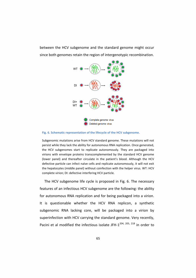
65
between the HCV subgenome and the standard genome might occur
since both genomes retain the region of intergenotypic recombination.
Fig. 6. Schematic representation of the lifecycle of the HCV subgenome.
Subgenomic mutations arise from HCV standard genome. These mutations will not
persist while they lack the ability for autonomous RNA replication. Once generated,
the HCV subgenomes start to replicate autonomously. They are packaged into
virions with envelope proteins transcomplemented by the standard HCV genome
(lower panel) and thereafter circulate in the patient’s blood. Although the HCV
defective particle can infect naïve cells and replicate autonomously, it will not exit
the hepatocytes (middle panel) without coinfection with the helper virus. WT: HCV
complete virion; DI: defective interfering HCV particle.
The HCV subgenome life cycle is proposed in Fig. 6. The necessary
features of an infectious HCV subgenome are the following: the ability
for autonomous RNA replication and for being packaged into a virion.
It is questionable whether the HCV RNA replicon, a synthetic
subgenomic RNA lacking core, will be packaged into a virion by
superinfection with HCV carrying the standard genome. Very recently,
Pacini et al modified the infectious isolate JFH-1204, 205, 218 in order to
66
recapitulate in vitro the genetic structure of two of the most
representative in-frame natural subgenomic deletion-containing RNAs
found circulating in patients206. Using this system, they analyzed
natural subgenomic variants for their ability to replicate autonomously
and demonstrated that the natural subgenomic deletion mutants are
replication competent and are trans-packaged into infectious virions
when coexpressed together with the wild-type virus. Furthermore,
their data suggest that the presence of the NS2 protease domain is
required in order to generate the correct NS3 N terminus, required for
RNA replication. Unexpectedly, the presence of NS2 generates, in turn,
a strict cis requirement for the core region in order to allow efficient
trans-packaging of the subgenomic RNA, revealing a complex interplay
between the NS2 and the core viral genes.
5. Host-pathogen interactions
Viral lifecycle as that of HCV completely relies on host cell
infrastructure, presupposing that the virus has evolved mechanisms to
utilize and control all cellular molecules or pathways required for viral
lifecycle. Hence, HCV must have acquired the ability to gain access to
key pathways controlling processes, such as cell growth, apoptosis and
protein synthesis, which are all considered to also be crucial for liver
regeneration. This occurs in a balanced way permitting persistent
replication of viral genomes and production of infectious particles
without endangering host cell viability and survival. In particular during
the last decade, accumulating evidence indicates that HCV utilizes
signaling pathways of the host with major impact on cellular growth,
67
viability, cell cycle or cellular metabolism, such as epidermal growth
factor-receptor mediated signals, the PI3K/Akt cascade or the family of
Src kinases. Furthermore, HCV specifically interacts with parts of the
cellular machinery involved in protein translation, processing,
maturation and transport, such as components of the translation
complex, the heat shock protein family, the immunophilins or the
vesicle-associated membrane protein-associated proteins A and B
(VAMP-A and B)219.
5.1. Lipid metabolism
A particular hallmark of HCV is the intimate link with the host lipid
metabolism. HCV virions were found to circulate in the blood in
complex with lipoproteins220. These lipoviroparticles (LVP) are highly
enriched in triglycerides and contain apoB and apoE210. The circulating
LVPs are a heterogeneous population with buoyant densities ranging
between 1.03 to 1.2 g/ml. The infectivity of HCV-positive inocula is
inversely correlated with the buoyant density of LVPs221.
Since HCV virions are associated with host lipoproteins, it seems
logical that HCV entry is connected to the host lipid metabolism.
Lipoprotein receptors (LDL receptor, SR-BI receptor) have been
reported to be involved in HCV entry. Furthermore, ligands of SR-BI
can modulate HCV entry. For instance, HDL enhance HCV entry,
whereas oxidized LDL inhibits HCV entry222. Also the lipid composition
of the plasma membrane influences HCV entry. Cholesterol depletion
from the plasma membrane decreases HCV entry by reducing the
expression of CD81 receptors at the cell surface223.

68
HCV exploits the VLDL assembly and secretion pathway to be
released from hepatocytes. Furthermore, it has been shown that lipid
droplets (LDs) play an important role in HCV virion assembly. LDs are
intracellular storage vesicles that contain triacylglycerols and
cholesteryl esters surrounded by a phospholipid monolayer. HCV core
protein accumulates around these LDs. This association is a peculiarity
of the Hepacivirus genus in the Flaviviridae family and is essential for
the recruitment of other viral proteins like NS5A and NS3 and for virus
production224.
HCV also requires elements of the cholesterol and fatty acids
biosynthetic pathways for efficient replication. Fatty acids can
stimulate or inhibit HCV replication depending on their degree of
saturation. Polyunsatured fatty acids inhibit HCV replication, whereas
saturated and mono-unsaturated fatty acids stimulate it225. Fatty acids
can be used as prosthetic groups that may facilitate the anchoring of
viral non-structural proteins to membranes. For example, NS4B
undergoes palmitoylation at two cysteine residues in the C-terminal
region. These lipid modifications were shown to be necessary for
protein-protein interactions226. Thus, palmitoylation can be a post-
translational modification required for the life cycle of HCV.
Statins, which are widely used for the treatment of
hypercholesterolemia, have also been reported to inhibit HCV
replication. Statins inhibit 3-hydroxy-3-methylglutaryl coenzyme A
(HMG-CoA) reductase, the rate-limiting enzyme in cholesterol
biosynthesis in the liver. The HMG-CoA reductase catalyzes the
conversion of HMG-CoA into mevalonic acid. Besides their cholesterol-

69
lowering effect, statins were shown to inhibit the replication of HCV
subgenomic genotype 1b replicons and to suppress RNA replication of
infectious JFH-1 HCV227, 228. The precise mechanism of the anti-HCV
activity of statins has not yet been unraveled. The anti-HCV activity of
statins may result from inhibition of geranylgeranylation of cellular
proteins rather than the inhibition of cholesterol synthesis225.
Geranylgeranylation is a post-translational modification that covalently
attaches geranylgeranyl groups to various cellular proteins to facilitate
their membrane association. These geranylgeranyl groups are
isoprenoids synthesized in the cholesterol biosynthesis pathway. More
recently, FBL2 has been reported to be a host target for
geranylgeranylation229. Geranylgeranylation of FBL2 appears to be
critical for HCV replication because the association between FBL2 and
NS5A, an interaction that is a prerequisite for HCV replication, depends
on geranylgeranylation of FBL2. In vivo results of statin monotherapy
for the treatment of HCV infected patients have yielded conflicting
results so far. Neither atorvastatin (after conventional 12-week
therapy) nor rosuvastatin resulted in a reduction in viral load230.
Nevertheless, fluvastatin was found to inhibit HCV RNA replication in
HCV infected patients in a recently published study. The drug was well
tolerated and resulted at relatively low doses (20-80 mg daily) in a
transient reduction in viral load (-0.5 to -1.75 log10, 2-5 weeks).
However, higher doses o fluvastatin did not reduce viral load231. A
recent retrospective analysis demonstrated that statin use in
combination with PEG-IFN and ribavirin was associated with a
significantly higher sustained virological response (SVR) and with a

70
higher viral response at week 4 and 12232. A pilot study evaluating the
combination of fluvastatin (20 mg daily) with PEG-IFN/ribavirin
revealed that fluvastatin could be used safely to increase the response
to PEG-IFN and ribavirin233. There is today obviously no compelling
evidence that statins, used in monotherapy, may result in a marked
reduction in HCV viral load in chronically infected patients. However,
statins may have the potential to increase the efficacy of current or
future HCV therapy.
The importance of HCV and host lipid interactions has also been
demonstrated clinically. Hepatitis C infection is associated with
decreased cholesterol and LDL levels. When HCV is cleared by antiviral
treatment, serum cholesterol levels are elevated again234. It is
hypothesized that HCV, by interrupting cholesterol synthesis and using
host lipids for replication, decreases circulating lipids. Furthermore,
the prevalence of steatosis in chronic hepatitis C is approximately
40%235.
5.2. Cellular stress, apoptosis, cell cycle
Apoptosis is central for the control and elimination of viral
infections. In HCV-infected liver, however, despite enhanced
hepatocyte apoptosis, viral persistence is observed.
Viral proteins interfere with the cellular apoptotic signaling pathway
and block key cellular elements of the host cell. Until recently, the lack
of an infectious HCV tissue culture system did not allow studying the
impact of HCV infection on hepatocyte apoptosis. Overall, the data
regarding the role of different HCV proteins are controversial and
71
ascribe to a given viral protein pro- and anti-apoptotic effects,
depending on the experimental system used. Since in most models
viral proteins are overexpressed by non-viral promoters, for virtually
all HCV proteins a pro-apoptotic effect has been described. Apart from
the unphysiological expression of viral proteins, these models further
lack the balance of intracellular viral expression of the different HCV
proteins and their interactions. Especially in HCV infection, intracellular
viral protein expression is very low236.
Further, HCV is genetically highly variable and exists as quasispecies
in a given patient. Different pro- and anti-apoptotic effects of the HCV
core protein from an individual patient have been described237,
suggesting special properties of different quasispecies proteins. These
protein differences may explain in part the different effects of viral
proteins on apoptosis. Studies of the contribution of genotypes or
quasispecies to the effects on apoptosis are largely missing. Further,
experiments designed to study the impact of HCV infection on
hepatocyte apoptosis must also consider the interactions between the
different HCV proteins. Therefore, only models based on the complete
and infectious virus may reflect to some extent the in vivo situation.
Very recently, gene expression profiling during multiple time-points
of acute HCV infection of cultured Huh-7.5 cells was performed to gain
insight into the cellular mechanism of HCV-associated cytopathic
effect. Maximal induction of cell-death–related genes and appearance
of activated caspase-3 in HCV-infected cells coincided with peak viral
replication, suggesting a link between viral load and apoptosis. Gene
ontology analysis revealed that many of the cell-death genes function

72
to induce apoptosis in response to cell cycle arrest. Labeling of dividing
cells in culture followed by flow cytometry also demonstrated the
presence of significantly fewer cells in S-phase in HCV-infected relative
to mock cultures, suggesting HCV infection is associated with delayed
cell cycle progression. Regulation of numerous genes involved in anti-
oxidative stress response and TGF-b1 signaling suggest these as
possible causes of delayed cell cycle progression. Significantly, a subset
of cell-death genes regulated during in vitro HCV infection was
similarly regulated specifically in liver tissue from a cohort of HCV-
infected liver transplant patients with rapidly progressive fibrosis.
Collectively, these data suggest that HCV mediates direct cytopathic
effects through deregulation of the cell cycle and that this process may
contribute to liver disease progression238.
5.3. Other host factors important for HCV lifecycle: results from
siRNA screenings and other studies – The emerging role of
PI4KIIIα
Like all viruses, HCV relies heavily on the host cell to replicate. As
such, there is significant interest in trying to identify cellular genes that
are required for HCV infection, both to understand the basic biology of
the HCV life cycle and to unearth potential new therapeutic targets.
Small or short interfering RNAs (siRNAs) mediate the degradation of
complementary mRNA that results in a specific silencing of gene
expression. siRNA mimics cellular miRNA that consists of small double
stranded RNA strands and regulates gene expression by degrading the
complementary mRNA. A key protein for the generation of these
73
molecules is DICER, an RNase enzyme that is responsible for the
production of short interfering RNAs and microRNAs239. Specific siRNAs
targeting the expression of individual cellular cofactors are successfully
applied as tools to study the impact of defined host factors for HCV
entry176 and replication240. Furthermore, functional RNA interference
(RNAi) screens have been shown to represent a powerful tool to
identify novel cellular cofactors for pathogen-host interactions241.
Up to now, nine limited and two genome-wide siRNA screens
designed to identify cellular cofactors required for HCV infection and
based on different HCV replication models have been performed159-162,
240, 242-247. However, the overlap of identified genes is very low, which
may be due to different experimental conditions, reagents, or hit
calling criteria.
There are numerous reasons for the varied results, including
differences in RNAi libraries, cells, transfection protocols, HCV
genotypes and HCV replication systems (sub-genomic replicons versus
infectious HCV).
The most consistent hit identified by all siRNA screens as HCV
dependency factor was phosphatidylinositol 4-kinase IIIa (PI4KIIIα)159-
162, 243, 245, 247, 248. Encoded by the PIK4CA gene and localized primarily
to the endoplasmic reticulum (ER), this enzyme is one of four kinases
(PI4KIIα, PI4KIIβ, PI4KIIIα, and PI4KIIIβ) in mammalian cells that
catalyze the synthesis of PI4P163. All four enzymes have different
subcellular localizations and regulation mechanisms of their activity
state, thus producing distinct PI4P pools inside the cell164.
74
It was proposed that PI4KIIIα plays a role in establishing HCV
replication complexes, which consist of the replication machinery
married to unique membranous structures induced by HCV
infection159. Membrane rearrangements are hallmarks of all positive-
stranded RNA virus infections249. In the case of HCV infection, these
structures are clusters of intracellular, cytosolic membranes and are a
major pathology associated with replication of the viral RNA genome in
vivo147. Similar membranous structures are observed using in vitro HCV
replication and viral protein expression systems and have been termed
“membranous webs”. Evidence that HCV replication proteins and de
novo synthesized viral RNA localize to the membranous web, which
forms in close proximity to the ER, implicates the webs as sites of viral
replication and the ER as a potential membrane source166. It is unclear
whether membrane rearrangements serve primarily to provide a high
local concentration of replication components, provide a scaffold for
replication, or shield the viral RNAs from recognition by the innate
immune system. The mechanism of HCV replication complex
formation, including intracellular membrane rearrangements, remains
to be determined.
The proposal that PI4KIIIα functions in HCV replication complex
formation was based on the following data. Treatment with siRNAs
and pharmacological inhibition of PI4KIIIα reduces HCV replication.
PI4KIIIα is not required for HCV entry or initial translation of the viral
genomic RNA. It co-localizes with markers of the HCV replicase, and
most importantly, membranous webs fail to accumulate in cells that
have been silenced for PI4KIIIα expression159. A role for PI4KIIIα in
75
forming replication complexes is also supported by elegant studies
from Tai et al. who show that the HCV replicase-associated NS5A
protein has aberrant localization in cells that have inducible expression
of the full HCV polyprotein and have been silenced for PI4KIIIα
expression161. On the basis of these results, it was proposed a model in
which infecting RNA genomes are translated at the rough ER, giving
rise to high amounts of polyprotein188. NS5A generated by polyprotein
cleavage binds to the kinase in a domain I-dependent manner and
recruits the enzyme to ER-derived membranes. Binding of NS5A to the
kinase stimulates its activity, resulting in high levels of PI4P at these
membrane sites. Those membranes thus obtain a PIP signature, which
might contribute directly to membrane properties or recruit viral or
host factors required for proper architecture and functionality of the
membranous web248.
The main question remaining is the mechanism of HCV replication
complex formation. RNAi analysis has identified a number of cellular
candidates that may be involved in constructing membrane-associated
sites of replication.
These include early endosomes (EEA1, RAB5 and PIK3C2G)159, late
endosomes (RAB7A, RABEPK)161, vesicles associated with the Golgi
apparatus (COPI components161 and RAB7L1159), and the ER (PIK4CA159,
161, 162, TBC1D20 and RAB1B250). Components of vesicular trafficking
and membrane fusion (VAP-A and VAPB)244 and regulators of actin
reorganization (CDC42159,161 and ROCK2159) may also be involved. A
role in replication complex formation for EEA1, RAB5, RAB7, COPI
subunits, PIK4CA, TBC1D20, VAP-A, VAP-B and CDC42 is further

76
supported by microscopy, proteomic and/or protein biochemistry
studies158, 159, 251. These cofactors, in addition to others, may work in
combination to establish the membrane-associated replication
complexes in infected cells.
One hypothesis is that phosphorylation of PI molecules by PI4KIIIα
and subsequent downstream modifications of PI4P attract cellular
and/or viral proteins to phospholipids. This serves to nucleate
replication proteins and membrane-bound vesicles, potentially
establishing the membranous web, which appears to be a non-
uniform, heterogeneous mix of vesicles. Additionally, PI4KIIIα itself
may be directly required for establishing and maintaining an intimate
interaction of the HCV replicase with cellular membranes. This is
suggested from yeast two-hybrid analysis wherein PI4KIIIα interacted
with HCV NS5A158.
In addition to the interest in the biology of PI4KIIIα in HCV infection,
it is possible that PI4KIIIα may be a legitimate drug target for treating
HCV infection. Pharmacological inhibitors of PI kinase activity prevent
HCV replication in vitro159, 161 and PI-3 kinase inhibitors have been
successful therapies against certain cancers252. We speculate that
inhibitors specific to PI4KIIIα may be successful therapeutics for HCV
with fewer issues of resistance than is observed for drugs targeting
viral enzymes.
5.4. Inhibition of HCV by compounds that target host factors
The viral replication cycle has three stages: entry, replication, and
assembly/release114. All stages depend on various host encoded

77
factors219. Targeting these host factors is an attractive option for
future HCV therapies. The intention is to minimize development of
HCV resistance and to enable efficient treatment of all HCV genotypes.
This approach has been pursued successfully in the HIV field, where
maraviroc (Selzentry, Celsentri), a small molecule compound that binds
to the essential HIV co-receptor chemokine receptor type 5 (CCR5) and
prevents its interaction with HIV, is now in clinical use253.
Cell entry. It has been shown that neutralizing antibodies against
CD81 can block HCV infection in vitro and in vivo and are currently in
preclinical development254. Prophylactic injection of monoclonal anti-
CD81 antibodies prevented infection of human liver-uPA-SCID mice.
However once infection had been established, no significant difference
in viral load was observed between anti-CD81-treated and control
animals (irrelevant antibody). Another approach to inhibiting HCV
entry is to develop small molecules targeting one of the entry factors.
ITX 5061 is an orally bioavailable small molecule compound that
enhances HDL levels in animals and patients by targeting the SR-BI. ITX
5061 has a good safety profile in animal toxicology studies and in
clinical studies involving over 250 subjects. This compound exhibits
picomolar potency in inhibiting both genotype 1 and genotype 2 HCV
presumably by blocking the interaction between the HCV glycoprotein
E2 and SR-BI255. Currently, the safety of ITX 5061 is being evaluated in
HCV-treatment naive patients (phase 1b). In March 2011, the company
iTherx announced that it has also commenced patient recruitment in
an open-label, proof-of-concept phase 1b study of ITX 5061 in liver
transplant patients with HCV. For CLDN1 and OCLN, efficient
78
neutralizing antibodies have not yet been developed but due to their
essential function in HCV entry both are potential targets analogous to
CD81 antibodies.
Such entry blockers are especially interesting in the context of liver
transplant where an efficient means of blocking cell entry might help
to prevent re-infection of the liver graft after transplantation.
Very recently, it has been reported that erlotinib can block HCV
entry at post-binding steps at similar timepoints as anti-CD81
antibodies by inhibition of the activity of EGFR181. Using FRET analyses
and tagged entry factors expressed in polarized HepG2 cells it was
suggested that EGFR activity is required for the formation of CD81-
claudin-1 co-receptor associations in liver-derived cell lines.
Furthermore, erlotinib also could control viral spread and
dissemination in cell culture. Importantly, treatment of animals with
the EGFR inhibitor erlotinib, which is in clinical use for the treatment of
non-small cell lung cancer and pancreatic cancer, resulted in a
somewhat delayed HCV infection and decreased viral load. However,
whether erlotinib has a clinical benefit in the setting of HCV infection
that outweighs its side effects has to be evaluated in the future.
HCV RNA replication. By the use of siRNA-based and other
screening approaches, several dozens of host factors involved in or, in
some cases, even essential for HCV RNA replication have been
identified in recent years. A representative factor that generated a lot
of interest is cyclophilin A (CypA), a cellular cis-trans-prolyl isomerase
that is required for HCV RNA replication and probably also assembly256,
257. It has been shown in vitro that CypA binds to HCV NS5A and can
79
facilitate replication through an unknown mechanism. Moreover, it
has been shown that HCV is much more sensitive to CypA inhibitors in
the presence of an intact NS2/3 junction, indicating that important
interactions between HCV and CypA may occur outside NS5A258.
Importantly, CypA seems to be important for all HCV genotypes.
Interestingly, the first compound exerting an inhibitory effect on HCV
replication by targeting CypA was the immunosuppressant
cyclosporine A256. Subsequently, cyclosporine A derivatives without
immunosuppressive properties were developed as potential antivirals.
The CypA inhibitor alisporivir, formerly known as Debio-025259, is
currently in a phase III trial for the treatment of treatment-naive HCV
genotype 1 patients; patient recruitment for this study is ongoing. At
the EASL International Liver Congress in April 2011, results of a phase II
study were presented: a triple regimen consisting of PEG-IFN/RBV plus
alisporivir for 48 weeks achieved SVR in 76% of the patients compared
to 55% in the control arm treated with PEG-IFN/RBV alone. The study
involved nearly 300 previously untreated patients infected with HCV
genotype 1. Treatment with alisporivir resulted in a low incidence of
adverse events, with discontinuation rates comparable between
intervention and control groups. As all HCV genotypes seem to be
similarly dependent on CypA, alisporivir may offer an effective
treatment option for a broad range of HCV genotypes. In fact,
alisporivir has also shown antiviral activity against other common HCV
genotypes (2-4) in clinical studies. Interestingly, this drug might also
have the potential to be important for an interferon-free regimen
since cases of SVR after 29 days of alisporivir monotherapy were
80
reported in HCV genotype 3 infected individuals. Moreover, the
genetic barrier for development of viral resistance seems to be very
high in comparison to HCV polymerase or protease inhibitors both in
vitro and in vivo. Thus alisporivir arguably represents the most
advanced and most promising anti-HCV drug targeting a host factor at
this point.
Another host factor critical for HCV genome replication has recently
been described by several groups: the enzyme phosphatidyinositol-4-
kinase III alpha (PI4KIIIα) binds to HCV NS5A and its enzymatic activity
is required for efficient HCV RNA replication159-161, 243. Recent data
suggest that the direct activation of this lipid kinase by HCV NS5A
contributes critically to the integrity of the membranous viral
replication complex248. Whether this kinase is also a suitable antiviral
target for the treatment of HCV and if it is possible to design a potent
inhibitor with tolerable side effects in vivo remain to be determined.
An unusual and scientifically intriguing host encoded antiviral target
is the microRNA 122 (miR-122)93. miR-122 is an abundant liver-specific
miRNA which is crucial for efficient HCV RNA replication in cultured
Huh7 cells stably expressing HCV replicons. It stimulates HCV RNA
replication and translation through interaction with two adjacent sites
downstream of stem loop I within the HCV 5′ untranslated region.
Moreover, a recent study found that among chronically HCV-infected
individuals pre-treatment intrahepatic miR-122 levels were
significantly lower among patients who responded poorly to interferon
therapy. Santaris Pharma has developed a locked nucleic acid-modified
oligonucleotide (miravirsen or SPC3649) complementary to the 5′-end
81
of miR-122 that resulted in functional inactivation of miRNA-122.
Miravirsen was shown to be active in HCV positive chimpanzees,
markedly reducing HCV RNA replication and showing no significant
side effects except for a profound decrease in serum cholesterol
levels94. Moreover, miravirsen-induced miR-122 antagonism had a
potent antiviral effect against HCV genotypes 1-6 in vitro260. Thus
miravirsen holds promise as a new antiviral therapy with a high barrier
to resistance and a tolerable side effect profile. Moreover, functional
inactivation of a miRNA to treat an infectious disease represents a
truly novel therapeutic paradigm far beyond the HCV or virology field.
A phase II trial of miravirsen in chronically HCV infected individuals is
currently recruiting patients.
Finally, HCV RNA replication seems to be intricately linked to the
cholesterol and fatty acid biosynthesis pathways. Statins, a widely used
group of drugs that target cholesterol metabolism by inhibition of 3-
hydroxyl-3-methylglutary coenzyme A (HMG CoA) reductase, have
been reported to inhibit HCV RNA replication in vitro, albeit the exact
mechanism is still under investigation227. Interestingly, the addition of
fluvastatin to PEG-IFN /RBV has been reported to improve rapid viral
response rates (RVR) but not SVR in HIV/HCV genotype 1 co-infected
patients and SVR rates in diabetic patients with chronic hepatitis C261.
However, in another study atorvastatin showed no effect on HCV RNA
and statins are currently not considered part of the standard of care230.
Assembly and release. The final stages, i.e., assembly and release of
progeny virions, are as yet the least understood part of the HCV
replication cycle. Several laboratories worldwide are involved in the

82
hunt for the critical host factors required for HCV assembly and
release. Any factor identified would then become a potential target for
future antiviral strategies. An early example of a compound targeting
the final replication cycle stages was celgosivir, an oral prodrug of the
natural product castanospermine which is derived from the Australian
chestnut. Celgosivir is a potent inhibitor of alpha-glucosidase I, a host
enzyme required for viral assembly, release, and infectivity262. Alpha-
glucosidase I inhibitors have been shown to inhibit the replication of a
broad range of enveloped viruses by preventing the correct folding of
their envelope glycoproteins. The drug demonstrated broad antiviral
activity in vitro and although the agent was not active as a
monotherapy for HCV infection, it demonstrated a synergistic effect in
combination with the current standard of care, both in vitro and in
phase II clinical trials. However, development of celgosivir has recently
been terminated due to safety concerns. Another cellular system
emerging as central to HCV virion production is the LDL and VLDL
pathways with apoE and microsomal triglyceride transfer protein being
thought to have central roles193, 194. However, this interesting and
rapidly moving research field has yet to produce promising therapeutic
footholds.
6. Experimental models to study HCV
The study of HCV replication and lifecycle has been hampered for a
decade by the fact that HCV cannot be cultured in vitro and by the lack
of appropriate HCV models. The discovery of subgenomic HCV
replicons and the infectious virus JFH-1 clone were major
83
breakthroughs for the knowledge of the HCV lifecycle and drug
discovery.
6.1. HCV subgenomic replicon
A major problem for the study of HCV was the ineffective cell
culture replication of primary and molecular clones of HCV. To
overcome this problem, the group of Bartenschlager created
antibiotic-resistant HCV genomes, allowing the selection of replication-
competent clones by antibiotic pressure202. To this purpose, the
sequence coding for the structural proteins was replaced by a
neomycin resistance gene. Additionally, a second heterologous EMCV
IRES was introduced to promote translation of the non-structural
proteins, thereby creating a so-called bicistronic subgenomic replicon.
A replicon is defined as a genetic element that car replicate under its
own control. When transfected in the human hepatoma cell line Huh7,
a low number of neomycin resistant colonies could be established that
carried stably replicating HCV RNAs.
The efficiency of the replicon model depends on cell culture
adaptive mutations and the selection of a highly permissive cell line.
Cell culture adaptive mutations enhance RNA replication and emerge
under neomycin selection. They cluster in three regions: the N-
terminus of the NS3 helicase, two distinct positions in NS4B and in the
centre of NS5A. Most adaptive mutations are found in NS5A, often
affecting a serine residue that is involved in NS5A
hyperphosphorylation.

84
An important drawback of the subgenomic replicon model is the
inability to produce infectious viral particles. This may be the result of
the presence of adaptive mutations, which seem to block viral
assembly. In chimpanzees adaptive mutations were found to attenuate
in vivo infectivity of HCV genomes263. Moreover, these adaptive
mutations were never observed in HCV infected patients or in
experimentally infected chimpanzees. Furthermore, since Huh7 cells
are immortalized tumor cells that differ from regular hepatocytes,
caution may be required when studying processes that are dominated
by the host cell (e. g. response to IFN). Despite these short-comings,
HCV replicons remain a major tool to study the replication of HCV and
to characterize new antiviral inhibitors that target the non-structural
proteins of HCV.
Several variations on the initial bicistronic replicon were created
including replicons of different genotypes264 and replicons containing
reporter genes such as fluorescent proteins or the firefly luciferase265,
allowing their use in high-throughput screening.
6.2. HCV cell cultured infectious model (HCVcc)
In 2005 a cell culture model enabling efficient propagation of HCV
was developed (HCV produced in cell culture; HCVcc). This model is
based on the HCV isolate from a Japanese patient with fulminant
hepatitis (JFH-1, genotype 2a)204, 205. This isolate was used to construct
subgenomic JFH-1 replicons. The JFH-1 subgenomic replicon replicated
efficiently in cell culture without the need of cell culture adaptive
mutations266. Moreover, when an in vitro transcribed full length
85
genome was transfected in Huh cells, secretion of HCV virions was
observed. These secreted virions proved to be infectious in cultured
cells, chimeric uPA/SCID mice and chimpanzees. The infectivity of
secreted virions was improved by using highly permissive cell lines
such as Huh7.5 cells and by generating chimeric constructs with
structural genes, p7 and NS2 of another genotype 2a clone, J6218.
Furthermore, the HCVcc virions can be neutralized by antibody against
E2 and the CD81 receptor as well as sera of HCV infected patients.
Several variants of JFH-1 were constructed, including chimeric JFH-1
based genomes of all HCV genotypes. In these chimeric genomes, the
structural genes, p7 and NS2 of JFH-1 were replaced by genotype-
specific sequences132. Hence, the development of the HCVcc model
proved a milestone in HCV research, since all steps in HCV lifecycle can
be studied, including HCV assembly and secretion.
6.3. Additional models for HCV
A model that has proven to be very useful to study the early steps
of HCV infection is the HCV pseudoparticle model. In this model,
unmodified HCV envelope glycoproteins are incorporated onto
lentiviral or retroviral cores such as MLV or HIV267, 268. HCVpp are
produced by transiently transfecting human 293T cells with expression
vectors encoding the HCV glycoproteins and an envelope-defective
MLV or HIV proviral genome. Characterization of the HCV
pseudoparticles revealed that they preferentially infect hepatocytes
and hepatocarcinoma cells, mimicking the tropism of HCV infection268.
Furthermore, HCVpp are neutralized by anti-E2 antibodies as well as
86
sera of HCV infected patients267. Multiple entry receptors were
identified using HCVpp, such as glycosaminoglycans, LDLR, claudin-1
etc.
The development of animal models to study HCV infection is
challenging because HCV can only infect humans and chimpanzees.
The chimpanzees model has been a cornerstone in HCV research and
studies in this model yielded important insights in virus-host
interactions. Many features of HCV infection are similar between
human and chimpanzees; however, the clinical course of infection is
milder compared to humans. The use of this model is limited because
of the restricted availability of chimpanzees, the high costs and ethical
concerns.
The construction of a small-rodent model for HCV has proven to be
very complex. Because of the narrow host tropism of HCV, rodents
have to be transplanted with human cells and therefore require a
constitutive lack of immune rejection toward these xenografts. This
goal can be achieved using the uPA/SCID chimeric mouse model. In
this model, the overexpression of the urokinase plasminogen activator
(uPA) gene results in severe hepatotoxicity, supporting the
repopulation of diseased liver by transplanted donor hepatocytes of
murine origin. By backcrossing the uPA transgenic mice with
immunodeficient mice (SCID, severe combined immune deficient), it
was possible to replace the diseased liver by human hepatocytes to
mimic human liver. These transplanted uPA/SCID mice are fully
permissive for HCV269. The uPA/SCID chimeric model is physiologically
closest to a natural human HCV infection and is therefore the most
87
successful small animal model for HCV infection at this moment.
However, there are some major drawbacks: (I) this model is not
suitable to study adaptive immune responses, immunopathology and
vaccine strategies because of the immunodeficiency of the chimeric
mice; (II) the transplantation technique is very challenging; (III) good
quality of primary human hepatocytes is required; (IV) the success of
repopulation of the liver after xenotransplantation is very variable.
88
C. SCOPE OF THE THESIS
Virus-host interactions are crucial for the pathogenesis of Hepatitis
C. Disease progression and response to therapy depends from viral and
host factors and from their mutual interactions. The study of host and
viral factors is also of primary importance for the development of new
antiviral therapies.
The goal of this work was to investigate some of the most relevant
viral and host factors in order to improve their knowledge and the
possibility to translate this knowledge to a useful clinical application.
CHAPTER 2: Metabolism of Phosphatidylinositol 4-Kinase IIIα-
Dependent PI4P is Subverted by HCV and Is Targeted by a 4-Amino
Quinazoline with Antiviral Activity
The enzymatic activity of PI4KIIIα is required for efficient HCV RNA
replication159-161, 243 and the direct activation of this lipid kinase by HCV
is critical for the integrity of the viral replication complex248.
Since we demonstrated that the anti-HCV compound AL9 is an
inhibitor of PI4KIIIα, this kinase is a suitable antiviral target for the
treatment of HCV.

89
CHAPTERS 3-4: Unraveling host responses to the emergence of
hepatitis C virus particles with defective RNA genomes
HCV particles with defective RNA genomes have been recently
identified in the serum of some patients with chronic HCV infection
and represent a significant proportion of viral load.
In order to investigate whether HCV defective genomes could play a
role in any of the hepatic disease manifestations associated with
chronic HCV infection, or affect response to antiviral therapy, we
adopted a two-fold ex vivo/in vitro approach.
On one hand, we performed a retrospective screening campaign
aiming at assessing the presence of defective genomes in the serum of
HCV-infected individuals stratified according to disease severity as well
as response to PEG-IFN/RBV combination therapy (CHAPTER 3).
On another hand, we studied the direct role of defective HCV
genomes in hepatocyte injury using an infectivity model system in
vitro. (CHAPTER 4).
90
Chapter 1 – References 1. Choo, Q.L. et al. Isolation of a cDNA clone derived from a
blood-borne non-A, non-B viral hepatitis genome. Science 244, 359-62 (1989).
2. Houghton, M. The long and winding road leading to the identification of the hepatitis C virus. J Hepatol 51, 939-48 (2009).
3. Robertson, B. et al. Classification, nomenclature, and database development for hepatitis C virus (HCV) and related viruses: proposals for standardization. International Committee on Virus Taxonomy. Arch Virol 143, 2493-503 (1998).
4. Shepard, C.W., Finelli, L. & Alter, M.J. Global epidemiology of hepatitis C virus infection. Lancet Infect Dis 5, 558-67 (2005).
5. Cooreman, M.P. & Schoondermark-Van de Ven, E.M. Hepatitis C virus: biological and clinical consequences of genetic heterogeneity. Scand J Gastroenterol Suppl 218, 106-15 (1996).
6. Hepatitis C: global prevalence. Wkly Epidemiol Rec 72, 341-4 (1997).
7. Armstrong, G.L. et al. The prevalence of hepatitis C virus infection in the United States, 1999 through 2002. Ann Intern Med 144, 705-14 (2006).
8. Marcellin, P. Hepatitis C: the clinical spectrum of the disease. J Hepatol 31 Suppl 1, 9-16 (1999).
9. Michielsen, P.P., Francque, S.M. & van Dongen, J.L. Viral hepatitis and hepatocellular carcinoma. World J Surg Oncol 3, 27 (2005).
10. Kato, Y. et al. Most of the patients with cirrhosis in Japan die from hepatocellular carcinoma. Oncol Rep 6, 1273-6 (1999).
11. Sanchez-Quijano, A. et al. Influence of human immunodeficiency virus type 1 infection on the natural course of chronic parenterally acquired hepatitis C. Eur J Clin Microbiol Infect Dis 14, 949-53 (1995).
12. Soto, B. et al. Human immunodeficiency virus infection modifies the natural history of chronic parenterally-acquired
91
hepatitis C with an unusually rapid progression to cirrhosis. J Hepatol 26, 1-5 (1997).
13. Vento, S. et al. Fulminant hepatitis associated with hepatitis A virus superinfection in patients with chronic hepatitis C. N Engl J Med 338, 286-90 (1998).
14. Zarski, J.P. et al. Characteristics of patients with dual infection by hepatitis B and C viruses. J Hepatol 28, 27-33 (1998).
15. Wasley, A. & Alter, M.J. Epidemiology of hepatitis C: geographic differences and temporal trends. Semin Liver Dis 20, 1-16 (2000).
16. Hepatitis C--global prevalence (update). Wkly Epidemiol Rec 75, 18-9 (2000).
17. Chowdhury, A. et al. Hepatitis C virus infection in the general population: a community-based study in West Bengal, India. Hepatology 37, 802-9 (2003).
18. Mujeeb, S.A., Shahab, S. & Hyder, A.A. Geographical display of health information: study of hepatitis C infection in Karachi, Pakistan. Public Health 114, 413-5 (2000).
19. Sulaiman, H.A. et al. Prevalence of hepatitis B and C viruses in healthy Indonesian blood donors. Trans R Soc Trop Med Hyg 89, 167-70 (1995).
20. Frank, C. et al. The role of parenteral antischistosomal therapy in the spread of hepatitis C virus in Egypt. Lancet 355, 887-91 (2000).
21. Dusheiko, G. et al. Hepatitis C virus genotypes: an investigation of type-specific differences in geographic origin and disease. Hepatology 19, 13-8 (1994).
22. Simmonds, P. Genetic diversity and evolution of hepatitis C virus--15 years on. J Gen Virol 85, 3173-88 (2004).
23. Berg, T. et al. Distribution of hepatitis C virus genotypes in German patients with chronic hepatitis C: correlation with clinical and virological parameters. J Hepatol 26, 484-91 (1997).
24. Pawlotsky, J.M. et al. Relationship between hepatitis C virus genotypes and sources of infection in patients with chronic hepatitis C. J Infect Dis 171, 1607-10 (1995).
92
25. Nakano, T., Lu, L., Liu, P. & Pybus, O.G. Viral gene sequences reveal the variable history of hepatitis C virus infection among countries. J Infect Dis 190, 1098-108 (2004).
26. Pybus, O.G. et al. The epidemic behavior of the hepatitis C virus. Science 292, 2323-5 (2001).
27. Ndjomou, J., Pybus, O.G. & Matz, B. Phylogenetic analysis of hepatitis C virus isolates indicates a unique pattern of endemic infection in Cameroon. J Gen Virol 84, 2333-41 (2003).
28. Simmonds, P. Reconstructing the origins of human hepatitis viruses. Philos Trans R Soc Lond B Biol Sci 356, 1013-26 (2001).
29. Cochrane, A. et al. A genetic analysis of hepatitis C virus transmission between injection drug users. J Infect Dis 186, 1212-21 (2002).
30. Yang, J.D. & Roberts, L.R. Hepatocellular carcinoma: A global view. Nat Rev Gastroenterol Hepatol 7, 448-58.
31. Donahue, J.G. et al. The declining risk of post-transfusion hepatitis C virus infection. N Engl J Med 327, 369-73 (1992).
32. Yeung, L.T., King, S.M. & Roberts, E.A. Mother-to-infant transmission of hepatitis C virus. Hepatology 34, 223-9 (2001).
33. Terrault, N.A. Sexual activity as a risk factor for hepatitis C. Hepatology 36, S99-105 (2002).
34. Sulkowski, M.S., Ray, S.C. & Thomas, D.L. Needlestick transmission of hepatitis C. Jama 287, 2406-13 (2002).
35. Hoofnagle, J.H. Course and outcome of hepatitis C. Hepatology 36, S21-9 (2002).
36. Di Bisceglie, A.M. Natural history of hepatitis C: its impact on clinical management. Hepatology 31, 1014-8 (2000).
37. Thomas, D.L. & Seeff, L.B. Natural history of hepatitis C. Clin Liver Dis 9, 383-98, vi (2005).
38. Bartosch, B., Thimme, R., Blum, H.E. & Zoulim, F. Hepatitis C virus-induced hepatocarcinogenesis. J Hepatol 51, 810-20 (2009).
39. Seeff, L.B. Natural history of chronic hepatitis C. Hepatology 36, S35-46 (2002).
40. Lefkowitch, J.H. Liver biopsy assessment in chronic hepatitis. Arch Med Res 38, 634-43 (2007).

93
41. Friedman, S.L. Hepatic fibrosis -- overview. Toxicology 254, 120-9 (2008).
42. Friedman, S.L. Mechanisms of hepatic fibrogenesis. Gastroenterology 134, 1655-69 (2008).
43. Mann, J. & Mann, D.A. Transcriptional regulation of hepatic stellate cells. Adv Drug Deliv Rev 61, 497-512 (2009).
44. Tsukada, S., Parsons, C.J. & Rippe, R.A. Mechanisms of liver fibrosis. Clin Chim Acta 364, 33-60 (2006).
45. Blobe, G.C., Schiemann, W.P. & Lodish, H.F. Role of transforming growth factor beta in human disease. N Engl J Med 342, 1350-8 (2000).
46. Gordon, K.J. & Blobe, G.C. Role of transforming growth factor-beta superfamily signaling pathways in human disease. Biochim Biophys Acta 1782, 197-228 (2008).
47. Alter, M.J. et al. Testing for hepatitis C virus infection should be routine for persons at increased risk for infection. Ann Intern Med 141, 715-7 (2004).
48. Alter, M.J. Prevention of spread of hepatitis C. Hepatology 36, S93-8 (2002).
49. Conry-Cantilena, C. et al. Routes of infection, viremia, and liver disease in blood donors found to have hepatitis C virus infection. N Engl J Med 334, 1691-6 (1996).
50. Hwang, L.Y. et al. Relationship of cosmetic procedures and drug use to hepatitis C and hepatitis B virus infections in a low-risk population. Hepatology 44, 341-51 (2006).
51. Scott, J.D. & Gretch, D.R. Molecular diagnostics of hepatitis C virus infection: a systematic review. Jama 297, 724-32 (2007).
52. Saldanha, J., Lelie, N. & Heath, A. Establishment of the first international standard for nucleic acid amplification technology (NAT) assays for HCV RNA. WHO Collaborative Study Group. Vox Sang 76, 149-58 (1999).
53. Pawlotsky, J.M. et al. Standardization of hepatitis C virus RNA quantification. Hepatology 32, 654-9 (2000).
54. Germer, J.J., Rys, P.N., Thorvilson, J.N. & Persing, D.H. Determination of hepatitis C virus genotype by direct sequence analysis of products generated with the Amplicor HCV test. J Clin Microbiol 37, 2625-30 (1999).
94
55. Ghany, M.G., Strader, D.B., Thomas, D.L. & Seeff, L.B. Diagnosis, management, and treatment of hepatitis C: an update. Hepatology 49, 1335-74 (2009).
56. Kleiner, D.E. The liver biopsy in chronic hepatitis C: a view from the other side of the microscope. Semin Liver Dis 25, 52-64 (2005).
57. Desmet, V.J., Gerber, M., Hoofnagle, J.H., Manns, M. & Scheuer, P.J. Classification of chronic hepatitis: diagnosis, grading and staging. Hepatology 19, 1513-20 (1994).
58. Bedossa, P. & Poynard, T. An algorithm for the grading of activity in chronic hepatitis C. The METAVIR Cooperative Study Group. Hepatology 24, 289-93 (1996).
59. Ishak, K. et al. Histological grading and staging of chronic hepatitis. J Hepatol 22, 696-9 (1995).
60. Rubbia-Brandt, L. et al. Steatosis affects chronic hepatitis C progression in a genotype specific way. Gut 53, 406-12 (2004).
61. Poynard, T. et al. Effect of treatment with peginterferon or interferon alfa-2b and ribavirin on steatosis in patients infected with hepatitis C. Hepatology 38, 75-85 (2003).
62. Olynyk, J.K. et al. Hepatic iron concentration as a predictor of response to interferon alfa therapy in chronic hepatitis C. Gastroenterology 108, 1104-9 (1995).
63. Westin, J. et al. Impact of hepatic steatosis on viral kinetics and treatment outcome during antiviral treatment of chronic HCV infection. J Viral Hepat 14, 29-35 (2007).
64. Fontana, R.J. et al. Iron reduction before and during interferon therapy of chronic hepatitis C: results of a multicenter, randomized, controlled trial. Hepatology 31, 730-6 (2000).
65. Crockett, S.D., Kaltenbach, T. & Keeffe, E.B. Do we still need a liver biopsy? Are the serum fibrosis tests ready for prime time? Clin Liver Dis 10, 513-34, viii (2006).
66. Rockey, D.C. & Bissell, D.M. Noninvasive measures of liver fibrosis. Hepatology 43, S113-20 (2006).
67. Fried, M.W. et al. Peginterferon alfa-2a plus ribavirin for chronic hepatitis C virus infection. N Engl J Med 347, 975-82 (2002).
95
68. Hadziyannis, S.J. et al. Peginterferon-alpha2a and ribavirin combination therapy in chronic hepatitis C: a randomized study of treatment duration and ribavirin dose. Ann Intern Med 140, 346-55 (2004).
69. Benhamou, Y. et al. Liver fibrosis progression in human immunodeficiency virus and hepatitis C virus coinfected patients. The Multivirc Group. Hepatology 30, 1054-8 (1999).
70. Harris, D.R. et al. The relationship of acute transfusion-associated hepatitis to the development of cirrhosis in the presence of alcohol abuse. Ann Intern Med 134, 120-4 (2001).
71. Fattovich, G. et al. Morbidity and mortality in compensated cirrhosis type C: a retrospective follow-up study of 384 patients. Gastroenterology 112, 463-72 (1997).
72. Kobayashi, S. et al. Development of hepatocellular carcinoma in patients with chronic hepatitis C who had a sustained virological response to interferon therapy: a multicenter, retrospective cohort study of 1124 patients. Liver Int 27, 186-91 (2007).
73. Yu, J.W., Wang, G.Q., Sun, L.J., Li, X.G. & Li, S.C. Predictive value of rapid virological response and early virological response on sustained virological response in HCV patients treated with pegylated interferon alpha-2a and ribavirin. J Gastroenterol Hepatol 22, 832-6 (2007).
74. Shiffman, M.L. et al. Peginterferon alfa-2a and ribavirin for 16 or 24 weeks in HCV genotype 2 or 3. N Engl J Med 357, 124-34 (2007).
75. Ferenci, P. et al. Predicting sustained virological responses in chronic hepatitis C patients treated with peginterferon alfa-2a (40 KD)/ribavirin. J Hepatol 43, 425-33 (2005).
76. Zeuzem, S., Welsch, C. & Herrmann, E. Pharmacokinetics of peginterferons. Semin Liver Dis 23 Suppl 1, 23-8 (2003).
77. Khuroo, M.S., Khuroo, M.S. & Dahab, S.T. Meta-analysis: a randomized trial of peginterferon plus ribavirin for the initial treatment of chronic hepatitis C genotype 4. Aliment Pharmacol Ther 20, 931-8 (2004).
78. Cotler, S.J. et al. Pretreatment symptoms and dosing regimen predict side-effects of interferon therapy for hepatitis C. J Viral Hepat 7, 211-7 (2000).
96
79. Schaefer, M. et al. Hepatitis C treatment in "difficult-to-treat" psychiatric patients with pegylated interferon-alpha and ribavirin: response and psychiatric side effects. Hepatology 46, 991-8 (2007).
80. Airoldi, J. & Berghella, V. Hepatitis C and pregnancy. Obstet Gynecol Surv 61, 666-72 (2006).
81. Camarero, C. et al. Hepatitis C virus infection acquired in childhood. Eur J Pediatr 167, 219-24 (2008).
82. Neumann, U.P. et al. Long-term outcome of liver transplants for chronic hepatitis C: a 10-year follow-up. Transplantation 77, 226-31 (2004).
83. Pessione, F. et al. Effect of alcohol consumption on serum hepatitis C virus RNA and histological lesions in chronic hepatitis C. Hepatology 27, 1717-22 (1998).
84. Westin, J. et al. Moderate alcohol intake increases fibrosis progression in untreated patients with hepatitis C virus infection. J Viral Hepat 9, 235-41 (2002).
85. Hourigan, L.F. et al. Fibrosis in chronic hepatitis C correlates significantly with body mass index and steatosis. Hepatology 29, 1215-9 (1999).
86. Tsai, J.F. et al. Independent and additive effect modification of hepatitis C and B viruses infection on the development of chronic hepatitis. J Hepatol 24, 271-6 (1996).
87. Su, A.I. et al. Genomic analysis of the host response to hepatitis C virus infection. Proc Natl Acad Sci U S A 99, 15669-74 (2002).
88. Patel, K., Zekry, A. & McHutchison, J.G. Steatosis and chronic hepatitis C virus infection: mechanisms and significance. Clin Liver Dis 9, 399-410, vi (2005).
89. McHutchison, J.G. et al. Telaprevir for previously treated chronic HCV infection. N Engl J Med 362, 1292-303.
90. Kwo, P.Y. et al. Efficacy of boceprevir, an NS3 protease inhibitor, in combination with peginterferon alfa-2b and ribavirin in treatment-naive patients with genotype 1 hepatitis C infection (SPRINT-1): an open-label, randomised, multicentre phase 2 trial. Lancet 376, 705-16.

97
91. Poordad, F. et al. Virologic response rates of weight-based taribavirin versus ribavirin in treatment-naive patients with genotype 1 chronic hepatitis C. Hepatology 52, 1208-15.
92. Gane, E.J. et al. Oral combination therapy with a nucleoside polymerase inhibitor (RG7128) and danoprevir for chronic hepatitis C genotype 1 infection (INFORM-1): a randomised, double-blind, placebo-controlled, dose-escalation trial. Lancet 376, 1467-75.
93. Jopling, C.L., Yi, M., Lancaster, A.M., Lemon, S.M. & Sarnow, P. Modulation of hepatitis C virus RNA abundance by a liver-specific MicroRNA. Science 309, 1577-81 (2005).
94. Lanford, R.E. et al. Therapeutic silencing of microRNA-122 in primates with chronic hepatitis C virus infection. Science 327, 198-201.
95. Patel, H. & Heathcote, E.J. Sustained virological response with 29 days of Debio 025 monotherapy in hepatitis C virus genotype 3. Gut 60, 879.
96. Gad, H.H. et al. Interferon-lambda is functionally an interferon but structurally related to the interleukin-10 family. J Biol Chem 284, 20869-75 (2009).
97. Robek, M.D., Boyd, B.S. & Chisari, F.V. Lambda interferon inhibits hepatitis B and C virus replication. J Virol 79, 3851-4 (2005).
98. Marcello, T. et al. Interferons alpha and lambda inhibit hepatitis C virus replication with distinct signal transduction and gene regulation kinetics. Gastroenterology 131, 1887-98 (2006).
99. Diegelmann, J. et al. Comparative analysis of the lambda-interferons IL-28A and IL-29 regarding their transcriptome and their antiviral properties against hepatitis C virus. PLoS One 5, e15200.
100. Muir, A.J. et al. Phase 1b study of pegylated interferon lambda 1 with or without ribavirin in patients with chronic genotype 1 hepatitis C virus infection. Hepatology 52, 822-32.
101. Ge, D. et al. Genetic variation in IL28B predicts hepatitis C treatment-induced viral clearance. Nature 461, 399-401 (2009).

98
102. Suppiah, V. et al. IL28B is associated with response to chronic hepatitis C interferon-alpha and ribavirin therapy. Nat Genet 41, 1100-4 (2009).
103. Tanaka, Y. et al. Genome-wide association of IL28B with response to pegylated interferon-alpha and ribavirin therapy for chronic hepatitis C. Nat Genet 41, 1105-9 (2009).
104. Thomas, D.L. et al. Genetic variation in IL28B and spontaneous clearance of hepatitis C virus. Nature 461, 798-801 (2009).
105. Rauch, A. et al. Genetic variation in IL28B is associated with chronic hepatitis C and treatment failure: a genome-wide association study. Gastroenterology 138, 1338-45, 1345 e1-7.
106. McCarthy, J.J. et al. Replicated association between an IL28B gene variant and a sustained response to pegylated interferon and ribavirin. Gastroenterology 138, 2307-14.
107. Urban, T.J. et al. IL28B genotype is associated with differential expression of intrahepatic interferon-stimulated genes in patients with chronic hepatitis C. Hepatology 52, 1888-96.
108. Honda, M. et al. Hepatic ISG expression is associated with genetic variation in interleukin 28B and the outcome of IFN therapy for chronic hepatitis C. Gastroenterology 139, 499-509.
109. Missiha, S.B., Ostrowski, M. & Heathcote, E.J. Disease progression in chronic hepatitis C: modifiable and nonmodifiable factors. Gastroenterology 134, 1699-714 (2008).
110. Feld, J.J. & Liang, T.J. Hepatitis C -- identifying patients with progressive liver injury. Hepatology 43, S194-206 (2006).
111. Kau, A., Vermehren, J. & Sarrazin, C. Treatment predictors of a sustained virologic response in hepatitis B and C. J Hepatol 49, 634-51 (2008).
112. Ghany, M.G., Nelson, D.R., Strader, D.B., Thomas, D.L. & Seeff, L.B. An update on treatment of genotype 1 chronic hepatitis C virus infection: 2011 practice guideline by the American Association for the Study of Liver Diseases. Hepatology 54, 1433-44.
113. Ciesek, S. & Manns, M.P. Hepatitis in 2010: the dawn of a new era in HCV therapy. Nat Rev Gastroenterol Hepatol 8, 69-71.
99
114. Moradpour, D., Penin, F. & Rice, C.M. Replication of hepatitis C virus. Nat Rev Microbiol 5, 453-63 (2007).
115. Friebe, P., Boudet, J., Simorre, J.P. & Bartenschlager, R. Kissing-loop interaction in the 3' end of the hepatitis C virus genome essential for RNA replication. J Virol 79, 380-92 (2005).
116. Targett-Adams, P., Hope, G., Boulant, S. & McLauchlan, J. Maturation of hepatitis C virus core protein by signal peptide peptidase is required for virus production. J Biol Chem 283, 16850-9 (2008).
117. Perlemuter, G. et al. Hepatitis C virus core protein inhibits microsomal triglyceride transfer protein activity and very low density lipoprotein secretion: a model of viral-related steatosis. Faseb J 16, 185-94 (2002).
118. Branch, A.D., Stump, D.D., Gutierrez, J.A., Eng, F. & Walewski, J.L. The hepatitis C virus alternate reading frame (ARF) and its family of novel products: the alternate reading frame protein/F-protein, the double-frameshift protein, and others. Semin Liver Dis 25, 105-17 (2005).
119. Fimia, G.M., Tripodi, M. & Alonzi, T. Transgenic models for Hepatitis C virus pathogenesis. Cell Death Differ 10 Suppl 1, S16-8 (2003).
120. McLauchlan, J. Lipid droplets and hepatitis C virus infection. Biochim Biophys Acta 1791, 552-9 (2009).
121. Abid, K. et al. An in vitro model of hepatitis C virus genotype 3a-associated triglycerides accumulation. J Hepatol 42, 744-51 (2005).
122. Boulant, S. et al. Hepatitis C virus core protein induces lipid droplet redistribution in a microtubule- and dynein-dependent manner. Traffic 9, 1268-82 (2008).
123. Farinati, F. et al. Hepatitis C virus: from oxygen free radicals to hepatocellular carcinoma. J Viral Hepat 14, 821-9 (2007).
124. Dubuisson, J. Folding, assembly and subcellular localization of hepatitis C virus glycoproteins. Curr Top Microbiol Immunol 242, 135-48 (2000).
125. Goffard, A. et al. Role of N-linked glycans in the functions of hepatitis C virus envelope glycoproteins. J Virol 79, 8400-9 (2005).
100
126. Op De Beeck, A. et al. Characterization of functional hepatitis C virus envelope glycoproteins. J Virol 78, 2994-3002 (2004).
127. Luik, P. et al. The 3-dimensional structure of a hepatitis C virus p7 ion channel by electron microscopy. Proc Natl Acad Sci U S A 106, 12712-6 (2009).
128. Carrere-Kremer, S. et al. Subcellular localization and topology of the p7 polypeptide of hepatitis C virus. J Virol 76, 3720-30 (2002).
129. Steinmann, E. et al. Hepatitis C virus p7 protein is crucial for assembly and release of infectious virions. PLoS Pathog 3, e103 (2007).
130. Wozniak, A.L. et al. Intracellular proton conductance of the hepatitis C virus p7 protein and its contribution to infectious virus production. PLoS Pathog 6, e1001087.
131. Lorenz, I.C., Marcotrigiano, J., Dentzer, T.G. & Rice, C.M. Structure of the catalytic domain of the hepatitis C virus NS2-3 protease. Nature 442, 831-5 (2006).
132. Pietschmann, T. et al. Construction and characterization of infectious intragenotypic and intergenotypic hepatitis C virus chimeras. Proc Natl Acad Sci U S A 103, 7408-13 (2006).
133. Jirasko, V. et al. Structural and functional studies of nonstructural protein 2 of the hepatitis C virus reveal its key role as organizer of virion assembly. PLoS Pathog 6, e1001233.
134. Popescu, C.I. et al. NS2 protein of hepatitis C virus interacts with structural and non-structural proteins towards virus assembly. PLoS Pathog 7, e1001278.
135. Morikawa, K. et al. Nonstructural protein 3-4A: the Swiss army knife of hepatitis C virus. J Viral Hepat 18, 305-15.
136. Foy, E. et al. Regulation of interferon regulatory factor-3 by the hepatitis C virus serine protease. Science 300, 1145-8 (2003).
137. Raney, K.D., Sharma, S.D., Moustafa, I.M. & Cameron, C.E. Hepatitis C virus non-structural protein 3 (HCV NS3): a multifunctional antiviral target. J Biol Chem 285, 22725-31.
138. Ma, Y., Yates, J., Liang, Y., Lemon, S.M. & Yi, M. NS3 helicase domains involved in infectious intracellular hepatitis C virus particle assembly. J Virol 82, 7624-39 (2008).

101
139. Dumont, S. et al. RNA translocation and unwinding mechanism of HCV NS3 helicase and its coordination by ATP. Nature 439, 105-8 (2006).
140. Gu, M. & Rice, C.M. Three conformational snapshots of the hepatitis C virus NS3 helicase reveal a ratchet translocation mechanism. Proc Natl Acad Sci U S A 107, 521-8.
141. Failla, C., Tomei, L. & De Francesco, R. Both NS3 and NS4A are required for proteolytic processing of hepatitis C virus nonstructural proteins. J Virol 68, 3753-60 (1994).
142. Brass, V. et al. Structural determinants for membrane association and dynamic organization of the hepatitis C virus NS3-4A complex. Proc Natl Acad Sci U S A 105, 14545-50 (2008).
143. Shiryaev, S.A., Chernov, A.V., Shiryaeva, T.N., Aleshin, A.E. & Strongin, A.Y. The acidic sequence of the NS4A cofactor regulates ATP hydrolysis by the HCV NS3 helicase. Arch Virol 156, 313-8.
144. Lindenbach, B.D. et al. The C terminus of hepatitis C virus NS4A encodes an electrostatic switch that regulates NS5A hyperphosphorylation and viral replication. J Virol 81, 8905-18 (2007).
145. Gouttenoire, J., Penin, F. & Moradpour, D. Hepatitis C virus nonstructural protein 4B: a journey into unexplored territory. Rev Med Virol 20, 117-29.
146. Welsch, C. et al. Structural and functional comparison of the non-structural protein 4B in flaviviridae. J Mol Graph Model 26, 546-57 (2007).
147. Egger, D. et al. Expression of hepatitis C virus proteins induces distinct membrane alterations including a candidate viral replication complex. J Virol 76, 5974-84 (2002).
148. Tellinghuisen, T.L., Marcotrigiano, J., Gorbalenya, A.E. & Rice, C.M. The NS5A protein of hepatitis C virus is a zinc metalloprotein. J Biol Chem 279, 48576-87 (2004).
149. Tellinghuisen, T.L., Marcotrigiano, J. & Rice, C.M. Structure of the zinc-binding domain of an essential component of the hepatitis C virus replicase. Nature 435, 374-9 (2005).

102
150. Love, R.A., Brodsky, O., Hickey, M.J., Wells, P.A. & Cronin, C.N. Crystal structure of a novel dimeric form of NS5A domain I protein from hepatitis C virus. J Virol 83, 4395-403 (2009).
151. Tellinghuisen, T.L., Foss, K.L., Treadaway, J.C. & Rice, C.M. Identification of residues required for RNA replication in domains II and III of the hepatitis C virus NS5A protein. J Virol 82, 1073-83 (2008).
152. Brillet, R. et al. The nonstructural 5A protein of hepatitis C virus genotype 1b does not contain an interferon sensitivity-determining region. J Infect Dis 195, 432-41 (2007).
153. Appel, N. et al. Essential role of domain III of nonstructural protein 5A for hepatitis C virus infectious particle assembly. PLoS Pathog 4, e1000035 (2008).
154. Evans, M.J., Rice, C.M. & Goff, S.P. Phosphorylation of hepatitis C virus nonstructural protein 5A modulates its protein interactions and viral RNA replication. Proc Natl Acad Sci U S A 101, 13038-43 (2004).
155. Tellinghuisen, T.L., Foss, K.L. & Treadaway, J. Regulation of hepatitis C virion production via phosphorylation of the NS5A protein. PLoS Pathog 4, e1000032 (2008).
156. Masaki, T. et al. Interaction of hepatitis C virus nonstructural protein 5A with core protein is critical for the production of infectious virus particles. J Virol 82, 7964-76 (2008).
157. Macdonald, A. & Harris, M. Hepatitis C virus NS5A: tales of a promiscuous protein. J Gen Virol 85, 2485-502 (2004).
158. Ahn, J. et al. Systematic identification of hepatocellular proteins interacting with NS5A of the hepatitis C virus. J Biochem Mol Biol 37, 741-8 (2004).
159. Berger, K.L. et al. Roles for endocytic trafficking and phosphatidylinositol 4-kinase III alpha in hepatitis C virus replication. Proc Natl Acad Sci U S A 106, 7577-82 (2009).
160. Borawski, J. et al. Class III phosphatidylinositol 4-kinase alpha and beta are novel host factor regulators of hepatitis C virus replication. J Virol 83, 10058-74 (2009).
161. Tai, A.W. et al. A functional genomic screen identifies cellular cofactors of hepatitis C virus replication. Cell Host Microbe 5, 298-307 (2009).
103
162. Vaillancourt, F.H. et al. Identification of a lipid kinase as a host factor involved in hepatitis C virus RNA replication. Virology 387, 5-10 (2009).
163. Balla, A. & Balla, T. Phosphatidylinositol 4-kinases: old enzymes with emerging functions. Trends Cell Biol 16, 351-61 (2006).
164. D'Angelo, G., Vicinanza, M., Di Campli, A. & De Matteis, M.A. The multiple roles of PtdIns(4)P -- not just the precursor of PtdIns(4,5)P2. J Cell Sci 121, 1955-63 (2008).
165. Vicinanza, M., D'Angelo, G., Di Campli, A. & De Matteis, M.A. Function and dysfunction of the PI system in membrane trafficking. Embo J 27, 2457-70 (2008).
166. Gosert, R. et al. Identification of the hepatitis C virus RNA replication complex in Huh-7 cells harboring subgenomic replicons. J Virol 77, 5487-92 (2003).
167. Moradpour, D. et al. Membrane association of hepatitis C virus nonstructural proteins and identification of the membrane alteration that harbors the viral replication complex. Antiviral Res 60, 103-9 (2003).
168. Zhong, W., Uss, A.S., Ferrari, E., Lau, J.Y. & Hong, Z. De novo initiation of RNA synthesis by hepatitis C virus nonstructural protein 5B polymerase. J Virol 74, 2017-22 (2000).
169. Bressanelli, S. et al. Crystal structure of the RNA-dependent RNA polymerase of hepatitis C virus. Proc Natl Acad Sci U S A 96, 13034-9 (1999).
170. Ferrer-Orta, C., Arias, A., Escarmis, C. & Verdaguer, N. A comparison of viral RNA-dependent RNA polymerases. Curr Opin Struct Biol 16, 27-34 (2006).
171. Choi, K.H. et al. The structure of the RNA-dependent RNA polymerase from bovine viral diarrhea virus establishes the role of GTP in de novo initiation. Proc Natl Acad Sci U S A 101, 4425-30 (2004).
172. Moradpour, D. et al. Membrane association of the RNA-dependent RNA polymerase is essential for hepatitis C virus RNA replication. J Virol 78, 13278-84 (2004).
173. Neumann, A.U. et al. Hepatitis C viral dynamics in vivo and the antiviral efficacy of interferon-alpha therapy. Science 282, 103-7 (1998).
104
174. Simmonds, P. et al. Consensus proposals for a unified system of nomenclature of hepatitis C virus genotypes. Hepatology 42, 962-73 (2005).
175. Kuiken, C., Hraber, P., Thurmond, J. & Yusim, K. The hepatitis C sequence database in Los Alamos. Nucleic Acids Res 36, D512-6 (2008).
176. Zeisel, M.B., Fofana, I., Fafi-Kremer, S. & Baumert, T.F. Hepatitis C virus entry into hepatocytes: molecular mechanisms and targets for antiviral therapies. J Hepatol 54, 566-76.
177. Scarselli, E. et al. The human scavenger receptor class B type I is a novel candidate receptor for the hepatitis C virus. Embo J 21, 5017-25 (2002).
178. Pileri, P. et al. Binding of hepatitis C virus to CD81. Science 282, 938-41 (1998).
179. Evans, M.J. et al. Claudin-1 is a hepatitis C virus co-receptor required for a late step in entry. Nature 446, 801-5 (2007).
180. Ploss, A. et al. Human occludin is a hepatitis C virus entry factor required for infection of mouse cells. Nature 457, 882-6 (2009).
181. Lupberger, J. et al. EGFR and EphA2 are host factors for hepatitis C virus entry and possible targets for antiviral therapy. Nat Med 17, 589-95.
182. Coyne, C.B. & Bergelson, J.M. Virus-induced Abl and Fyn kinase signals permit coxsackievirus entry through epithelial tight junctions. Cell 124, 119-31 (2006).
183. Blanchard, E. et al. Hepatitis C virus entry depends on clathrin-mediated endocytosis. J Virol 80, 6964-72 (2006).
184. Modis, Y., Ogata, S., Clements, D. & Harrison, S.C. Structure of the dengue virus envelope protein after membrane fusion. Nature 427, 313-9 (2004).
185. Meertens, L., Bertaux, C. & Dragic, T. Hepatitis C virus entry requires a critical postinternalization step and delivery to early endosomes via clathrin-coated vesicles. J Virol 80, 11571-8 (2006).
186. Singh, I. & Helenius, A. Role of ribosomes in Semliki Forest virus nucleocapsid uncoating. J Virol 66, 7049-58 (1992).
105
187. Welsch, S. et al. Composition and three-dimensional architecture of the dengue virus replication and assembly sites. Cell Host Microbe 5, 365-75 (2009).
188. Quinkert, D., Bartenschlager, R. & Lohmann, V. Quantitative analysis of the hepatitis C virus replication complex. J Virol 79, 13594-605 (2005).
189. Bartenschlager, R., Penin, F., Lohmann, V. & Andre, P. Assembly of infectious hepatitis C virus particles. Trends Microbiol 19, 95-103.
190. Jones, C.T., Murray, C.L., Eastman, D.K., Tassello, J. & Rice, C.M. Hepatitis C virus p7 and NS2 proteins are essential for production of infectious virus. J Virol 81, 8374-83 (2007).
191. Boson, B., Granio, O., Bartenschlager, R. & Cosset, F.L. A concerted action of hepatitis C virus p7 and nonstructural protein 2 regulates core localization at the endoplasmic reticulum and virus assembly. PLoS Pathog 7, e1002144.
192. Counihan, N.A., Rawlinson, S.M. & Lindenbach, B.D. Trafficking of hepatitis C virus core protein during virus particle assembly. PLoS Pathog 7, e1002302.
193. Huang, H. et al. Hepatitis C virus production by human hepatocytes dependent on assembly and secretion of very low-density lipoproteins. Proc Natl Acad Sci U S A 104, 5848-53 (2007).
194. Gastaminza, P. et al. Cellular determinants of hepatitis C virus assembly, maturation, degradation, and secretion. J Virol 82, 2120-9 (2008).
195. Olofsson, S.O. et al. Lipid droplets as dynamic organelles connecting storage and efflux of lipids. Biochim Biophys Acta 1791, 448-58 (2009).
196. Yagi, S. et al. Identification of novel HCV subgenome replicating persistently in chronic active hepatitis C patients. J Med Virol 77, 399-413 (2005).
197. Roux, L., Simon, A.E. & Holland, J.J. Effects of defective interfering viruses on virus replication and pathogenesis in vitro and in vivo. Adv Virus Res 40, 181-211 (1991).
198. Kupfermann, H., Thiel, H.J., Dubovi, E.J. & Meyers, G. Bovine viral diarrhea virus: characterization of a cytopathogenic
106
defective interfering particle with two internal deletions. J Virol 70, 8175-81 (1996).
199. Tautz, N., Thiel, H.J., Dubovi, E.J. & Meyers, G. Pathogenesis of mucosal disease: a cytopathogenic pestivirus generated by an internal deletion. J Virol 68, 3289-97 (1994).
200. Aoki, H. et al. Characterization of classical swine fever virus associated with defective interfering particles containing a cytopathogenic subgenomic RNA isolated from wild boar. J Vet Med Sci 63, 751-8 (2001).
201. Lancaster, M.U., Hodgetts, S.I., Mackenzie, J.S. & Urosevic, N. Characterization of defective viral RNA produced during persistent infection of Vero cells with Murray Valley encephalitis virus. J Virol 72, 2474-82 (1998).
202. Lohmann, V. et al. Replication of subgenomic hepatitis C virus RNAs in a hepatoma cell line. Science 285, 110-3 (1999).
203. Appel, N., Herian, U. & Bartenschlager, R. Efficient rescue of hepatitis C virus RNA replication by trans-complementation with nonstructural protein 5A. J Virol 79, 896-909 (2005).
204. Wakita, T. et al. Production of infectious hepatitis C virus in tissue culture from a cloned viral genome. Nat Med 11, 791-6 (2005).
205. Zhong, J. et al. Robust hepatitis C virus infection in vitro. Proc Natl Acad Sci U S A 102, 9294-9 (2005).
206. Pacini, L., Graziani, R., Bartholomew, L., De Francesco, R. & Paonessa, G. Naturally occurring hepatitis C virus subgenomic deletion mutants replicate efficiently in Huh-7 cells and are trans-packaged in vitro to generate infectious defective particles. J Virol 83, 9079-93 (2009).
207. Matsuo, E. et al. Characterization of HCV-like particles produced in a human hepatoma cell line by a recombinant baculovirus. Biochem Biophys Res Commun 340, 200-8 (2006).
208. Nugent, C.I., Johnson, K.L., Sarnow, P. & Kirkegaard, K. Functional coupling between replication and packaging of poliovirus replicon RNA. J Virol 73, 427-35 (1999).
209. Maillard, P. et al. Nonenveloped nucleocapsids of hepatitis C virus in the serum of infected patients. J Virol 75, 8240-50 (2001).

107
210. Andre, P. et al. Characterization of low- and very-low-density hepatitis C virus RNA-containing particles. J Virol 76, 6919-28 (2002).
211. Shimoike, T., Mimori, S., Tani, H., Matsuura, Y. & Miyamura, T. Interaction of hepatitis C virus core protein with viral sense RNA and suppression of its translation. J Virol 73, 9718-25 (1999).
212. Kunkel, M., Lorinczi, M., Rijnbrand, R., Lemon, S.M. & Watowich, S.J. Self-assembly of nucleocapsid-like particles from recombinant hepatitis C virus core protein. J Virol 75, 2119-29 (2001).
213. Sakai, A. et al. The p7 polypeptide of hepatitis C virus is critical for infectivity and contains functionally important genotype-specific sequences. Proc Natl Acad Sci U S A 100, 11646-51 (2003).
214. Nagy, P.D. & Simon, A.E. New insights into the mechanisms of RNA recombination. Virology 235, 1-9 (1997).
215. Becher, P., Orlich, M., Konig, M. & Thiel, H.J. Nonhomologous RNA recombination in bovine viral diarrhea virus: molecular characterization of a variety of subgenomic RNAs isolated during an outbreak of fatal mucosal disease. J Virol 73, 5646-53 (1999).
216. Tuplin, A., Evans, D.J. & Simmonds, P. Detailed mapping of RNA secondary structures in core and NS5B-encoding region sequences of hepatitis C virus by RNase cleavage and novel bioinformatic prediction methods. J Gen Virol 85, 3037-47 (2004).
217. Kalinina, O., Norder, H. & Magnius, L.O. Full-length open reading frame of a recombinant hepatitis C virus strain from St Petersburg: proposed mechanism for its formation. J Gen Virol 85, 1853-7 (2004).
218. Lindenbach, B.D. et al. Complete replication of hepatitis C virus in cell culture. Science 309, 623-6 (2005).
219. Bode, J.G., Brenndorfer, E.D., Karthe, J. & Haussinger, D. Interplay between host cell and hepatitis C virus in regulating viral replication. Biol Chem 390, 1013-32 (2009).
108
220. Thomssen, R. et al. Association of hepatitis C virus in human sera with beta-lipoprotein. Med Microbiol Immunol 181, 293-300 (1992).
221. Hijikata, M. et al. Equilibrium centrifugation studies of hepatitis C virus: evidence for circulating immune complexes. J Virol 67, 1953-8 (1993).
222. von Hahn, T. et al. Oxidized low-density lipoprotein inhibits hepatitis C virus cell entry in human hepatoma cells. Hepatology 43, 932-42 (2006).
223. Kapadia, S.B., Barth, H., Baumert, T., McKeating, J.A. & Chisari, F.V. Initiation of hepatitis C virus infection is dependent on cholesterol and cooperativity between CD81 and scavenger receptor B type I. J Virol 81, 374-83 (2007).
224. Hope, R.G. & McLauchlan, J. Sequence motifs required for lipid droplet association and protein stability are unique to the hepatitis C virus core protein. J Gen Virol 81, 1913-25 (2000).
225. Kapadia, S.B. & Chisari, F.V. Hepatitis C virus RNA replication is regulated by host geranylgeranylation and fatty acids. Proc Natl Acad Sci U S A 102, 2561-6 (2005).
226. Yu, G.Y., Lee, K.J., Gao, L. & Lai, M.M. Palmitoylation and polymerization of hepatitis C virus NS4B protein. J Virol 80, 6013-23 (2006).
227. Ikeda, M. et al. Different anti-HCV profiles of statins and their potential for combination therapy with interferon. Hepatology 44, 117-25 (2006).
228. Amemiya, F. et al. Targeting lipid metabolism in the treatment of hepatitis C virus infection. J Infect Dis 197, 361-70 (2008).
229. Wang, C. et al. Identification of FBL2 as a geranylgeranylated cellular protein required for hepatitis C virus RNA replication. Mol Cell 18, 425-34 (2005).
230. O'Leary, J.G., Chan, J.L., McMahon, C.M. & Chung, R.T. Atorvastatin does not exhibit antiviral activity against HCV at conventional doses: a pilot clinical trial. Hepatology 45, 895-8 (2007).
231. Bader, T. et al. Fluvastatin inhibits hepatitis C replication in humans. Am J Gastroenterol 103, 1383-9 (2008).
109
232. Harrison, S.A. et al. Serum cholesterol and statin use predict virological response to peginterferon and ribavirin therapy. Hepatology 52, 864-74.
233. Sezaki, H. et al. An open pilot study exploring the efficacy of fluvastatin, pegylated interferon and ribavirin in patients with hepatitis C virus genotype 1b in high viral loads. Intervirology 52, 43-8 (2009).
234. Corey, K.E. et al. Hepatitis C virus infection and its clearance alter circulating lipids: implications for long-term follow-up. Hepatology 50, 1030-7 (2009).
235. Asselah, T., Rubbia-Brandt, L., Marcellin, P. & Negro, F. Steatosis in chronic hepatitis C: why does it really matter? Gut 55, 123-30 (2006).
236. Fischer, R., Baumert, T. & Blum, H.E. Hepatitis C virus infection and apoptosis. World J Gastroenterol 13, 4865-72 (2007).
237. Pavio, N. et al. Hepatitis C virus core variants isolated from liver tumor but not from adjacent non-tumor tissue interact with Smad3 and inhibit the TGF-beta pathway. Oncogene 24, 6119-32 (2005).
238. Walters, K.A. et al. Genomic analysis reveals a potential role for cell cycle perturbation in HCV-mediated apoptosis of cultured hepatocytes. PLoS Pathog 5, e1000269 (2009).
239. Sarnow, P., Jopling, C.L., Norman, K.L., Schutz, S. & Wehner, K.A. MicroRNAs: expression, avoidance and subversion by vertebrate viruses. Nat Rev Microbiol 4, 651-9 (2006).
240. Ng, T.I. et al. Identification of host genes involved in hepatitis C virus replication by small interfering RNA technology. Hepatology 45, 1413-21 (2007).
241. Lupberger, J., Brino, L. & Baumert, T.F. RNAi: a powerful tool to unravel hepatitis C virus-host interactions within the infectious life cycle. J Hepatol 48, 523-5 (2008).
242. Supekova, L. et al. Identification of human kinases involved in hepatitis C virus replication by small interference RNA library screening. J Biol Chem 283, 29-36 (2008).
243. Trotard, M. et al. Kinases required in hepatitis C virus entry and replication highlighted by small interference RNA screening. Faseb J 23, 3780-9 (2009).
110
244. Randall, G. et al. Cellular cofactors affecting hepatitis C virus infection and replication. Proc Natl Acad Sci U S A 104, 12884-9 (2007).
245. Li, Q. et al. A genome-wide genetic screen for host factors required for hepatitis C virus propagation. Proc Natl Acad Sci U S A 106, 16410-5 (2009).
246. Coller, K.E. et al. RNA interference and single particle tracking analysis of hepatitis C virus endocytosis. PLoS Pathog 5, e1000702 (2009).
247. Jones, D.M., Domingues, P., Targett-Adams, P. & McLauchlan, J. Comparison of U2OS and Huh-7 cells for identifying host factors that affect hepatitis C virus RNA replication. J Gen Virol 91, 2238-48.
248. Reiss, S. et al. Recruitment and activation of a lipid kinase by hepatitis C virus NS5A is essential for integrity of the membranous replication compartment. Cell Host Microbe 9, 32-45.
249. Miller, S. & Krijnse-Locker, J. Modification of intracellular membrane structures for virus replication. Nat Rev Microbiol 6, 363-74 (2008).
250. Sklan, E.H. et al. TBC1D20 is a Rab1 GTPase-activating protein that mediates hepatitis C virus replication. J Biol Chem 282, 36354-61 (2007).
251. Berger, K.L. & Randall, G. Potential roles for cellular cofactors in hepatitis C virus replication complex formation. Commun Integr Biol 2, 471-3 (2009).
252. Ma, W.W. & Adjei, A.A. Novel agents on the horizon for cancer therapy. CA Cancer J Clin 59, 111-37 (2009).
253. Gilliam, B.L., Riedel, D.J. & Redfield, R.R. Clinical use of CCR5 inhibitors in HIV and beyond. J Transl Med 9 Suppl 1, S9.
254. Meuleman, P. et al. Anti-CD81 antibodies can prevent a hepatitis C virus infection in vivo. Hepatology 48, 1761-8 (2008).
255. Syder, A.J. et al. Small molecule scavenger receptor BI antagonists are potent HCV entry inhibitors. J Hepatol 54, 48-55.
256. Watashi, K. et al. Cyclophilin B is a functional regulator of hepatitis C virus RNA polymerase. Mol Cell 19, 111-22 (2005).
111
257. Yang, F. et al. Cyclophilin A is an essential cofactor for hepatitis C virus infection and the principal mediator of cyclosporine resistance in vitro. J Virol 82, 5269-78 (2008).
258. Kaul, A. et al. Essential role of cyclophilin A for hepatitis C virus replication and virus production and possible link to polyprotein cleavage kinetics. PLoS Pathog 5, e1000546 (2009).
259. Flisiak, R. et al. The cyclophilin inhibitor Debio 025 combined with PEG IFNalpha2a significantly reduces viral load in treatment-naive hepatitis C patients. Hepatology 49, 1460-8 (2009).
260. Li, Y.P., Gottwein, J.M., Scheel, T.K., Jensen, T.B. & Bukh, J. MicroRNA-122 antagonism against hepatitis C virus genotypes 1-6 and reduced efficacy by host RNA insertion or mutations in the HCV 5' UTR. Proc Natl Acad Sci U S A 108, 4991-6.
261. Rao, G.A. & Pandya, P.K. Statin therapy improves sustained virologic response among diabetic patients with chronic hepatitis C. Gastroenterology 140, 144-52.
262. Durantel, D. Celgosivir, an alpha-glucosidase I inhibitor for the potential treatment of HCV infection. Curr Opin Investig Drugs 10, 860-70 (2009).
263. Bukh, J. et al. Mutations that permit efficient replication of hepatitis C virus RNA in Huh-7 cells prevent productive replication in chimpanzees. Proc Natl Acad Sci U S A 99, 14416-21 (2002).
264. Blight, K.J., McKeating, J.A., Marcotrigiano, J. & Rice, C.M. Efficient replication of hepatitis C virus genotype 1a RNAs in cell culture. J Virol 77, 3181-90 (2003).
265. Vrolijk, J.M. et al. A replicon-based bioassay for the measurement of interferons in patients with chronic hepatitis C. J Virol Methods 110, 201-9 (2003).
266. Kato, T. et al. Efficient replication of the genotype 2a hepatitis C virus subgenomic replicon. Gastroenterology 125, 1808-17 (2003).
267. Bartosch, B., Dubuisson, J. & Cosset, F.L. Infectious hepatitis C virus pseudo-particles containing functional E1-E2 envelope protein complexes. J Exp Med 197, 633-42 (2003).

112
268. Hsu, M. et al. Hepatitis C virus glycoproteins mediate pH-dependent cell entry of pseudotyped retroviral particles. Proc Natl Acad Sci U S A 100, 7271-6 (2003).
269. Meuleman, P. et al. Morphological and biochemical characterization of a human liver in a uPA-SCID mouse chimera. Hepatology 41, 847-56 (2005).
113
CHAPTER 2
Metabolism of Phosphatidylinositol 4-Kinase IIIα-
Dependent PI4P is Subverted by HCV and Is
Targeted by a 4-Anilino Quinazoline with Antiviral
Activity
A. Bianco1, V. Reghellin1, L. Donnici1, S. Fenu1, R. Alvarez1,
C. Baruffa2, F. Peri2, M. Pagani1, S. Abrignani1, P.
Neddermann1 and R. De Francesco1
1 Department of Genomics and Molecular Biology – Virology Program
Istituto Nazionale Genetica Molecolare (INGM) – Milano, Italy
2 Department of Biotechnology and Biosciences – University of
Milano-Bicocca – Milano, Italy
Bianco A, Reghellin V, Donnici L, Fenu S, Alvarez R, et al. (2012)
Metabolism of Phosphatidylinositol 4-Kinase IIIα-Dependent PI4P Is
Subverted by HCV and Is Targeted by a 4-Anilino Quinazoline with
Antiviral Activity.
PLoS Pathog 8(3): e1002576. doi:10.1371/journal.ppat.1002576

114
A. Abstract
4-anilino quinazolines have been identified as inhibitors of HCV
replication. The target of this class of compounds was proposed to be
the viral protein NS5A, although unequivocal proof has never been
presented. A 4-anilino quinazoline moiety is often found in kinase
inhibitors, leading us to formulate the hypothesis that the anti-HCV
activity displayed by these compounds might be due to inhibition of a
cellular kinase. Type III phosphatidylinositol 4-kinase α (PI4KIIIα) has
recently been identified as a host factor for HCV replication. We
therefore evaluated AL-9, a compound prototypical of the 4-anilino
quinazoline class, on selected phosphatidylinositol kinases. AL-9
inhibited purified PI4KIIIα and, to a lesser extent, PI4KIIIβ. In Huh7.5
cells, PI4KIIIα is responsible for the phosphatidylinositol-4 phosphate
(PI4P) pool present in the plasma membrane. Accordingly, we
observed a gradual decrease of PI4P in the plasma membrane upon
incubation with AL-9, indicating that this agent inhibits PI4KIIIα also in
living cells. Conversely, AL-9 did not affect the level of PI4P in the Golgi
membrane, suggesting that the PI4KIIIβ isoform was not significantly
inhibited under our experimental conditions. Incubation of cells
expressing HCV proteins with AL9 induced abnormally large clusters of
NS5A, a phenomenon previously observed upon silencing PI4KIIIα by
RNA interference. In light of our findings, we propose that the antiviral
effect of 4-anilino quinazoline compounds is mediated by the
inhibition of PI4KIIIα and the consequent depletion of PI4P required
for the HCV membranous web. In addition, we noted that HCV has a

115
profound effect on cellular PI4P distribution, causing significant
enrichment of PI4P in the HCV-membranous web and a concomitant
depletion of PI4P in the plasma membrane. This observation implies
that HCV – by recruiting PI4KIIIα in the RNA replication complex –
hijacks PI4P metabolism, ultimately resulting in a markedly altered
subcellular distribution of the PI4KIIIα product.
B. Introduction
Hepatitis C virus (HCV) is an enveloped, single-stranded RNA virus
classified as member of the Hepacivirus genus within the Flaviviridae
family. The 9.6 kb positive-sense RNA genome contains a single open
reading frame encoding a polyprotein of about 3,000 amino acids,
flanked by highly structured 5’ and 3’ untranslated (UTR) regions.
Following its release into the cytoplasm of the host cell, viral RNA is
translated via an internal ribosome entry site (IRES), giving rise to a
single polypeptide that is cleaved into 10 different mature protein
products: Core, gpE1, gpE2, p7, NS2, NS3, NS4A, NS4B, NS5A, and
NS5B. HCV RNA replication takes place in the cytoplasm, in association
with a virus-induced intracellular membrane structure termed
“membranous web”, onto which NS proteins assemble to form the so-
called RNA replication complexes.
It is estimated that 3% of the world’s population are chronically
infected by the hepatitis C virus (HCV). Most infections become chronic
and over time evolve into chronic hepatitis. The most unwanted
complication of chronic hepatitis is cirrhosis, a massive liver fibrosis,
which can lead to liver failure and hepatocellular carcinoma.
116
Since the discovery of hepatitis C virus (HCV) in the late 1980’s
much progress has been made in the understanding of the viral life
cycle of HCV. Nonetheless, to date no vaccines are available and the
current standard of care, involving lengthy treatment with a
combination of ribavirin and pegylated interferon- (PEG-IFN-),
eradicates the infection in half of treated patients. A large effort has
been made in the past two decades in order to develop novel anti-HCV
therapies with greater efficacy. Two oral direct-acting antiviral agents
(DAA) targeting the HCV NS3/4 protease, boceprevir and telaprevir,
have recently reached the market and more are being developed {}.
While the initial efforts to the discovery of DAA focused almost
exclusively on the best characterized HCV enzymes required for viral
replication – the NS3/4A protease and the NS5B polymerase – in the
past few years the NS5A viral protein has been attracting more and
more attention as a target for drug development1, 2. NS5A possesses
no known enzymatic activity. It is a multifunctional non-structural
protein important for viral replication3-6 as well as viral assembly7-9. It
is a phosphoprotein consisting of three domains10. Domain I is highly
conserved and forms a dimeric structure11, 12, whereas domain II and III
are believed to adopt a “natively unfolded” conformation13, 14.
In recent years, several anti-HCV compounds identified using cell-
based replicon screens were indicated to target NS5A based on the
analysis of the mutations associated with emergence of resistance in
the replicon system15, 16.
The most studied series of these “NS5A inhibitors” is represented
by BMS-790052, an agent that is leading the field, having
117
demonstrated potent antiviral activity in clinical studies. Compounds in
this class are characterized by a complex, dimeric or pseudo-dimeric
structure and a high molecular weight, when compared with
conventional “drug-like” small molecules. Resistance mutations against
these compounds emerge readily in Domain I of NS5A17, with the most
recurrent of these changes corresponding to variant of tyrosine at
position 9317. Although direct interaction with purified NS5A has not
been demonstrated, compelling reverse genetic experiments17 as well
as molecular models15, 18 strongly support the notion that NS5A is the
direct target of these compounds.
A less characterized series of compounds, belonging to a different
chemical class, was also initially indicated to target NS5A on the basis
of the mutation pattern observed in resistant replicons18. The common
structural element of this latter class of inhibitors is a 4-anilino
quinazoline core. A representative member of this class of compounds
is A-831/AZD-2836, an experimental antiviral agent that entered
clinical trials but was later discontinued due to the lack of adequate
exposure. For these agents, the mutations reported to be associated
with resistance were found to be different from those expected for the
NS5A inhibitor described above, pointing to a different mechanism of
action: a few mutations were found at the C-terminal end of NS5A
Domain I (E212D, L199F and T200P), whereas most mutations occurred
in NS5A Domains II and III (P299L, S370P, V388D, V362A, S390G and
S370P). Additional mutations were also found in NS4B (S258T) and
NS5B (S76A)18. Reverse genetics studies in which these mutations were
reintroduced in the replicon, however, did not recapitulate the
118
resistant phenotype observed in the original cellular clones, leaving
thus the possibility open that these compounds act through a different
viral or cellular target.
Interestingly, many kinase inhibitors, including some approved
antitumoral drugs (gefitinib, lapatinib, erlotinib) are 4-anilino
quinazoline derivatives19-21. Altogether, these considerations led us to
investigate whether the anti-HCV activity displayed by these
compounds might be due to inhibition of a cellular kinase.
Recently, several small-interfering RNA (siRNA) screening campaigns
have identified type III phosphatidylinositol 4-kinases (PI4K) as crucial
host factors for HCV replication. In particular, PI4KIIIα was found to be
required for HCV RNA replication in a cell line- and genotype-
independent manner, whereas the requirement for the isoform was
observed to be less dramatic and limited to Con-1 (genotype 1b)
replicons22-25. It was shown that the catalytic activity of PI4KIIIα is
required to rescue HCV replication in cells with a stable knock-down of
PI4KIIIα. In addition, it has been proposed that NS5A stimulates
PI4KIIIα activity by direct interaction via Domain I26-28. All these
observations taken together made us consider the
phosphatidylinositol 4-kinases a potential alternative candidate target
for 4-anilino quinazoline inhibitors of HCV replication.
In this paper, we present evidence that AL-9, a member of this class
of compounds previously reported to target NS5A, inhibits PI4P
formation by direct inhibition of phosphatidylinositol 4-kinase IIIα
(PI4KIIIα). In addition, we provide evidence that pharmacological
inhibition of PI4KIIIα with AL-9 results in altered subcellular
119
distribution of NS5A similar to that observed after RNAi knock-down of
the PI4KIIIα mRNA, strongly supporting a mechanism of HCV inhibition
mediated by the inhibition of PI4KIIIα. Moreover, we show that HCV
subverts components of the phosphatidylinositol-4 phosphate (PI4P)
pathway to function in favor of its own life cycle, thereby enriching the
PI4P concentration in the membranous web while depleting the
plasma membrane PI4P pool.
C. Results
1. Compound AL-9 inhibits HCV replication in vitro
AL-9 is a member of 4-anilino quinazoline-containing HCV
replication inhibitors described previously (18; Fig. 1). In order to
confirm its anti-HCV activity, we tested the effect of this compound on
HCV replication in Huh7.5 cells stably expressing genotype 1b or 2a
subgenomic replicons (Con1-SR and JFH-A4, respectively). The EC50
values, calculated by measuring viral RNA after incubation with AL-9
for three days, are reported in Table 1. Replicon EC50 values for AL-9
were found to be 0.29 µM and 0.75 µM for genotype 1b and 2a,
respectively. In order to prove that AL-9 inhibits HCV replication not
only in the context of a HCV subgenomic replicon, but also in the
context of the complete viral life-cycle, we determined the inhibitory
activity using the J6/JFH-1 HCV virus. In this case, the EC50 value was
found to be 1.2 µM, a figure comparable with the result obtained with
genotype 2a subgenomic replicon. CC50 values are shown for Con1-SR,
JFH-A4 and Huh7.5 cells, respectively. In summary, AL-9 inhibits HCV
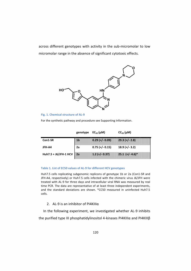
120
across different genotypes with activity in the sub-micromolar to low
micromolar range in the absence of significant cytotoxic effects.
Fig. 1. Chemical structure of AL-9
For the synthetic pathway and procedure see Supporting Information.
genotype EC50 (µM) CC50 (µM)
Con1-SR 1b 0.29 (+/- 0.09) 29.3 (+/- 2.8)
JFH-A4 2a 0.75 (+/- 0.15) 18.9 (+/- 3.2)
Huh7.5 + J6/JFH-1 HCV 2a 1.2 (+/- 0.37) 25.1 (+/- 4.6)*
Table 1. List of EC50 values of AL-9 for different HCV genotypes
Huh7.5 cells replicating subgenomic replicons of genotype 1b or 2a (Con1-SR and JFH-A4, respectively) or Huh7.5 cells infected with the chimeric virus J6/JFH were treated with AL-9 for three days and intracellular viral RNA was measured by real time PCR. The data are representative of at least three independent experiments, and the standard deviations are shown. *CC50 measured in uninfected Huh7.5 cells.
2. AL-9 is an inhibitor of PI4KIIIα
In the following experiment, we investigated whether AL-9 inhibits
the purified type III phosphatidylinositol 4-kinases PI4KIIIα and PI4KIIIβ
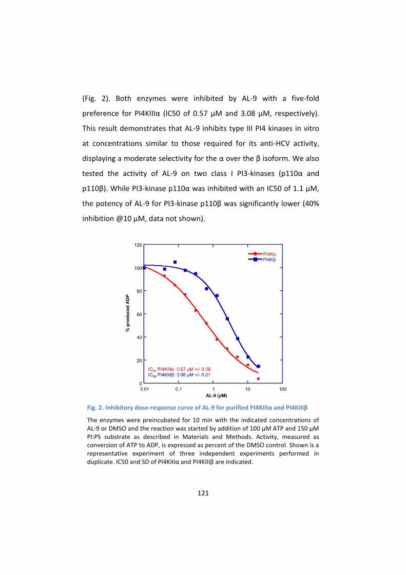
121
(Fig. 2). Both enzymes were inhibited by AL-9 with a five-fold
preference for PI4KIIIα (IC50 of 0.57 µM and 3.08 µM, respectively).
This result demonstrates that AL-9 inhibits type III PI4 kinases in vitro
at concentrations similar to those required for its anti-HCV activity,
displaying a moderate selectivity for the α over the β isoform. We also
tested the activity of AL-9 on two class I PI3-kinases (p110α and
p110β). While PI3-kinase p110α was inhibited with an IC50 of 1.1 µM,
the potency of AL-9 for PI3-kinase p110β was significantly lower (40%
inhibition @10 µM, data not shown).
Fig. 2. Inhibitory dose-response curve of AL-9 for purified PI4KIIIα and PI4KIIIβ
The enzymes were preincubated for 10 min with the indicated concentrations of AL-9 or DMSO and the reaction was started by addition of 100 µM ATP and 150 µM PI:PS substrate as described in Materials and Methods. Activity, measured as conversion of ATP to ADP, is expressed as percent of the DMSO control. Shown is a representative experiment of three independent experiments performed in duplicate. IC50 and SD of PI4KIIIα and PI4KIIIβ are indicated.

122
Our hypothesis is that AL-9 inhibits HCV replication via inhibition of
PI4KIIIα. Thus, we wanted to assess whether AL-9 also inhibited
PI4KIIIα in living cells. To this aim, we needed to set up an assay that
allowed us to monitor the activity of this kinase in intact cells. PI4KIIIα
is primarily localized to the ER, whereas PI4KIIIβ is localized to the
Golgi membranes29. It was shown that PI4KIIIβ contributes to the
synthesis of PI4P at the Golgi membranes30, 31. Subcellular localization
of the enzymes, however, does not always coincide with their function.
Thus, PI4KIIIα, considered to be an ER-resident enzyme, has previously
been shown to be critical for the generation and maintenance of the
plasma membrane PI4P pool during phospholipase C activation and
Ca2 signaling in HEK-293 or Cos-7 cells31, 32 as well as in resting Cos-7
cells33. Whether PI4KIIIα is responsible for the maintenance of the
plasma membrane PI4P pool under normal cell culture conditions in
hepatoma cells is currently not known. Hammond et al33 have
developed immunocytochemical techniques that enable selective
staining of the PI4P pool present in the plasma membrane (plasma
membrane staining protocol) or in the intracellular membranes (Golgi
staining protocol), respectively. We used this technique, in
combination with RNA gene silencing or pharmacological inhibition, to
decipher which of the type III enzymes participates in the synthesis of
the Golgi- or plasma membrane PI4P -pools in Huh7.5 hepatoma cells.
To address which type III PI4 kinase is responsible for the synthesis
of the different cellular PI4P pools, HuH7.5 cells were treated with
siRNAs targeting PI4KIIIα, PI4KIIIβ or an unrelated siRNA (mock-siRNA)
as described in the Materials and Methods section. Immunoblots
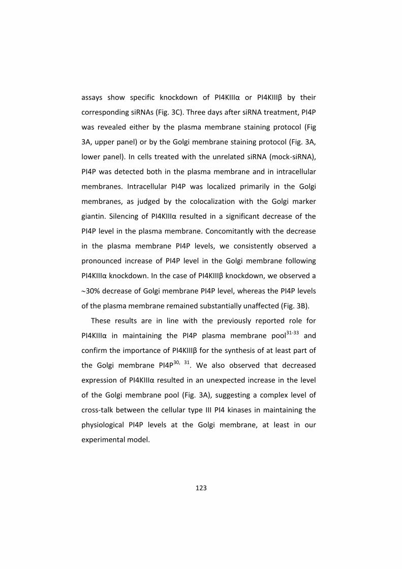
123
assays show specific knockdown of PI4KIIIα or PI4KIIIβ by their
corresponding siRNAs (Fig. 3C). Three days after siRNA treatment, PI4P
was revealed either by the plasma membrane staining protocol (Fig
3A, upper panel) or by the Golgi membrane staining protocol (Fig. 3A,
lower panel). In cells treated with the unrelated siRNA (mock-siRNA),
PI4P was detected both in the plasma membrane and in intracellular
membranes. Intracellular PI4P was localized primarily in the Golgi
membranes, as judged by the colocalization with the Golgi marker
giantin. Silencing of PI4KIIIα resulted in a significant decrease of the
PI4P level in the plasma membrane. Concomitantly with the decrease
in the plasma membrane PI4P levels, we consistently observed a
pronounced increase of PI4P level in the Golgi membrane following
PI4KIIIα knockdown. In the case of PI4KIIIβ knockdown, we observed a
30% decrease of Golgi membrane PI4P level, whereas the PI4P levels
of the plasma membrane remained substantially unaffected (Fig. 3B).
These results are in line with the previously reported role for
PI4KIIIα in maintaining the PI4P plasma membrane pool31-33 and
confirm the importance of PI4KIIIβ for the synthesis of at least part of
the Golgi membrane PI4P30, 31. We also observed that decreased
expression of PI4KIIIα resulted in an unexpected increase in the level
of the Golgi membrane pool (Fig. 3A), suggesting a complex level of
cross-talk between the cellular type III PI4 kinases in maintaining the
physiological PI4P levels at the Golgi membrane, at least in our
experimental model.
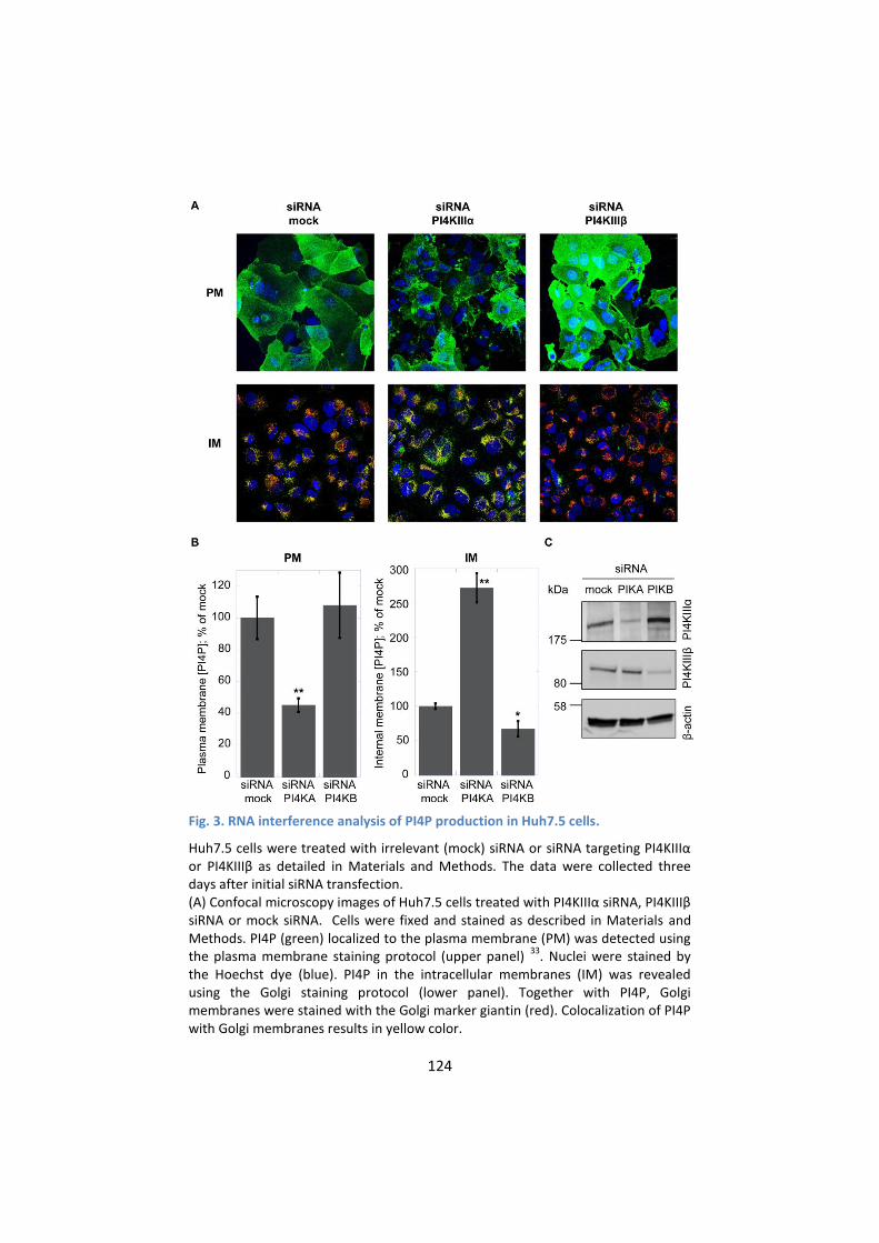
124
Fig. 3. RNA interference analysis of PI4P production in Huh7.5 cells.
Huh7.5 cells were treated with irrelevant (mock) siRNA or siRNA targeting PI4KIIIα or PI4KIIIβ as detailed in Materials and Methods. The data were collected three days after initial siRNA transfection. (A) Confocal microscopy images of Huh7.5 cells treated with PI4KIIIα siRNA, PI4KIIIβ siRNA or mock siRNA. Cells were fixed and stained as described in Materials and Methods. PI4P (green) localized to the plasma membrane (PM) was detected using the plasma membrane staining protocol (upper panel)
33. Nuclei were stained by
the Hoechst dye (blue). PI4P in the intracellular membranes (IM) was revealed using the Golgi staining protocol (lower panel). Together with PI4P, Golgi membranes were stained with the Golgi marker giantin (red). Colocalization of PI4P with Golgi membranes results in yellow color.

125
(B) Quantification of PI4P levels by immunofluorescence analysis. Changes in mean fluorescence intensity relative to the control (mock siRNA) are shown. Four randomly picked fields were analyzed per each condition, as described in Materials
and Methods. Data are presented as averages SEM. *, P < 0.05; **, P < 0.01. (C) Immunoblot analysis of protein expression after RNAi silencing. Lysates prepared from Huh 7.5 cells transfected with irrelevant siRNA (mock), PI4KIIIα siRNA (PIKA) or PI4KIIIβ siRNA (PIKB) were analyzed by immunoblotting with PI4KIIIα, PI4KIIIβ or β-actin antibodies as indicated in the figure. Positions of the protein molecular weight markers are shown on the left side.
In order to confirm and extend the results described above, we
utilized a known pharmacological inhibitor of the type III PI4 kinases.
PIK93 was previously exploited to distinguish between the roles of the
two PI4KIII isoforms34, 35. In particular, a concentration of 0.5 µM PIK93
is expected to affect only PI4KIIIβ, whereas 30 µM PIK93 should inhibit
both PI4KIIIβ and PI4KIIIα. Thus, Huh7.5 cells were treated with 0.5 µM
or 30 µM PIK93 or with DMSO as control. After two hours of
incubation, PI4P was revealed either by the plasma membrane staining
protocol (Fig 4A, upper panel) or by the Golgi staining protocol (Fig. 4A,
lower panel). PI4P levels associated with the Golgi membranes
decreased by 25% after incubation with 0.5 µM PIK93 (Fig. 4B). This is
in line with PI4KIIIβ contributing to the production of PI4P present in
the Golgi membranes (PI4KIIα, another contributor of Golgi-localized
PI4P is not inhibited by PIK9334, 35). Increasing PIK93 concentration to
30 µM further increased the inhibition of the intracellular membrane
PI4P pool, to 65% (Fig. 4B). This could be due to a more complete
inhibition of PI4KIIIβ; however, based on this experiment, we cannot
rule out a contribution of PI4KIIIα activity to the maintenance of the
Golgi membrane PI4P pool. In contrast to what observed in the Golgi-
associated membranes, the plasma membrane PI4P level was not
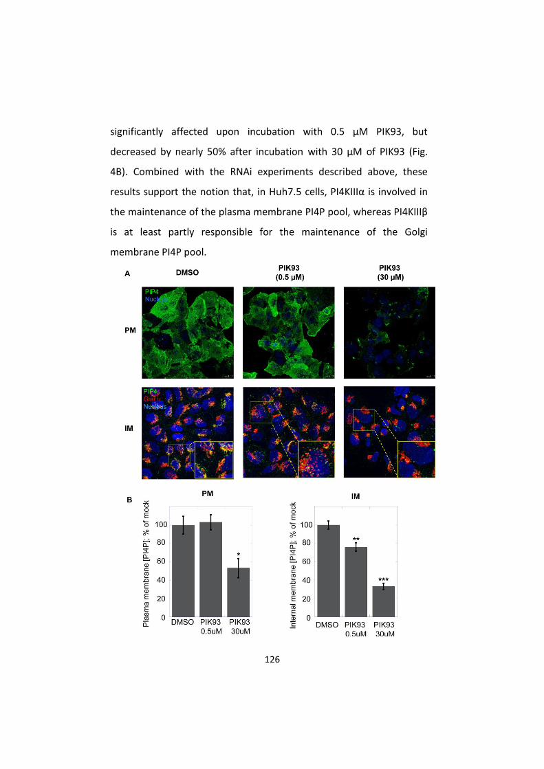
126
significantly affected upon incubation with 0.5 µM PIK93, but
decreased by nearly 50% after incubation with 30 µM of PIK93 (Fig.
4B). Combined with the RNAi experiments described above, these
results support the notion that, in Huh7.5 cells, PI4KIIIα is involved in
the maintenance of the plasma membrane PI4P pool, whereas PI4KIIIβ
is at least partly responsible for the maintenance of the Golgi
membrane PI4P pool.
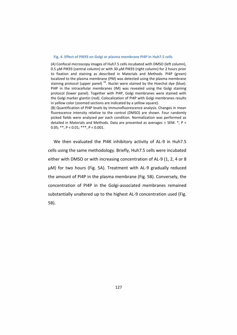
127
Fig. 4. Effect of PIK93 on Golgi or plasma membrane PI4P in Huh7.5 cells
(A) Confocal microscopy images of Huh7.5 cells incubated with DMSO (left column), 0.5 µM PIK93 (central column) or with 30 µM PIK93 (right column) for 2 hours prior to fixation and staining as described in Materials and Methods. PI4P (green) localized to the plasma membrane (PM) was detected using the plasma membrane staining protocol (upper panel)
33. Nuclei were stained by the Hoechst dye (blue).
PI4P in the intracellular membranes (IM) was revealed using the Golgi staining protocol (lower panel). Together with PI4P, Golgi membranes were stained with the Golgi marker giantin (red). Colocalization of PI4P with Golgi membranes results in yellow color (zoomed sections are indicated by a yellow square). (B) Quantification of PI4P levels by immunofluorescence analysis. Changes in mean fluorescence intensity relative to the control (DMSO) are shown. Four randomly picked fields were analyzed per each condition. Normalization was performed as
detailed in Materials and Methods. Data are presented as averages SEM. *, P < 0.05; **, P < 0.01; ***, P < 0.001.
We then evaluated the PI4K inhibitory activity of AL-9 in Huh7.5
cells using the same methodology. Briefly, Huh7.5 cells were incubated
either with DMSO or with increasing concentration of AL-9 (1, 2, 4 or 8
µM) for two hours (Fig. 5A). Treatment with AL-9 gradually reduced
the amount of PI4P in the plasma membrane (Fig. 5B). Conversely, the
concentration of PI4P in the Golgi-associated membranes remained
substantially unaltered up to the highest AL-9 concentration used (Fig.
5B).
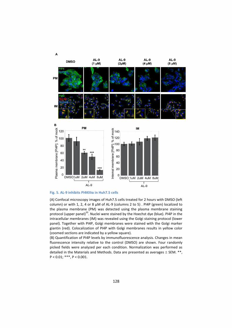
128
Fig. 5. AL-9 inhibits PI4KIIIα in Huh7.5 cells
(A) Confocal microscopy images of Huh7.5 cells treated for 2 hours with DMSO (left column) or with 1, 2, 4 or 8 µM of AL-9 (columns 2 to 5) . PI4P (green) localized to the plasma membrane (PM) was detected using the plasma membrane staining protocol (upper panel)
33. Nuclei were stained by the Hoechst dye (blue). PI4P in the
intracellular membranes (IM) was revealed using the Golgi staining protocol (lower panel). Together with PI4P, Golgi membranes were stained with the Golgi marker giantin (red). Colocalization of PI4P with Golgi membranes results in yellow color (zoomed sections are indicated by a yellow square). (B) Quantification of PI4P levels by immunofluorescence analysis. Changes in mean fluorescence intensity relative to the control (DMSO) are shown. Four randomly picked fields were analyzed per each condition. Normalization was performed as
detailed in the Materials and Methods. Data are presented as averages SEM. **, P < 0.01; ***, P < 0.001.

129
In all, the results described above suggest that AL-9 inhibits PI4KIIIα
also in living cells, while not appreciably affecting the activity of
PI4KIIIβ. This is in line with the selectivity for PI4KIIIα over PI4KIIIβ
observed in the biochemical assays.
3. HCV alters the intracellular and plasma membrane
distribution of PI4P
Viral infection induces modification of intracellular membrane
structures36 and, for some RNA viruses including HCV, it has been
shown that these induced membranous structures are highly enriched
for PI4P28, 37. Before testing the activity of AL-9 in HCV-infected cells,
we wanted to know what the impact of HCV on cellular membrane
structures was, with special regard to the subcellular membrane
distribution of PI4P.
Naïve Huh7.5 cells or cells actively replicating the genotype 2a or 1b
HCV subgenomic replicon were investigated for their PI4P
concentration in internal membranes or plasma membranes,
respectively (Fig. 6A). As previously shown, cells expressing the HCV
replicon form a membranous web that is highly enriched for PI4P (Fig.
6A, lower panel). The level of PI4P in these virus-specific membrane
structures is markedly higher in JFH-A4 cells, containing the very
efficient genotype 2a JFH-1 replicon, compared to the Con1-SR cells,
which are based on the genotype 1b Con1 replicon, possibly mirroring
the different RNA replication efficiency. It is well established that the
kinase responsible for the production of the PI4P pool present in these
structures is PI4KIIIα. In the current model, PI4KIIIα interacts with the

130
viral protein NS5A, leading to up-regulation of the kinase activity and
accumulation of PI4P in the virus-specific membranous web26-28.
Conversely, the results shown in Figures 3-5 suggest that – in absence
of viral replication – a major function of PI4KIIIα is the synthesis of the
PI4P pool in the plasma membrane. We therefore asked ourselves
whether the presence of HCV could not only influence distribution and
enrichment of PI4P in internal membranes, but also alter the PI4P
plasma membrane pool. In Fig. 6A, we show that, concomitantly with
the increase of PI4P in the internal membranes (lower panel), HCV
replication promotes a marked decrease of PI4P concentration in the
plasma membrane (upper panel). Relative quantification of the PI4P
levels in the different experimental conditions is shown in Fig. 6B. This
experiment demonstrates that the presence of HCV causes a dramatic
change of PI4P localization in cellular membranes, whereby the
increase of PI4P concentration in the virus-specific membranous
structures appears to be accompanied by a depletion of the PI4P pool
normally present in the plasma membrane.
131
Fig. 6. HCV impacts subcellular PI4P distribution
(A) Huh7.5 cells, JFH-A4 and Con1-SR cells were analyzed by confocal microscopy for the presence of PI4P (green) in the plasma membranes (PM, upper panel) or in the intracellular membrane (IM, lower panel) using the protocols described in Materials and Methods. Nuclei were stained by the Hoechst dye (blue). For internal membrane staining, giantin (red) was used as a specific marker for Golgi membranes. (B) Quantification of PI4P levels by immunofluorescence analysis. Changes in mean fluorescence intensity relative to the control (Huh7.5 cells) are shown. Four randomly picked fields were analyzed per each condition. Normalization was
performed as detailed in Materials and Methods. Data are presented as averages SEM. *, P < 0.05; **, P < 0.01; ***, P < 0.001.

132
We next investigated whether HCV-associated changes in PI4P
distribution could be reverted upon cure of the HCV replicon by
specific inhibitors. We treated JFH-A4 cells for two weeks either with
the HCV RdRp inhibitor HCV-796 or with the HCV NS3/4A protease
inhibitor MK-5172 and followed PI4P localization in internal
membranes and in the plasma membrane (Fig. 7). Independent of the
type of inhibitor used, the result shows that the HCV-induced PI4P –
enriched membranous web in JFH-A4 cells disappeared upon
suppression of HCV replication and that the intracellular PI4P
localization returned to the Golgi-localization as observed in the naïve
Huh7.5 cells (left column). In parallel, the plasma membrane
concentration of PI4P increases to the levels observed in naïve cells
(middle column). NS5A staining (right column) as well as real-time RT-
PCR (not shown) indicated that the prolonged treatment with HCV-
inhibitor led to complete and stable suppression of viral protein
expression and undetectable level of HCV RNA. Thus, removal of HCV
RNA brings PI4P synthesis and distribution back to a level comparable
to naïve Huh7.5 cells. It is worth of note, however, that the previous
presence of HCV replicons in the cured cells induced some irreversible
morphological changes of unknown nature, such as a smaller cell size.

133
Fig. 7. Reversibility of HCV-induced changes in PI4P subcellular distribution
JFH-A4 cells were incubated for 14 days with the HCV RdRp inhibitor HCV-796 (2 µM) or the HCV NS3/4A protease inhibitor MK-5172 (0.2 µM). Cure from HCV was controlled by detection of NS5A with a specific NS5A antibody (red, right column). As control, untreated Huh7.5 cells or JFH-A4 cells were used. Cells were fixed and PI4P (green) was detected in the internal membranes (IM, left column) or in the plasma membrane (PM, central column). For internal membrane staining giantin (red) was used as a specific marker for Golgi membranes. Nuclei were stained by the Hoechst dye (blue).
134
4. AL-9 inhibits PI4KIIIα in HCV-replicating cells
We have shown that PI4KIIIα is inhibited by AL-9 in naïve Huh7.5
cells. As discussed above, in HCV-replicating cells, the kinase activity of
PI4KIIIα is up-regulated by a direct protein-protein interaction with the
viral protein NS5A. In the following experiment (Fig. 8), we explored
whether AL-9 is able to inhibit PI4KIIIα also in this context. JFH-A4 cells
were incubated with increasing concentration of AL-9 for 4 hours and
PI4P concentration in the HCV membranous web was followed by
immunostaining (Golgi staining protocol). Treatment of cells with AL-9
lead to clear inhibition of PI4P accumulation in the HCV membranous
web. Incubation with 8 µM AL-9 depleted as much as 70% of the PI4P
present in the intracellular membranes of replicon-containing cells.
This result confirms that AL-9 inhibits PI4KIIIα independent of its
membranous localization and suggests that this inhibition could be
responsible for the observed antiviral effect. Since AL-9 has anti-HCV
activity in the concentration range used here, longer incubation of HCV
replicons with AL-9 results in inhibition of HCV- RNA and -protein
synthesis. As a consequence the PI4P –enriched HCV membranous web
would disintegrate. In this case loss of PI4P in the internal membranes
could be not a direct consequence of PI4KIIIα inhibition, but a
consequence of disintegration of the HCV membranous web.
In order to rule out this possibility, we checked localization of NS5A,
a presumed marker for HCV replication sites, after 4 hours of
incubation with AL-9. Localization of NS5A does not change, suggesting
that AL-9 does not significantly change the structure of the HCV
membranous web upon 4 hours of treatment. Moreover, incubating

135
the same replicon cells for 4 hours with HCV-796, an HCV polymerase
inhibitor, did not lead to appreciable depletion of the membranous
web PI4P pool indicating that the loss of PI4P in the HCV-induced
intracellular membranes is the direct consequence of inhibition of
PI4KIIIα, and not the consequence of inhibition of HCV replication.
Additional evidence is provided in the experiment below, in which
expression of the HCV polyprotein, and consequently formation of a
membranous web, was driven by cDNA plasmid rather than by
autonomously replicating HCV RNA.
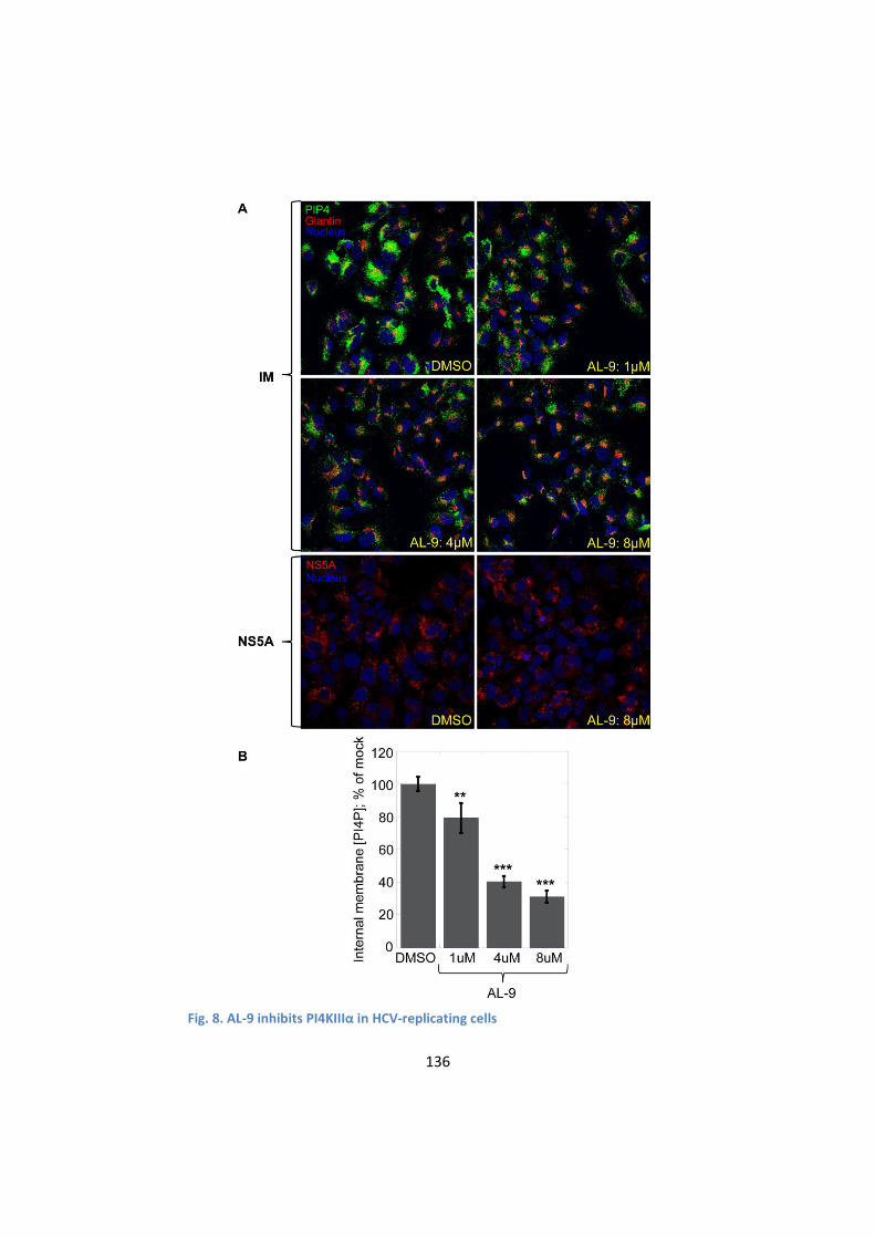
136
Fig. 8. AL-9 inhibits PI4KIIIα in HCV-replicating cells
137
(A) JFH-A4 cells were treated with DMSO or AL-9 for 4 hours and internal membranes were stained for PI4P (green) and the Golgi marker giantin (red) using the Golgi staining protocol, as described in Materials and Methods. DMSO or AL-9 concentrations are indicated within the image. Alternatively, cells were stained for NS5A as described in Materials and Methods (indicated as NS5A). Nuclear DNA was stained with Hoechst dye (blue). PI4KIIIα, associated with the HCV-associated membranous web is inhibited by AL-9. The decrease of PI4P is not due to inhibition of the HCV replication indicated by unchanged NS5A expression and localization (lower panel). (B) Quantification of PI4P levels by immunofluorescence analysis. Changes in mean fluorescence intensity relative to the control (DMSO) are shown. Four randomly picked fields were analyzed per each condition. Normalization was performed as
detailed in Materials and Methods. Data are presented as averages SEM. **, P < 0.01; ***, P < 0.001.
5. PI4KIIIα inhibition by AL-9 alters sub-cellular distribution of
NS5A
It was previously shown that knock-down of PI4KIIIα expression by
RNAi resulted in the production of large NS5A clusters. This was
achieved in an experimental setting where the HCV polyprotein was
expressed from DNA constructs, thus avoiding potential confounding
effects due to inhibition of HCV RNA replication23, 28. We wanted to
assess whether pharmacological inhibition of PI4KIIIα kinase activity
would lead to similar effects on NS5A subcellular localization. Thus we
followed the effect of AL-9 on NS5A localization after transient DNA
transfection in Lunet-T7 cells with a plasmid expressing genotype 2a
nonstructural proteins NS3-NS5B under the control of a T7 promoter38.
Cells were treated either with DMSO (upper panels) or with 8 µM AL-9
(lower panels) for 2, 8 or 16 hours and localization of NS5A as well as
PI4P were followed by indirect fluorescence microscopy (Fig. 9). Cells
successfully transfected with the HCV polyprotein expressed NS5A and
induced the PI4P -enriched membranous web. After 8 hours of
138
treatment with AL-9, changes in NS5A localization in form of larger
clusters become visible. At the same time, PI4P concentration in the
membranous web started to decrease. After 16 hours of incubation
with AL-9, NS5A was concentrated almost exclusively in large clusters.
At this time-point, PI4P in the internal membranes had completely
vanished.
In summary, this experiment shows that, in cells expressing the HCV
polyprotein from cDNA, prolonged treatment with AL-9 results in a
redistribution of NS5A into large clustered structures with high
resemblance to the structures previously observed after silencing of
the PI4KIIIα gene by RNAi23, 28. Concomitantly, we observed a depletion
of the PI4P pool present in the HCV-induced membranous structures.
These results indicate that the catalytic activity of PI4KIIIα is directly or
indirectly required for the proper localization of HCV NS5A protein into
the membranous web. Furthermore, the experiment just described
lands additional support to the notion that the antiviral effect of AL-9
is mediated by the inhibition of PI4KIIIα.
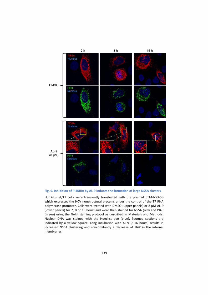
139
Fig. 9. Inhibition of PI4KIIIα by AL-9 induces the formation of large NS5A clusters
Huh7-Lunet/T7 cells were transiently transfected with the plasmid pTM-NS3-5B which expresses the HCV nonstructural proteins under the control of the T7 RNA polymerase promoter. Cells were treated with DMSO (upper panels) or 8 µM AL-9 (lower panels) for 2, 8 or 16 hours and were then stained for NS5A (red) and PI4P (green) using the Golgi staining protocol as described in Materials and Methods. Nuclear DNA was stained with the Hoechst dye (blue). Zoomed sections are indicated by a yellow square. Long incubation with AL-9 (8-16 hours) results in increased NS5A clustering and concomitantly a decrease of PI4P in the internal membranes.
140
D. Discussion
In the present paper, we show that a compound belonging to the
class of 4-anilino quinazoline inhibitors of HCV replication is an
inhibitor of PI4KIIIα, a cellular lipid kinase required for viral replication.
PI4KIIIα belongs to the family of type III phosphatidylinositol 4-
kinases, enzymes that catalyze the conversion of phosphatidylinositol
to phosphatidylinositol 4-phosphate (PI4P). PI4P is the most abundant
monophosphorylated inositol phospholipid in mammalian cells and the
importance of this phospholipid is just started to be unraveled39. In
addition to playing important roles in intracellular signaling and
membrane trafficking, phosphatidylinositol lipids and their
metabolizing enzymes are also exploited by many different viruses in
order to transform cellular membranes in structures supporting their
replication36, 40, 41. PI4KIIIβ was shown to be a host factor required for
enterovirus replication37, whereas several reports have demonstrated
that PI4KIIIα is crucial for HCV replication22-25. Owing to the importance
of this pathway, the need for specific inhibitors of PI4III kinases is
increasing. Only recently, some enviroxime-like compounds with anti-
viral activity against enterovirus have been demonstrated to target
PI4KIIIβ. One of these agents is a very specific inhibitor of the β-
isoform of the type III PI4-kinases42. So far, no such compound exists
for the PI4KIII-α isoform. A commonly used inhibitor for type III
phosphatidylinositol 4-kinases is PIK93, which has originally been
designed to inhibit class I PI3-kinases34. This compound allows
differential inhibition of PI4KIIIβ alone or PI4KIIIα and PI4KIIIβ together
depending on the concentration used. In this paper, we show that a 4-

141
anilino quinazoline derivative, termed AL-9 (Fig. 1 and S1), is able to
inhibit PI4KIIIα in a test tube as well as in living cells. AL-9 inhibited
purified PI4KIIIα, with a moderate (5-fold) selectivity over the
isoform (Fig. 2). In cell culture, we observed that treatment with AL-9
efficiently inhibits the maintenance of the plasma membrane PI4P pool
in Huh7.5 cells while not significantly affecting the Golgi membrane
pool at the highest concentration used (Fig. 5). This finding is in line
with the moderate selectivity observed in the biochemical assay. Thus,
AL-9 represents a lead candidate for the development of more potent
and more specific inhibitors of PI4KIIIα.
Anti-HCV compounds of the 4-anilino quinazoline class were
previously assumed to exert their antiviral effect via inhibition of the
viral protein NS5A. This conclusion rested on analysis of the mutations
found in the HCV replicon in association with resistance to these
agents18. Mutations generated against 4-anilino quinazolines where
localized mainly in NS5A, in triplets that occurred all in NS5A or
appeared concomitant with changed in NS4B or NS5B (see also
Introduction). Reverse genetic experiments, in which these mutations
were reintroduced in the replicon (single, double and triple
combinations), however, did not support a role for these mutations in
conferring resistance to 4-anilino quinazolines. In order to assess
whether the reported mutations conferred any level of resistance to
AL-9, we independently performed reverse genetics studies in which
selected mutations triplets, reported to be associated with the higher
level of resistance, were reintroduced in a genotype 1b replicon with
the same genetic background as the one reported in the original

142
resistance study (Fig. S2). These mutation triplets are: FAG:
L199F+V362A+S390G (NS5A), DLD: E212D+P299L+V388D (NS5A), and
PPA: T200P+S370P(NS5A)+S76A(NS5B). We observed that the replicon
containing the first triplet lost the ability to replicate at significant
level. For replicons containing the latter two combinations of
mutations, RNA replication could be measured, although at a lower
level compared to the parental construct (35% and 20%, respectively).
These replicons, however, remained equally sensitive to AL-9 as the
parental replicon (Fig. S2), opening the question as to which really is
the target of this compound class. We are currently trying to select
HCV replicons resistant to AL-9. So far we were unable to identify
mutations that confer resistance to AL-9.
Our new data on AL-9 suggest that inhibition of HCV replication by
4-anilino quinazoline compounds is a consequence of PI4KIIIα
inhibition. Our conclusion rests on a number of experimental findings.
First of all, we showed that AL-9 is an inhibitor of purified type III PI4
kinases. Furthermore, we clearly demonstrated that AL-9 inhibits
PI4KIIIα both in naïve Huh7.5 cells (Fig. 5, discussed above) as well in
cells harboring actively replicating HCV RNA (Fig. 8). In cells where HCV
replication occurs, PI4KIIIα interacts physically with HCV NS5A. This
interaction, in turn, leads to the stimulation of PI4P synthesis at the
HCV replication sites28. Treatment of replicon-harboring cells with AL-9
leads to efficient suppression of the PI4P pool at the HCV replication
sites and does so independently of inhibition of HCV replication. This
indicates that – although the enzymatic activity of PI4KIIIα is

143
modulated by the interaction with an HCV protein NS5A – it remains
sensitive to the action of the 4-anilino quinazoline inhibitor.
We also investigated whether the dramatic changes observed in
PI4P membrane levels by treatment with AL-9 could be associated with
alteration in the subcellular distribution of type III PI4 kinases. To this
aim, we analyzed the subcellular distribution of the type III PI4 kinases
in Huh7.5 or Luc-A4 cells following incubation with AL-9 (Fig. S3). We
observed no major effect of AL-9 on the localization of either PI4KIIIα
or PI4KIII, in line with the notion that the observed effects are
primarily due to the inhibition of the kinase activity rather than to an
altered protein subcellular distribution.
In cells that express the HCV polyprotein from a trans-gene, knock-
down of PI4KIIIα by RNAi was previously shown to cause a dramatic
change in NS5A subcellular distribution, from a pattern consistent with
localization in the membranous web replication complexes to
abnormally large cytoplasmic clusters23, 26, 28. In Fig. 9, we show that
AL-9 treatment of cells ectopically expressing the HCV nonstructural
proteins results in a time-dependent depletion of PI4P and a
concomitant change of NS5A localization to the large-clustered
structures discussed above, reinforcing the notion that the anti-HCV
effect of AL-9 and related compounds are likely to be mediated by the
inhibition of PI4KIIIα.
We also found that PI3K p110α is inhibited by AL-9 in vitro at
concentration similar to those needed to inhibit type III PI4-kinases.
However, no Class I PI3-kinase has been shown to influence HCV
replication thus inhibition of HCV replication by AL-9 is not due to
144
inhibition of Class I PI3-kinases. So far, the only PI3-kinase that resulted
as positive hit for HCV replication inhibition in siRNA screens is PI3-
kinase C2 gamma24. Future work will have to address whether AL-9
inhibits PI3KC2G in addition to Type III PI4-kinases.
During the characterization of AL-9 we focused our attention on
various aspects of PI4P metabolism in Huh7.5 cells with and without
replicating HCV. We observed a typical Golgi localization of PI4P in
intracellular membranes of naïve Huh7.5 cells and confirmed a role for
PI4KIIIβ in maintaining at least part of this pool. In order to get the
complete picture we also investigated the PI4P pool present in the
plasma membrane. In yeast, Stt4p, the ortholog to the mammalian
PI4KIIIα, is localized at the plasma membrane and it is the major
contributor for the synthesis of the plasma membrane-localized PI4P39.
In mammalian cells, the role of PI4KIIIα for the maintenance of the
plasma membrane PI4P pool has been demonstrated in HEK-293 and
Cos-7 cells31-33. Here we demonstrate that liver-derived Huh7.5 cells
are endowed with a rich PI4P pool in the plasma membrane and that
the enzyme responsible for its maintenance is PI4KIIIα. In HCV-
replicating cells, the subcellular PI4P distribution is profoundly altered.
As already reported previously, the presence of HCV causes the
induction of a membranous web highly enriched for PI4KIIIα-
syntesized PI4P. In accordance, several reports demonstrate that NS5A
recruits PI4KIIIα to the membranous web by direct protein-protein
interaction, thereby stimulating its enzymatic activity26-28.
Concomitantly with the induction of highly PI4P -enriched internal
membranes, we observe a marked decrease of PI4P in the plasma

145
membrane. One possible explanation could be that – by hijacking
PI4KIIIα - HCV might be able to enrich PI4P in the virus-induce
membranous web not only by directly activating the enzymatic activity
of PI4KIIIα recruited into the HCV RNA replication compartment, but
also by preventing transport of the PI4KIIIα-synthesized PI4P from the
synthesis site to the plasma membrane. How PI4KIIIα, localized at the
ER, synthesizes the PI4P pool present in the plasma membrane it is still
an enigma. This topological discrepancy can partially be resolved
assuming that PI4KIIIα-dependent PI4P production occurs on ER-PM
contact sites, that is, sites of close apposition between ER and PM. In
yeast it has been demonstrated that a complex interplay between
different proteins regulate the PI4P metabolism at the plasma
membrane43. Among these proteins are Osh, the yeast ortholog of the
human OSBP and the ER membrane VAP proteins Scs2 and Scs22, the
yeast orthologs of human VAP proteins. Interestingly, h-VAP-33 and
OSBP have been shown to be important for HCV replication44-46. It may
be possible that recruitment of PI4KIIIα to the HCV membranous web
through NS5A prevents interaction of PI4KIIIα with its cellular protein
partners required to direct PI4P to the plasma membrane. Upon
withdrawal of HCV from the cells (Fig. 7) PI4KIIIα is again free for
interaction with the adequate partners. A possible role of PI4KIIIα in
PI4P trafficking between the plasma- and intracellular membranes is
suggested by our finding that RNAi silencing of this PI4 kinase results in
decreased concentration of PI4P in the plasma membrane with a
concomitant increase in the level of PI4P in the endomembranes (Fig.
3). Such a function of PI4KIIIα would have to be independent of the
146
kinase activity, since pharmacological inhibition (with PIK93 or AL-9)
does not recapitulate this phenomenon observed by knocking down
the protein expression.
In summary, the presence of HCV may change PI4P metabolism not
only by activating the catalytic activity of PI4KIIIα by NS5A but also by
modulating the PI4P distribution between different membrane
compartments. The net result is an enrichment of the PI4P pool in the
HCV-induced membranous web with a concomitant depletion of the
plasma membrane PI4P pool.
Concluding, in this paper we demonstrate that a class of HCV
inhibitors originally proposed to target NS5A does in fact target the
host factor PI4KIIIα. Compounds targeting host factors may have the
general advantage of imposing a higher genetic barrier to the
development of resistance. AL-9, a member of this class of
compounds, inhibits PI4KIIIα and to our knowledge, it is the first
compound with a clear preference for PI4KIIIα over PI4KIIIβ. For this
reason, AL-9 offers a good candidate as lead compound for the
development of more potent and specific pharmacological inhibitors of
PI4KIIIα to be used both as important research tools as well as leads
for initial drug discovery.
147
E. Materials and Methods
1. Reagents and plasmids
The HCV RNA polymerase inhibitor HCV-796 and the PI kinase
inhibitor PIK93 were a gift from Arrow Pharmaceuticals. The HCV
protease inhibitor MK-5172 was purchased from Selleck Chemicals.
Nucleic acids were manipulated according to standard protocols.
Plasmid FBac-His-CD-PI4KA was constructed as follows: the catalytic
domain of PI4KIIIα was amplified by PCR using the oligonucleotides 5’-
cactgcggatccataatggggatgatgcagtgtgtgattg-3’ (sense), 5’-
cctgcgaattctcagtaggggatgtcattc-3’ (antisense) and the plasmid pEF1A-
PIK4CA untagged (a kind gift from G. Randall, Department of
Microbiology, University of Chicago) as template. The resulting PCR
fragment was subcloned into the vector pCR-Blunt II-Topo (Invitrogen)
and finally cloned into the BamH1–XhoI cloning sites of the plasmid
vector pFastBac THT-B. The resulting protein expressed from this
plasmid contains an N-terminal hexa-histidine tag and starts at PI4KIIIα
amino acid G873 (reference sequence NM_058004). pTM-NS3-5B
expression vector expressing the HCV genotype 2a nonstructural
proteins under the control of the T7 promoter was a generous gift
from V. Lohmann (Department of Molecular Virology, University of
Heidelberg)38. Synthesis of compound AL-9 is described in Supporting
Information.
2. Cell lines and culture conditions
The human hepatoma-derived cell line Huh7.547 were grown in
Dulbecco’s modified Eagle’s medium (DMEM) supplemented with 10%
148
fetal bovine serum, 100 U/ml penicillin, 100 μg/ml Streptomycin and 2
mM L-glutamine; G418 (0.8 mg/ml) was added to cell lines containing
the HCV replicon. Stable cell lines expressing HCV genotype 1b or 2a
subgenomic replicons were generated by electroporation of in vitro-
transcribed RNA into Huh7.5 cells48 and following selection with G418
(0.8 mg/ml) for three weeks. Con1-SR: Huh7.5 cells replicating the
Con1 subgenomic replicon with the adaptive mutations E1202G in NS3
and S2204R in NS5A. JFH-A4: Huh7.5 cells replicating the JFH-1
subgenomic replicon together with the luciferase reporter gene
constructed as described previously49. JFH-A4 cells were cured from
the HCV replicon by two weeks of treatment with the protease
inhibitor MK-5172 (0.2 µM) or the HCV RNA polymerase inhibitor HCV-
796 (2 µM), respectively. Huh-LUNET/T7 cells were a kind gift from V.
Lohmann (Department of Molecular Virology, University of Heidelberg,
Germany).
3. Replication and infection assays
For replication assays, JFH-A4 or Con1-SR cells were plated at the
density of 3x10⁴ or 6x10⁴ cells/well, respectively, in 24-well dishes the
day before the experiment. Cells were treated with AL-9 resulting in a
final concentration of 1 % DMSO in the cell medium. After three days
of treatment, RNA was extracted using the RNeasy Mini Kit (Qiagen)
and HCV RNA was quantified by real time PCR using the following
oligonucleotide and probe set designed for the HCV IRES as described
previously 48: sense (5’-GCGAAAGGCCTTGTGGTACT-3’), antisense (5’-
CACGGTCTACGAGACCTCCC-3’), and probe (5’-
149
CCTGATAGGGTGCTTGCGAGTGCC-3’, 5’ 6-carboxyfluorescein [FAM]/3’
6-carboxytetramethylrhodamine [TAMRA]). GAPDH mRNA was used as
internal control for data normalization.
Production of infectious virus was performed as follows: J6/JFH-1
chimeric RNA (1-846(J6CF)/847-3034(JFH1) was electroporated into
Huh7.5 cells using the protocol described previously48. Briefly, 2x10⁶
cells were electroporated with 10 µg of RNA in a final volume of 200 µl
and 4x10⁶ cells were plated in a T-75 flask. Three days post
electroporation, medium was harvested and stored at -20°C in small
aliquots. Calculation of EC50 of AL-9 using the infectious HCV virus was
performed as follows: Huh7.5 cells were plated at 4x10⁴ cells/well in
24-well plates the day before infection. Infection was started by
addition of 10 µl of cell medium containing infectious virus (see above)
at an MOI of 50 in a final volume of 400 µl. After 6 hours of incubation,
medium was removed and replaced with 400 µl of fresh medium
containing serial dilutions of AL-9. RNA was collected after 72 hours of
incubation and quantified by real time PCR. Cell cytotoxicity (CC50) of
AL-9 was calculated using the cell viability assay CellTiter-Blue
(Promega). Huh7.5, JFH-A4, or Con1-SR cells (5x10³ cells/well in 96-
well dishes) were plated the day before treatment. AL-9 was added
and cell viability was measured after four days of treatment.
4. Expression and purification of the catalytic domain of PI4KIIIα
Recombinant baculovirus was generated with the plasmid FBac-His-
CD-PI4KA using the Bac-to-Bac system following the instructions of the
manufacturer (Invitrogen). For protein expression, Sf9 cells were
150
infected with recombinant baculovirus at a density of 2x10⁶ Sf9
cells/ml for 3 days at 20°C. To prepare cell extract (1.5x10⁸ cells), cells
were incubated in hypotonic buffer (10 mM HEPES (pH 7.5), 10 mM
NaCl, 1 mM Tris(2-carboxyethyl)phosphine (TCEP) and EDTA-free
protease inhibitor cocktail (Complete, Roche) for 30 min in ice and
mechanically broken by 20 strokes of a Dounce homogenizer. After
homogenizing, cells were incubated in lysis buffer (50 mM HEPES (pH
7.5), 500 mM NaCl, 10 % glycerol, 1 % Triton-X100, 1 mM TCEP and
EDTA-free protease inhibitor cocktail (Complete, Roche) for further 30
min in ice and cell extract was cleared by centrifugation for 45 min at
20.000g. The cleared supernatant was incubated in batch with Ni-
Sepharose High Performance (GE Healthcare) for 2 hours at 4°C with
continuous shaking. The resin was first washed with 10 resin-volumes
of wash buffer (50 mM HEPES (pH 7.5), 10 % glycerol, 0.4 % Triton X-
100, 150 mM NaCl and 20 mM imidazol) followed by elution with wash
buffer containing 250 mM imidazole. Active fraction (0.5 ml) were
dialyzed against 50 mM HEPES (pH 7.5), 150 mM NaCl, 1 mM DTT, 0.4
% Triton X-100 and 10% glycerol and stored at -80°C in small aliquots.
5. In vitro kinase assay
PI4K kinase activity was assayed with the ADP-Glo Kinase Assay
(Promega), according to the manufacturer’s instructions. Briefly, 0.5 µl
of PI4KIIIα-CD or 0.05 µl PI4KIIIβ (32 ng, Invitrogen) were preincubated
with DMSO or AL-9 in reaction buffer (20 mM Tris (pH 7.5), 5 mM
MgCl2, 2 mM DTT, 0.5 mM EGTA, 0.4% Triton X-100) for 10 min at
room temperature in a final volume of 8 µl. The reaction was started
151
by addition of 2 µl of ATP and PI:PS Lipid Kinase Substrate (Invitrogen)
to give a final concentration of 100 µM and 150 µM, respectively. After
1 hour of incubation at room temperature the reaction was stopped
and further processed as described by the manufacturer. In parallel
the reaction was performed without PI:PS substrate in order to detect
contaminating ATPase activity present in the protein fractions. This
activity was subtracted from the measured kinase activity. Kinase
activity of PI3Kα (p110α/p85α) and PI3Kβ (p110β/p85α) was assayed
as above using 5 ng or 20 ng, respectively (Millipore). Reaction buffer
was changed to 50 mM HEPES pH7.5, 10 mM MgCl2 and 1 mM DTT.
6. Indirect Immunofluorescence
Cells were plated one day before the experiment in 24-well plates
(5x10⁴ cells/well for Huh7.5 and JFH-A4 cells, 7x10⁴ cells/well for Con1-
SR and 1x10⁵ cells/well for cured JFH-A4 cells). Cells were either
untreated or treated with compounds for the time as indicated in the
figure legend. PI4P staining of the plasma membrane or internal
membranes was performed exactly as described previously33. Primary
antibodies used were: anti- PI4P IgM (Cat. No. Z-P004, 1:300, Echelon),
anti-Giantin antibody (Cat. No. PRB-114C-200 1:1000, Covance),
affinity-purified rabbit anti NS5A antibody (1:2000), anti-PI4KIIIα
kinase (Cat. No. 4902, 1:50, Cell Signaling), anti-PI4KIIIβ kinase (Cat.
No, 611817, 1:500, BD Transduction). Secondary antibodies used were
goat anti-mouse IgM Alexa Fluor 488 (Cat. No. A-21042, 1:600,
Invitrogen) and goat anti-rabbit Alexa Fluor 568 (Cat. No. A-11011,
1:600, Invitrogen). For type III PI4K kinases or NS5A staining, all
152
incubations were performed at room temperature. Cells were washed
once with PBS and fixed with 300 μl of 4% PFA for 15 min. Cells were
washed three times with PBS and permeabilized with 500 μl of 0.1 %
Triton X-100 (or 0.5% for PI4KIIIα kinase staining) in PBS for 10 min.
Unspecific binding was blocked by incubation with 3 % BSA in PBS (for
PI4KIIIα staining no blocking was performed). After incubation with the
primary antibody in blocking buffer, cell were washed with PBS and
subsequently incubated with goat secondary antibodies conjugated to
Alexa-Fluor 568, or Alexa-Fluor 488 at a dilution of 1:600. Nuclei were
stained with Hoechst dye 33342 (Sigma; 1:4000). Slides were then
mounted with 5 μl ProLong Gold Antifade (Invitrogen) and analyzed by
using an inverted Leica TCS SP5 scanning laser confocal microscope.
Digital images were taken using LAS AF software (Leica) and processed
using Volocity software (Perkin Elmer). Quantification of fluorescence
intensity was determined from multiple images using Volocity. Relative
changes in fluorescence intensity mean values where obtained from
four randomly picked fields for each condition (150 ~ 300 cells). For
plasma membrane staining, total PI4P fluorescence intensity obtained
in each condition was normalized to the number of cells present in
each field. For the quantification of relative PI4P levels in internal
membranes, PI4P fluorescence intensity was normalized using the
fluorescence intensity of the Golgi marker giantin. Quantitative
immunofluorescence data are presented as means the standard
error of the mean (SEM). For the calculation of statistical significance,
a two-tailed, unpaired t-test was performed.
153
7. siRNA silencing
3x10⁴ Huh7.5 cells/well were seeded in 24-well plates on
microscope cover glasses and transfected with 50 nM of siRNAs in
serum-free Opti-MEM (Invitrogen) using Lipofectamine RNAiMAX
(Invitrogen), according to the manufacturer’s protocol. For western
blot analysis, the transfection reaction was proportionally scaled up to
6-well plates. In order to maximize the silencing efficiency, 24 hours
after the first transfection, the cells were subjected to a second round
of siRNA transfection. siRNA sequences were the following (5’->3’
sense strand): mock siRNA, GUA UGA CCG ACU ACG CGU ATT (custom,
Sigma-Aldrich); PI4KIIIα siRNA, CCG CCA UGU UCU CAG AUA ATT
(custom, Sigma-Aldrich); and PI4KIII siRNA, GCA CUG UGC CCA ACU
AUG ATT (Silencer Validated siRNA s10543; Ambion). Three days after
the initial transfection, cells were stained for PI4P as described
previously33, or subjected to western blot analysis. For immunoblot
analysis of protein expression, cells were harvested with TEN buffer
(10mM Tris/HCl pH 8.0, 1 mM EDTA, 100 mM NaCl), washed once with
PBS and lysed with 2X protein sample buffer (125 mM Tris-HCl pH 6.8,
10 mM EDTA, 0,003 gr bromophenol blue, 20% glycerol, 4% SDS and
10% β-mercaptoethanol; 200l). The samples were then sonicated,
heated at 95°C and loaded onto 7.5% polyacrylamide-SDS page
(Criterion, Biorad). After electrophoresis proteins were transferred to a
nitrocellulose membrane and unspecific binding was blocked by PBS
supplemented with 0.5% Tween (PBS-T) and 5% milk. Membranes
were then incubated overnight at 4°C with primary antibodies (anti-
PI4KIIIα, cat no. 4902, 1:250 Cell Signaling, anti-PI4KIIIβ, cat. No.
154
611817, 1:3000 BD Transduction Laboratories, mαβ-actin, cat. No.
A1978, 1:5000, Sigma). HRP-conjugated secondary antibodies (donkey
anti-rabbit, Cat. No. 9341 and sheep anti-mouse, Cat. No. 9311, GE
Healthcare) were incubated for 1 hour at room temperature and
detection was performed using SuperSignal-Femto chemiluminescent
substrate (Pierce-Thermo Scientific).
8. T7-driven HCV polyprotein expression
1.5 x 10⁶ Huh7-Lunet/T7 cells/100 mm dish were transfected with
20 µg pTM-NS3-5B using the transfection reagent Lipofectamine 2000
(Invitrogen). Six hours after transfection, cells were seeded in 24-well
plates on microscope cover glasses for indirect immunofluorescence.
After 5 hours, cells were treated either with DMSO or with 8 µM AL-9
for 2, 8 or 16 hours and co-staining of NS5A and PI4P was performed
using the Golgi staining protocol, as described previously33.
F. Supporting Information
1. Figure S1 (see p. 155). Synthetic pathway for compound AL-9
2. Figure S2 (see p. 156). HCV replicons harboring putative 4-
anilino quinazoline resistance mutations retain sensitivity to
inhibition by AL-9
3. Figure S3 (see p. 157). Effect of AL-9 on subcellular
distribution of type III PI kinases

155
Fig. S1. Synthetic pathway for compound AL-9
Reagents and conditions: (a) n-BuLi, dry THF, 278uC 1 h, 20uC 3 h, Bu3SnCl, 278uC 2 h, RT overnight; (b) Formamide, 155uC, 16 h; (c) SOCl2, dry DMF, reflux, 5 h, 4-morpholinoaniline, dry CH3CN, reflux, 16 h; (d) compound 1, bis(triphenylphosphine) palladiumdichloride, dry THF, reflux; (e)HCl 2M, THF/H2O 1 : 1, RT; (f) NaBH(OAc)3, CH2Cl2/AcOH (15:1), RT.

156
Fig. S2. HCV replicons harboring putative 4-anilino quinazoline resistance
mutations retain sensitivity to inhibition by AL-9.
Huh7.5 cells where transiently transfected with genotype 1b subgenomic replicons carrying mutation triplets reported to be associated to resistance to 4-anilino quinazolines (ET-FAG, ET-PPA or ET-DLD) or with the parental replicon (ET). The ET replicon is a derivative of the Con-1 replicon that contains adaptive mutations at positions E1202G, T1280I, and K1846T, i.e, the same genetic background used in the original resistance study. The putative resistance mutations triplets engineered in this replicon were as follows: ET-FAG (L199F, V362A, S390G in NS5A); ET-PPA (T200P, S370P in NS5A and S76A in NS5B); ET-DLD (E212D, P299L, V388D in NS5A). Transfected cells were treated with AL-9 for three days. Inhibitory dose-response curve of AL-9 are shown. Transient HCV replication was measured by Luciferase activity and is expressed as % of the DMSO control. The data are averages from of three experimental replicates. EC50 values +/21 SD are shown in the figure inset. Replicon ET-FAG did not replicate at appreciable levels.
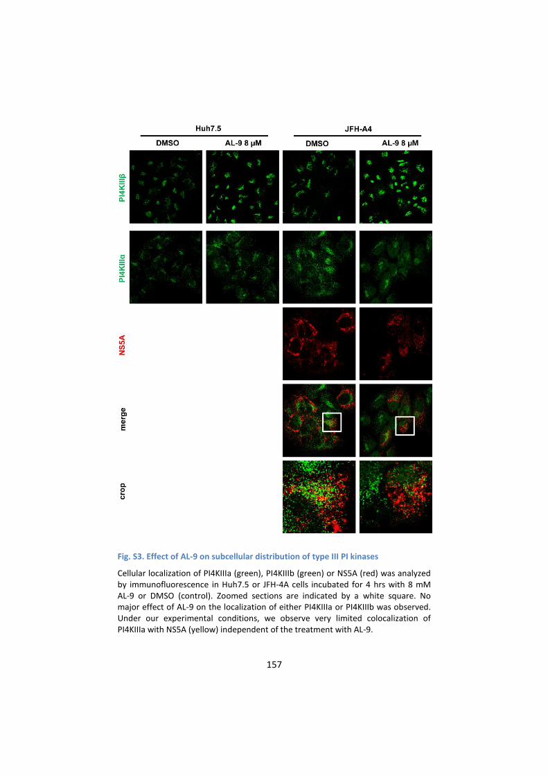
157
Fig. S3. Effect of AL-9 on subcellular distribution of type III PI kinases
Cellular localization of PI4KIIIa (green), PI4KIIIb (green) or NS5A (red) was analyzed by immunofluorescence in Huh7.5 or JFH-4A cells incubated for 4 hrs with 8 mM AL-9 or DMSO (control). Zoomed sections are indicated by a white square. No major effect of AL-9 on the localization of either PI4KIIIa or PI4KIIIb was observed. Under our experimental conditions, we observe very limited colocalization of PI4KIIIa with NS5A (yellow) independent of the treatment with AL-9.

158
4. Protocol S1. Chemical synthesis of compound AL-9
All the reagents were commercially available and used without
further purification unless indicated otherwise. All solvents were
anhydrous grade unless indicated otherwise. When dry conditions
were required, the reactions were carried out in oven-dried glassware
under a slight pressure of argon. Reaction were magnetically stirred
and monitored by thin-layer chromatography (TLC) on silica gel. TLC
was performed on Silica Gel 60 F254 plates (Merck) with UV detection,
or using a developing solution of 0.5% orcinol in EtOH/H2SO4 (3%),
followed by heating at 180°C. Flash column chromatography was
performed on silica gel 230–400 mesh (Merck). The petroleum ether
used as eluent in chromatography has boiling range of 40–60°C. 1H and
13C-NMR spectra were recorded on a Varian 400 MHz MERCURY
instrument at 300 K. Chemical shifts are reported in ppm downfield
from TMS as internal standard. Mass spectra were recorded on ESI-MS
triple quadrupole (model API2000 QTrap™, Applied Biosystems).
tributyl-(5-(diethoxymethyl)furan-2-yl)stannane (1)
To a solution of 2-furaldehyde diethyl acetal (500µL, 2.9 mmol) in
diethyl ether (3 mL) cooled to -78°C, n-butyllitium (3.9 mmol) was
added under Ar atmosphere and the solution was stirred at -78°C for
1h and then for 3 h at 20°C. The solution was cooled again to -78°C and
tributyltin chloride (1 mL, 3.9 mmol) was added dropwise; stirring for 2
h at -78°C and 12 h at room temperature.
The reaction was quenched by adding aqueous NH4Cl and product
extracted with diethyl ether. The organic layer was dried over Na2SO4
159
and concentrated in vacuo. The crude product was used without any
further purification (brown oil, Rf = 0.74, AcOEt/hexane 0.2:9.8).
6-bromoquinazolin-4-ol (2)
To a solution of 2-ammino-5-bromobenzoic acid (5 g, 23.1 mmol),
formamide (15 mL, 377.3 mmol) was added and the mixture was
refluxed under Ar atmosphere for 16 h. Then 6 mL of water were
carefully added (warning! boiling solution) and product immediately
precipitated as a white solid. The mixture was then cooled at RT and
12 mL of water were added slowly. The mixture was stirred 30 min and
the resulting precipitate was filtered and re-crystallized from cold
ethanol obtaining product (white solid, yield: 53%, Rf = 0.51, AcOEt/
petroleum ether 9.4:0.6)
1H-NMR (400 MHz; CDCl3/CD3OD 8:1) δ = 8.34 (d, 1H, J = 2 Hz), 7.97
(s, 1H), 7.83 (dd, 1H, J = 8.7 Hz, J = 2.3 Hz), 7.55 (d, 1H, J = 8.7 Hz).
6-bromo-N-(4-morpholinophenyl)quinazolin-4-amine (3)
To a solution of 6-bromoquinazolin-4-ol (2.84 g, 12.6 mmol) in DMF
(0.84 mL) thionyl chloride (28 mL) was added and the mixture was
refluxed for 5 h. The solvent was evaporated in vacuo by adding some
toluene (3 x 28 mL) to completely remove thionyl chloride. The residue
was dissolved in CH3CN (38 mL), 4-morphoaniline (2.48 mg, 13.9
mmol) was added and the mixture was refluxed for 72 h. Crude
product precipitated upon cooling the solution and the solid
precipitate was recrystallized from diethyl ether obtaining pure
product (orange solid, yield: 79%, Rf = 0.37, AcOEt).

160
1H-NMR (400 Mhz, DMSO-d6) δ = 11.30 (bs, 1H, NH), 9.04 (bs, 1H),
8.87 (s, 1H), 8.19 (bd, 1H, J = 8.8 Hz), 7.79 (d, 1H, J = 8.8 Hz), 7.57-7.04
(AA’XX’ system, 4H, J = 9.0 Hz), 3.74 (m, 4H), 3.14 (m, 4H).
6-(5-(diethoxymethyl)furan-2-yl)-N(4-morpholinophenyl)quinazolin-
4-amine (4)
To a solution of 6-bromo-N-(4-morpholinophenyl)quinazolin-4-
amine (3) (300 mg, 0.78 mmol ) in dry THF (7 mL), tributyl-(5-
(diethoxymethyl)furan-2-yl)stannane (1) (1.4 g, 3.1 mmol) and
bis(triphentlphosphine) palladium dichloride (44 mg, 0.06 mmol) were
added. The reaction mixture was heated to reflux for 25 h under Ar
atmosphere. The solvent was evaporated in vacuo, the crude product
was dissolved in AcOEt and washed with water and brine. The organic
layer was dried over Na2SO4 and concentrated in vacuo. The crude
product obtained was purified with flash chromatography on silica gel
(AcOEt/exane 8:2) obtaining pure product (yellow solid, yield 74%, Rf =
0.24, AcOEt /petroleum ether 9.4:0.6).
1H-NMR (400 MHz, CDCl3) δ= 8.65 (s, 1H), 8.20 (d, 1H, J = 1.5 Hz),
7.97 (dd, 1H, J = 8.8 Hz, J=1.5 Hz), 7.84 (d, 1H, J = 8.8 Hz), 7.56-6.93
(AA’XX’ system, 4H, J = 8.5 Hz), 6.70 (d, 1H, J = 2.7 Hz), 6.53 (d, 1H, J =
2.7 Hz), 5.57 (s, 1H), 3.87 (m, 4H), 3.60 (m,4H), 3.13 (m, 4H), 1.25 (m,
6H).
13C-NMR (400 MHz, CDCl3) δ = 158.02, 155.05, 152.70, 151.97,
149.30, 148.73, 130.24, 129.03, 128.89, 128.48, 124.21, 116.19,
115.31, 114.91, 110.71, 106.86, 96.27, 66.88, 61.54, 49.51, 15.15.
ESI-MS: calculated 474.34, found: 475.21 [M+H+]; 497.21 [M+Na+].
161
5-(4-((4-morpholinophenyl)amino)quinazolin-6-yl)furan-2-
carbaldehyde (5)
To a solution of 6-(5-(diethoxymethyl)furan-2-yl)-N-(4-
morpholinophenyl)quinazolin-4-amine (4) (87 mg, 0.18 mmol) in THF
(8 mL), HCl 2M (1.6 mL) was added and the mixture was reacted for 2 h
at room temperature. The reaction mixture was basified to pH 8 with
NaOH 2M. Organic solvent was evaporated in vacuo and the aqueous
residue was extracted with AcOEt. The organic layer was dried over
Na2SO4 and concentrated in vacuo. Product (orange solid, yield 98%, Rf
= 0.61, AcOEt /MeOH 9:1) was used without any further purification.
1H-NMR (400 MHz, CDCl3) δ = 9.60 (s, 1H), 8.65 (1H, s), 8.41 (1H, d, J
=1.3 Hz), 8.01 (dd, 1H, J= 8.8 Hz, 1.5 Hz ), 7.85-7.57 (AA’XX’ system, 4H,
J = 8.8 Hz), 7.32 (d, 1H, J = 3.7 Hz), 6.92 (m, 3H), 3.82 (t, 4H, J = 4.8 Hz),
3.11 (t, 4H, J = 4.8 Hz).
13C-NMR (400 MHz, CDCl3) δ = 177.21, 158.30, 156.05, 152.12,
150.55, 148.81, 130.03, 129.35, 129.31, 126.35, 124.28, 117.92,
116.03, 115.47, 108.67, 66.84, 49.39.
ESI-MS: calculated 400.15, found: 401.13 [M+H+].
(5-(4-((morpholinophenyl)amino)quinazolin-6-yl)furan-2-
yl)methanol (6)
To a solution of 5-(4-((4-morpholinophenyl)amino)quinazolin-6-
yl)furan-2-carbaldehyde (5) (140 mg, 0.35 mmol) in dry CH2Cl2 (5mL)
and AcOH glacial (0.3 mL), sodium triacetoxyborohydride (148 mg,
0.70 mmol) was added. The solution was reacted for 3 h at room
temperature. Fresh CH2Cl2 was added and washed with water and
brine. Aqueous layer was washed again with fresh AcOEt. The
162
combined organic layers were dried over Na2SO4 and concentrated in
vacuo. The crude product obtained was purified with flash
chromatography on silica gel (AcOEt) obtaining pure product (yellow
solid, yield 35%, Rf = 0.53 AcOEt/MeOH 9:1).
1H-NMR (400 MHz, DMSO-d6) δ = 9.84 (s, 1H), 8.74 (d, 1H, J = 1.4
Hz), 8.44 (s, 1H), 8.11 (dd, 1H, J = 8.8 Hz, J = 1.4 Hz), 7.74-7.61 (AA’XX’
system, 4H, J = 8.8 Hz), 7.03 (d, 1H, J = 3.2 Hz), 6.98 (d, 2H, J = 8.8 Hz),
6.48 (d, 1H, J = 3.2 Hz), 5.32 (t, 1H, J = 5.7 Hz), 4.50 (d, 2H, J = 5.7 Hz),
3.74 (t, 4H, J = 4.7 Hz), 3.09 (t, 4H, J = 4.7 Hz).
13C-NMR (100 MHz, DMSO-d6) δ= 157.80, 156.04, 154.53, 151.79,
148.88, 147.90, 130.79, 128.36, 128.09, 124.14, 116.59, 115.43,
115.07, 109.59, 107.65, 66.13, 55.84, 48.81.
ESI-MS: calculated, 402.17, found: 403.19 [M+H+].
5. Protocol S2. Construction and assays of HCV replicons
harboring putative resistance mutations
The mutants pFKi341-PiLuc-NS3-3’/ET-DLD (E212D, P299L, V388D in
NS5A) and pFKi341-PiLuc-NS3-3’/ET-FAG (L199F, V362A, S390G in
NS5A) were generated starting from pFKi341-PiLuc-NS3-3’/ET
(provided by V. Lohman). Two synthetic fragments (737 or 767 bp,
respectively) containing the indicated triple mutations were provided
from MWG-Eurofins. Using Crossover PCR-based method SalI-BamHI
fragments carrying mutations were obtained and transferred into
pFKi341-PiLuc-NS3-3’-ET backbone. The mutant pFKi341-PiLuc-NS3-3’-
ET-PPA (T200P, S370P in NS5A and S76A in NS5B) was obtained using
Multi Site-Directed Mutagenesis Kit (Agilent Technologies). In
163
particular, the MluI-SpeI fragment of pFKi341-PiLuc-NS3-3’-ET was
subcloned into pCR2.1 vector, mutated according to manifacturer’s
protocol and transferred back into the pFKi341-PiLuc-NS3-3’-ET
backbone. All numbers refer to aminoacid position of HCV proteins
(HCV Con-1; EMBL database accession number AJ238799).
The pFKi341-PiLuc-NS3-3’/ET construct and the mutants pFKi341-
PiLuc-NS3-3’/ET-DLD, pFKi341-PiLuc-NS3-3’/ET-FAG and pFKi341-PiLuc-
NS3-3’/ET-PPA were linearized at the 3’ end of the viral genome by
ScaI digestion and purified by Sodium Acetate precipitation. RNA
transcripts were generated and purified by MEGAscript T7 kit (Ambion)
according to the manufacturer’s protocol. 2x10⁶ Huh7.5 cells were
electroporated with 10 μg of each RNA construct in a final volume of
200 μl as described previously48. Electroporated cells were plated at
the density of 7.5x10³ cells/well in 96-well plates. 6 hours after
electroporation, cells were treated with serial dilutions of AL-9. After
three days of treatment, cell viability was measured by CellTiter-Blue®
(Promega) and luciferase activity was measured by Bright-Glo™
Luciferase Assay System (Promega), according to the manufacturer’s
protocols.
G. Acknowledgments
We wish to thank Glenn Randall, Volker Lohmann and Charles M.
Rice for providing us with essential constructs and cells. We express
our gratitude to Roberto Cighetti for his contribution to the synthesis
of the compound AL-9 and to Manila Boca for her assistance and
training in operating the confocal microscope. We also would like to

164
thank Richard Angell and Magdalena Laver from Arrow Therapeutics
for helpful discussions during this research.
This work/research was partially supported by the Italian Ministry
of Education, University and Research (MIUR) (grant number
RBAP10TPXK to INGM). FP acknowledges the Regione Lombardia for
financial support (grant number 14546, Network Enabled Drug Design,
NEDD). The funders have no role in the study design, data collection
and analysis, decision to publish, or preparation of the manuscript.
165
H. Chapter 2 – References 1. Vermehren, J. & Sarrazin, C. New HCV therapies on the
horizon. Clin Microbiol Infect 17, 122-34. 2. Lemon, S.M. et al. Development of novel therapies for
hepatitis C. Antiviral Res 86, 79-92. 3. Blight, K.J., Kolykhalov, A.A. & Rice, C.M. Efficient initiation of
HCV RNA replication in cell culture. Science 290, 1972-4 (2000).
4. Tellinghuisen, T.L., Foss, K.L., Treadaway, J.C. & Rice, C.M. Identification of residues required for RNA replication in domains II and III of the hepatitis C virus NS5A protein. J Virol 82, 1073-83 (2008).
5. Huang, L. et al. Hepatitis C virus nonstructural protein 5A (NS5A) is an RNA-binding protein. J Biol Chem 280, 36417-28 (2005).
6. Lohmann, V., Korner, F., Dobierzewska, A. & Bartenschlager, R. Mutations in hepatitis C virus RNAs conferring cell culture adaptation. J Virol 75, 1437-49 (2001).
7. Tellinghuisen, T.L., Foss, K.L. & Treadaway, J. Regulation of hepatitis C virion production via phosphorylation of the NS5A protein. PLoS Pathog 4, e1000032 (2008).
8. Appel, N. et al. Essential role of domain III of nonstructural protein 5A for hepatitis C virus infectious particle assembly. PLoS Pathog 4, e1000035 (2008).
9. Hughes, M., Griffin, S. & Harris, M. Domain III of NS5A contributes to both RNA replication and assembly of hepatitis C virus particles. J Gen Virol 90, 1329-34 (2009).
10. Tellinghuisen, T.L., Marcotrigiano, J., Gorbalenya, A.E. & Rice, C.M. The NS5A protein of hepatitis C virus is a zinc metalloprotein. J Biol Chem 279, 48576-87 (2004).
11. Tellinghuisen, T.L., Marcotrigiano, J. & Rice, C.M. Structure of the zinc-binding domain of an essential component of the hepatitis C virus replicase. Nature 435, 374-9 (2005).
12. Love, R.A., Brodsky, O., Hickey, M.J., Wells, P.A. & Cronin, C.N. Crystal structure of a novel dimeric form of NS5A domain I protein from hepatitis C virus. J Virol 83, 4395-403 (2009).
166
13. Hanoulle, X. et al. Domain 3 of non-structural protein 5A from hepatitis C virus is natively unfolded. Biochem Biophys Res Commun 381, 634-8 (2009).
14. Liang, Y., Ye, H., Kang, C.B. & Yoon, H.S. Domain 2 of nonstructural protein 5A (NS5A) of hepatitis C virus is natively unfolded. Biochemistry 46, 11550-8 (2007).
15. Conte, I. et al. Synthesis and SAR of piperazinyl-N-phenylbenzamides as inhibitors of hepatitis C virus RNA replication in cell culture. Bioorg Med Chem Lett 19, 1779-83 (2009).
16. Lemm, J.A. et al. Identification of hepatitis C virus NS5A inhibitors. J Virol 84, 482-91.
17. Fridell, R.A. et al. Genotypic and phenotypic analysis of variants resistant to HCV NS5A replication complex inhibitor BMS-790052: In Vitro and In Vivo correlations. Hepatology.
18. Schmitz, U. & Tan, S.L. NS5A--from obscurity to new target for HCV therapy. Recent Pat Antiinfect Drug Discov 3, 77-92 (2008).
19. Dancey, J. & Sausville, E.A. Issues and progress with protein kinase inhibitors for cancer treatment. Nat Rev Drug Discov 2, 296-313 (2003).
20. Moy, B., Kirkpatrick, P., Kar, S. & Goss, P. Lapatinib. Nat Rev Drug Discov 6, 431-2 (2007).
21. Sebolt-Leopold, J.S. & English, J.M. Mechanisms of drug inhibition of signalling molecules. Nature 441, 457-62 (2006).
22. Borawski, J. et al. Class III phosphatidylinositol 4-kinase alpha and beta are novel host factor regulators of hepatitis C virus replication. J Virol 83, 10058-74 (2009).
23. Tai, A.W. et al. A functional genomic screen identifies cellular cofactors of hepatitis C virus replication. Cell Host Microbe 5, 298-307 (2009).
24. Berger, K.L. et al. Roles for endocytic trafficking and phosphatidylinositol 4-kinase III alpha in hepatitis C virus replication. Proc Natl Acad Sci U S A 106, 7577-82 (2009).
25. Vaillancourt, F.H. et al. Identification of a lipid kinase as a host factor involved in hepatitis C virus RNA replication. Virology 387, 5-10 (2009).
167
26. Berger, K.L., Kelly, S.M., Jordan, T.X., Tartell, M.A. & Randall, G. Hepatitis C virus stimulates the phosphatidylinositol 4-kinase III alpha-dependent phosphatidylinositol 4-phosphate production that is essential for its replication. J Virol.
27. Lim, Y.S. & Hwang, S.B. Hepatitis C virus NS5A protein interacts with phosphatidylinositol 4-kinase type IIIalpha and regulates viral propagation. J Biol Chem 286, 11290-8.
28. Reiss, S. et al. Recruitment and activation of a lipid kinase by hepatitis C virus NS5A is essential for integrity of the membranous replication compartment. Cell Host Microbe 9, 32-45.
29. Wong, K., Meyers dd, R. & Cantley, L.C. Subcellular locations of phosphatidylinositol 4-kinase isoforms. J Biol Chem 272, 13236-41 (1997).
30. Weixel, K.M., Blumental-Perry, A., Watkins, S.C., Aridor, M. & Weisz, O.A. Distinct Golgi populations of phosphatidylinositol 4-phosphate regulated by phosphatidylinositol 4-kinases. J Biol Chem 280, 10501-8 (2005).
31. Balla, A., Tuymetova, G., Tsiomenko, A., Varnai, P. & Balla, T. A plasma membrane pool of phosphatidylinositol 4-phosphate is generated by phosphatidylinositol 4-kinase type-III alpha: studies with the PH domains of the oxysterol binding protein and FAPP1. Mol Biol Cell 16, 1282-95 (2005).
32. Balla, A. et al. Maintenance of hormone-sensitive phosphoinositide pools in the plasma membrane requires phosphatidylinositol 4-kinase IIIalpha. Mol Biol Cell 19, 711-21 (2008).
33. Hammond, G.R., Schiavo, G. & Irvine, R.F. Immunocytochemical techniques reveal multiple, distinct cellular pools of PtdIns4P and PtdIns(4,5)P(2). Biochem J 422, 23-35 (2009).
34. Knight, Z.A. et al. A pharmacological map of the PI3-K family defines a role for p110alpha in insulin signaling. Cell 125, 733-47 (2006).
35. Toth, B. et al. Phosphatidylinositol 4-kinase IIIbeta regulates the transport of ceramide between the endoplasmic reticulum and Golgi. J Biol Chem 281, 36369-77 (2006).
168
36. Miller, S. & Krijnse-Locker, J. Modification of intracellular membrane structures for virus replication. Nat Rev Microbiol 6, 363-74 (2008).
37. Hsu, N.Y. et al. Viral reorganization of the secretory pathway generates distinct organelles for RNA replication. Cell 141, 799-811.
38. Backes, P. et al. Role of annexin A2 in the production of infectious hepatitis C virus particles. J Virol 84, 5775-89.
39. D'Angelo, G., Vicinanza, M., Di Campli, A. & De Matteis, M.A. The multiple roles of PtdIns(4)P -- not just the precursor of PtdIns(4,5)P2. J Cell Sci 121, 1955-63 (2008).
40. Heaton, N.S. & Randall, G. Multifaceted roles for lipids in viral infection. Trends Microbiol 19, 368-75.
41. Salonen, A., Ahola, T. & Kaariainen, L. Viral RNA replication in association with cellular membranes. Curr Top Microbiol Immunol 285, 139-73 (2005).
42. Arita, M. et al. Phosphatidylinositol 4-kinase III beta is a target of enviroxime-like compounds for antipoliovirus activity. J Virol 85, 2364-72.
43. Stefan, C.J. et al. Osh proteins regulate phosphoinositide metabolism at ER-plasma membrane contact sites. Cell 144, 389-401.
44. Gao, L., Aizaki, H., He, J.W. & Lai, M.M. Interactions between viral nonstructural proteins and host protein hVAP-33 mediate the formation of hepatitis C virus RNA replication complex on lipid raft. J Virol 78, 3480-8 (2004).
45. Hamamoto, I. et al. Human VAP-B is involved in hepatitis C virus replication through interaction with NS5A and NS5B. J Virol 79, 13473-82 (2005).
46. Amako, Y., Sarkeshik, A., Hotta, H., Yates, J., 3rd & Siddiqui, A. Role of oxysterol binding protein in hepatitis C virus infection. J Virol 83, 9237-46 (2009).
47. Blight, K.J., McKeating, J.A. & Rice, C.M. Highly permissive cell lines for subgenomic and genomic hepatitis C virus RNA replication. J Virol 76, 13001-14 (2002).
48. Pacini, L., Graziani, R., Bartholomew, L., De Francesco, R. & Paonessa, G. Naturally occurring hepatitis C virus subgenomic deletion mutants replicate efficiently in Huh-7 cells and are

169
trans-packaged in vitro to generate infectious defective particles. J Virol 83, 9079-93 (2009).
49. Hopkins, S. et al. SCY-635, a novel nonimmunosuppressive analog of cyclosporine that exhibits potent inhibition of hepatitis C virus RNA replication in vitro. Antimicrob Agents Chemother 54, 660-72.

170
171
CHAPTER 3
The presence of hepatitis C virus deletion mutants
is associated with necro-inflammatory activity and
pattern of response to therapy in HCV1 patients
C. Cheroni#1, L. Donnici#1, A. Aghemo2, S. Noppornpanth1, V.
Valveri1, A. Bianco1, P. Neddermann1, M. Pagani1, R.
Soffredini2, R. D’Ambrosio2, M.G. Rumi3, M. Colombo2, S.
Abrignani1 and R. De Francesco1
1 Department of Genomics and Molecular Biology – Virology Program
Istituto Nazionale Genetica Molecolare (INGM) – Milano, Italy
2 A.M. Migliavacca Center for Liver Disease, First Division of
Gastroenterology, Fondazione IRCCS Ca’ Granda Ospedale Maggiore
Policlinico Milano, Universita` degli Studi di Milano, Milan, Italy
3 Division of Hepatology, Ospedale San Giuseppe IRCCS Multimedica,
Università degli Studi di Milano, Milan, Italy
# These authors contributed equally to the manuscript
This work is going to be submitted for publication to Hepatology.
172
A. Abstract
Hepatitis C virus (HCV) variants characterized by genomic deletions
in the region of structural proteins have been sporadically detected in
the serum and liver of hepatitis C patients. These defective genomes
are capable of autonomous RNA replication and are packaged into
infectious viral particles in cells co-infected with the wild-type virus.
The frequency of such forms in HCV infected population, as well as
their impact on the progression of liver disease and antiviral therapy
outcome, are currently unknown.
To investigate the incidence of HCV defective variants and their
correlations with clinical characteristics, a screening campaign was
performed on pre-therapy serum samples from a well-characterized
cohort of HCV1 naïve patients who received treatment with Pegylated
Interferon alpha (PEG-IFNα) and Ribavirin (RBV). 132 HCV1 subjects
were successfully analyzed for the presence of defective species
exploiting a long-distance nested PCR assay spanning from Core to
NS3.
HCV forms with deletions affecting prevalently E1, E2 and p7
proteins were found in a high fraction (25/132, 19%) of the subjects.
Their presence was associated with patient older age, higher viral load
and increased inflammatory activity. While the presence of
subgenomic HCV did not appear to influence significantly virological
decline nor sustained virological response rates to PEG-IFNα and RBV
(24% vs. 38%) it resulted to be an independent predictor of relapse in
173
the population reaching the viral clearance at the end of the treatment
(OR:3.35).
In conclusion, in chronic HCV1 patients, the presence of HCV with
defective genomes was associated with hepatic necro-inflammatory
lesions. Furthermore, defective HCV RNA may represent a novel
predictor of relapse after initial virological response following PEG-
IFNα/RBV therapy.
B. Introduction
Hepatitis C virus (HCV) is an enveloped virus belonging to the family
of Flaviviridae. Its genome consists of a positive-stranded RNA
molecule of approximately 9.6 Kb that is structured in a 5’ non-coding
region (5’UTR), a long open reading frame encoding a polyprotein
precursor of about 3010 amino acids and a 3’UTR region. The
polyprotein is processed by the action of both viral and cellular
proteases to produce structural (Core and E1-E2 glycoproteins) and
non-structural proteins (p7, NS2, NS3, NS4A-B and NS5A-B). The RNA-
dependent RNA polymerase NS5B produces with a high frequency of
incorrect nucleotide insertion; this characteristic, coupled with the lack
of proofreading ability and the high in-vivo productive rate of new
virions, is believed to be the driving force of HCV genetic diversity1, 2.
HCV infection affects an estimated 170 million people worldwide
and is the major cause of chronic liver disease, liver cirrhosis and
hepatocellular carcinoma3. Standard antiviral therapy is based on long-
term administration of Pegylated Interferon alpha (PEG-IFNα) and
174
ribavirin (RBV), but permits to achieve viral clearance only in about 40-
60% of cases.
Both viral and host genetic variability are thought to influence
treatment success rates. Recently, whole-genome association studies
identified host single nucleotide polymorphisms (SNPs) near the
genomic region encoding IL28B as strongly associated with both
spontaneous and treatment-induced viral clearance4-6: under PEG-
IFNα/RBV therapy, HCV1 patients having the rs12979860 CT or TT
genotype display reduced viral load decline and lower SVR rates.
Regarding viral genetics, HCV genotypes 1 and 4 are considered more
difficult to treat than genotypes 2 and 37.
HCV variants characterized by genomic deletions have been
highlighted in the serum and liver of chronic HCV patients8-10 as well as
in the serum of immunosilent infections11 and liver transplant
recipients with recurrent HCV infection12. These defective genomes
contain large in-frame deletions affecting prevalently the Envelope (E1
and E2) region, but retain the Core region in addition to the genome
portions that are essential for autonomous HCV replication (5’UTR and
NS3-NS5-3’UTR)8. It has recently been demonstrated9, 10 in in vitro
infectivity models that subgenomic HCV RNAs with the architecture of
the naturally occurring ones are capable of autonomous replication
and are expressed at the protein levels, display a higher replication
fitness compared to the full-length genome and are efficiently
packaged into infectious viral particles when co-expressed together
with full-length viral genome. Their structural peculiarities confer to
HCV defective particles remarkable features: although they require the
175
complementation of structural proteins by a wild type virus to
complete virion assembly and exit, upon infection of a naïve
hepatocyte they will transduce self-replicating HCV subgenomes,
resulting in the persistent expression of HCV proteins even in the
absence of co-infection with the wild-type helper virus.
Deleted HCV species are reminiscent of defective interfering (DI)
forms that are reported for other RNA viruses, including Flaviviridae
family members pestivirus (Classical Swine Fever Virus – CSFV – and
Bovine Viral Diarrhea Virus – BVDV – 13-15) and flavivirus (Murray Valley
encephalitis virus16). DI variants are characterized by large genomic
deletions, but retain the regions that are critically required for genome
replication and encapsidation17; therefore, they are both dependent
on and competitive with the parental virus.
While a key pathogenic role of DI forms is already well-established
for flaviviruses such as CSFV and BVDV, to date the clinical relevance of
HCV defective particles, as well as their incidence in general HCV
infected population, have never been systematically evaluated. To
address this point, we undertook a screening campaign for HCV
defective genomes on pre-therapy serum samples of a well-
characterized cohort of chronic HCV1 patients undergoing standard
therapy treatment.

176
C. Results
1. HCV particles with defective genomes are found in the serum
of a large fraction of chronic hepatitis C genotype 1 patients
Pre-therapy serum samples of 132 subjects fulfilling the selection
criteria were screened for the presence of HCV defective species
exploiting a long-distance nested PCR assay spanning from core to
NS3.
Demographic and clinical characteristics of the patients are
described in Tables 1A and 1B. Both genders were well represented
(52.3% males) and the median age was 56 years (IQR 19.25). 30.3% of
patients had moderate to severe histological activity (Grading 9) and
37.1% of them showed advanced fibrosis (Staging 4).
RVR (rapid virological response) and cEVR (complete early
virological response) were obtained respectively in 34.1% and 58.3% of
the cohort. At the end of treatment and follow-up period, 47 subjects
(35.6%) achieved SVR, while 27 experienced a viral relapse and 58
were non-responders.
Large fragment PCR amplification, from nucleotide 127 (5’UTR
region) to 3649 (NS3 protein), followed by agarose gel analysis
revealed in a portion of the subjects the presence of amplicons
consistently smaller than those expected from the amplification of full-
length HCV genome (ranging from 1200 to 2000 bps, compared to
3522), thus suggesting the occurrence of large deletions (Fig. 1A). This
screening strategy, being based on a very sensitive nested PCR
technique that gives a preferential amplification of shorter amplicons,
is able to detect the defective variant even if more rare than the full-
177
length variant. However, we cannot completely rule out the possibility
to have classified as full-length patients in which the % of defective is
very low.
Subsequent cloning and nucleotide sequencing investigation of the
shorter amplicons was employed in each putative defective sample to
confirm and further characterize the deletion events. Defective HCV
genomes were confirmed in a high fraction of the cohort: we observed
defective variants in 25/132 (18.9%) of the patients. Sequencing
analysis highlighted the presence of in-frame deletions affecting
prevalently envelope proteins and p7 (Figure 1C). In fact, in all the
defective variants, E2 was completely deleted and E1 and p7 were at
least partially missing; the initial portion of NS2 was lost in 21 (84%) of
them. Two deletions in a single genome were observed in 3 patients, in
which a small residual fragment ranging from 28 to 43 bps was
localized between the two large deletions.
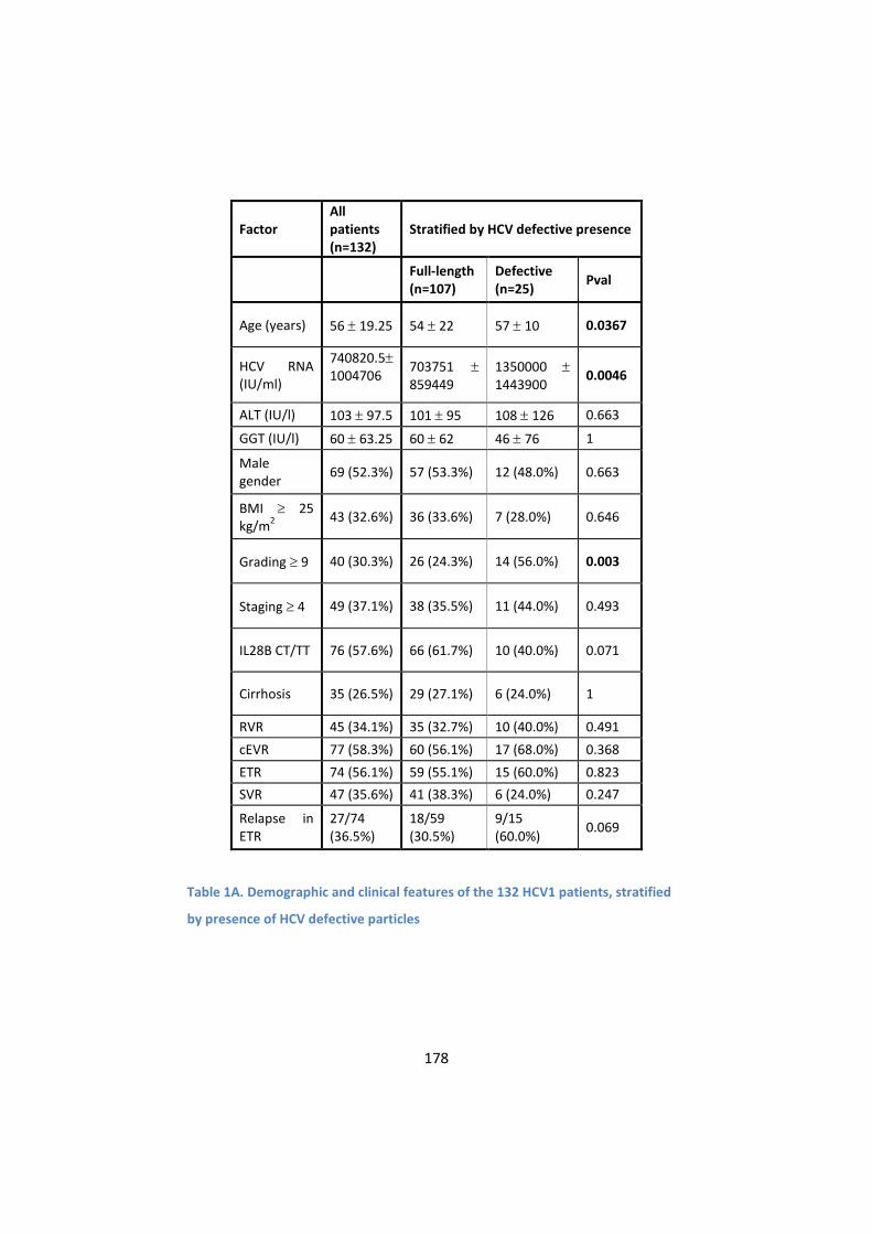
178
Factor All patients (n=132)
Stratified by HCV defective presence
Full-length (n=107)
Defective (n=25)
Pval
Age (years) 56 19.25 54 22 57 10 0.0367
HCV RNA (IU/ml)
740820.5 1004706
703751 859449
1350000 1443900
0.0046
ALT (IU/l) 103 97.5 101 95 108 126 0.663
GGT (IU/l) 60 63.25 60 62 46 76 1
Male gender
69 (52.3%) 57 (53.3%) 12 (48.0%) 0.663
BMI 25 kg/m
2
43 (32.6%) 36 (33.6%) 7 (28.0%) 0.646
Grading 9 40 (30.3%) 26 (24.3%) 14 (56.0%) 0.003
Staging 4 49 (37.1%) 38 (35.5%) 11 (44.0%) 0.493
IL28B CT/TT 76 (57.6%) 66 (61.7%) 10 (40.0%) 0.071
Cirrhosis 35 (26.5%) 29 (27.1%) 6 (24.0%) 1
RVR 45 (34.1%) 35 (32.7%) 10 (40.0%) 0.491
cEVR 77 (58.3%) 60 (56.1%) 17 (68.0%) 0.368
ETR 74 (56.1%) 59 (55.1%) 15 (60.0%) 0.823
SVR 47 (35.6%) 41 (38.3%) 6 (24.0%) 0.247
Relapse in ETR
27/74 (36.5%)
18/59 (30.5%)
9/15 (60.0%)
0.069
Table 1A. Demographic and clinical features of the 132 HCV1 patients, stratified
by presence of HCV defective particles
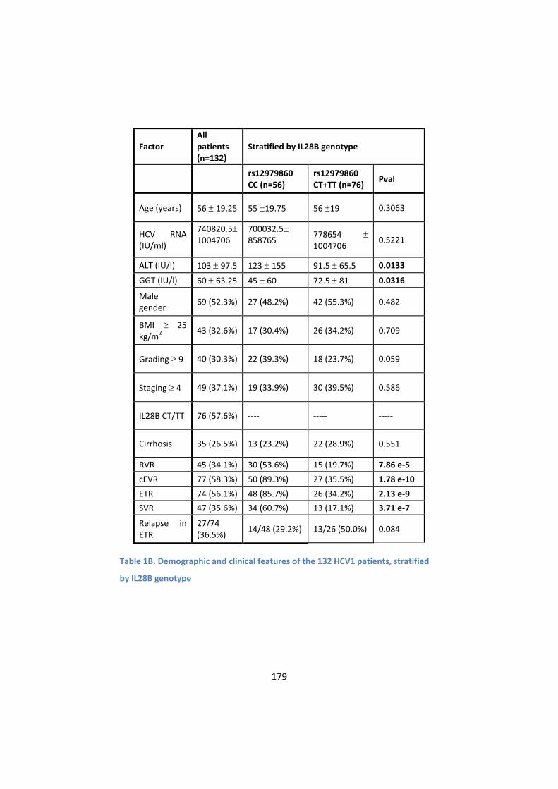
179
Factor All patients (n=132)
Stratified by IL28B genotype
rs12979860 CC (n=56)
rs12979860 CT+TT (n=76)
Pval
Age (years) 56 19.25 55 19.75 56 19 0.3063
HCV RNA (IU/ml)
740820.5 1004706
700032.5 858765
778654 1004706
0.5221
ALT (IU/l) 103 97.5 123 155 91.5 65.5 0.0133
GGT (IU/l) 60 63.25 45 60 72.5 81 0.0316
Male gender
69 (52.3%) 27 (48.2%) 42 (55.3%) 0.482
BMI 25 kg/m
2
43 (32.6%) 17 (30.4%) 26 (34.2%) 0.709
Grading 9 40 (30.3%) 22 (39.3%) 18 (23.7%) 0.059
Staging 4 49 (37.1%) 19 (33.9%) 30 (39.5%) 0.586
IL28B CT/TT 76 (57.6%) ---- ----- -----
Cirrhosis 35 (26.5%) 13 (23.2%) 22 (28.9%) 0.551
RVR 45 (34.1%) 30 (53.6%) 15 (19.7%) 7.86 e-5
cEVR 77 (58.3%) 50 (89.3%) 27 (35.5%) 1.78 e-10
ETR 74 (56.1%) 48 (85.7%) 26 (34.2%) 2.13 e-9
SVR 47 (35.6%) 34 (60.7%) 13 (17.1%) 3.71 e-7
Relapse in ETR
27/74 (36.5%)
14/48 (29.2%) 13/26 (50.0%) 0.084
Table 1B. Demographic and clinical features of the 132 HCV1 patients, stratified
by IL28B genotype

180
Fig. 1. Viral genome architecture of the defective forms identified in the serum of
HCV1 patients
Representative agarose-gel pictures showing full-length (lanes 1-2, 4-5 of the first gel and 1-5 of the second gel) and defective bands (lanes 3, 6 of the first gel and 6, 7 of the second gel) after the second round of nested PCR (A). Usually, in samples showing the shorter band, the full-length genome could not be detected, given the preferential amplification of the deletion mutant. Representative agarose-gel picture showing amplification of a small core segment from the same RNA (B). Schematic representation of the architecture of the 25 defective variants identified in the sera of HCV1 patients (C).
181
2. The presence of defective genomes is associated with patient
older age, higher viral load and increased necro-inflammatory
activity
We then examined the correlations between the presence of
defective HCV forms and clinical characteristics or viral kinetics on the
132 HCV1 patients of the cohort.
As visible in Table 1A, the presence of HCV defective genomes was
found to be associated with older patient age (Wilcoxon test Pval
0.0367), higher viremic load (Wilcoxon test Pval 0.0046) and mild to
severe hepatic necro-inflammatory activity (histological activity index
≥9; Fisher’s test Pval 0.003).
Analyzing viral kinetics during PEG-IFNα/RBV treatment, we could
not detect substantial differences in the decline of viral load at early
time-points (RVR and cEVR rates) in carriers of deleted variants; also
ETR percentages were unchanged. SVR rates tended to be lower for
patients with defective genomes (24% compared to 38.3%), but the
difference was far from statistic significance (Fisher test Pval 0.247, Fig
2A).
Strikingly, however, among 74 patients who were HCV RNA-
negative at the end of the treatment, virological relapse was observed
in 60% (9/15) of the subjects carrying HCV defective genomes
compared to 30% (18/59) of those infected with only the full-length
virus (Fig. 2B). The association is characterized by a strong odds ratio
(3.35) and a moderate statistic significance in univariable analysis (Chi-
test Pval 0.034, Fisher’s test Pval 0.069).
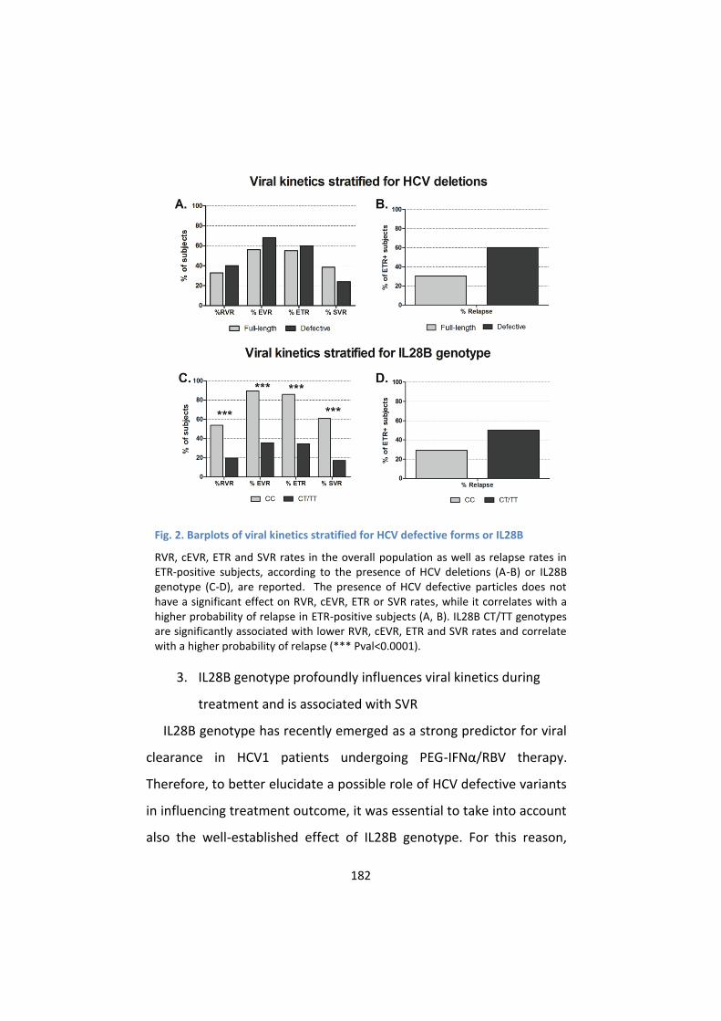
182
Fig. 2. Barplots of viral kinetics stratified for HCV defective forms or IL28B
RVR, cEVR, ETR and SVR rates in the overall population as well as relapse rates in ETR-positive subjects, according to the presence of HCV deletions (A-B) or IL28B genotype (C-D), are reported. The presence of HCV defective particles does not have a significant effect on RVR, cEVR, ETR or SVR rates, while it correlates with a higher probability of relapse in ETR-positive subjects (A, B). IL28B CT/TT genotypes are significantly associated with lower RVR, cEVR, ETR and SVR rates and correlate with a higher probability of relapse (*** Pval<0.0001).
3. IL28B genotype profoundly influences viral kinetics during
treatment and is associated with SVR
IL28B genotype has recently emerged as a strong predictor for viral
clearance in HCV1 patients undergoing PEG-IFNα/RBV therapy.
Therefore, to better elucidate a possible role of HCV defective variants
in influencing treatment outcome, it was essential to take into account
also the well-established effect of IL28B genotype. For this reason,
183
rs12979860 polymorphism was determined in the patients of our
cohort. SNP genotype frequencies in HCV1 population were 42.4% CC,
28% CT and 29.6% TT; therefore, 57.6% of the subjects were carriers of
the “non-responder” T allele.
The relationships between IL28B genotype and clinical
characteristics and viral kinetics are listed in Table 1B. IL28B
unfavorable genotype resulted associated with lower ALT and higher
GGT pre-therapy levels (Wilcoxon test Pval 0.0133 and 0.0316
respectively). Moreover, a weak association (Fisher’s test Pval 0.059)
was noticeable between IL28B unfavorable genotype and lower
grading scores.
Analyzing the therapy outcome, IL28B unfavorable genotype
strongly correlated with treatment failure (OR 7.35, Pval 3.71e-7); the
effect is noticeable already at early time points (RVR and cEVR rates,
Fig2C-D).
4. The association between HCV defective particles and higher
histological grading is independent from IL28B genotype
In order to confirm the correlation between increased necro-
inflammatory activity and the presence of HCV defective particles by
also taking in account the possible role of IL28B genotype and other
confounding factors, a stepwise multivariable logistic regression model
was exploited. The stepwise approach was used for the selection of
the variables and a final model was established on the basis of the
minimum Akaike Information Criterion (AIC). As shown in Table 2, the
model confirmed the role of HCV deletions (Pval 0.028, OR 2.89), along
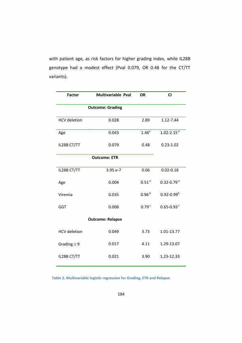
184
with patient age, as risk factors for higher grading index, while IL28B
genotype had a modest effect (Pval 0.079, OR 0.48 for the CT/TT
variants).
Factor Multivariable Pval OR CI
Outcome: Grading
HCV deletion 0.028 2.89 1.12-7.44
Age 0.043 1.46a 1.02-2.15 a
IL28B CT/TT 0.079 0.48 0.23-1.02
Outcome: ETR
IL28B CT/TT 3.95 e-7 0.06 0.02-0.18
Age 0.004 0.51 a 0.32-0.79 a
Viremia 0.035 0.96 b 0.92-0.99b
GGT 0.008 0.79 c 0.65-0.93 c
Outcome: Relapse
HCV deletion 0.049 3.73 1.01-13.77
Grading 9 0.017 4.11 1.29-13.07
IL28B CT/TT 0.021 3.90 1.23-12.33
Table 2. Multivariable logistic regression for Grading, ETR and Relapse
185
OR: odds ratio – CI: 95% confidence interval a: odds ratio and confidence intervals have been calculated considering an interval of 10 years. b: odds ratio and confidence intervals have been calculated considering an interval of 10
5 units of viremia
c: odds ratio and confidence intervals have been calculated considering an interval of 20 units of GGT
5. Defective HCV particles and IL28B genotype are independent
predictors of relapse in ETR-positive patients
Using the same approach, we exploited a multivariable logistic
regression model to pinpoint the parameters associated with viral
clearance at the end of treatment (ETR). In the final model,
summarized in Table 2, IL28B CC genotype was the variable most
strongly and positively associated with ETR (Pval 3.95e-7, OR 0.06 for
CT/TT variants, corresponding to an OR of 16.6 for CC genotype).
Younger patient age and lower pre-treatment viral load and GGT levels
also resulted significantly associated with ETR. No impact on ETR
obtainment was found for the presence of defective HCV particles,
that therefore was not included as co-variate in the model.
Similarly, a model was constructed to evaluate the explanatory
variables predicting virological relapse in the 74 subjects who where
HCV-negative at the end of treatment. As described in Table 2, the
presence of deletions in HCV genome represented a risk factor for
relapse with an odds ratio of 3.73 (Pval 0.049), along with mild-to-
severe histological grading (OR 4.11, Pval 0.017) and IL28B CT/TT
genotypes (OR 3.90, Pval 0.021).

186
D. Discussion
In this study, we analyzed the presence of viral defective forms in a
well-characterized cohort of HCV1 patients. The screening campaign
was designed to detect deletion events occurring in the region of Core-
NS3 and revealed the presence of large in-frame deletions affecting
prevalently E1, E2 and p7 proteins. This finding is in line with already
published results; in fact, the subgenomic HCV species reported in
previous studies conserved the regions essential for RNA replication
(5’UTR, 3’UTR and from NS3 to NS5B), intact Core and the protease
domain of NS29, 10, 12, 18.
Analyzing pre-therapy sera of 132 patients, defective genomes were
found in about 20% of them; to our knowledge, this is the first
systematic analysis done on such a large number of subjects, and it
permits to infer that the emerging of deletion mutants, far from being
a sporadic phenomenon, occurs in a relevant portion of HCV1-infected
subjects.
Defective interfering forms are reported for a variety of viruses, and
their clinical impact is variegate19. In fact, as a consequence of DI
particles insurgence, a reduction in the titers of the parental virus20
and attenuation of disease severity21 have been reported; on the other
hand, it has been proposed that DI viruses may play a role in the
establishment and maintenance of persistent infection, as described
for Murray Valley Encephalitis Virus and Japanese Encephalitis Virus16,
22, 23. Moreover, a crucial pathogenic role has been established for
some DI viruses belonging to the Flaviviridae family. In the case of
BVDV, a highly cytopathogenic viral isolate was shown to contain a DI
187
virus lacking the genes encoding the structural proteins14. In fact, the
pathogenesis of lethal mucosa disease in animals persistently infected
with non-cytopathogenic BVDV has been ascribed to the insurgence
cytopathogenic variants containing genomic rearrangements, among
which internal deletions24. Similarly, cytopathogenicity of CSFV was
demonstrated to be associated to a DI variant lacking the structural
protein-coding region25.
Regarding HCV, the ability of defective particles to influence the
clinical course of the disease is largely unexplored. Focalizing on the
clinical characteristics of our cohort, we observed that the presence of
HCV defective variants correlates with higher viral load, older patient
age and higher degree of hepatic inflammation. Since defective viruses
are thought to be generated from the wild type through errors of the
polymerase, the probability of their insurgence increases
proportionally with the number of replication events. This may be at
the basis of the association with older patient age (which probably
could be considered as a proxy of the duration of infection) and higher
viremia. From an etiopathological point of view, the association with
the grading score appears more intriguing, raising the possibility that
HCV defective particles exacerbate the liver inflammation processes.
Given their high replication fitness and inability to autonomously exit
from the cell, deleted variants are likely more prone to lead to the
accumulation of viral proteins inside the cell; therefore, the persistent
expression of HCV defective genomes in liver hepatocytes could have
important pathological consequences. From this point of view, the
pathogenic activity of defective forms could results from the balance
188
between the ability to accumulate in host cells (in absence of the
helper virus) and, when complemented with the wild type structural
proteins, to spread to other cells. Since the presence of an intact Core
is one of the hallmarks of defective HCV genomes we found, it is
conceivable that cells infected with subgenomic HCV will accumulate
very high level of intracellular Core protein that, in contrast to the case
of cells infected with the wild type virus, will not be counterbalanced
by the formation of mature virions and virus exit. HCV Core protein is
normally localized on the surface of cytoplasmic lipid droplets (LDs),
which are believed to represent sites of virus assembly and production
of nascent virions26. Overexpression of intracellular Core has been
reported to cause abnormal accumulation and redistribution of LDs27,
28, as well as to induce the production for free radicals and apoptotic
cell death29. Moreover, Core protein has been indicated to be involved
in liver steatosis, fibrosis and carcinogenesis in various mouse model
systems30.
Altered expression of other viral proteins may also negatively
impact the hepatocyte function. As already described, all the deleted
variants found were characterized by the partial or total loss of E1, E2
and p7. While the involvement of E1 and E2 envelope glycoproteins in
viral entry is well-established, the function of p7 in HCV life cycle is less
understood, although it has been reported to be fundamental for virus
infection31 but not necessary for RNA replication32, 33. More recently, it
has been demonstrated that p7 plays a crucial role in re-directing Core
from the LDs to the ER, probably favoring virus assembly34, 35; more
precisely, the interactions between Core, p7 and the first

189
transmembrane domain of NS2 determine the degree of localization of
core in ER or LD36. In this context, the partial or complete lack of p7
may be another mechanism that favors the accumulation of Core on
the lipid droplets. Finally, also the fusion protein produced by the
deletion event may be able to exacerbate the pathological changes in
the cell. Induction of ER stress has been reported in both in vitro
models of HCV and biopsies of HCV patients37; since fusion proteins
are probably more prone to be unfolded or misfolded, it could be
speculated that ER stress is intensified in patients carriers of defective
variants. A growing body of evidence suggests that ER stress and the
inflammatory response are strictly interconnected38.
We did not observe a correlation between the presence of
defective forms and the staging score. However, it should be
considered that, for this cohort, the lack of knowledge of the time of
infection and of insurgence of the defective variants prevents us to
correctly estimate the fibrosis progression rate and the impact of the
subgenomic virus on it.
We then evaluated the viral kinetics during standard therapy
treatment in HCV1 patients carrying the defective forms compared to
those in which only the full-length variant was detected. No difference
was found in RVR, cEVR and ETR rates, thus indicating that defective
particles do not seem to play a key role in determining viral load
decline during treatment. This result was confirmed also by logistic
regression analysis, in which IL28B genotype, patient age and pre-
treatment viremia, but not the presence of defective HCV, were
identified as independent predictors of ETR. Refining the analysis on

190
the ETR-positive population, however, we observed that defective
particle carriers displayed a higher probability to experience virological
relapse during the follow-up period. This result is particularly
interesting, since it has been proposed that defective virus belonging
to the Flaviviridae family may play a role in the establishment and
maintenance of chronic infections, although the majority of the data
comes from in-vitro experiments16, 23. A possible role in viral
persistence has been suggested also for deleted variants of hepatitis B
virus (HBV). Deletions in the surface antigen regions S1 and S2 of HBV
genome have been reported in sera and liver of HBV patients39, 40 and,
since pre-S region contains several epitopes for T or B cells41, the
emergence of deleted HBV variants may result in an impairment of
viral clearance and therefore potentially be a mechanism of evasion of
immune surveillance. Moreover, the incidence of these variants
increases with the progression of the disease42.
Multivariable analysis confirmed the role of subgenomic HCV as
independent risk factor for relapse, with an effect magnitude (OR 3.73)
comparable to that of IL28B genotype (OR 3.90). Interestingly in our
cohort, 3 out of 4 (75%) of ETR-positive subjects carriers of defective
forms and IL28B unfavorable genotype experienced virological relapse,
while only 22% (8/37) of patients with the IL28B favorable genotype
and lacking defective forms had a post-treatment relapse.
Although this might suggest a role for both IL28B genotype and HCV
with defective genomes in determining the pattern of response to
PEG-IFNα/RBV, we acknowledge that external validation of our data is
necessary. Indeed, although our cohort is the largest one to date to be
191
analyzed for the presence of defective forms of HCV, it is still quite
limited in size to provide conclusive evidence of their role in modifying
treatment response. Further reinforcing the need for external
validations is the fact that our treatment cohort might not reflect well
the everyday clinical practice as all our patients were caucasian, with a
relatively high prevalence of the IL28B CC genotype and in all cases
received PEG-IFNα-2b.
In conclusion, our study indicated that, in pre-treatment sera of
chronic HCV1 patients, HCV particles with large in frame-deletions in
E1-NS2 region are detected in about 20% of the subjects and correlate
with a higher degree of liver inflammation measured by the Ishak
score. While RVR and ETR rates were not affected, the presence of
defective particles was more frequent in subject experiencing a viral
relapse after standard PEG-IFNα-2b/RBV therapy.
E. Materials and Methods
1. Patients
152 Patients infected by HCV genotype 1 who received PEG-IFNα-
2b/RBV therapy in a previous study conducted at the Center for Liver
Disease at Maggiore Hospital (Milan, Italy), were recalled and offered
participation in the current study.
Treatment exclusion criteria were Hepatitis B Virus (HBV) or Human
Immunodeficiency Virus (HIV) co-infection, decompensated liver
disease, drug dependence or >40 g/day alcohol intake. Subjects
affected by poorly controlled diabetes, severe depression,

192
autoimmune disease or concomitant malignant neoplastic diseases
were also excluded.
A diagnostic liver biopsy performed prior to antiviral treatment was
available for each subject. Liver biopsies were evaluated by a single
expert pathologist and scored according to the Ishak system in
separate reports for grading (from 0 to 18, ranging from 0 to 4 for
piecemeal necrosis, focal necrosis and portal inflammation, and from 0
to 6 for confluent necrosis) and staging (from 0 -no fibrosis- to 6 -
cirrhosis-).
Serum HCV RNA was assessed by in-house nested reverse
transcriptase (RT)-PCR, using specific primers for 5’UTR and quantified
by Versant HCV RNA 3.0 assays (bDNA 3.0; Bayer Corporation,
Emeryville, CA, USA).
Excluded from the current analysis were 12 patients who
discontinued anti-HCV treatment prematurely for non virological
reasons, as well as 8 patients who were lost to follow-up. A total of
132 HCV-1 were enrolled and analyzed.
2. Treatment
Patients were treated for 48 weeks with PEG-IFNα-2b
(PEGINTRON®; Schering–Plough now MSD, Kenilworth, NJ, USA) at
doses of 1.5 μg/kg once per week subcutaneously. RBV (REBETOL®;
Schering–Plough now MSD) was dosed according to baseline weight
(800 mg for weight <65 kg, 1000 mg for 65–85 kg and 1200 mg for >85
kg).

193
Clearance of serum HCV RNA was assessed at week 4 (RVR: rapid
virological response), at week 12 (cEVR: complete early virological
response) and at week 48 of treatment (ETR: end of treatment
response). Sustained virological response (SVR) was defined as
undetectable HCV RNA at post-treatment week 24. ETR-positive
patients who became HCV-RNA positive during the follow-up were
classified as relapsers. Patients who had any other virologic response
were considered as non-responders.
3. Viral RNA extraction and amplification of HCV genomes
Total RNA was extracted from 200μl of serum by High Pure Viral
RNA kit (Roche Diagnostics GmbH) and eluted in 35μl of nuclease free
water. Reverse transcription was performed with Transcriptor HiFi
cDNA Synthesis kit (Roche Diagnostics GmbH) using degenerate HCV1-
specific reverse primer 3636R (5’-TGGTCYACATTGGTRTACATYTG, nts
3636-3658) according to manufacturer’s instructions. cDNA fragments
were amplified by nested PCR with Expand High Fidelity PCR System
(Roche Diagnostics GmbH). The first round of amplification was
performed with 4.5μl of template and primers 32F (sense, 5’-
ATCACTCCCCTGTGAGGAAC, nts 36-55) and 3636R (antisense, 5’-
TGGTCYACATTGGTRTACATYTG, nts 3636-3658) in 25 μl of reaction
volume. For the second round of amplification, 1 μl of template and
primers 66F (sense, TCCCGCGAGAGCCATAGT, nts 127-144) and 3627R
(antisense, TTGGTRTACATYTGGRTGAYHGG, nts 3627-3649) were used.
PCR cycling conditions consisted of an initial denaturation step of 94°C
for 2 min followed by 35 cycles of 15 sec at 94°C, 90 sec at annealing

194
temperature (from 48°C to 55°C), 4 min at 68°C and final elongation 10
min at 68°C. To improve the yield, the first 10 PCR cycles were
performed at a lower annealing temperature and the following 25
cycles by improving the elongation time for 5 sec for every cycle. As
positive control for first round amplification, part of core sequence
was amplified with AmpliTaq Gold DNA Polymerase (Applied
Biosystems) using primers 953F (sense, 5’-
AGGTCTCGTAGACCGTGCATCATG, nts 321-344) and 951R (antisense,
5’-CAYGTRAGGGTATCGATGAC, nts 705-724).
PCR products were examined on 1% agarose gel stained with
ethidium bromide. Amplified products showing lower height compared
to HCV full length genome were prepared for sequencing analysis: PCR
amplicons were gel purified with Wizard® SV Gel and PCR Clean-Up
System (Promega), cloned into pCR®-Blunt II- TOPO® (Zero Blunt®
TOPO® PCR Cloning Kit; Invitrogen) following manufacturer’s
instructions and sequenced. Sequences were provided by Primm s.r.l.
and analyzed by BioEdit and Vector NTI softwares. HCV genome of H77
strain (accession NC_004102) was used as reference sequence for
primers and deletion nucleotide positions.
4. Genomic DNA extraction and determination of IL28B
genotype
Genomic DNA (gDNA) was extracted from 100 µl of serum using
QIAamp DNA Micro kit (Qiagen) and pre-amplified with Genoplex
Whole-genome Amplification kit (Sigma) following manufacturer’s
instruction.
195
rs12979860 TaqMan SNP Genotyping assays were run starting from
30 ng of pre-amplified DNA on a 7900HT real time PCR instrument
(Applied Biosystems, Carlsbad, CA), following manufacturer’s
instructions.
5. Statistical analysis
Continuous variables were expressed as median interquartile
range (IQR), while categorized variables were expressed as
frequencies; statistical comparisons between groups were performed
by the Wilcoxon test (also known as Mann-Withney test) and Fisher’s
test respectively. The threshold of significance was set at 0.05 for all
the analyses.
Multivariable logistic regression models were established exploiting
an automatic stepwise procedure; the final model was set up on the
basis of the minimum AIC (Akaike Information Criterion). All statistical
analyses were performed in R43, using MASS library for the stepwise
procedure44.
F. Acknowledgments
The authors thank Prof. Mario Comelli for the statistical support
and help in writing the manuscript.
This work/research was partially supported by the Italian Ministry
of Education, University and Research (MIUR) (grant number
RBAP10TPXK to INGM).

196
G. Chapter 3 – References 1. Okamoto, H. et al. Genetic drift of hepatitis C virus during an
8.2-year infection in a chimpanzee: variability and stability. Virology 190, 894-9 (1992).
2. Smith, D.B. et al. The origin of hepatitis C virus genotypes. J Gen Virol 78 ( Pt 2), 321-8 (1997).
3. Perz, J.F., Armstrong, G.L., Farrington, L.A., Hutin, Y.J. & Bell, B.P. The contributions of hepatitis B virus and hepatitis C virus infections to cirrhosis and primary liver cancer worldwide. J Hepatol 45, 529-38 (2006).
4. Ge, D. et al. Genetic variation in IL28B predicts hepatitis C treatment-induced viral clearance. Nature 461, 399-401 (2009).
5. Suppiah, V. et al. IL28B is associated with response to chronic hepatitis C interferon-alpha and ribavirin therapy. Nat Genet 41, 1100-4 (2009).
6. Tanaka, Y. et al. Genome-wide association of IL28B with response to pegylated interferon-alpha and ribavirin therapy for chronic hepatitis C. Nat Genet 41, 1105-9 (2009).
7. Kau, A., Vermehren, J. & Sarrazin, C. Treatment predictors of a sustained virologic response in hepatitis B and C. J Hepatol 49, 634-51 (2008).
8. Noppornpanth, S. et al. Characterization of hepatitis C virus deletion mutants circulating in chronically infected patients. J Virol 81, 12496-503 (2007).
9. Sugiyama, K. et al. Genetic analysis of hepatitis C virus with defective genome and its infectivity in vitro. J Virol 83, 6922-8 (2009).
10. Yagi, S. et al. Identification of novel HCV subgenome replicating persistently in chronic active hepatitis C patients. J Med Virol 77, 399-413 (2005).
11. Bernardin, F. et al. High levels of subgenomic HCV plasma RNA in immunosilent infections. Virology 365, 446-56 (2007).
12. Iwai, A. et al. Identification of novel defective HCV clones in liver transplant recipients with recurrent HCV infection. J Viral Hepat 13, 523-31 (2006).
197
13. Aoki, H. et al. Characterization of classical swine fever virus associated with defective interfering particles containing a cytopathogenic subgenomic RNA isolated from wild boar. J Vet Med Sci 63, 751-8 (2001).
14. Kupfermann, H., Thiel, H.J., Dubovi, E.J. & Meyers, G. Bovine viral diarrhea virus: characterization of a cytopathogenic defective interfering particle with two internal deletions. J Virol 70, 8175-81 (1996).
15. Tautz, N., Thiel, H.J., Dubovi, E.J. & Meyers, G. Pathogenesis of mucosal disease: a cytopathogenic pestivirus generated by an internal deletion. J Virol 68, 3289-97 (1994).
16. Lancaster, M.U., Hodgetts, S.I., Mackenzie, J.S. & Urosevic, N. Characterization of defective viral RNA produced during persistent infection of Vero cells with Murray Valley encephalitis virus. J Virol 72, 2474-82 (1998).
17. Roux, L., Simon, A.E. & Holland, J.J. Effects of defective interfering viruses on virus replication and pathogenesis in vitro and in vivo. Adv Virus Res 40, 181-211 (1991).
18. Pacini, L., Graziani, R., Bartholomew, L., De Francesco, R. & Paonessa, G. Naturally occurring hepatitis C virus subgenomic deletion mutants replicate efficiently in Huh-7 cells and are trans-packaged in vitro to generate infectious defective particles. J Virol 83, 9079-93 (2009).
19. Marriott, A.C. & Dimmock, N.J. Defective interfering viruses and their potential as antiviral agents. Rev Med Virol 20, 51-62 (2010).
20. de la Torre, J.C. & Holland, J.J. RNA virus quasispecies populations can suppress vastly superior mutant progeny. J Virol 64, 6278-81 (1990).
21. Aaskov, J., Buzacott, K., Thu, H.M., Lowry, K. & Holmes, E.C. Long-term transmission of defective RNA viruses in humans and Aedes mosquitoes. Science 311, 236-8 (2006).
22. Poidinger, M., Coelen, R.J. & Mackenzie, J.S. Persistent infection of Vero cells by the flavivirus Murray Valley encephalitis virus. J Gen Virol 72 ( Pt 3), 573-8 (1991).
23. Yoon, S.W. et al. Characterization of homologous defective interfering RNA during persistent infection of Vero cells with Japanese encephalitis virus. Mol Cells 21, 112-20 (2006).
198
24. Fricke, J., Gunn, M. & Meyers, G. A family of closely related bovine viral diarrhea virus recombinants identified in an animal suffering from mucosal disease: new insights into the development of a lethal disease in cattle. Virology 291, 77-90 (2001).
25. Meyers, G. & Thiel, H.J. Cytopathogenicity of classical swine fever virus caused by defective interfering particles. J Virol 69, 3683-9 (1995).
26. McLauchlan, J. Lipid droplets and hepatitis C virus infection. Biochim Biophys Acta 1791, 552-9 (2009).
27. Abid, K. et al. An in vitro model of hepatitis C virus genotype 3a-associated triglycerides accumulation. J Hepatol 42, 744-51 (2005).
28. Boulant, S. et al. Hepatitis C virus core protein induces lipid droplet redistribution in a microtubule- and dynein-dependent manner. Traffic 9, 1268-82 (2008).
29. Farinati, F. et al. Hepatitis C virus: from oxygen free radicals to hepatocellular carcinoma. J Viral Hepat 14, 821-9 (2007).
30. Fimia, G.M., Tripodi, M. & Alonzi, T. Transgenic models for Hepatitis C virus pathogenesis. Cell Death Differ 10 Suppl 1, S16-8 (2003).
31. Sakai, A. et al. The p7 polypeptide of hepatitis C virus is critical for infectivity and contains functionally important genotype-specific sequences. Proc Natl Acad Sci U S A 100, 11646-51 (2003).
32. Grakoui, A., Wychowski, C., Lin, C., Feinstone, S.M. & Rice, C.M. Expression and identification of hepatitis C virus polyprotein cleavage products. J Virol 67, 1385-95 (1993).
33. Lohmann, V. et al. Replication of subgenomic hepatitis C virus RNAs in a hepatoma cell line. Science 285, 110-3 (1999).
34. Shavinskaya, A., Boulant, S., Penin, F., McLauchlan, J. & Bartenschlager, R. The lipid droplet binding domain of hepatitis C virus core protein is a major determinant for efficient virus assembly. J Biol Chem 282, 37158-69 (2007).
35. Steinmann, E. et al. Hepatitis C virus p7 protein is crucial for assembly and release of infectious virions. PLoS Pathog 3, e103 (2007).

199
36. Boson, B., Granio, O., Bartenschlager, R. & Cosset, F.L. A concerted action of hepatitis C virus p7 and nonstructural protein 2 regulates core localization at the endoplasmic reticulum and virus assembly. PLoS Pathog 7, e1002144 (2011).
37. Asselah, T. et al. In vivo hepatic endoplasmic reticulum stress in patients with chronic hepatitis C. J Pathol 221, 264-74 (2010).
38. Zhang, K. Integration of ER stress, oxidative stress and the inflammatory response in health and disease. Int J Clin Exp Med 3, 33-40 (2010).
39. Fernholz, D. et al. Infectious hepatitis B virus variant defective in pre-S2 protein expression in a chronic carrier. Virology 194, 137-48 (1993).
40. Melegari, M., Bruno, S. & Wands, J.R. Properties of hepatitis B virus pre-S1 deletion mutants. Virology 199, 292-300 (1994).
41. Chisari, F.V. & Ferrari, C. Hepatitis B virus immunopathogenesis. Annu Rev Immunol 13, 29-60 (1995).
42. Fan, Y.F. et al. Prevalence and significance of hepatitis B virus (HBV) pre-S mutants in serum and liver at different replicative stages of chronic HBV infection. Hepatology 33, 277-86 (2001).
43. 3-900051-07-0, R.D.C.T.I. (2009). 44. Venables, W.N. & Ripley, B.D. Modern Applied Statistics with
S. (Springer, New York, 2002).

200
201
CHAPTER 4
The expression of HCV genomes resembling
natural subgenomic deletions is cytopathic and
alters the expression of cholesterol biosynthesis
and stress responses genes
A. Bianco, C. Cheroni, L. Donnici, P. Neddermann, S.
Abrignani and R. De Francesco
Department of Genomics and Molecular Biology – Virology Program
Istituto Nazionale Genetica Molecolare (INGM) – Milano, Italy
The data presented in this chapter are part of an ongoing project.
202
A. Abstract
Chronic infection with hepatitis C virus (HCV) is the leading cause of
chronic liver disease, including primary liver cancer. HCV particles with
defective RNA genomes have been recently identified in the serum of
some patients with chronic HCV infection. The HCV defective genomes
are characterized by large in-frame deletions encompassing the region
encoding the viral envelope proteins and retaining all the essential
proteins for RNA replication. It was recently demonstrated that HCV
defective genomes are capable of autonomous RNA replication, and
they are efficiently packaged into infectious viral particles when co-
expressed together with full-length viral genome. We found HCV
defective genomes circulating in the serum of an unexpectedly high
fraction of patients with chronic hepatitis C of different genotypes
(about 20%).
In order to investigate whether HCV defective genomes could play a
role in any of the hepatic disease manifestations associated with HCV
infection, we studied whether defective HCV genomes may have a
direct role in hepatocyte lesions using an infectivity model system in
vitro.
We used the HCV infectivity system based on the HCV strain JFH-1,
that is able to replicate and generate viral particles in a competent cell
line, i.e. the hepatoma Huh7.5 cells. These cells were transfected with
two defective genomes J6/JFH-Δ284-736 and J6/JFH-Δ212-886 and the
cellular effects of their replication were compared to those elicited by
the HCV2a full-length chimera J6/JFH-1. Notably, we observed a
203
significant decrease in cell viability after expression of the HCV
genomes containing genomic deletions, compared to cells expressing
the full-length chimera.
In addition, we observed by confocal microscopy that the
expression of the HCV defective genomes – but not of the
corresponding full-length genome – induced abnormal accumulation
of peri-nuclear, Core-decorated LDs. This phenomenon could be
recapitulated by a dicistronic replicon expressing Core separately from
the HCV polyprotein (J6-Core-JFH-1-replicon), thus supporting a
causative role for the unbalanced expression of Core protein.
In order to dissect further the cytopathic effect associated with
expression of defective HCV genomes, we performed a whole-genome
expression profiling of Huh7.5 cells transfected with either full-length
or defective HCV RNA. Gene ontology analysis of the differentially
expressed genes revealed significant functional enrichment of
pathways related to Biosynthesis of Steroids.
In addition to the Cholesterol Biosynthesis genes, other pathways
were found to be differentially represented in cells expressing HCV
defective genome that are related to inflammatory/immune response
(TNFR2, Acute Phase Response signaling) or carcinogenesis (p53
Signaling).
In summary, our data suggest that HCV particles with defective
genomes have the potential to be implicated in the alterations of
lipid/cholesterol metabolism and/or liver steatosis often observed in
hepatitis C patients.
204
B. Introduction
Hepatitis C virus (HCV) is an enveloped virus belonging to the family
Flaviviridae. The virus genome is a positive-stranded RNA of about
9,600 nucleotides, which contains a single open reading frame (ORF)
encoding both structural (core, E1, E2, and p7) and nonstructural (NS2,
NS3, NS4A, NS4B, NS5A, and NS5B) proteins. Two highly conserved
untranslated regions (UTRs) are found at the 5’ and 3’ ends, which play
critical roles in both viral translation and replication1. HCV is estimated
to infect 170 million people worldwide2 and in a high percentage of
individuals causes a chronic liver infection that frequently evolves into
an array of diseases, including cirrhosis and hepatocellular carcinoma3,
4.
The HCV RNA-dependent RNA polymerase (NS5B) has a high
frequency of incorrect nucleotide insertions, in the range of 104 to 105
base substitutions per site, which can result in the rapid generation of
HCV quasispecies5. Because of this huge genetic diversity, HCV is
currently classified into six major genotypes and more than 80
subtypes6. Recombination may be another mechanism exploited by
HCV to increase genetic diversity: naturally occurring intergenotypic
recombinant viruses that often have their recombination points in the
trans-membrane domains of NS2 were recently identified7-10. Recent
publications have reported the presence of natural HCV subgenomic
RNAs in serum and liver of infected patients, mostly containing large
in-frame deletions from E1 up to NS2, always found together with the
full-length wild-type (wt) RNAs11-14. These mutant viral genomes
205
persist for a long time, and sequence analysis suggests that
subgenomic (the predominant species during this period) and full-
length HCV evolve independently14. In the first screening campaign for
HCV defective particles, we found HCV defective genomes circulating
in the serum of an unexpectedly high fraction of patients with chronic
hepatitis C (20%) (see Chapter 3 of this thesis).
Analysis of the genetic structure of independent subgenomic
deletion-containing RNAs11-14 strongly suggests that the possibility of
recombination and/or deletion is restricted to specific regions. As
expected, the 5’ UTR, the 3’ UTR, and the region coding from NS3 to
NS5B are always conserved, in line with the notion that these regions
are the minimal requirements for RNA replication. In addition to the
regions required for RNA replication, however, naturally occurring
subgenomic HCV RNA invariably contains an intact core region and the
protease domain of NS2.
Very recently, Pacini et al modified the infectious isolate JFH-115-17
in order to recapitulate in vitro the genetic structure of two of the
most representative in-frame natural subgenomic deletion-containing
RNAs found circulating in patients18. Using this system, they analyzed
natural subgenomic variants for their ability to replicate autonomously
and demonstrated that the natural subgenomic deletion mutants are
replication competent and are trans-packaged into infectious virions
when coexpressed together with the wild-type virus. Furthermore,
their data suggest that the presence of the NS2 protease domain is
required in order to generate the correct NS3 N terminus, required for
RNA replication. Unexpectedly, the presence of NS2 generates, in turn,

206
a strict cis requirement for the core region in order to allow efficient
trans-packaging of the subgenomic RNA, revealing a complex interplay
between the NS2 and the core viral genes.
HCV deleted genomes are “exit-defective”: thus, it is conceivable
that cells infected with defective HCV will accumulate very high level of
intracellular Core protein. HCV Core is localized on the surface of
cytoplasmic lipid droplets (LDs), believed to represent sites of virus
assembly and production of nascent virions. Overexpression of
intracellular Core protein is associated with abnormal accumulation
and redistribution of LDs, a phenomenon that could be linked to
hepatic steatosis and altered lipid metabolism.
In the present study, we observed a significant decrease in cell
viability and a clear reorganization of lipid droplets after expression in
hepatoma cells of HCV genomes containing genomic deletions similar
to those observed in HCV-infected patients. In order to dissect further
the cytopathic effect associated with the expression of defective HCV
genomes, we performed whole-genome expression profiling of Huh7.5
cells transfected with either full-length or defective HCV RNA. Gene
ontology analysis of the differentially expressed genes revealed
significant functional enrichment of pathways related to Biosynthesis
of Steroids, inflammatory/immune response (TNFR2, Acute Phase
Response signaling) or carcinogenesis (p53 Signaling).
207
C. Results
1. Expression of HCV defective RNA genomes in Huh7.5 cells
In order to study the host response to the replication of HCV
defective RNA genomes, Huh-7.5 cells were transfected with in vitro
transcribed HCV RNA as described in Materials and Methods. The
effect of HCV defective RNA genomes replication was investigated by
comparing the cellular responses to the transfection of the HCV-2a
full-length chimera J6/JFH-1 and the defective genomes J6/JFH-Δ284-
736 and J6/JFH-Δ212-886 (Fig. 1A). These mutants recapitulate two
representative natural subgenomic deletions13 in the context of the
J6/JFH-1 chimera. The J6/JFH-1 chimera was originally generated to
improve the exit efficiency and the titer of secreted virions by fusing
the nonstructural genes of JFH-1 with structural genes, p7 and NS2 of
another genotype 2a clone, J615. The J6/JFH-1 construct was used as
reference in our study.
The construct J6-core-JFH-rep is a dicistronic JFH-1 replicon
expressing HCV NS3-5B and Core under the ECMV or HCV IRES,
respectively (Fig. 1A). This construct was transfected in parallel to FL-
J6/JFH-1 and its defective mutants in order to distinguish the cellular
effects due to Core accumulation from those specifically raised by the
presence of the fusion protein encoded across the deletion (Fig. 1A).
The GND mutant of the J6/JFH-1 construct (J6/JFH-1-GND) was used as
mock. Following transfection, HCV-positive cells were visualized by
NS5A staining. As shown in Figure 1B, the majority of the cells
expressed the viral antigen NS5A by 48 hrs post-transfection, thus
indicating a transfection efficiency above 80%.

208
Fig. 1. Constructs used for cell culture experiments and transfection efficiency
(A) Huh7.5 cells were transfected with different HCV RNA constructs: the HCV-2a full-length chimera J6/JFH-1, the defective genomes J6/JFH-Δ284-736 and J6/JFH-Δ212-886, and the J6-core-JFH-rep construct. (B) Confocal microscopy analysis of cells transfected with the construct indicated in (A). Two days post transfection, cells were stained as described in Materials and Methods and analyzed by confocal microscopy (40X magnification). Transfection efficiency was determined counting NS5A positive cells (red) per total number of nuclei, stained by the Hoechst dye (blue), for at least 200 cells per sample.
209
2. Replication of HCV defective RNA genomes into Huh7.5
impairs cell viability
In order to measure the cell viability under expression of HCV
mutants resembling natural subgenomic deletions, Huh7.5 cells were
transfected with the constructs described in Fig. 1A and cell viability
was measured 48, 72 and 96hrs after transfection. As controls, the
constructs lacking p7 (J6/JFH Δp7-Rluc2A) or lacking E1-E2 (J6/JFH ΔE1-
E2-Rluc2A), in addition to their full-length control (J6/JFH Rluc2A),
were transfected in parallel. These defective genomes differ from the
two HCV mutants resembling natural subgenomic deletions because
they do not express a fusion protein encoded across the deletion. The
expression of HCV defective genomes resembling natural subgenomic
deletions induced a strong decrease of cell viability, that was not
observed with the other constructs considered. The cell viability is
reduced to the 60% of mock already at 48hrs post transfection. The
cytopathic effect became more prominent at 72 hrs and appeared to
affect about the 70% of the cells by 96 hrs post-transfection (Fig. 2).
These findings support the hypothesis that the toxicity of HCV
defective genomes resembling natural deletions could be due to the
expression of a fusion protein across the deletion (presumably
unfolded), rather than to the accumulation of Core: in fact,
accumulation of Core can also occur in cells expressing the other exit-
defective constructs (J6/JFH Δp7-Rluc2A, J6/JFH ΔE1-E2-Rluc2A and J6-
core-JFH-rep), where cell viability is not significantly impaired.
210
Fig. 2. The replication of HCV mutants resembling natural subgenomic deletions
decreases the viability of hepatoma cells
Cell viability was measured by CellTiterBlue at the indicated timepoints after transfection. The cells were incubated with the CellTiterBlue reagent for four hours at 37°C following the instructions of the manufacturer. Cell viability was calculated as percentage of FL-J6/JFH-1 GND. Shown is a representative experiment of three independent experiments performed in triplicates. The cell viability of cells transfected with J6/JFH-Δ284-736 and J6/JFH-Δ212-886 was the 60% of mock already at 48 hrs post transfection, went to 40% at 72 hrs and fell to 30% 96hrs after transfection. No significant toxicity was observed in cells expressing the other constructs considered.
211
3. The expression of HCV defective genomes induces a typical
reorganization of lipid droplets that co-localize with
intracellular Core
The replication efficiency of HCV defective genomes was
investigated by Pacini et al and compared to the parental construct
J6/JFH-1. At 48hrs the HCV defective genomes replicate at four-fold
higher levels than the FL genome, while after this timepoint the
replication of the HCV defective genomes fall down dramatically18,
presumably due to the inherent cytopathic effect. In order to clarify if
replication of the HCV defective genomes could lead to Core
accumulation into the hepatoma cells, a confocal microscopy analysis
of Core and lipid droplets-associated Core was performed.
Lipid droplets (LD) are sites of HCV assembly. Soon after synthesis
and processing, the HCV core protein is transferred to the lipid
monolayer of the LD surface. The core protein also directs the transfer
of the HCV replication complex to the LD surface where the assembly
of virions is supposedly taking place19. It is well established that Core
expression in hepatoma cells induces a typical reorganization of lipid
droplets that cluster around the endoplasmic reticulum and co-localize
with Core. This clustering was observed when Core is expressed alone
or as part of a polyprotein encoding HCV structural proteins (Core-E1-
E2)20, as well as for exit-defective HCV constructs, such as those
variants lacking p7 or E1-E2 (21 and personal observations). The same
LD distribution is also observed in cells infected with the original JFH-1
strain: it can be considered an “exit-defective” virus since its exit

212
efficiency is very low if compared with the J6/JFH-1 chimera (21 and
personal observations).
In our experimental conditions, a clear reorganization of lipid
droplets was observed in Huh7.5 cells expressing either J6/JFH-Δ212-
886 or J6/JFH-Δ284-736 (Fig. 3) and also with J6-core-JFH-rep (data not
shown), concomitantly with the accumulation of Core in these cells.
Since Core was found to be cytotoxic in vitro and in animal
models22, 23, we wondered if Core accumulation in cells expressing HCV
defective genomes could change the host transcriptional pattern and
lead to cytotoxicity.
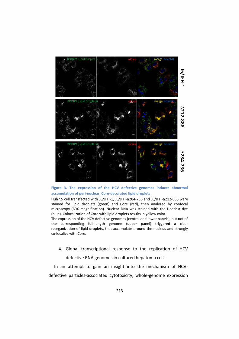
213
Figure 3. The expression of the HCV defective genomes induces abnormal
accumulation of peri-nuclear, Core-decorated lipid droplets
Huh7.5 cell transfected with J6/JFH-1, J6/JFH-Δ284-736 and J6/JFH-Δ212-886 were stained for lipid droplets (green) and Core (red), then analyzed by confocal microscopy (60X magnification). Nuclear DNA was stained with the Hoechst dye (blue). Colocalization of Core with lipid droplets results in yellow color. The expression of the HCV defective genomes (central and lower panels), but not of the corresponding full-length genome (upper panel) triggered a clear reorganization of lipid droplets, that accumulate around the nucleus and strongly co-localize with Core.
4. Global transcriptional response to the replication of HCV
defective RNA genomes in cultured hepatoma cells
In an attempt to gain an insight into the mechanism of HCV-
defective particles-associated cytotoxicity, whole-genome expression

214
profiling of the cells expressing defective HCV RNA genomes was set
up. The constructs FL-J6/JFH-1-GND (mock), FL-J6/JFH-1, J6/JFH-Δ284-
736 and J6/JFH-Δ212-886 were electroporated as described in
Materials and Methods. Cells were harvested at 48 hours post-
transfection and total RNA was isolated for global gene expression
profiling as described in Materials and Methods. At this timepoint cell
sufferance started to be evident (Fig. 2). Genome-wide gene
expression was analyzed by the Illumina microarray platform.
Experiments were performed in quintuplicate and 9332 transcripts
were found to be expressed in at least one of the four cellular
conditions in all the biological replicates. 208 genes that showed at
least 1.5 fold change with a Pval<0.01 (calculated as described in the
method session) in both the defective genomes compared to the full-
length were selected for further investigation (Fig. 4A).
The list of 208 genes generated from the differential expression
analysis was analyzed for biological function and pathway enrichment
by Ingenuity Pathway Analysis software (IPA). This analysis was used to
identify the cellular processes represented by the changes in steady-
state abundance of transcripts associated to the replication of HCV
defective RNA genomes, compared to the expression patterns
associated with the replication of FL-J6/JFH-1. Functional analysis by
IPA reveals a significant functional enrichment of the canonical
pathways indicated in Fig. 4B: the pathway of Steroid Biosynthesis was
the most significantly altered in cells expressing either J6/JFH-Δ212-
886 or J6/JFH-Δ284-736 (Fig. 4B).
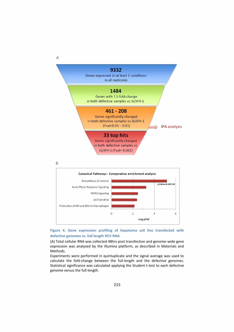
215
Figure 4. Gene expression profiling of hepatoma cell line transfected with
defective genomes vs. full length HCV RNA
(A) Total cellular RNA was collected 48hrs post transfection and genome-wide gene expression was analyzed by the Illumina platform, as described in Materials and Methods. Experiments were performed in quintuplicate and the signal average was used to calculate the fold-change between the full-length and the defective genomes. Statistical significance was calculated applying the Student t-test to each defective genome versus the full-length.

216
Genes showing at least 1.5 fold change and a Pval<0.01 in both the defective genomes were selected for further investigation. (B) The analysis of pathways and biological function was performed using IPA software (Ingenuity Pathway Analysis, Ingenuity System) and the enrichment was determined taking into account the built-in Fisher exact test. This statistic test gave a p value of significance for each pathway included in the Gene Ontology database: low p values indicate that a significant number of genes belonging to a given pathway are differentially expressed in cells expressing the HCV defective genomes versus the cells expressing the FL genome. In this graph, p values are indicated as –Log: the higher –Log Pval, the stronger is the statistical significance of pathway enrichment. Gene ontology analysis of genes that are differentially expressed in cells transfected with either defective genomes reveal the involvement of pathways related to synthesis of steroids and response to stress.
5. Genes of Cholesterol Biosynthesis pathway are
downregulated in cells expressing either J6/JFH-Δ212-886 or
J6/JFH-Δ284-736
Since the intimate connection between HCV lifecycle and
cholesterol is well known19, the expression of the enzymes involved in
cholesterol biosynthesis in our experimental conditions was
investigated more in detail (Fig. 5). Genes crucial for cholesterol
biosynthesis, as SQLE (Squalene Epoxidase), HMGCR
(Hydroxymethylglutaryl-CoA Reductase), EBP (Emopamil Binding
Protein - Sterol Isomerase) and IDI1 (Isopentenyl-diphosphate Delta-
Isomerase 1) are stable or upregulated in J6/JFH-1 expressing cells but
are downregulated in cells expressing defective HCV genome. This
expression pattern could be induced by a regulation system that acts
upstream to the expression of many cholesterol biosynthesis genes,
such as for example a regulation mediated by a transcription factor
(i.e. SREBP-2).
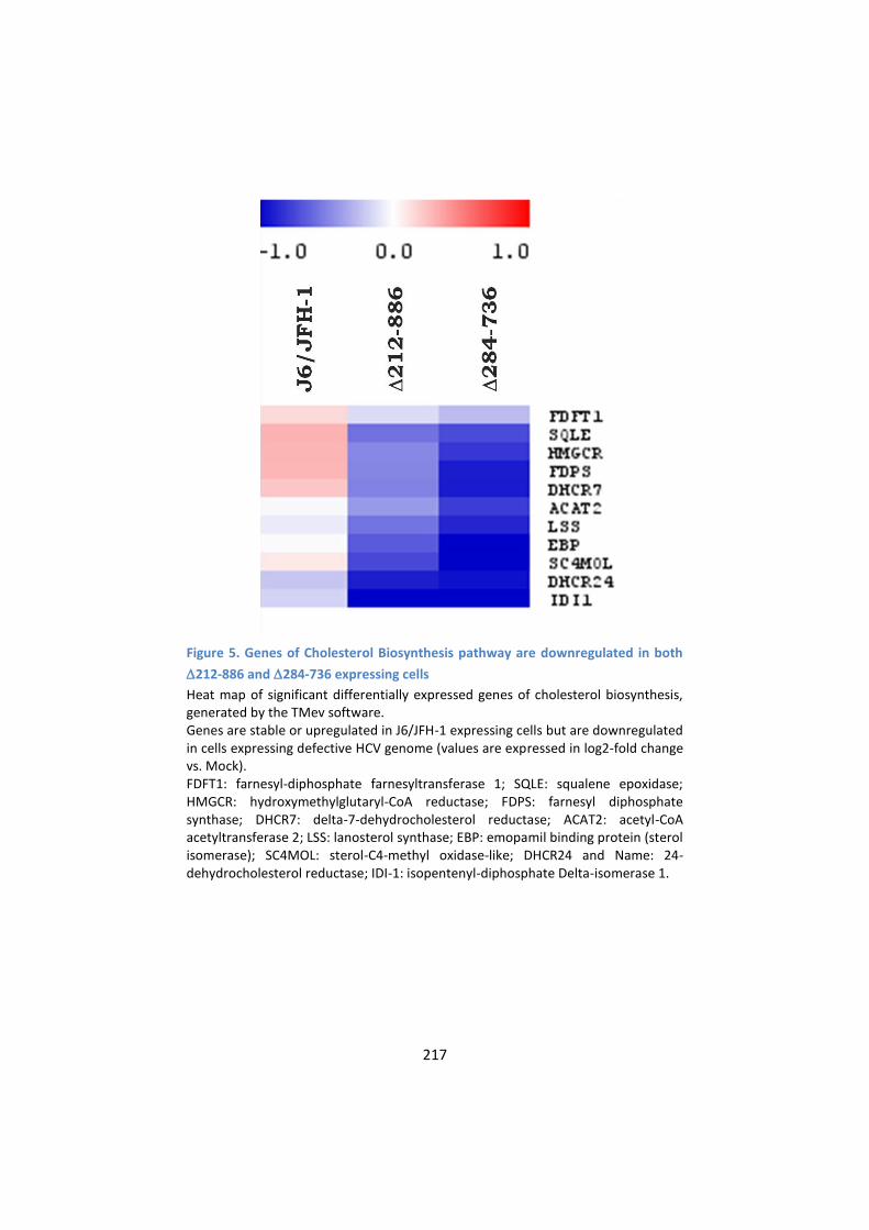
217
Figure 5. Genes of Cholesterol Biosynthesis pathway are downregulated in both
212-886 and 284-736 expressing cells
Heat map of significant differentially expressed genes of cholesterol biosynthesis, generated by the TMev software. Genes are stable or upregulated in J6/JFH-1 expressing cells but are downregulated in cells expressing defective HCV genome (values are expressed in log2-fold change vs. Mock). FDFT1: farnesyl-diphosphate farnesyltransferase 1; SQLE: squalene epoxidase; HMGCR: hydroxymethylglutaryl-CoA reductase; FDPS: farnesyl diphosphate synthase; DHCR7: delta-7-dehydrocholesterol reductase; ACAT2: acetyl-CoA acetyltransferase 2; LSS: lanosterol synthase; EBP: emopamil binding protein (sterol isomerase); SC4MOL: sterol-C4-methyl oxidase-like; DHCR24 and Name: 24-dehydrocholesterol reductase; IDI-1: isopentenyl-diphosphate Delta-isomerase 1.
218
6. The cellular stress sensor gene ATF3 is strongly induced by the
HCV defective genomes
We wondered if the expression of markers of cell stress was linked
to accumulation of Core protein, or to other features of HCV deleted
genomes resembling naturally occurring HCV defective particles.
Among the 461 significant differentially expressed genes from
whole-genome expression profiling, there were two members of the
ATF family of transcription factors: ATF3 and ATF6. The ATF proteins
are sensors of cellular stress signals, including oxidative stress and
unfolded protein response (UPR)24, 25.
The expression of Activating transcription factor 3 (ATF3) was
investigated by Real-Time PCR as described in Materials and Methods
in cells expressing the FL-J6/JFH-1, its dead mutant FL-J6/JFH-1 GND,
the defective genomes J6/JFH-Δ284-736 and J6/JFH-Δ212-886, the
minimal replicon JFH-neo-rep, the J6-core-JFH-rep construct described
above, or both FL-J6/JFH-1 and J6/JFH-Δ284-736. ATF3 was previously
found to be upregulated in cells infected by J6/JFH-126. We observed a
much stronger induction in cells expressing defective genomes
compared to cells expressing the JFH-1 replicon or the dicistronic
replicon expressing Core separately from the HCV polyprotein (J6-
Core-JFH-rep). The observed overexpression did not appear to be
related to the block of HCV particles exit, since it is present also when
the HCV defective genome is transfected together with the full-length
genome (Fig. 6), that acts as helper virus and allows the exit of HCV
defective particles harboring deleted genomes18. These observations
support the hypothesis that Core accumulation alone is not sufficient
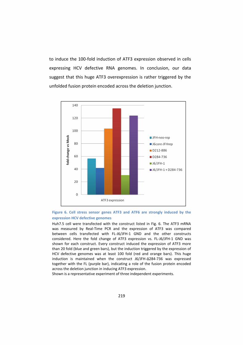
219
to induce the 100-fold induction of ATF3 expression observed in cells
expressing HCV defective RNA genomes. In conclusion, our data
suggest that this huge ATF3 overexpression is rather triggered by the
unfolded fusion protein encoded across the deletion junction.
Figure 6. Cell stress sensor genes ATF3 and ATF6 are strongly induced by the
expression HCV defective genomes
Huh7.5 cell were transfected with the construct listed in Fig. 6. The ATF3 mRNA was measured by Real-Time PCR and the expression of ATF3 was compared between cells transfected with FL-J6/JFH-1 GND and the other constructs considered. Here the fold change of ATF3 expression vs. FL-J6/JFH-1 GND was shown for each construct. Every construct induced the expression of ATF3 more than 20 fold (blue and green bars), but the induction triggered by the expression of HCV defective genomes was at least 100 fold (red and orange bars). This huge induction is maintained when the construct J6/JFH-Δ284-736 was expressed together with the FL (purple bar), indicating a role of the fusion protein encoded across the deletion junction in inducing ATF3 expression. Shown is a representative experiment of three independent experiments.

220
D. Discussion
In this study, we have investigated the host responses to the in vitro
replication of two representative in-frame subgenomic deletion
mutants found circulating in patients. The infectious isolate J6/JFH-1
was modified in order to recapitulate in vitro the genetic structure of
two of the most representative in-frame natural subgenomic deletion-
containing RNAs found circulating in patients18. We observed a clear
sufferance of cells harboring either defective genomes J6/JFH-Δ284-
736 and J6/JFH-Δ212-886, this was not observed in cells where the
HCV 2a full-length chimera was replicating (Fig. 2). Similarly, no toxicity
was detected in cells harboring other exit-defective constructs (J6/JFH
Δp7-Rluc2A, J6/JFH ΔE1-E2-Rluc2A and J6-core-JFH-rep). The two
defective constructs resembling naturally occurring defective HCV
genomes share some peculiarities with the J6-core-JFH-rep construct
and the Δp7 or ΔE1-E2 constructs: (I) they all replicate autonomously;
(II) they produce complete HCV Core and non-structural proteins; (III)
they cannot produce viral particles without the presence of an helper
virus or without trans-complementation of the missing proteins18, 27, so
(IV) the viral proteins accumulates into the hepatocytes. Conversely,
the HCV genomes resembling natural subgenomic deletions have the
unique characteristic of expressing a fusion protein encoded across the
deletion: the HCV polyprotein processing of J6/JFH-Δ284-736
generates an E1-E2 fusion protein, while the processing of J6/JFH-
Δ212-886 generates an E1-NS2 fusion protein. These proteins are likely
221
to be unfolded, so they may trigger an unfolded protein response
and/or an Endoplasmic Reticulum (ER) stress.
In an attempt to elucidate the pathogenic relevance of the HCV
defective particles, we investigated the formation and distribution of
cytoplasmic lipid droplets in cells transfected with defective HCV
genomes. The relationship between the in vivo steatogenic properties
of HCV and the ability of HCV Core protein to associate with
intracellular lipid droplets and to induce lipid accumulation in vitro is
well established for different HCV genotypes, especially for HCV
genotype 3. The steatogenic potential of Core correlated with a
specific distribution of lipid droplets. Core protein binding was
associated with an accumulation of lipid droplets in the perinuclear
area28. Here we found that the expression of HCV defective RNAs
resembling naturally occurring defective particles induces a typical
reorganization of lipid droplets that co-localize with intracellular Core
around the nucleus (Fig. 3). This clustering was also observed when
Core is expressed alone or as part of a polyprotein encoding HCV
structural proteins (Core-E1-E2)20, as well as for other exit-defective
HCV constructs, such as those variants lacking p7 or E1-E2 (21 and
personal observations). The same LD distribution was also observed in
cells infected with the original JFH-1 strain, which can be considered
an “exit-defective” virus since its exit efficiency is very low if compared
with the J6/JFH-1 chimera (21 and personal observations).
Morphological changes in intracellular LD are induced by different HCV
genotype Core sequences; however, it has been reported that LD
induced by genotype 3a Core are larger and contain more neutral
222
lipids than those induced by genotype 1b sequences28. Since the
infection with HCV genotype 3 is strongly correlated with steatosis in
HCV-infected patients, the alterations of LD could be linked to altered
lipid metabolism in hepatoma cells.
In summary, we demonstrated that naturally occurring HCV
defective particles are exit-defective HCV particles that are able to
induce the reorganization of lipid droplets around the nucleus in vitro.
This particular lipid droplets pattern has been correlated with the
steatogenic potential of HCV in vivo28.
Further, we investigated the host transcriptional responses to the
expression of defective HCV RNA genomes by a whole-genome
expression profiling of the cells expressing defective HCV RNA
genomes (Fig. 4A), in order to gain an insight into the mechanism of
HCV-defective particles-associated cytotoxicity.
Gene ontology analysis of the differentially expressed genes
revealed significant functional enrichment of pathways related to
Biosynthesis of Steroids, Acute Phase Response Signaling, TNFR2
Signaling and p53 Signaling (Fig. 4B). In particular, we found that many
genes of Cholesterol Biosynthesis pathway were downregulated in
cells expressing defective HCV genomes. Conversely, these genes were
stable or upregulated in cells expressing the full-length J6/JFH-1
genome (Fig. 5). This phenomenon could be related to a negative feed-
back regulation due to excessive intracellular lipid accumulation
and/or impaired lipoprotein secretion. Downregulation of genes
involved in the Cholesterol Biosynthesis pathway, concomitant with
223
the altered morphology of LDs, was related to the block of HCV
particles exit.
In addition to the Cholesterol Biosynthesis, other pathways were
found to be differentially represented in cells expressing HCV defective
genome that may be related to inflammatory/immune response
(Interferon, TNFR2, Acute Phase Response signaling) or carcinogenesis
(p53 Signaling).
Among the 461 significant differentially expressed genes from
whole-genome expression profiling, there were two members of the
ATF family of transcription factors: ATF3 and ATF6. The ATF proteins
are sensors of cellular stress signals, including oxidative stress and
unfolded protein response (UPR). In our gene expression profiling,
ATF6 and ATF3 showed similar expression pattern. ATF6 is a
recognized ER-stress sensor in hepatocytes and recent studies
demonstrated its activation in liver of patients with chronic hepatitis
C29.
Here we showed that HCV defective particles trigger substantial
overexpression of Activating transcription factor 3 (ATF3). This gene
was previously found to be upregulated in cells infected by J6/JFH-126.
We observed a much stronger induction in cells expressing defective
genomes compared to cells expressing the JFH-1 replicon or the
dicistronic replicon expressing Core separately from the HCV
polyprotein (J6-Core/JFH-1replicon). The observed overexpression did
not appear to be related to the block of HCV particles exit, as
demonstrated by the fact that co-transfection of a deleted genome
with a full-length genome did not rescue the induction of ATF3
224
overexpression. Our data suggest that ATF3 overexpression is triggered
by the unfolded fusion protein encoded across the deletion junction.
Increased ATF3 expression accounts for suppression of p53-
dependent senescence and enhanced tumorigenic potential30. ATF3
was found to be also relevant in modulating innate immune response,
inflammation and cell cycle regulation24, 31, but its role in these
mechanisms is still controversial and needs further investigation.
Taken together, our data suggest that HCV particles with defective
genomes could represent important determinants of steatosis and cell
sufferance in hepatocytes infected by HCV.

225
E. Materials and Methods
1. Reagents and plasmids
The generation of the HCV defective genomes J6/JFH-Δ284-736 and
J6/JFH-Δ212-886 was described previously18. The construct J6-core-
JFH-rep was cloned as follows. HCV-IRES-Core was amplified by PCR
from the FL-J6/JFH-1 with the primers DeltaF1new
(CGACGGCCAGTGAATTCTAATACGACTC, containing the EcoRI site) and
Core-stop-PmeI-R
(CCGGCGCGCCGTTTAAACAAAGCTTCAAGCGGAGACCGGGGTGGTGATGC
AGGACAGC, containing the flanking bases, the PmeI site and the stop
codon). The PCR product and the pUC19-JFH-neo-rep were digested
and cloned with EcoRI and PmeI (this digest excises the HCV-IRES-neo
fragment from the pUC19-JFH-neo-rep plasmid). All nucleic acids
manipulations were performed according to standard protocols.
The constructs FL- J6/JFH-1, FL- J6/JFH-1 GND, J6/JFH Rluc2A, J6/JFH
Δp7-Rluc2A and J6/JFH ΔE1-E2-Rluc2A were a generous gift from C.
Rice (The Rockefeller University).
2. Cell culture, transfection and measure of cell viability
The human hepatoma-derived cell line Huh7.532 were grown in
Dulbecco’s modified Eagle’s medium (DMEM) supplemented with 10%
fetal bovine serum, 100 U/ml penicillin, 100 μg/ml Streptomycin and 2
mM L-glutamine. Electroporation of in vitro-transcribed HCV genomic
RNA into Huh7.5 cells was performed as described previously18. 2x10⁶
cells were electroporated with 10 μg of RNA in a final volume of 200
μl.
226
1.4x10⁴ electroporated cells were plated in triplicates in 96-well
plates and cell viability was measured at the indicated timepoints using
the cell viability assay CellTiter-Blue (Promega), following the
instructions of the manufacturer.
3. Indirect immunofluorescence
After electroporation, cells were plated in 24-well plates with glass
coverslides (8x10⁴ cells/well) and stained for HCV markers and/or lipid
droplets at the timepoints indicated in the figure legends. Primary
antibodies used were: mouse anti-NS5A antibody (Cat. No. HCM-131-
5, 1:1000, Austral Biologicals) and human anti-Core antibody (1:1000, a
generous gift by M. Mondelli, University of Pavia). Secondary
antibodies used were goat anti-mouse IgG Alexa Fluor 568 (Cat. No.
A11004, 1:600, Invitrogen) and goat anti-human IgG Alexa Fluor 568
(Cat. No. A21090, 1:600, Invitrogen). Lipid droplets were stained by
4,4-difluoro-1,3,5,7,8-pentamethyl-4-bora-3a,4a-diaza-s-indacene
(BODIPY 493/503, Cat. No. D-3922, 1:300, Invitrogen).
Cells were washed once with PBS and fixed with 300 μl of 4% PFA
for 10 min. Cells were washed three times with PBS and permeabilized
with 500 μl of 0.1 % Triton X-100 in PBS for 10 min. Unspecific binding
was blocked by incubation with 3 % BSA in PBS. After incubation with
the primary antibody in blocking buffer, cell were washed with PBS
and subsequently incubated with goat secondary antibodies
conjugated to Alexa-Fluor 568 and BODIPY 493/503. Nuclei were
stained with Hoechst dye 33342 (Sigma; 1:4000). Slides were then
mounted with 5 μl ProLong Gold Antifade (Invitrogen) and analyzed by
227
using an inverted Leica TCS SP5 scanning laser confocal microscope.
Digital images were taken using LAS AF software (Leica).
4. Isolation of cellular RNA, expression microarray format and
data analysis
Total RNA was isolated from approximately 106 cells using mirVana
Isolation Kit (Ambion) following the instructions of the manufacturer.
Extracted RNA was quantified by Nanodrop. All extracted RNA samples
were quality controlled for integrity with 2100 Bioanalyzer (Agilent
Technologies) and samples with RIN lower than 8 were discarded.
Illumina Direct Hybridization Assays were performed according to
the standard protocol (Illumina Inc.). For each sample, 250 ng of total
RNA were reverse transcribed according to the Illumina TotalPrep RNA
Amplification kit (Ambion) and cRNA was generated after a 14 hours of
in vitro transcription. Washing, staining and hybridization were
performed according to the standard Illumina protocol: briefly, 750 ng
of cRNA of each sample in a final volume of 15 μl were hybridized onto
Illumina Expression BeadChip arrays (HumanHT-12 v3 Expression
BeadChip). Hybridization and scanning were performed according to
the manufacturer’s indications on an Illumina iScan System and data
were processed with BeadStudio v.3; arrays were quantile normalized,
with no background subtraction, and average signals were calculated
on probe-level data for genes whose detection p-value was lower than
0.0001 in at least one of the experimental conditions examines (9332
probes). After logarithmic transformation, the expression ratio
between full length or defective constructs and mock condition was
228
calculated in each experimental session and then used for the
calculation of the mean among experimental sessions; the statistic
significance was determined by applying an unpaired two-tailed t test.
Transcripts were defined as differentially expressed if there was a 1.5-
fold or greater difference in the gene expression levels and a Pval<0.05
in both the defective constructs versus the full-length.
Pathway and biological function analyses were performed using IPA
software (Ingenuity Pathway Analysis, Ingenuity System) and the
enrichment was determined taking into account the built-in Fisher
exact test.
Global visualization of differentially expressed genes by heat map
was obtained using the TMev software.
5. Quantitative Real-Time PCR
Quantitative Real-Time PCR (qPCR) was used to validate gene
expression changes. Total RNA was extracted by mirVana as described
above. Quantitative PCR was performed by the QuantiTect Probe RT-
PCR Kit (Qiagen), using a one-step strategy, according to the
manufacturer’s instructions. 50 ng of total RNA extracted from cells
were assayed and GAPDH expression was measured as internal
reference (Applied Biosystems). Relative quantification was obtained
by the DDCt method of qPCR data analysis. The probe used to measure
the expression of ATF3 was the Hs00231069_m1* (Applied
Biosystems).
229
F. Chapter 4 – References
1. Moradpour, D., Penin, F. & Rice, C.M. Replication of hepatitis C virus. Nat Rev Microbiol 5, 453-63 (2007).
2. Hepatitis C--global prevalence (update). Wkly Epidemiol Rec 75, 18-9 (2000).
3. Blonski, W. & Reddy, K.R. Hepatitis C virus infection and hepatocellular carcinoma. Clin Liver Dis 12, 661-74, x (2008).
4. Hoofnagle, J.H. Course and outcome of hepatitis C. Hepatology 36, S21-9 (2002).
5. Simmonds, P. Genetic diversity and evolution of hepatitis C virus--15 years on. J Gen Virol 85, 3173-88 (2004).
6. Simmonds, P. et al. Consensus proposals for a unified system of nomenclature of hepatitis C virus genotypes. Hepatology 42, 962-73 (2005).
7. Kageyama, S. et al. A natural inter-genotypic (2b/1b) recombinant of hepatitis C virus in the Philippines. J Med Virol 78, 1423-8 (2006).
8. Kalinina, O., Norder, H., Mukomolov, S. & Magnius, L.O. A natural intergenotypic recombinant of hepatitis C virus identified in St. Petersburg. J Virol 76, 4034-43 (2002).
9. Legrand-Abravanel, F. et al. New natural intergenotypic (2/5) recombinant of hepatitis C virus. J Virol 81, 4357-62 (2007).
10. Noppornpanth, S. et al. Identification of a naturally occurring recombinant genotype 2/6 hepatitis C virus. J Virol 80, 7569-77 (2006).
11. Bernardin, F. et al. High levels of subgenomic HCV plasma RNA in immunosilent infections. Virology 365, 446-56 (2007).
12. Iwai, A. et al. Identification of novel defective HCV clones in liver transplant recipients with recurrent HCV infection. J Viral Hepat 13, 523-31 (2006).
13. Noppornpanth, S. et al. Characterization of hepatitis C virus deletion mutants circulating in chronically infected patients. J Virol 81, 12496-503 (2007).
14. Yagi, S. et al. Identification of novel HCV subgenome replicating persistently in chronic active hepatitis C patients. J Med Virol 77, 399-413 (2005).
230
15. Lindenbach, B.D. et al. Complete replication of hepatitis C virus in cell culture. Science 309, 623-6 (2005).
16. Wakita, T. et al. Production of infectious hepatitis C virus in tissue culture from a cloned viral genome. Nat Med 11, 791-6 (2005).
17. Zhong, J. et al. Robust hepatitis C virus infection in vitro. Proc Natl Acad Sci U S A 102, 9294-9 (2005).
18. Pacini, L., Graziani, R., Bartholomew, L., De Francesco, R. & Paonessa, G. Naturally occurring hepatitis C virus subgenomic deletion mutants replicate efficiently in Huh-7 cells and are trans-packaged in vitro to generate infectious defective particles. J Virol 83, 9079-93 (2009).
19. Negro, F. Abnormalities of lipid metabolism in hepatitis C virus infection. Gut 59, 1279-87.
20. Boulant, S. et al. Hepatitis C virus core protein induces lipid droplet redistribution in a microtubule- and dynein-dependent manner. Traffic 9, 1268-82 (2008).
21. Shavinskaya, A., Boulant, S., Penin, F., McLauchlan, J. & Bartenschlager, R. The lipid droplet binding domain of hepatitis C virus core protein is a major determinant for efficient virus assembly. J Biol Chem 282, 37158-69 (2007).
22. Moriya, K. et al. The core protein of hepatitis C virus induces hepatocellular carcinoma in transgenic mice. Nat Med 4, 1065-7 (1998).
23. Moriya, K. et al. Hepatitis C virus core protein induces hepatic steatosis in transgenic mice. J Gen Virol 78 ( Pt 7), 1527-31 (1997).
24. Thompson, M.R., Xu, D. & Williams, B.R. ATF3 transcription factor and its emerging roles in immunity and cancer. J Mol Med (Berl) 87, 1053-60 (2009).
25. Verfaillie, T., Salazar, M., Velasco, G. & Agostinis, P. Linking ER Stress to Autophagy: Potential Implications for Cancer Therapy. Int J Cell Biol 2010, 930509.
26. Walters, K.A. et al. Genomic analysis reveals a potential role for cell cycle perturbation in HCV-mediated apoptosis of cultured hepatocytes. PLoS Pathog 5, e1000269 (2009).
27. Steinmann, E., Brohm, C., Kallis, S., Bartenschlager, R. & Pietschmann, T. Efficient trans-encapsidation of hepatitis C

231
virus RNAs into infectious virus-like particles. J Virol 82, 7034-46 (2008).
28. Piodi, A., Chouteau, P., Lerat, H., Hezode, C. & Pawlotsky, J.M. Morphological changes in intracellular lipid droplets induced by different hepatitis C virus genotype core sequences and relationship with steatosis. Hepatology 48, 16-27 (2008).
29. Asselah, T. et al. In vivo hepatic endoplasmic reticulum stress in patients with chronic hepatitis C. J Pathol 221, 264-74.
30. Wu, X. et al. Opposing roles for calcineurin and ATF3 in squamous skin cancer. Nature 465, 368-72.
31. Hai, T., Wolford, C.C. & Chang, Y.S. ATF3, a hub of the cellular adaptive-response network, in the pathogenesis of diseases: is modulation of inflammation a unifying component? Gene Expr 15, 1-11.
32. Blight, K.J., McKeating, J.A. & Rice, C.M. Highly permissive cell lines for subgenomic and genomic hepatitis C virus RNA replication. J Virol 76, 13001-14 (2002).

232

233
CHAPTER 5
SUMMARY, CONCLUSIONS AND
TRANSLATIONAL IMPACT OF THE PROJECTS

234
A. Discovery of HCV inhibitors targeting host factors
Most novel drugs directed against HCV, including the recently
approved NS3/4A protease inhibitors boceprevir and telaprevir, are
inhibitors of viral enzymes. Since HCV is an RNA virus with a short and
highly variable genome, these direct-acting antivirals (DAAs) are prone
to rapidly inducing the emergence of resistant HCV variants. This
problem could be mitigated by the development of drugs that target
host factors that the virus depends on during the various stages of its
replication cycle. An increasing understanding of the molecular
interactions between the virus and its host cell has allowed the
identification of promising host targets for anti-HCV therapy and host-
targeting agents (HTAs) are currently under development. The most
advanced compound is an inhibitor of cyclophilin A, a host factor
known to be critical for viral RNA replication and possible virion
assembly or release. This compound, alisporivir, has demonstrated in
vivo efficacy and is now in a phase 3 trial. Several other HTAs with very
different host targets are further upstream in the development
pipeline.
In CHAPTER 2 of this thesis we demonstrated that the 4-Amino
Quinazoline compound named AL-9 inhibits HCV replication by
targeting PI4KIIIα1. The importance of this enzyme for HCV replication
has recently been described by several groups: it binds to HCV NS5A
and its enzymatic activity is required for efficient HCV RNA replication2-
6. PI4KIIIα belongs to the family of type III phosphatidylinositol 4-
kinases, enzymes that catalyze the conversion of phosphatidylinositol
235
to phosphatidylinositol 4-phosphate (PI4P). PI4P is the most abundant
monophosphorylated inositol phospholipid in mammalian cells and the
importance of this phospholipid is just started to be unraveled7. In
addition to playing important roles in intracellular signaling and
membrane trafficking, phosphatidylinositol lipids and their
metabolizing enzymes are also exploited by many different viruses in
order to transform cellular membranes in structures supporting their
replication8, 9. PI4KIIIβ was shown to be a host factor required for
enterovirus replication10, whereas several reports have demonstrated
that PI4KIIIα is crucial for HCV replication2, 3, 5, 11. Owing to the
importance of this pathway, the need for specific inhibitors of PI4III
kinases is increasing. Only recently, some enviroxime-like compounds
with antiviral activity against enterovirus have been demonstrated to
target PI4KIIIβ. One of these agents is a very specific inhibitor of the β-
isoform of the type III PI4-kinases12. So far, no such compound exists
for the PI4KIIIα isoform.
We demonstrated that a class of HCV inhibitors originally proposed
to target NS5A does in fact target the host factor PI4KIIIα. Compounds
targeting host factors may have the general advantage of imposing a
higher genetic barrier to the development of resistance. AL-9, a
member of this class of compounds, inhibits PI4KIIIα and to our
knowledge, it is the first compound with a clear preference for PI4KIIIα
over PI4KIIIβ. For this reason, AL-9 is a good lead compound for the
development of more potent and specific pharmacological inhibitors of
PI4KIIIα to be used both as important research tools as well as leads
for initial drug discovery1.
236
B. HCV-defective RNA genomes as biomarkers of disease severity
and response to anti-HCV therapy
Among the unmet clinical needs in the field of Hepatitis C there is
the necessity to find useful biomarkers of disease progression and
response to therapies.
Understanding which are the patients at risk of developing a faster
progressive liver disease would have great advantages. From the
clinical point of view, antiviral therapy would be the most beneficial in
patients at higher risk of developing progressive liver disease and
would therefore be given as soon as possible. Furthermore, the gold
standard for assessing hepatic fibrosis, that is liver biopsy, is invasive,
subject to sampling errors, and has rare but occurring potentially life
threatening complications, thus limiting its acceptability in patients
with mild or moderate disease. There is therefore the medical need to
develop non-invasive and reliable serum markers that accurately
reflect hepatic fibrotic and cirrhotic disease. Moreover, effective
surveillance of patients at high risk of developing HCC (i.e., patients
with cirrhosis) could potentially decrease HCC-related mortality rate,
and the availability of early and reliable serological markers of HCC
remains a huge unmet medical need. Finally, from the basic research
point of view, the study of how host and viral factors could affect
disease progression may shed light on fundamental pathogenic
mechanisms in the path from fibrosis to cirrhosis to cancer, which still
remain poorly defined.
237
Another important need in the Hepatitis C field regards the
prediction of the rate of response to therapy for each patient. To date,
the standard antiviral therapy, based on long-term administration of
PEG-IFNα/RBV, permits to achieve viral clearance only in about 40-60%
of cases. Both viral and host genetic variability are thought to influence
treatment success rates. SNPs near the IL28B gene are strongly
associated with both spontaneous and treatment-induced viral
clearance13-15. Regarding viral genetics, HCV genotypes 1 and 4 are
considered more difficult to treat than genotypes 2 and 316. Although
IL28B genotype is a significant pretreatment predictor of response to
therapy, consideration should be given to ordering the test when it is
likely to influence either the physician’s or patient’s decision to initiate
therapy17. There is therefore the medical need for additional novel
predictors of response to therapy.
In this study, we analyzed pre-therapy sera of 132 patients and we
found defective genomes in about 20% of them; to our knowledge,
this is the first systematic analysis done on such a large number of
subjects, and it permits to infer that the emerging of deletion mutants,
far from being a sporadic phenomenon, occurs in a relevant portion of
HCV1-infected subjects. Defective interfering forms are reported for a
variety of viruses, and their clinical impact is variegate18. Regarding
HCV, the ability of defective particles to influence the clinical course of
the disease is largely unexplored. We observed that the presence of
HCV defective variants correlates with higher viral load, older patient
age and higher degree of hepatic inflammation. Since defective viruses
are thought to be generated from the wild type through errors of the

238
polymerase, the probability of their insurgence increases
proportionally with the number of replication events. This may be at
the basis of the association with older patient age (which can be
considered as a surrogate of the duration of infection) and higher
viremia. From an etiopathological point of view, the association with
the grading score appears more intriguing, raising the possibility that
HCV defective particles exacerbate the liver inflammation processes.
We then evaluated the viral kinetics during standard therapy
treatment in HCV1 patients carrying the defective forms compared to
those in which only the full-length variant was detected. No difference
was found in RVR, cEVR and ETR rates, thus indicating that defective
particles do not seem to play a key role in determining viral load
decline during treatment. This result was confirmed also by logistic
regression analysis, in which IL28B genotype, patient age and pre-
treatment viremia, but not the presence of defective HCV, were
identified as independent predictors of ETR. Refining the analysis on
the ETR-positive population, however, we observed that defective
particle carriers displayed a higher probability to experience virological
relapse during the follow-up period. This result is particularly
interesting, since it has been proposed that defective virus belonging
to the Flaviviridae family may play a role in the establishment and
maintenance of chronic infections, although the majority of the data
comes from in-vitro experiments19, 20. In conclusion, our study
indicated that, in pre-treatment sera of chronic HCV1 patients, HCV
particles with large in frame-deletions in E1-NS2 region are detected in
about 20% of the subjects and correlate with a higher degree of liver
239
inflammation measured by the Ishak score. While RVR and ETR rates
were not affected, the presence of defective particles was more
frequent in subject experiencing a viral relapse after standard PEG-
IFNα-2b/RBV therapy. The finding that the presence of circulating HCV
defective genomes determines a different pattern of treatment failure
and potentially a higher risk for liver damage in patients with genotype
1 HCV should be taken into account in the design of new algorithms
aimed at maximizing HCV therapy efficacy (CHAPTER 3).
From the molecular point of view, we demonstrated that the
expression of HCV genomes resembling natural subgenomic deletions
is cytopathic and alters the expression of genes related to
carcinogenesis, inflammation and stress responses (CHAPTER 4). These
properties could be mechanistically linked to the increased
inflammatory activity found in the liver of patients harboring HCV
defective particles (as described in CHAPTER 3).
Another pathway differentially regulated in cells harboring HCV
defective genomes regards the biosynthesis of cholesterol. These
genes are upregulated in cells infected with wild-type FL-J6/JFH-1 but
downregulated in cells where HCV defective RNA genomes actively
replicate. Concomitantly, perinuclear lipid droplets accumulate in cells
which express the deleted HCV genomes. This phenotype supports the
notion that the expression of HCV defective genomes profoundly alters
lipid metabolism in infected cells. In the future, we will investigate
whether the presence of HCV defective particles is correlated with
steatosis and/or other markers of dysregulation of lipid metabolism in
HCV-infected patients.

240
In conclusion, our study is the first to elucidate the host response
elicited by the expression of defective RNA genomes in hepatocytes
and to investigate the role of defective virus in the HCV-associated
liver disease.
241
C. Chapter 5 – References
1. Bianco, A. et al. Metabolism of Phosphatidylinositol 4-Kinase IIIalpha-Dependent PI4P Is Subverted by HCV and Is Targeted by a 4-Anilino Quinazoline with Antiviral Activity. PLoS Pathog 8, e1002576.
2. Berger, K.L. et al. Roles for endocytic trafficking and phosphatidylinositol 4-kinase III alpha in hepatitis C virus replication. Proc Natl Acad Sci U S A 106, 7577-82 (2009).
3. Borawski, J. et al. Class III phosphatidylinositol 4-kinase alpha and beta are novel host factor regulators of hepatitis C virus replication. J Virol 83, 10058-74 (2009).
4. Reiss, S. et al. Recruitment and activation of a lipid kinase by hepatitis C virus NS5A is essential for integrity of the membranous replication compartment. Cell Host Microbe 9, 32-45.
5. Tai, A.W. et al. A functional genomic screen identifies cellular cofactors of hepatitis C virus replication. Cell Host Microbe 5, 298-307 (2009).
6. Trotard, M. et al. Kinases required in hepatitis C virus entry and replication highlighted by small interference RNA screening. Faseb J 23, 3780-9 (2009).
7. D'Angelo, G., Vicinanza, M., Di Campli, A. & De Matteis, M.A. The multiple roles of PtdIns(4)P -- not just the precursor of PtdIns(4,5)P2. J Cell Sci 121, 1955-63 (2008).
8. Miller, S. & Krijnse-Locker, J. Modification of intracellular membrane structures for virus replication. Nat Rev Microbiol 6, 363-74 (2008).
9. Heaton, N.S. & Randall, G. Multifaceted roles for lipids in viral infection. Trends Microbiol 19, 368-75.
10. Hsu, N.Y. et al. Viral reorganization of the secretory pathway generates distinct organelles for RNA replication. Cell 141, 799-811.
11. Vaillancourt, F.H. et al. Identification of a lipid kinase as a host factor involved in hepatitis C virus RNA replication. Virology 387, 5-10 (2009).
242
12. Arita, M. et al. Phosphatidylinositol 4-kinase III beta is a target of enviroxime-like compounds for antipoliovirus activity. J Virol 85, 2364-72.
13. Ge, D. et al. Genetic variation in IL28B predicts hepatitis C treatment-induced viral clearance. Nature 461, 399-401 (2009).
14. Suppiah, V. et al. IL28B is associated with response to chronic hepatitis C interferon-alpha and ribavirin therapy. Nat Genet 41, 1100-4 (2009).
15. Tanaka, Y. et al. Genome-wide association of IL28B with response to pegylated interferon-alpha and ribavirin therapy for chronic hepatitis C. Nat Genet 41, 1105-9 (2009).
16. Kau, A., Vermehren, J. & Sarrazin, C. Treatment predictors of a sustained virologic response in hepatitis B and C. J Hepatol 49, 634-51 (2008).
17. Ghany, M.G., Nelson, D.R., Strader, D.B., Thomas, D.L. & Seeff, L.B. An update on treatment of genotype 1 chronic hepatitis C virus infection: 2011 practice guideline by the American Association for the Study of Liver Diseases. Hepatology 54, 1433-44.
18. Marriott, A.C. & Dimmock, N.J. Defective interfering viruses and their potential as antiviral agents. Rev Med Virol 20, 51-62.
19. Lancaster, M.U., Hodgetts, S.I., Mackenzie, J.S. & Urosevic, N. Characterization of defective viral RNA produced during persistent infection of Vero cells with Murray Valley encephalitis virus. J Virol 72, 2474-82 (1998).
20. Yoon, S.W. et al. Characterization of homologous defective interfering RNA during persistent infection of Vero cells with Japanese encephalitis virus. Mol Cells 21, 112-20 (2006).
243
PUBLICATIONS
Bianco A, Reghellin V, Donnici L, Fenu S, Alvarez R, et al. (2012)
Metabolism of Phosphatidylinositol 4-Kinase IIIα-Dependent PI4P Is
Subverted by HCV and Is Targeted by a 4-Anilino Quinazoline with
Antiviral Activity.
PLoS Pathog 8(3): e1002576. doi:10.1371/journal.ppat.1002576

244

245