Embed Size (px)
Citation preview

BioMed CentralBMC Cancer
ss
Open AcceResearch articleAPRIL is a novel clinical chemo-resistance biomarker in colorectal adenocarcinoma identified by gene expression profilingRussell D Petty*1,2, Leslie M Samuel1,2, Graeme I Murray1, Graham MacDonald1,2, Terrence O'Kelly3, Malcolm Loudon3, Norman Binnie3, Emad Aly3, Aileen McKinlay3, Weiguang Wang5, Fiona Gilbert1,4, Scot Semple1,4 and Elaina SR Collie-Duguid*1Address: 1Section of Translational Medical Sciences, Division of Applied Medicine, School of Medicine and Dentistry, University of Aberdeen, Aberdeen, UK, 2Departments of Oncology, Aberdeen Royal Infirmary, Aberdeen, UK, 3Surgery, Aberdeen Royal Infirmary, Aberdeen, UK, 4Clinical Radiology, Aberdeen Royal Infirmary, Aberdeen, UK and 5University of Wolverhampton, Wolverhampton, UK
Email: Russell D Petty* - [email protected]; Leslie M Samuel - [email protected]; Graeme I Murray - [email protected]; Graham MacDonald - [email protected]; Terrence O'Kelly - [email protected]; Malcolm Loudon - [email protected]; Norman Binnie - [email protected]; Emad Aly - [email protected]; Aileen McKinlay - [email protected]; Weiguang Wang - [email protected]; Fiona Gilbert - [email protected]; Scot Semple - [email protected]; Elaina SR Collie-Duguid* - [email protected]
* Corresponding authors
AbstractBackground: 5-Fluorouracil(5FU) and oral analogues, such as capecitabine, remain one of themost useful agents for the treatment of colorectal adenocarcinoma. Low toxicity and convenienceof administration facilitate use, however clinical resistance is a major limitation. Investigation hasfailed to fully explain the molecular mechanisms of resistance and no clinically useful predictivebiomarkers for 5FU resistance have been identified. We investigated the molecular mechanisms ofclinical 5FU resistance in colorectal adenocarcinoma patients in a prospective biomarker discoveryproject utilising gene expression profiling. The aim was to identify novel 5FU resistance mechanismsand qualify these as candidate biomarkers and therapeutic targets.
Methods: Putative treatment specific gene expression changes were identified in a transcriptomicsstudy of rectal adenocarcinomas, biopsied and profiled before and after pre-operative short-courseradiotherapy or 5FU based chemo-radiotherapy, using microarrays. Tumour from untreatedcontrols at diagnosis and resection identified treatment-independent gene expression changes.Candidate 5FU chemo-resistant genes were identified by comparison of gene expression data setsfrom these clinical specimens with gene expression signatures from our previous studies ofcolorectal cancer cell lines, where parental and daughter lines resistant to 5FU were compared. Acolorectal adenocarcinoma tissue microarray (n = 234, resected tumours) was used as anindependent set to qualify candidates thus identified.
Results: APRIL/TNFSF13 mRNA was significantly upregulated following 5FU based concurrentchemo-radiotherapy and in 5FU resistant colorectal adenocarcinoma cell lines but not inradiotherapy alone treated colorectal adenocarcinomas. Consistent withAPRIL's known functionas an autocrine or paracrine secreted molecule, stromal but not tumour cell protein expression by
Published: 11 December 2009
BMC Cancer 2009, 9:434 doi:10.1186/1471-2407-9-434
Received: 14 April 2009Accepted: 11 December 2009
This article is available from: http://www.biomedcentral.com/1471-2407/9/434
© 2009 Petty et al; licensee BioMed Central Ltd. This is an Open Access article distributed under the terms of the Creative Commons Attribution License (http://creativecommons.org/licenses/by/2.0), which permits unrestricted use, distribution, and reproduction in any medium, provided the original work is properly cited.
Page 1 of 11(page number not for citation purposes)

BMC Cancer 2009, 9:434 http://www.biomedcentral.com/1471-2407/9/434
immunohistochemistry was correlated with poor prognosis (p = 0.019) in the independent set.Stratified analysis revealed that protein expression of APRIL in the tumour stroma is associatedwith survival in adjuvant 5FU treated patients only (n = 103, p < 0.001), and is independentlypredictive of lack of clinical benefit from adjuvant 5FU [HR 6.25 (95%CI 1.48-26.32), p = 0.013].
Conclusions: A combined investigative model, analysing the transcriptional response in clinicaltumour specimens and cancers cell lines, has identified APRIL, a novel chemo-resistance biomarkerwith independent predictive impact in 5FU-treated CRC patients, that may represent a target fornovel therapeutics.
BackgroundSignificant progress has been made recently in the sys-temic treatment of colorectal adnocarcinoma (CRC).There are currently 8 agents licensed for use in the US andEurope 5-fluorouracil (5FU), floxuridine, capecitabine,irinotecan, oxaliplatin, cetuximab, panitumumab andbevacizumab [1]. Combination therapy is the standard ofcare for both early and advanced disease [1]. 5FU, or anoral analogue capecitabine, is a component of the major-ity of combination regimens and the low toxicity, easeand convenience of administration, favour its clinical use.However, a modest response rate due to clinical resistanceto 5FU is a major limitation. Older studies with 5FU mon-otherapy demonstrate that the majority of CRC patientstreated will not benefit from 5FU, for example the objec-tive response rate to 5FU or capecitabine monotherapy inadvanced CRC is 20% [1].
Identification of the clinically important mechanisms ofresistance to 5FU would allow better selection of patientsfor 5FU therapy and the rationale design of targeted ther-apeutics to overcome resistance, and thus increase theproportion of patients deriving benefit from 5FU. A pre-dictive biomarker for clinical 5FU resistance would clearlybe useful, but progress has been limited in this area andinvestigation has thus far failed to fully explain the molec-ular mechanisms that areimportant for clinical 5FU resist-ance [2-4]. Preclinical and clinical studies have mainlyfocussed upon molecules concerned with 5FU metabo-lism (Dihydropyrimidine dehydrogenase (DPD), Thymi-dine phosphorylase (TP)) or Thymidylate Synthase (TS),a well characterised 5FU target [3,4]. Clinical studies incolorectal cancer, assessing these molecules by a variety oftechniques (IHC, RT-PCR, ELISA, genotyping), whiledemonstrating correlation between benefit (such asresponse and survival) from 5FU or capecitabine, have sofar failed either to demonstrate genuine clinical utility aspredictive biomarkers or produce useful targeted agents[3]. Overall, given the widespread clinical use of 5FU or itsoral formulations, there is still a need for novel discoveryapproaches in this area.
The global perspective provided by gene expression profil-ing has provided novel insights into the molecular mech-
anisms of clinical response to therapy in human cancers[5], although few studies have specifically addressed clin-ical therapy response in colorectal adenocarcinomas [6-10] and only 1 has analysed serial biopsies before andafter treatment [8]. This report describes our prospectivelydesigned discovery study, Aberdeen Microarray in RectalCancer Study-1 (AMRECS1) using a combined approach,identifying candidate molecules from clinical specimensand comparing them with our 5FU chemo-resistance datafrom cell line model systems [11]. We aimed to identifynovel mechanisms of resistance to 5-fluorouracil (5FU)that are clinically relevant in CRC patients. Tumour biop-sies were collected before and after pre-operative therapyin rectal cancer patients following staging and stratifica-tion with magnetic resonance imaging (MRI), to identifygene expression changes that occur following either 'shortcourse' radiotherapy (SCRT) or 5FU-based concurrentchemo-radiotherapy (CRT). Gene expression profilesfrom these matched clinical specimens were comparedwith profiles generated from colorectal adenocarcinomacell lines, both sensitive parental and derived daughtercell lines with increasing resistance to 5FU. Data is pre-sented for the validation of one potential novel clinical5FU resistance candidate APRIL/TNFSF13 in an independ-ent set of 234 patients with colorectal cancer.
MethodsPatients, Follow up and TreatmentThe study was approved by the North of ScotlandResearch Ethics Committee. Patients provided informedconsent in accordance with the regulations and instruc-tions of the North of Scotland Research Ethics Committeefor study participation, including use and publication ofresults. Full clinicopathological details are provided intable 1 and 2 and in Additional File 1. Patients wereselected for either SCRT or CRT based upon MRI stagingfeatures [12]. All the radiotherapy was CT planned, usinga 3 field technique (posterior and two lateral fields), mul-tileaf collimation and with patients having a full bladderduring the radiotherapy. Surgery was performed either thefollowing week, for SCRT patients, or 6 to 8 weeks aftercompletion of chemo-radiotherapy.
Page 2 of 11(page number not for citation purposes)

BMC Cancer 2009, 9:434 http://www.biomedcentral.com/1471-2407/9/434
Gene expression profilingTumour biopsies were collected at the time of endoscopicdiagnosis of rectal adenocarcinoma and placed immedi-ately into RNAlater (800 μl) (Ambion, Austin, Texas).Tumour biopsies collected at time of curative surgicalresection were placed immediately into normal saline anda pathologist provided a representative tumour biopsy,which was placed immediately into RNAlater within 30minutes (800 μl). Tissues were stored in RNALater at 4°Covernight (16-18 hours), then washed in 500 μl ice coldRNase free PBS (Ambion, Austin, TX) and snap frozen inliquid nitrogen. Long-term storage of tissues was at -80°C.Before RNA extraction, histological diagnosis and featureswere confirmed by frozen section histology. Extractionand purification of total RNA was performed using TRI-ZOL reagent (Invitrogen, Carlsbad, CA) and RNeasyMicrokits (Qiagen, Venlo, The Netherlands), according tothe manufacturer's instructions. Quantification of totalRNA was performed by spectrophometry (260/280 ratio1.9 to 2.2 for all samples). Quality of total RNA and cRNAwas assessed using a BioAnalyser 2100 (Agilent technolo-gies, Palo Alto, CA). Target preparation for the Affymetrix
Genechips™ was according to manufacturer's instructions(Affymetrix, Santa Clara, CA). Specifically, 4 μg of totalRNA was used for reverse transcription and synthesis andamplification of biotin labelled cRNA using the One cycletarget labelling and control reagents. Clean-up of biotin-cRNA was performed with RNeasy Minikits (Qiagen,Venlo, The Netherlands). Fragmentation was performedusing 20 μg of biotin-labelled cRNA. A hybridisationcocktail was prepared from 15 μg which was first hybrid-ised to Test 3 GeneChips™ to assess sample quality(GAPDH 3':5' < 3 and Actin 3': 5' < 3) and then toHGU133 Plus2.0 GeneChips™ (10 μg) for gene expressionanalysis. Procedures for hybridisation, washing, stainingand scanning of chips were carried out according to stand-ard protocols (Affymetrix, Santa Clara, CA).
Analysis of gene expression profiling dataAnalysis of the gene expression data is described in detailin Additional file 2 and as described previously [11,13].Raw data for gene expression is provided in MIAME com-plaint format in Array express, accession number E-MEXP-1901
Table 1: Locally advanced rectal adenocarcinoma patients analysed by gene expression microarray.
Patient Treatment1 Stage at Diagnosis2
Diagnostic biopsy grade &
histology
Diagnostic biopsy
cellularity3
Surgical biopsy grade & histology
Surgical biopsy cellularity3
Pathological stage4
CRT1 CRT T2N1 M0 moderately differentiated
adenocarcinoma
60% poorly differentiated
adenocarcinoma
60% T3N2
CRT2 CRT T3N1 M0 moderately differentiated
adenocarcinoma
60% moderately differentiated
adenocarcinoma
60% T3N1
CRT3 CRT T3N0 M0 moderately differentiated
adenocarcinoma
60% moderately differentiated
adenocarcinoma
50% T3N0
CRT4 CRT T4N1 M0 moderately differentiated
adenocarcinoma
50% moderately differentiated
adenocarcinoma
50% T2N0
RT1 RT T2N0 M0 moderately differentiated
adenocarcinoma
60% moderately differentiated
adenocarcinoma
60% T3N0
RT2 RT T2N1 M0 moderately differentiated
adenocarcinoma
60% moderately differentiated
adenocarcinoma
60% T2N1
RT3 RT T2N0 M0 moderately differentiated
adenocarcinoma
50% moderately differentiated
adenocarcinoma
60% T3N2
RT4 RT T2N0 M0 moderately differentiated
adenocarcinoma
60% moderately differentiated
adenocarcinoma
60% T3N0
CON1 None T3N1 M0 moderately differentiated
adenocarcinoma
75% moderately differentiated
adenocarcinoma
70% T3N1
CON2 None T2N1 M0 moderately differentiated
adenocarcinoma
50% moderately differentiated
adenocarcinoma
50% T3N1
1 CRT = neoadjuvant concurrent chemoradiotherapy; RT = Short course pre-operative radiotherapy. 2 MRI and clinical stage. 3 % Tumour versus normal cells in biopsy profiled. 4 Pathological stage post-preoperative therapy
Page 3 of 11(page number not for citation purposes)
BMC Cancer 2009, 9:434 http://www.biomedcentral.com/1471-2407/9/434
ImmunohistochemistryDescription of the Tissue Microarray (TMA) is provided inprevious publications [14]. A total of 268 colorectaltumours and 50 normal colon cores are represented, with1 core per case. During the staining procedure 34 (13%)tumour cores were lost, leaving cores from 234 patientsavailable for assessment. Antigen retrieval was performedby microwaving in 10 mM citrate (pH 6.0) for 20 minutes.An autostainer (Dakocytomation, Glostrup, Denmark)was used for staining the sections using a mouse mono-clonal primary antibody for human APRIL/TNFSF13(1:60 dilution, Abcam, Cambridge, UK) and Chemate-Envision detection system (Dakocytomation, Glostrup,Denmark), according to the manufacturer's instructions.All sections were double scored by 2 independent investi-gators who were blinded to the clinical data. Scoring dis-crepancies were resolved by examination of sections at adouble-headed microscope. Sections were scored positiveor negative for tumour and/or stromal staining. In addi-tion tumour staining intensity was scored as weak, mod-erate or strong.
Statistical analysisContinuity corrected χ2 test, with Fisher's exact test whereappropriate, was used for binary categorical variables,Pearson's χ2 test for non-binary categorical variables andStudent's t-test for numerical variables. Kaplan-Meiercurves were constructed to assess survival and the log ranktest to assess statistical significance. The Cox proportionalhazards model was used for multivariate analysis of sur-vival. Two-sided p values of less than 0.05 were considered
significant. All analyses were performed using SPSS forWindows, version 13.0 (SPSS Inc, Chicago, IL).
ResultsChemo-radiotherapy or radiotherapy altered gene expression in rectal cancerIn a pilot transcriptomics study of rectal cancer patients,we used oligonucleotide microarrays to profile the expres-sion of over 47000 transcripts representing 38562 humangenes in rectal tumour biopsies before and after pre-oper-ative treatment with CRT (n = 4 patients); table 1). Rectaltumour biopsies before and after SCRT (n = 4 patients;table 1) were also analysed to enable comparison of geneexpression changes in patients treated with 5FU-basedchemo-radiotherapy with those observed in patientsreceiving radiotherapy alone. Rectal tumour biopsies, atdiagnosis and surgical resection, from two patients whodid not undergo any pre-operative treatment (table 1)were used to identify treatment-independent gene expres-sion changes.
SOPs were developed and validated to allow collection oftissues at endoscopic diagnosis and at surgical resection,whilst preserving RNA integrity. Total RNA extracted fromthese tissues (10-30 mg) in this pilot study provided suffi-cient yield (8 to 40 ug) and quality total RNA for geneexpression analysis on Affymetrix oligonucleotide micro-arrays. Raw gene expression data is provided in MIAMEcomplaint format in Array express, accession number E-MEXP-1901.
Threshold and probabilistic filtering of the data (see Addi-tional file 2) identified 86 genes (91 probe sets) consist-ently, significantly and specifically altered following 5FU-based CRT and 51 genes (58 probe sets) following SCRT(see Additional File 3 for details of genes and fold changefollowing therapy). Hierarchical cluster analysis, high-lights 2 distinct clusters of genes up-regulated or down-regulated following CRT (figure 1A) or SCRT (figure 1B).The expression profiles of each of these gene sets clearlyseparates pre- and post-treatment samples into two pri-mary clusters for each treatment group (figure 1). A matrixanalysis (DMTv1.0, Affymetrix, CA) of therapy-alteredgene sets identified using threshold filtering alone (seeAdditional file 2; 697 probe sets in CRT group and 570 inSCRT group, including 86 overlapping), reveals that thesegenes sets are significantly non-overlapping (p = 0.010),demonstrating highly distinct alterations to the tumourtranscriptome following treatment with SCRT or 5FU-based CRT.
The biological functions of the CRT and SCRT altered genesets were evaluated (additional file 4). While many of thesame key biological pathways are identified in each treat-ment group, consistent with a co-ordinated transcrip-
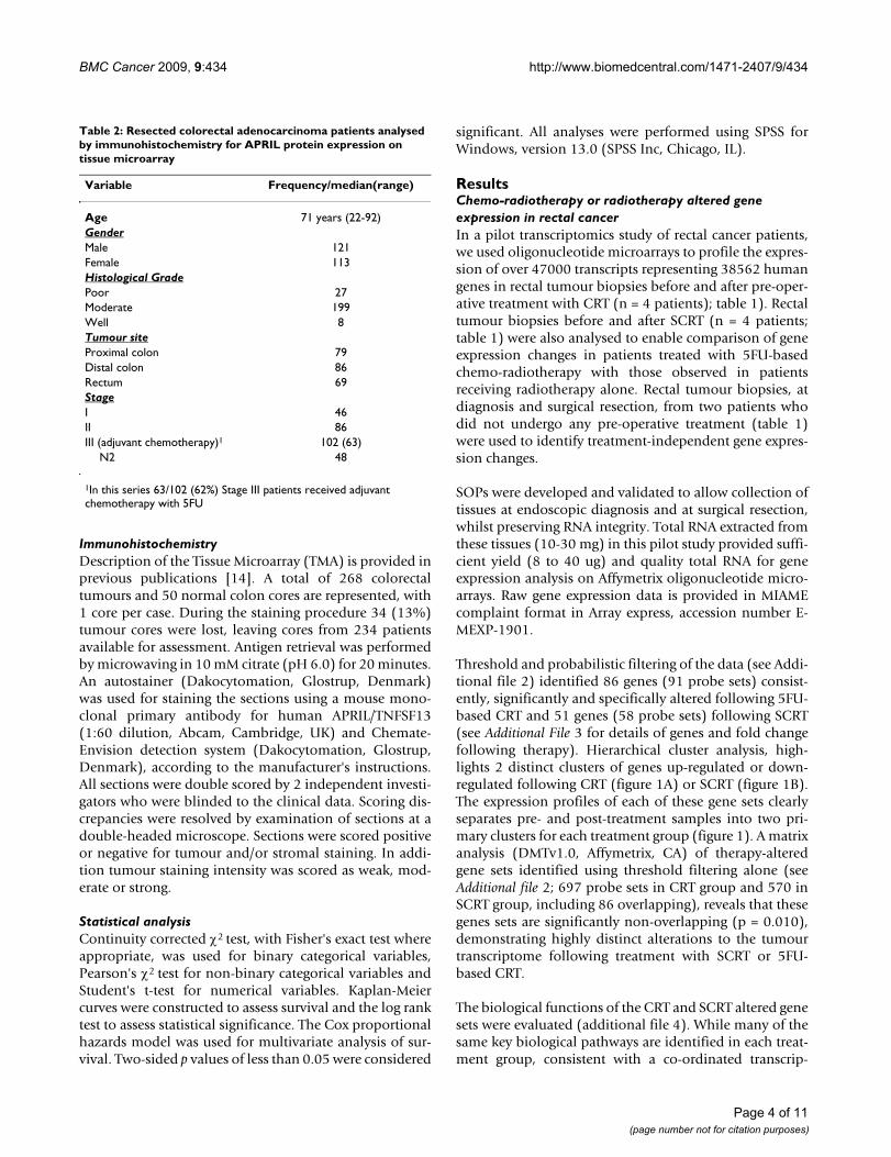
Table 2: Resected colorectal adenocarcinoma patients analysed by immunohistochemistry for APRIL protein expression on tissue microarray
Variable Frequency/median(range)
Age 71 years (22-92)GenderMale 121Female 113Histological GradePoor 27Moderate 199Well 8Tumour siteProximal colon 79Distal colon 86Rectum 69StageI 46II 86III (adjuvant chemotherapy)1 102 (63)
N2 48
1In this series 63/102 (62%) Stage III patients received adjuvant chemotherapy with 5FU
Page 4 of 11(page number not for citation purposes)

BMC Cancer 2009, 9:434 http://www.biomedcentral.com/1471-2407/9/434
tional response, there are some pathways only alteredfollowing CRT and some pathways (cell death and cellcycle) where there is numerically significantly morechange in gene expression in the CRT treated patients(additional file 4).
This represents an initial pilot study of the first samples inour rectal cancer patient cohort. It is important to notethat the small sample size, necessitates validation of thesecandidate gene expression changes in a larger cohort. Theprimary aim of this study was to identify candidate 5FUresistance markers in rectal tumours, in a pilot discoverystudy using a transcriptome-wide approach and to vali-date key candidate/s that may have mechanistic relevance
in a larger cohort. Identification and validation of onesuch marker is described below.
APRIL/TNFSF13 in colorectal cancerAs we were interested in potential mediators of 5FU resist-ance in rectal tumours in vivo, we further mined the geneexpression analysis using a pathway focussed analysis ofcell deaths pathways, including those involved in regula-tion or execution of caspase-dependent apoptotic, cas-pase-independent and necrotic cell death genes (n = 2177genes; additional file 5). Threshold and probabilistic fil-tering of the gene expression data identified 17 cell deathgenes consistently and significantly altered in rectaltumours following chemo-radiotherapy (additional file6). Several of these genes have been implicated in colorec-
Hierarchical cluster analysis of chemoradiotherapy or radiotherapy treated tumoursFigure 1Hierarchical cluster analysis of chemoradiotherapy or radiotherapy treated tumours. This analysis separates pre- and post-treatment biopsies using (a) 86 genes identified as changed in chemoradiotherapy treated patients and (b) 51 genes identified as changed in short course radiotherapy treated patients. (c) Post-treatment tumour biopsies, cluster according to treatment received with the combined set of 137 genes, but (d) pre-treatment tumour biopsies do not. Columns represent tumour samples and rows represent genes (red: up-regulated and green: down-regulated, radiotherapy [blue] or chemoradio-therapy [pink]).
(b)(a)
(d)(c)
Page 5 of 11(page number not for citation purposes)

BMC Cancer 2009, 9:434 http://www.biomedcentral.com/1471-2407/9/434
tal cancer pathogenesis and the pathogenesis of other can-cers, and also radioresistance, but none previously in 5FUchemoresistance (for more details see additional file 6).Comparison of the 17 cell death genes altered in responseto 5FU based CRT in tumours from rectal cancer patients,with gene expression changes identified in our previousstudy of 5FU resistant cancer cell lines [11], demonstrated4 of the 17 genes up-regulated following CRT (but notradiotherapy alone) in rectal cancer patients and in 5FU-resistant cancer cells compared to the sensitive parentallines(See additional file 6, Table S6.1). This included theTNF superfamily ligand, APRIL (TNFSF13).
APRIL has been characterised as promoting cell survivaland cell proliferation and this involves NFκB activation[15-19]. In addition, APRIL mRNA has been shown to beincreased in colorectal tumours compared to normalmucosa [17]. These data supported further investigationof a putative functional role for APRIL in clinical 5FUchemo-resistance.
APRIL protein expression was evaluated in 234 resectedcolorectal adenocarcinomas and 50 normal colon or rec-tal mucosa specimens (table 2). APRIL protein was notexpressed in normal colon tissues but was, as expected,expressed in both colorectal tumour cells and the tumourstroma (Table 3 and figure 2). Tumour cell staining wasobserved in the cytosol and membrane of tumour cells(figure 2). Stromal staining was evident in both the extra-cellular matrix and also in stromal cells (figure 2).
APRIL, a putative 5FU chemo-resistance factor and predictive biomarkerin 5FU treated colorectal cancer patientsWe examined the relationship between APRIL proteinexpression and survival after surgical resection. We pro-spectively determined that we would evaluate bothtumour cell and tumour stromal expression of APRIL pro-tein due to its characterized biological function as asecreted autocrine and/or paracrine molecule. There wasno significant relationship between APRIL protein expres-sion in tumour cells and survival (Additional file 7). Incontrast, expression of APRIL protein in the tumourstroma was associated with poor survival (n = 234, p =0.019, figure 3a), including in stage III patients (n = 102,p = 0.016, figure 3b), but was not associated with survivalin Stage I or II (n = 46 p = 0.601 and n = 86 p = 0.440,respectively, Additional File 7).
In light of our hypothesised role of APRIL in 5FU resist-ance, we stratified the Stage III patients according towhether or not they received adjuvant chemotherapy with5FU following surgical resection of their primary tumour.Stage I and II patients did not receive adjuvant chemother-apy in this series. Tumour stroma expression of APRILprotein is only associated with worse survival in those
patients treated with adjuvant 5FU and there is no rela-tionship with survival in Stage III patients not treated withadjuvant chemotherapy (n = 102, p < 0.001, figure 3c). In5FU treated Stage III patients (n = 63), median survival forstroma positive is 36 months with predicted 5 year sur-vival 42.0% (95% confidence interval 11.8% - 72.2%);median survival not yet reached for stroma negative andpredicted 5 year survival is 85% (95% confidence inter-vals 71.7%-98.6%). Multivariate analysis confirms expres-sion of APRIL protein in the tumour stroma as anindependent prognostic factor in chemotherapy treatedStage III patients, with a HR of 6.25 (95% CI 1.48-26.32,p = 0.013, table 4).
The survival of the 5FU treated Stage III colorectal cancerpatients who express APRIL protein in the tumour stromaparallels survival observed in Stage III patients who didnot receive adjuvant therapy (treatment decision due topatient or physician preference), irrespective of APRILprotein expression (figure 3c). In contrast, the APRIL neg-ative patients have an excellent predicted 5 year survivaland have a clear and statistically significant (p < 0.001)survival benefit compared to untreated or APRIL positive5FU treated patients (figure 3c). These data suggest thatAPRIL has no prognostic impact in colorectal cancertreated by surgical resection alone, but has predictiveimpact for benefit from adjuvant 5FU in colorectal cancerpatients.
DiscussionGlobal gene expression profiling of clinical response totherapy has provided a useful means for biomarker andnovel target discovery in several solid tumours [5,13]. The
Table 3: Tumour cell and stromal expression of APRIL protein in colorectal adenocarcinomas.
APRIL Immunohistochemistry
Tumour Cell Positive 130 (55.6%)Weak 70 (29.9%)Moderate 49 (20.9%)Strong 11 (4.7%)
Stroma Negative 104 (44.4%)Positive 121 (51.7%)Negative 113 (48.3%)
Immunohistochemical analysis of a rectal adenocarcinoma tissue microarray (n = 234 tumours) demonstrated that APRIL protein was expressed in tumour cells and/or tumour stroma. Positive stromal expression was strong. In tumour cells expressing APRIL, intensity was weak, moderate or strong. The number of rectal adenocarcinomas with positive staining for APRIL protein. Percentage of the total (n = 234) is in parentheses. There was a significant correlation between tumour cell and stromal expression (p = 0.048). There was no significant association between tumour cell or stromal staining and age, gender, histological grade, tumour site or Duke's stage (all p > 0.20. Data not shown).
Page 6 of 11(page number not for citation purposes)

BMC Cancer 2009, 9:434 http://www.biomedcentral.com/1471-2407/9/434
work described in this paper has used and extended thisexperimental approach to rectal adenocarcinomas. Thedata presented constitutes an analysis from gene expres-sion profiling of prospectively collected pre- and post-treatment tumour specimens from patients with rectaladenocarcinomas receiving pre-operative therapy.
Since a small number of rectal adenocarcinomas havebeen profiled (n = 10), stringent and focussed analysis ofthe microarray data was applied to identify leads for fur-ther investigation. This included hypothesis-driven focuson cell death pathways and comparison with our previ-ously published cell line work. The key candidate was sub-sequently validated a in larger independent set (n = 234)using a different technique (immunohistochemistry).
The biological validity of the experimental model and thedata is confirmed by the finding of significant alterationsin the gene expression of previously implicated moleculesand pathways, for example p21 which has been impli-cated in numerous studies [20-25]. The biological path-ways identified (information 3 and 4) suggest a co-ordinated transcriptional response to radiotherapy- andCRT- induced cellular stress, consistent with other reportsinvolving gene expression profiling in cell lines and sev-eral different cancer types [2,11,13,25-29]. We hypothe-size that this reflects distinct biological effects of these two
treatments. However, the possibility of effects due to timecourse differences in the tumour sampling in each groupcannot be excluded.
A supervised analysis of cell death genes, reveals sharedgenes and pathways. The analysis supports the hypothe-sise that initiation of cell death is a common final path-way resulting from a multitude of upstream responses tothe insult and resultant cellular stress of cytotoxic chemo-therapy or radiotherapy thereby accounting for geneexpression overlap seen.
The majority of the genes identified in our analysis repre-sent genes and pathways that have not previously beenimplicated in clinical response of rectal adenocarcinomaor as mechanisms of action or resistance to radiotherapyor 5FU or 5FU-based CRT. This is consistent with the find-ings of other gene expression profiling studies in rectaladenocarcinoma or other tumour types for radiotherapyor 5FU [6,8-11,26,28-30]. However, it is important tonote that this discovery phase utilised a small samplecohort and the candidate gene expression changes requirefurther validation in a lrger independent cohort.
APRIL/TNFSF13 was found to be upregulated followingCRT but not radiotherapy alone in rectal cancers and wasalso up-regulated in 5FU resistant cell lines in our previ-
Immunohistochemistry for APRIL in resected colorectal adenocarcinomasFigure 2Immunohistochemistry for APRIL in resected colorectal adenocarcinomas. Staining for APRIL was seen in the tumour cells (membrane and cytosol) and stroma (extracellular matrix and stromal cells) of colorectal adenocarcinomas. All combinations of tumour cell and stromal staining were seen. Tumour cell staining could be scored weak, moderate and strong. Examples show strong tumour cell staining and stromal staining.
Page 7 of 11(page number not for citation purposes)

BMC Cancer 2009, 9:434 http://www.biomedcentral.com/1471-2407/9/434
ous studies [11]. The biological function of APRIL as asecreted molecule that has autocrine and paracrine func-tions to promote cell survival and proliferation and itspreviously documented expression in colorectal adeno-carcinoma but not normal cells outside the immune sys-tem, supported it's further investigation as a novelmechanism of 5FU action and resistance, and as a predic-tive biomarker [15-19,31-35].
This study found that expression of APRIL protein incolorectal tumour stroma was associated with worse sur-vival, but only in those patient's treated with adjuvant
5FU chemotherapy. This relationship was also main-tained in a multivariate analysis of 5FU chemotherapytreated Stage III colorectal adenocarcinoma patients (HR6.25, 1.47-26.31, p = 0.013), in which the Hazard ratiocompares favourably to other previously published puta-tive 5FU predictive biomarkers in colorectal cancer [2-4].Tumour cell expression of APRIL was correlated with stro-mal staining but was not significantly associated with sur-vival. Overall, APRIL appears to have no therapyindependent prognostic impact in colorectal adenocarci-noma in this analysis.
APRIL protein expression in tumour stroma and survival of colorectal cancer patientsFigure 3APRIL protein expression in tumour stroma and survival of colorectal cancer patients. (a). Kaplan-Meier survival plots for tumour stroma APRIL protein expression analysed by immunohistochemistry of 234 colorectal cancer patients fol-lowing surgical resection.(b) Stromal staining for APRIL in Stage III patients following surgical resection (n = 102) (c) Combined analysis of stage III patients (n = 102) stratified according to adjuvant therapy and tumour stroma APRIL protein. P value is log rank test.
p=0.019
Negative
Positive
a b
Negative
Positive
p=0.016
c
p<0.001
Page 8 of 11(page number not for citation purposes)

BMC Cancer 2009, 9:434 http://www.biomedcentral.com/1471-2407/9/434
Within the limitations of a retrospective study, theseresults suggest that APRIL may have clinical utility as apredictive biomarker to select patients who would notbenefit from adjuvant 5FU monotherapy. For example,currently adjuvant 5FU is used clinically in an empiricalway without predictive biomarkers in stage III patientsand in this paradigm the majority of patients with Stage IIIcancers will not benefit from 5FU. Therefore, the ability toidentify some of these stage III patients who will not ben-efit from 5FU has clear potential clinical utility in optimis-ing and individualising clinical use of 5FU in this setting.An important question is whether APRIL confers crossresistance to other active agents used to treat colorectalcancer, especially Oxaliplatin and Irinotecan, this wouldbe potentially useful to guide 5FU combination adjuvanttherapy in stage III patients, but especially in stage IIpatients where 5FU alone appears to have limited benefit.
The data allows us to hypothesise that APRIL may providea useful novel therapeutic target. Morphological examina-tion has suggested that positively staining stromal cellsinclude lymphocytes and fibroblasts, but not endothelialcells. This is consistent with evidence indicating thatAPRIL is predominantly secreted and exerts it's effects viacell surface receptors, acting in a paracrine or autocrinefashion [15-19,31-35].
Our data indicate that APRIL might be secreted by tumourcells or stromal cells within the tumour. The APRIL signal-ling mechanisms that may mediate tumour cell survivalare not well characterised [32]. However, in vitro work inglioma cell lines and ex vivo studies in BCLL, has shownthat APRIL stimulates proliferation and inhibits apoptosisin response to a wide range of stimuli, including CD95L,TRAIL and cytotoxic drugs and survival in B-CLL cellsinvolves NFκB activation [15-19,31-34]. More recently ithas been suggested that tumour infiltrating neutrophils
may be an important source of APRIL production in solidtumours [35].
If APRIL is functional as an extracellular secreted moleculethis makes it amenable to targeting with either a smallmolecule inhibitor or monoclonal antibody, as has beenemployed successfully for other targets in solid tumourse.g. bevacizumab against VEGF. An anti-APRIL targetedtherapy may be useful in reversal of acquired 5FU resist-ance or in combination in patients whose tumours over-express the molecule.
The lack of therapy independent prognostic impact sug-gests that an anti-APRIL therapy may not have anticanceractivity on it's own, but the cell survivalpromoting activitymay be more generally applicable to other therapeutic celldeath stresses. Therefore, combination of an anti-APRILagent with agents other than 5FU may be active, and ourcell line data also suggest that they may be active in othertumour types, such as breast cancer.
ConclusionsIn this study we have used a combined investigativemodel, analysing the transcriptional response in clinicaltumour specimens from rectal adenocarcinomas and can-cer cell lines, to identify APRIL, as a novel 5FU chemo-resistance biomarker. We have validated its importance inan independent set of colorectal adenocarcinomas. Thisdata supports further investigation of the clinical utility ofAPRIL as a predictive biomarker for 5FU resistance incolorectal adenocarcinomas and other solid tumour typesand also as a target for novel therapeutics aimed atreversal of clinical resistance to 5FU and its oral ana-logues.
Competing interestsThe authors declare that they have no competing interests.
Table 4: Multivariate analysis using Cox proportional hazards regression model for adjuvant chemotherapy treated Stage III patients.
Variable HR 95% Confidence Interval p value
Age(>70 vs <70)
1.006 0.955-1.059 0.835
Gender(Female vs Male)
0.532 0.159-1.783 0.307
Grade(poor vs moderate vs well)
N/A N/A 0.873
Site(Proximal vs Distal)
5.015 0.695-36.191 0.110
APRILTumour Stroma Staining(positive vs negative)
6.250 1.471-26.316 0.013
APRILTumour Cell Staining(positive vs negative)
0.64 0.200-2.044 0.452
Page 9 of 11(page number not for citation purposes)

BMC Cancer 2009, 9:434 http://www.biomedcentral.com/1471-2407/9/434
Authors' contributionsRDP designed the study, completed ethical submission,consented patients for study, performed, analysed andinterpreted gene expression profiling and immunohisto-chemistry and wrote the manuscript. LMS participated instudy design, ethical submission, consenting of patientsfor study, interpretation of immunohistochemical data,and writing of manuscript GIM- participated in studydesign, histopathological review of specimens, provisionof tissue microarray, analysis and interpretation of immu-nohistochemical data, and writing of manuscript. GMac-participated in study design, and consenting of patientsfor the study. T O'K, NB, EA, AMc- Consented patients forstudy, and provided fresh tumour tissue for gene expres-sion profiling. WW- Assisted with analysis of gene expres-sion data. FG- participated in study design, performedMRIs and reported MRIs SS- participated in study designand reporting of MRIs. ECD - participated in study design,assisted with ethical submission, assisted with analysisand interpretation of gene expression and immunohisto-chemical data, and writing of manuscript. All authors readand approved the final manuscript.
Additional material
AcknowledgementsThis work was supported by The Friends of the Aberdeen and the North Centre for Oncology, Haematology and Radiotherapy (ANCHOR), Asso-
Additional file 1Further details of Patients and Treatments. Clinicopathological, selec-tion criteria, staging and chemotherapy and radiotherapy protocol details for patients in the study.Click here for file[http://www.biomedcentral.com/content/supplementary/1471-2407-9-434-S1.DOC]
Additional file 2Details of analysis of Gene Expression Profiling Data. Details of qual-ity control, normalisation and analysis for identification of genes whose expression is consistently and significantly altered as a consequence of chemoradiotherapy or radiotherapy. Figure S2. Schematic to illustrate bioinformatics analysis performed to identify genes whose expression was consistently and significantly altered as a result of either neoadjuvant chemoradiotherapy or short course radiotherapy.Click here for file[http://www.biomedcentral.com/content/supplementary/1471-2407-9-434-S2.DOC]
Additional file 3Details of genes identified in analysis of rectal adenocarcinomas. Details of genes identified in analysis of rectal adenocarcinomas whose expression is consistently and significantly changed after treatment with chemoradiotherapy or radiotherapy. Table S3.1-. List of 86 genes (91 probe sets) whose expression is consistently and significantly changed after treatment with chemoradiotherapy. Table S3.2 -List of 52 genes (58 probe sets) whose expression is consistently and significantly chaged after treatment with short course radiotherapy.Click here for file[http://www.biomedcentral.com/content/supplementary/1471-2407-9-434-S3.DOC]
Additional file 4Biological pathways altered following neoadjuvant radiotherapy or chemoradiotherapy in rectal tumours. The number of genes in each bio-logical pathway whose expression was altered following chemoradiother-apy or radiotherapy is shown. Gene ontologies (biological function) were assigned according to GO, Genespring v6.1, Netaffx, EntrezGene, RefSeq and literature searches using Medline and ISI. Table S4 - The number of genes in each biological pathway whose expression was altered following chemoradiotherapy or radiotherapy.Click here for file[http://www.biomedcentral.com/content/supplementary/1471-2407-9-434-S4.DOC]
Additional file 5Cell death gene list used for supervised gene expression analysis. Xcel file with list of identified 2177 genes involved in the control, regulation and execution of cell death (apoptotic and non-apoptotic forms) that were represented on the HGU133 Plus 2.0 GeneGhip, using databases (GO, Genespring v6.1, RefSeq, EntrezGene) and literature searches (Medline and ISI).Click here for file[http://www.biomedcentral.com/content/supplementary/1471-2407-9-434-S5.XLS]
Additional file 6Details of Supervised analysis of Cell Death Pathways. Schematic rep-resentations to explain supervised bionformatic analysis of cell death path-ways. Figure S6 Schematic illustrating the bioinformatics analysis performed for the supervised analysis of cell death genes. GCOSv1.2 and Genespring v6.1 were used for the analyses. Table S6. List of Cell death genes identified in this analysis in CRT and SCRT treated rectal adeno-carcinoma patients. Table S6.1. Genes identified as candidate novel mechanisms of 5FU chemoresistance or sensitivity from gene expression profiling experiments of 5FU resistant colorectal and breast cancer cell linesClick here for file[http://www.biomedcentral.com/content/supplementary/1471-2407-9-434-S6.DOC]
Additional file 7Additional survival analyses for APRIL protein expression in colorectal adenocarcinomas. Kaplan-Meier survival plots for APRIL protein expres-sion in tumour cells of colorectal adenocarcinom patients in stage I, II and II and APRIl stroma expression in Stage I and II Figure S7.1 Kaplan-Meier survival plots for APRIL immunohistochemistry showing no signif-icant relationship for tumour cell protein expression and survival. All patients (n = 234), analysed according to intensity of APRIL staining in tumour cells [weak, moderate or strong (b)] or positive versus negative tumour cell staining (a), or stratified according to stage Dukes A/Stage I, Dukes B/Stage II and Dukes C/Stage III (c). Figure S7.2. Kaplan-Meier survival plots for APRIL immuno-histochemistry showing that positive staining in the tumour stroma shows no association with survival in Duke's A/Stage I (n = 46) or B/Stage II tumours (n = 86).Click here for file[http://www.biomedcentral.com/content/supplementary/1471-2407-9-434-S7.DOC]
Page 10 of 11(page number not for citation purposes)

BMC Cancer 2009, 9:434 http://www.biomedcentral.com/1471-2407/9/434
ciation for International Cancer Research, NHS Grampian Research and Development Office, the James Alexander Mearns' Trust and University of Aberdeen Development Trust (Friends of Little Star).
References1. Davies JM, Goldberg RM: First-line therapeutic strategies in
metastatic colorectal cancer. Oncology (Williston Park) 2008,22(13):1470-1479.
2. Boyer J, Maxwell PJ, Longley DB, Johnston PG: 5-Fluorouracil:identification of novel downstream mediators of tumourresponse. Anticancer Res 2004, 24(2A):417-423.
3. Longley DB, Allen WL, Johnston PG: Drug resistance, predictivemarkers and pharmacogenomics in colorectal cancer. Bio-chim Biophys Acta 2006, 1766(2):184-196.
4. Zhang N, Yin Y, Xu SJ, Chen WS: 5-Fluorouracil: mechanisms ofresistance and reversal strategies. Molecules 2008,13(8):1551-1569.
5. Minna JD, Girard L, Xie Y: Tumor mRNA expression profilespredict responses to chemotherapy. J Clin Oncol 2007,25(28):4329-4336.
6. Ghadimi BM, Grade M, Difilippantonio MJ, Varma S, Simon R, Mon-tagna C, et al.: Effectiveness of Gene Expression Profiling forResponse Prediction of Rectal Adenocarcinomas to Preop-erative Chemoradiotherapy. J Clin Oncol 2005, 23(9):1826-1838.
7. Khambata-Ford S, Garrett CR, Meropol NJ, Basik M, Harbison CT,Wu S, et al.: Expression of epiregulin and amphiregulin and K-ras mutation status predict disease control in metastaticcolorectal cancer patients treated with cetuximab. J ClinOncol 2007, 25(22):3230-3237.
8. Inoue Y, Shirane M, Miki C, Hiro J, Tanaka K, Kobayashi M, Mori K,Yanagi H, Kusunoki M: Gene expression profiles of colorectalcarcinoma in response to neo-adjuvant chemotherapy. Inter-national Journal of Oncology 2004, 25(6):1641-1649.
9. Nagtegaal ID, Gaspar CG, Peltenburg LT, Marijnen CA, Kapiteijn E,Velde CJ van de, et al.: Radiation induces different changes inexpression profiles of normal rectal tissue compared withrectal carcinoma. Virchows Arch 2005, 446(2):127-135.
10. Nagtegaal I, Gaspar C, Marijnen C, Velde C Van De, Fodde R, VanKrieken H: Morphological changes in tumour type after radi-otherapy are accompanied by changes in gene expressionprofile but not in clinical behaviour. J Pathol 2004,204(2):183-192.
11. Wang W, Cassidy J, O'Brien V, Ryan KM, Collie-Duguid E: Mecha-nistic and predictive profiling of 5-Fluorouracil resistance inhuman cancer cells. Cancer Res 2004, 64(22):8167-8176.
12. MERCURY Study Group: Diagnostic accuracy of preoperativemagnetic resonance imaging in predicting curative resectionof rectal cancer: prospective observational study. BMJ 2006,333(7572):779.
13. Petty RD, Kerr KM, Murray GI, Nicolson MC, Rooney PH, Bissett D,et al.: Tumor transcriptome reveals the predictive and prog-nostic impact of lysosomal protease inhibitors in non-small-cell lung cancer. J Clin Oncol 2006, 24(11):1729-1744.
14. Carpenter B, McKay M, Dundas SR, Lawrie LC, Telfer C, Murray GI:Heterogeneous nuclear ribonucleoprotein K is overexpressed, aberrantly localised and is associated with poorprognosis in colorectal cancer. Br J Cancer 2006, 95(7):921-927.
15. Kern C, Cornuel JF, Billard C, Tang R, Rouillard D, Stenou V, et al.:Involvement of BAFF and APRIL in the resistance to apop-tosis of B-CLL through an autocrine pathway. Blood 2004,103(2):679-688.
16. Deshayes F, Lapree G, Portier A, Richard Y, Pencalet P, Mahieu-Caputo D, et al.: Abnormal production of the TNF-homologueAPRIL increases the proliferation of human malignant gliob-lastoma cell lines via a specific receptor. Oncogene 2004,23(17):3005-3012.
17. Kelly K, Manos E, Jensen G, Nadauld L, Jones DA: APRIL/TRDL-1,a tumor necrosis factor-like ligand, stimulates cell death.Cancer Res 2000, 60(4):1021-1027.
18. Rennert P, Schneider P, Cachero TG, Thompson J, Trabach L, HertigS, et al.: A soluble form of B cell maturation antigen, a recep-tor for the tumor necrosis factor family member APRIL,inhibits tumor cell growth. J Exp Med 2000, 192(11):1677-1684.
19. Roth W, Wagenknecht B, Klumpp A, Naumann U, Hahne M, TschoppJ, et al.: APRIL, a new member of the tumor necrosis factor
family, modulates death ligand-induced apoptosis. Cell DeathDiffer 2001, 8(4):403-410.
20. Fu CG, Tominaga O, Nagawa H, Nita ME, Masaki T, Ishimaru G, et al.:Role of p53 and p21/WAF1 detection in patient selection forpreoperative radiotherapy in rectal cancer patients. Dis ColonRectum 1998, 41(1):68-74.
21. Qiu H, Sirivongs P, Rothenberger M, Rothenberger DA, Garcia-Agui-lar J: Molecular prognostic factors in rectal cancer treated byradiation and surgery. Dis Colon Rectum 2000, 43(4):451-459.
22. Rau B, Sturm I, Lage H, Berger S, Schneider U, Hauptmann S, et al.:Dynamic expression profile of p21WAF1/CIP1 and Ki-67predicts survival in rectal carcinoma treated with preopera-tive radiochemotherapy. J Clin Oncol 2003, 21(18):3391-3401.
23. Sogawa N, Takiguchi N, Koda K, Oda K, Satomi D, Kato K, et al.:Value of expression of p21WAF1/CIP1 as a prognostic factorin advanced middle and lower rectal cancer patients treatedwith preoperative radio-chemotherapy. Int J Oncol 2002,21(4):787-793.
24. Yoshikawa R, Yanagi H, Kusunoki M, Fujiwara Y, Noda M, Hashimoto-Tamaoki T, et al.: Prognostic values of radiation-induced p53 inadjacent normal mucosa and p21WAF1/CIP1 expression inrectal cancer patients. Int J Oncol 2002, 21(6):1223-1228.
25. Wang W, McLeod HL, Cassidy J, Collie-Duguid ES: Mechanisms ofacquired chemoresistance to 5-fluorouracil and tomudex:thymidylate synthase dependent and independent networks.Cancer Chemother Pharmacol 2007, 59(6):839-845.
26. Maxwell PJ, Longley DB, Latif T, Boyer J, Allen W, Lynch M, et al.:Identification of 5-fluorouracil-inducible target genes usingcDNA microarray profiling. Cancer Res 2003, 63(15):4602-4606.
27. Jansen MP, Foekens JA, van Staveren IL, Dirkzwager-Kiel MM, RitstierK, Look MP, et al.: Molecular classification of tamoxifen-resist-ant breast carcinomas by gene expression profiling. J ClinOncol 2005, 23(4):732-740.
28. Rieger KE, Chu G: Portrait of transcriptional responses toultraviolet and ionizing radiation in human cells. Nucleic AcidsRes 2004, 32(16):4786-4803.
29. Rieger KE, Hong WJ, Tusher VG, Tang J, Tibshirani R, Chu G: Tox-icity from radiation therapy associated with abnormal tran-scriptional responses to DNA damage. Proc Natl Acad Sci USA2004, 101(17):6635-6640.
30. Ding LH, Shingyoji M, Chen F, Chatterjee A, Kasai KE, Chen DJ: Geneexpression changes in normal human skin fibroblastsinduced by HZE-particle radiation. Radiat Res 2005, 164(4 Pt2):523-526.
31. Kelsen DP, Ginsberg R, Pajak TF, Sheahan DG, Gunderson L, Mor-timer J, et al.: Chemotherapy Followed by Surgery Comparedwith Surgery Alone for Localized Esophageal Cancer. N EnglJ Med 1998, 339(27):1979-1984.
32. Mackay F, Ambrose C: The TNF family members BAFF andAPRIL: the growing complexity. Cytokine Growth Factor Rev 2003,14(3-4):311-324.
33. Hahne M, Kataoka T, Schroter M, Hofmann K, Irmler M, Bodmer JL,et al.: APRIL, a new ligand of the tumor necrosis factor family,stimulates tumor cell growth. J Exp Med 1998,188(6):1185-1190.
34. Stein JV, Lopez-Fraga M, Elustondo FA, Carvalho-Pinto CE, RodriguezD, Gomez-Caro R, et al.: APRIL modulates B and T cell immu-nity. J Clin Invest 2002, 109(12):1587-1598.
35. Mhawech-Fauceglia P, Allal A, Odunsi K, Andrews C, Herrmann FR,Huard B: Role of the tumour necrosis family ligand APRIL insolid tumour development: Retrospective studies in bladder,ovarian and head and neck carcinomas. Eur J Cancer 2008,44(15):2097-2100.
Pre-publication historyThe pre-publication history for this paper can be accessedhere:
http://www.biomedcentral.com/1471-2407/9/434/prepub
Page 11 of 11(page number not for citation purposes)