Embed Size (px)
Citation preview

BlurredVision
Janna Cottingame
Nikohl Ostermeier
Abby Perone

Blurred Vision Defined Blurred vision refers to a lack of sharpness of vision
resulting in the inability to see fine detail.
Blurred vision may result from abnormalities present at birth such as near- or farsightedness that require corrective lenses or it may signal the presence of eye disease.
Blurred vision can also be a symptom of numerous conditions that do not directly involve the eye, such as migraine or stroke

Normal Anatomy of the Eye

The 5 I’s of Eye DDXs Idiopathic Infectious Inflammatory Ischemic Infiltrative

Intro to Blurred Vision
Main Categories General Blurred Vision Distance Blurred Vision Near Blurred Vision
Near/ Distance Blur Differentials Distance:
Myopia/ nearsightedness- difficulty seeing object far away
Near: Diabetic retinopathy- retinal damage from chronic DM Hyperopia/ farsightedness- difficulty seeing objects
nearby Presbyopia- visual acuity changes associated with aging

General Blur Differentials Accomodative disorder Albinism Amblyopia Anirdia Asteroid hyalosis Astygmatism Cataract * Cellulitis

General Blur Differentials Central serous retinopathy Computer vision syndrome Contact lens problem Corneal abrasion Coreal atrophy Corneal erosion Cranial nerve palsy Drugs Diabetic Retinopathy*

General Blur Differentials Eye tumor Foreign body Fungal keratosis Glaucoma * Hypotony Keratoconus Lens Dislocation Macular Degeneration* Meibomianitis
General Blur Differentials Migraine Neuroretinitis Nystagmus Opthalmoplegia Optic nerve problems Photokeratitis Pterygium Retinal detachment* Retinitis

General Blur Differentials Retinoschisis Scleritis Stroke Surgery Trauma Temporal Arteritis Uveitis Vascular problems Vitreous hemorrhage White dot syndrome

Diabetic Retinopathy Defined: Damage to the eye's retina that
occurs with long-term diabetes. Symptoms may not present until significant damage is present.
The leading cause of blindness in work-age Americans.

Diabetic Retinopathy Mechanism of the Pathology
Caused by damage to blood vessels of the retina.
The retina is the layer of tissue at the back of the inner eye.
It changes light and images that enter the eye into nerve signals that are sent to the brain.
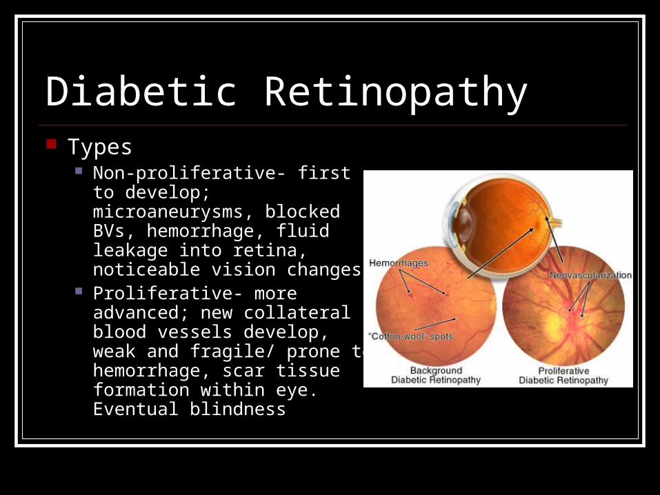
Diabetic Retinopathy Types
Non-proliferative- first to develop; microaneurysms, blocked BVs, hemorrhage, fluid leakage into retina, noticeable vision changes
Proliferative- more advanced; new collateral blood vessels develop, weak and fragile/ prone to hemorrhage, scar tissue formation within eye. Eventual blindness

Diabetic Retinopathy S/Sx Subjective
Blurred vision Gradual vision loss Floaters Shadows/ areas
missing from visual fields
Difficulty seeing at nighttime
Double vision Pain in one eye

Diabetic Retinopathy Objective
Hard exudates on retinal background Cotton Wool spots Hemorrhages
Preretinal Flame Intraretinal subretinal

Diabetic Retinopathy Risk Factors Poor control of blood sugar levels High blood pressure High cholesterol Pregnancy Black or hispanic Smokers

Testing for Diabetic Retinopathy Fluorescein angiopathy
Pictures will be taken as dye circulates through the eyes. Images pinpoint blood vessels that are closed, broken down or leaking fluid.
Optical coherence tomography
This imaging test provides cross-sectional images of the retina that show the thickness of the retina, which will help determine whether fluid has leaked into retinal tissue.

Diabetic Retinopathy DDX Branch retinal vein occulsion Central retinal vein occlusion Microaneurysm Macular edema, diabetic Valsalva retinopathy Hemoglobulinopathies

Retinal Detachment What is the Retina?
A very thin layer of nerve tissue that lines the back of the eye. When light strikes the retina a complex biochemical reaction takes place and sends information to the brain through the optic nerve for seeing and understanding.

Retinal Detachment S/SX Flashing lights and floaters are
usually the first signs of retinal detachment and is often ignored by patients. Blurred vision or a “curtain appearance” in their vision is commonly the next progression and one where patients may mention to you problems with their vision.

Retinal Detachment How does the retina
detach? It can get a hole, break, or a
tear. When this happens the vitreous gel will begin to leak between then retina and the back of the eye. This puts pressure on the retina and eventually pushes it away from the eye completely if not treated.

Retinal Detachment Risk Factors They occur most commonly in younger adults (25 to
50 years of age) who are highly nearsighted (myopic) and in older people following cataract surgery and trauma
Diseases/conditions that predispose to the development of a detachment: Lattice degeneration-thinning of the lateral margins of the retina
mostly associated with severe nearsightedness. Patients using pilocarpine (eye drops) for glaucoma Patients with chronic inflammation of the eye (uveitis) Cataract surgery

Retinal Detachment DDX Great history questions to help differentiate
if patient presents with blurred vision: Have you recently seen flashing lights/
floaters? Have you had any trauma to your
eye/head recently? Are you using any medications, especially
for glaucoma? When was the last time you say the
Opthomologist? For what? Are you nearsighted? Have you had cataract surgery?

Macular Degeneration Most common risk factors:
Age- The leading cause of vision loss and blindness in people over 65 years of age.
Obesity and inactivity Heredity High BP Smoking Lighter skin color Drug Side Effects (Aralen anti-
malarial & Phenothiazin anti-psychotic)

Screening for Macular Degeneration An Amsler grid can be used as a
preliminary screening If test positive they may send
you for a fluorescein angiography: During this test they inject dye into
the veins and when the dye reached the interior of the eye you can image the blood vessels. In advanced degeneration there will be abnormal growth of the blood vessels.
QuickTime™ and aTIFF (Uncompressed) decompressor
are needed to see this picture.

DDX Retinal Detachment and Macular Degeneration Location of Blurriness
Macular is central blurriness Retinal detachment is in the periphery.
Age Group Afflicted: Macular is m/c in over 65 years old Retinal detachment is m/c 25-50 years

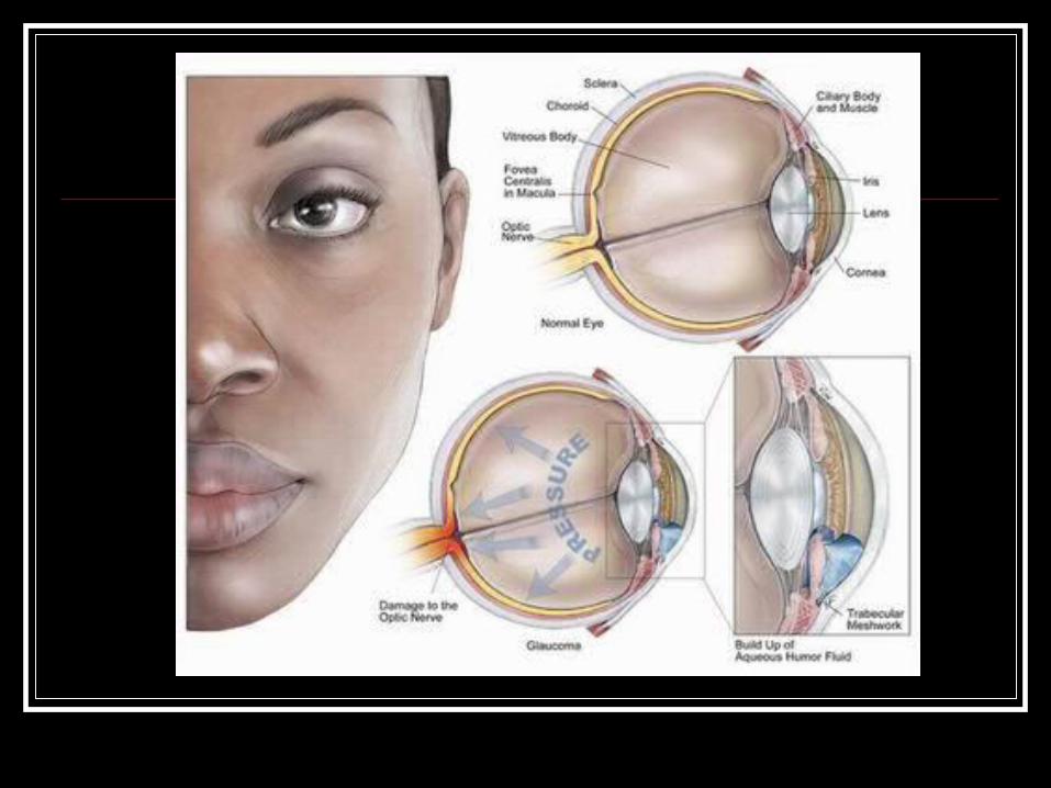
Glaucoma Not just one disease, but a group of conditions
resulting in optic nerve damage Abnormally high pressure inside your eye
(intraocular pressure) usually causes this damage
Can damage your vision so gradually you don't notice any loss of vision until the disease is at an advanced stage.

Glaucoma The most common
type of glaucoma, primary open-angle glaucoma, has no noticeable signs or symptoms except gradual vision loss

Glaucoma S/Sx The most common types of glaucoma, primary open-angle glaucoma and
acute angle-closure glaucoma, have completely different symptoms:
Primary open-angle glaucoma s/sx include:
- Gradual loss of peripheral vision, usually in both eyes- Tunnel vision in advanced stages
Acute-angle closure glaucoma s/sx include:
-Severe eye pain- Nausea and vomiting- sudden onset of viual disturbances often in low light- Blurred vision- Halos around lights- Reddening of the eyes

Primary Open-angle Glaucoma Drainage angle formed by the cornea and
the iris remains open, but the microscopic drainage channels in the angle - trabecular meshwork -are partially obstructed, causing the aqueous humor to drain out of the eye too slowly
Fluid backup and a gradual increase of pressure within your eye
Damage to the optic nerve is painless and so slow that a large portion of your vision can be lost before you're even aware of a problem
Exact cause of primary open-angle glaucoma remains unknown.

Open-angle Glaucoma

Closed-angle Glaucoma Occurs when the iris bulges forward
to narrow or block the drainage angle formed by the cornea and the iris aqueous fluid can no longer access
the trabecular meshwork at the angle, so the eye pressure increases abruptly
Angle-closure glaucoma usually occurs suddenly (acute angle-closure glaucoma), but it can also occur gradually (chronic angle-closure glaucoma)

Closed-angle Glaucoma

At Risk Populations for Glaucoma Routine eye checkups every two years if
you're between 18-60 years old, and every year if you're older than 60.
African-Americans have a much higher risk of glaucoma screened every three to five years from age 20 to
29 every two to four years from age 30 to 40 and every one to two years thereafter.

Glaucoma Risk Factors Elevated internal eye pressure (intraocular
pressure) Age Ethnic background Family history of glaucoma Medical conditions Other eye conditions Nearsightedness Prolonged corticosteroid use
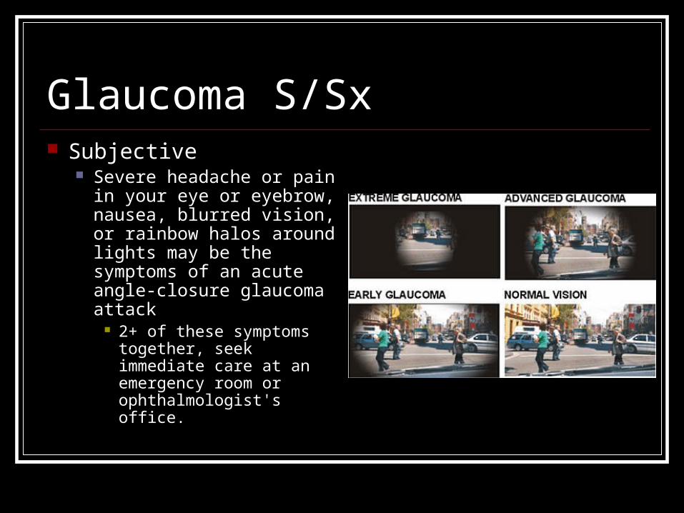
Glaucoma S/Sx Subjective
Severe headache or pain in your eye or eyebrow, nausea, blurred vision, or rainbow halos around lights may be the symptoms of an acute angle-closure glaucoma attack
2+ of these symptoms together, seek immediate care at an emergency room or ophthalmologist's office.

Glaucoma Mechanism Increased intraocular pressure is usually
associated with the optic nerve damage that characterizes glaucoma. From a buildup of aqueous humor, which is naturally
and continuously produced in the front of your eye. Aqueous humor normally exits your eye through
a drainage system at the angle where the iris and the cornea meet When the drainage system doesn't function properly,
the aqueous humor can't filter out of the eye at its normal rate, and pressure builds within your eye

Glaucoma Complications If left untreated,
progressive vision loss will occur, typically in these stages: Peripheral blind spots Tunnel visions Total blindness
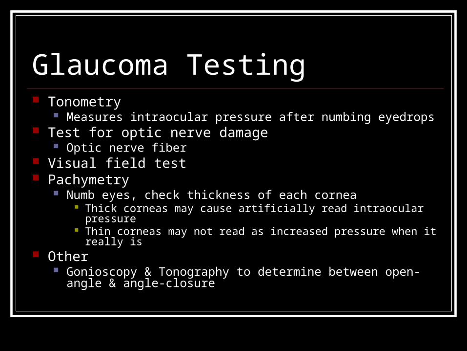
Glaucoma Testing Tonometry
Measures intraocular pressure after numbing eyedrops Test for optic nerve damage
Optic nerve fiber Visual field test Pachymetry
Numb eyes, check thickness of each cornea Thick corneas may cause artificially read intraocular pressure Thin corneas may not read as increased pressure when it really is
Other Gonioscopy & Tonography to determine between open-angle &
angle-closure

Cataracts
A cataract occurs when the lens of the eye becomes cloudy, and can eventually progress to the degree of this right eye

Cataract Causes Usually when aging or injury cause changes to
the lens The lens becomes less flexible, less transparent,
& thicker Tissues break down & clump, clouding small areas of lens
As cataracts progrss, the clouding becomes more dense & involves more of the lens

Cataract S/Sx At first, the cloudiness in vision caused by a cataract
might only affect a small enough part of the lens that it is unnoticeable
Grows, clouds more of the lens, distorting more light passing through the lens Clouded, blurred, dim vision Difficulty seeing at night Photosensitivity & sensitivity to glare “halos” Frequent changes in eyeglass/contact prescriptions Fading/yellowing of colors Double vision in a single eye

Cataract S/Sx
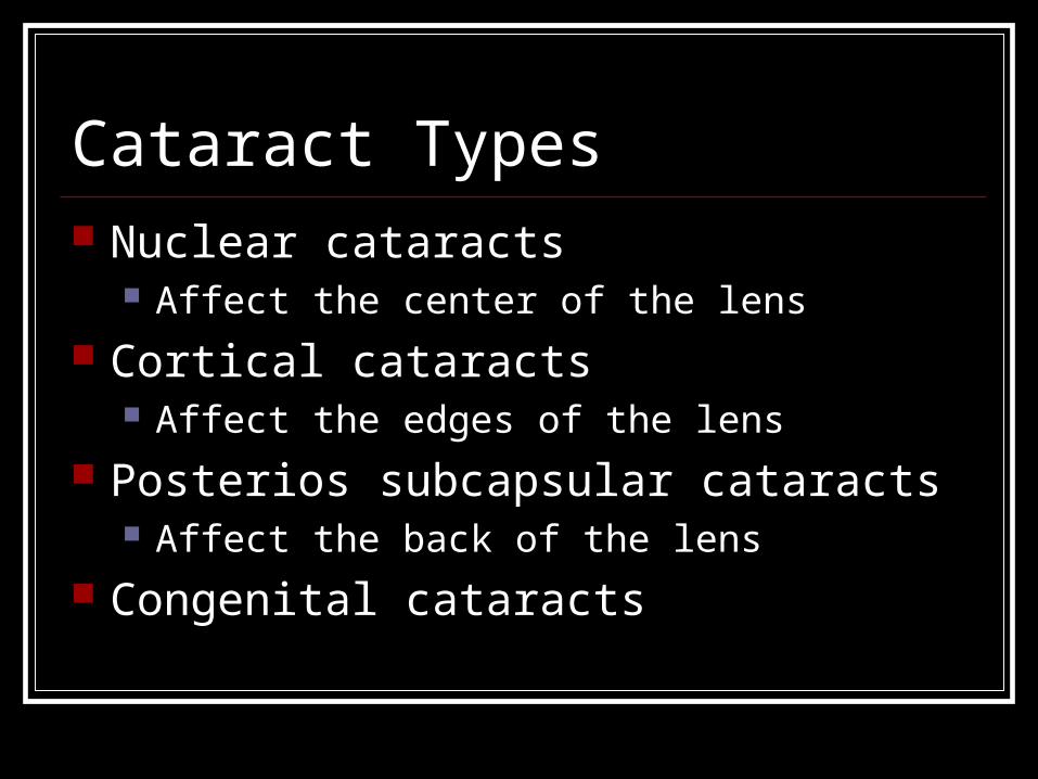
Cataract Types Nuclear cataracts
Affect the center of the lens
Cortical cataracts Affect the edges of the lens
Posterios subcapsular cataracts Affect the back of the lens
Congenital cataracts

Cataract Risk Factors Aging Diabetes Excessive alcohol consumption Excessive exposure to sunlight Exposure to ionizing radiation
Xrays, chemo Family history High blood pressure Obesity Previous eye injury/inflammation Previous eye surgery Prolonged use of corticosteroid medications Smoking

Cataract Testing Visual Acuity Test
Signs of impairment? Slit-lamp examination
Magnifies cornea, iris, lens, space btwn iris & cornea
Dilating eyes for retinal examination opthalmoscope

Glaucoma vs. Cataracts While glaucoma is most often a
problem with drainage, a cataract is a clouding of the eye’s lens allowing less light to pass through.
Both cataracts and glaucoma can be a natural part of the aging process. Many people over 60 may have both. Otherwise, the two are not associated.
With the exception of glaucoma due to secondary causes such as trauma or steroids, glaucoma does not cause cataracts and cataracts do not cause glaucoma
Both are serious conditions that can cause you to lose vision. However, loss of vision due to cataracts can be reversed with surgery. Loss of vision from glaucoma is irreversible.
Glaucoma
Cataracts

References http://www.mayoclinic.com/health/diabetic-retinopathy/
DS00447/DSECTION=tests%2Dand%2Ddiagnosis http://www.mayoclinic.com/health/cataracts/DS00050 http://www.mayoclinic.com/health/glaucoma/DS00283 http://emedicine.medscape.com/article/1225210-diagnosis https://www.onlinececreditsnow.com/assets/
Sample_Course.pdf https://health.google.com/health/ref/Diabetic+retinopathy http://www.allaboutvision.com/conditions/eye-problems.htm


![Janna medina[1]](https://img.dokumen.tips/doc/110x75/5588f5c2d8b42a4e138b46d2/janna-medina1.jpg)