Embed Size (px)
Citation preview

Blood...The River Blood...The River of Lifeof Life
(A delivery system of 5 (A delivery system of 5 liters of liquid fun)liters of liquid fun)
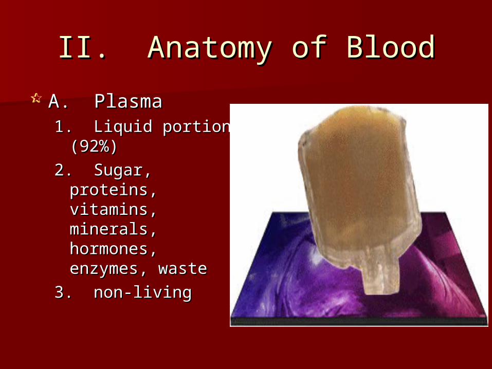
II. Anatomy of BloodII. Anatomy of Blood
A. PlasmaA. Plasma1. Liquid portion 1. Liquid portion
(92%)(92%)
2. Sugar, proteins, 2. Sugar, proteins, vitamins, minerals, vitamins, minerals, hormones, hormones, enzymes, wasteenzymes, waste
3. non-living3. non-living
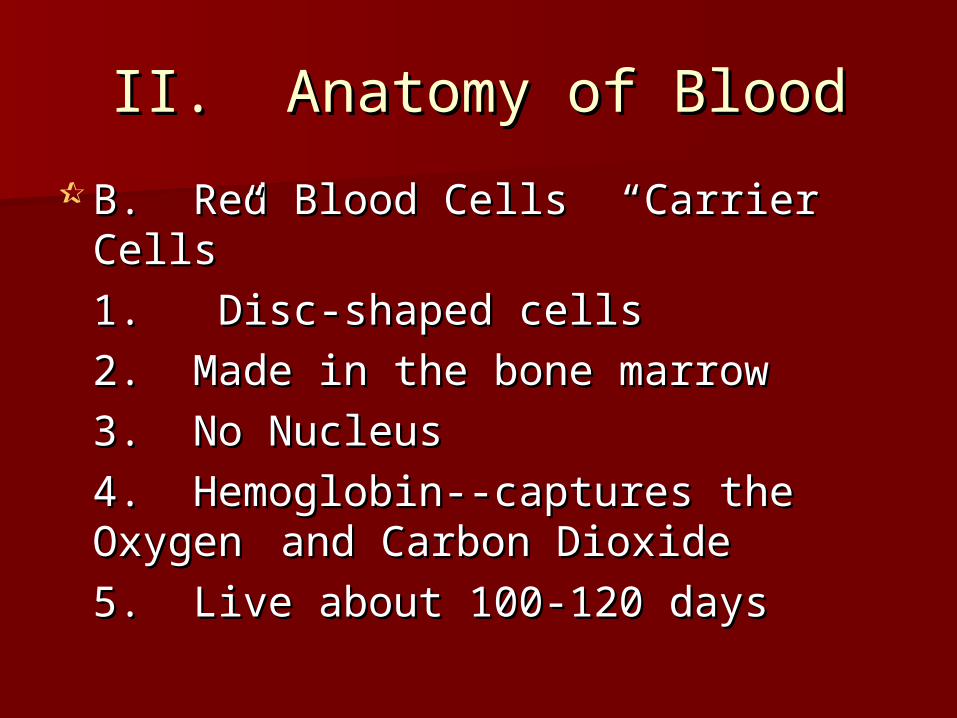
II. Anatomy of BloodII. Anatomy of Blood
B. Red Blood Cells “Carrier Cells”B. Red Blood Cells “Carrier Cells”
1. Disc-shaped cells 1. Disc-shaped cells
2. Made in the bone marrow2. Made in the bone marrow
3. No Nucleus3. No Nucleus
4. Hemoglobin--captures the 4. Hemoglobin--captures the Oxygen Oxygen and Carbon Dioxideand Carbon Dioxide
5. Live about 100-120 days5. Live about 100-120 days


ABO Rh
Carbohydrates
MARKERS ONCELL
A – A antigensB- B antigensAB – A & B
antigensO – no antigens
Rh+= antigen Rh - = no antigen
The Scoop on Blood TypesThe Scoop on Blood TypesAntigensAntigens
Used in ID

The Scoop on Blood TypeThe Scoop on Blood TypeAntibodiesAntibodies
ABO Rh
Proteins
Anti-AAnti-B
Anti Rh+Bind to Antigens
Find Invaders
In plasma


BlooBlood d TypeType
AntigenAntigens on s on RBCRBC
AntibodiAntibodies in es in PlasmaPlasma
Can Can DonatDonate To:e To:
Can Can ReceivReceive e From:From:
AA AA Anti-BAnti-B A, ABA, AB A, OA, O
BB BB Anti-AAnti-A B, ABB, AB B, OB, O
ABAB A, BA, B NoneNone ABAB AllAll
OO NoneNone Anti-A, Anti-A, Anti-BAnti-B
AllAll OO
Rh+Rh+ ++ NoneNone ++ +/-+/-
Rh -Rh - NoneNone Anti +Anti + +/-+/- --
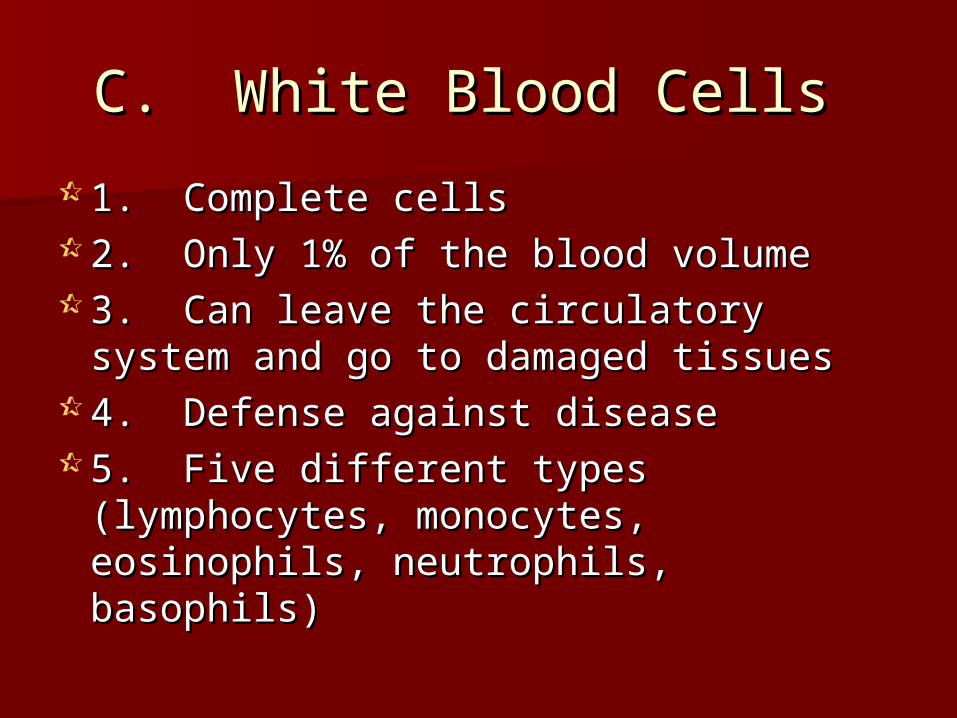
C. White Blood Cells C. White Blood Cells
1. Complete cells1. Complete cells 2. Only 1% of the blood volume2. Only 1% of the blood volume 3. Can leave the circulatory system 3. Can leave the circulatory system
and go to damaged tissuesand go to damaged tissues 4. Defense against disease4. Defense against disease 5. Five different types (lymphocytes, 5. Five different types (lymphocytes,
monocytes, eosinophils, neutrophils, monocytes, eosinophils, neutrophils, basophils)basophils)

D. Platelets D. Platelets
1. Cell fragments1. Cell fragments 2. Essential for 2. Essential for
blood blood clottingclotting 3. Form a 3. Form a
temporary temporary plug to seal a plug to seal a
break in the break in the skinskin

I. Functions of Blood I. Functions of Blood
A. Transports oxygen to the body cells A. Transports oxygen to the body cells B. Transports carbon dioxide and waste B. Transports carbon dioxide and waste
from the body cellsfrom the body cells C. Transports food to body cells (glucose C. Transports food to body cells (glucose
and other necessary things)and other necessary things) D. Contains disease fightersD. Contains disease fighters E. Regulates body temperature--E. Regulates body temperature--
remember the skin unit!!remember the skin unit!! F. Transports hormonesF. Transports hormones

Anemia:Anemia:
Low oxygen carrying capacityLow oxygen carrying capacity Low RBCs, Low Hemoglobin, Low RBCs, Low Hemoglobin,
Abnormal RBCsAbnormal RBCs

Polycythemia:Polycythemia:
Too many RBCsToo many RBCs Blood very thick and flows slowlyBlood very thick and flows slowly

Genetics of Blood TypesGenetics of Blood Types
Blood types are an example of Blood types are an example of CoDominanceCoDominance
A and B are codominant and code for the A and B are codominant and code for the antigens on Red Blood cellsantigens on Red Blood cells
If a person has AB blood, they have both If a person has AB blood, they have both antigensantigens
O is recessive and codes for NO antigensO is recessive and codes for NO antigens To be recessive, you must have two To be recessive, you must have two
recessive allelesrecessive alleles Try a practice problemTry a practice problem
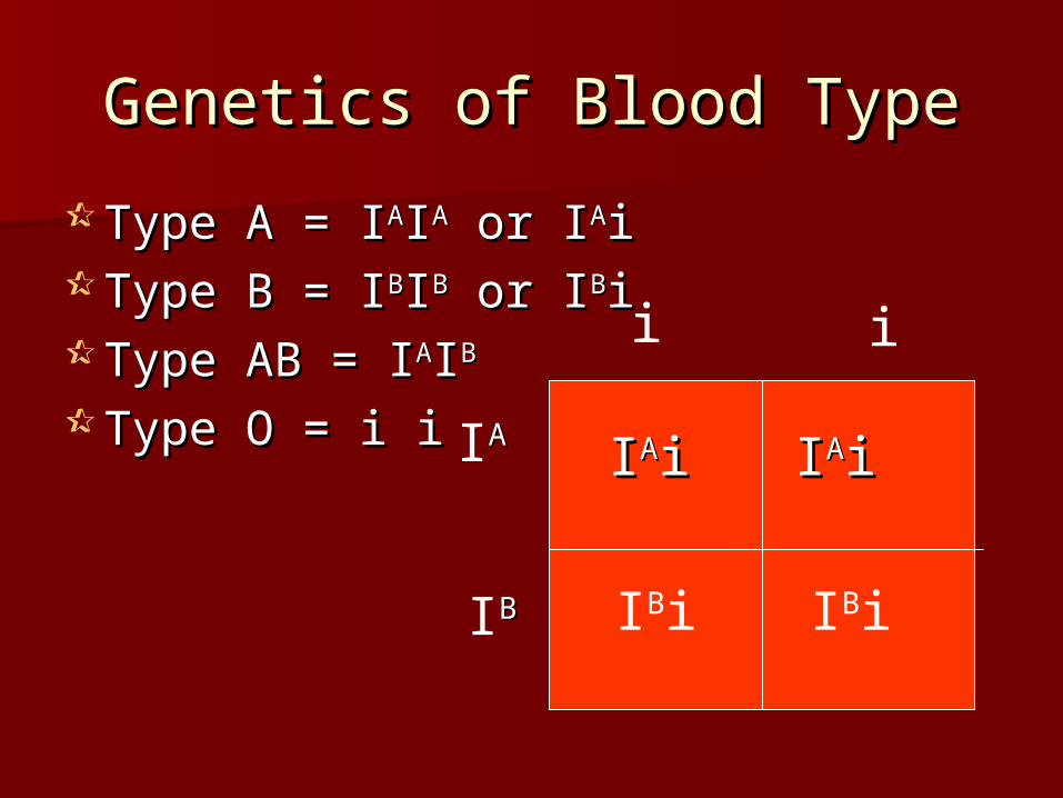
Genetics of Blood TypeGenetics of Blood Type
Type A = IType A = IAAIIAA or I or IAAii Type B = IType B = IBBIIBB or I or IBBii Type AB = IType AB = IAAIIBB
Type O = i iType O = i i
i i
IAA
IBB
IIAAii IIAAii
IBi IBi
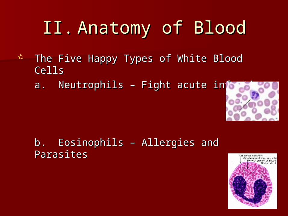
II.II. Anatomy of BloodAnatomy of Blood
The Five Happy Types of White Blood The Five Happy Types of White Blood CellsCells
a. Neutrophils – Fight acute infectiona. Neutrophils – Fight acute infection
b. Eosinophils – Allergies and Parasitesb. Eosinophils – Allergies and Parasites

(Cont)(Cont)
c. Basophils – Seen in inflammationc. Basophils – Seen in inflammation
d. Lymphocytes – Immune Response d. Lymphocytes – Immune Response – make antibodies, fights tumors– make antibodies, fights tumors
e. Monocytes – Fight chronic e. Monocytes – Fight chronic infections like TBinfections like TB

Leukemia: Leukemia:
Suppressed bone marrow Suppressed bone marrow functionfunction
Can’t fight diseaseCan’t fight disease

Mononucleosis: Mononucleosis:
Caused by the Epstein Barr VirusCaused by the Epstein Barr Virus Monocytes are called into actionMonocytes are called into action Tired, achy, sore throat, sore and Tired, achy, sore throat, sore and
swollen glandsswollen glands

Platelet ProblemsPlatelet Problems
Thombus/Embolus: Clot forms, can Thombus/Embolus: Clot forms, can cut off blood flow to organscut off blood flow to organs
Hemophilia: Bleeder’s disease, Hemophilia: Bleeder’s disease, missing 1 of 13 clotting factorsmissing 1 of 13 clotting factors
Platelet Deficiency: not enough Platelet Deficiency: not enough platelets, bleed for no reasonplatelets, bleed for no reason


III. HemostasisIII. Hemostasis The steps to forming a blood clot....The steps to forming a blood clot....
1. Platelets become sticky and cling to damaged 1. Platelets become sticky and cling to damaged sitesite
2. Anchored platelets release chemicals that 2. Anchored platelets release chemicals that attract more platelets - Forms a platelet plug attract more platelets - Forms a platelet plug (white thrombus)(white thrombus)
3. Platelets release serotonin, a chemical that 3. Platelets release serotonin, a chemical that causes the blood vessel to spasm. This narrows causes the blood vessel to spasm. This narrows the vessel and decreases blood loss until clotting.the vessel and decreases blood loss until clotting.
4. Chemical reactions galore result in fibrin, a 4. Chemical reactions galore result in fibrin, a meshwork that traps RBCs to make a clot.meshwork that traps RBCs to make a clot.
5. Clot squeezes serum from the mass to dry it 5. Clot squeezes serum from the mass to dry it out.out.
Fun Facts...Fun Facts...
This process takes 3-6 minutes. So This process takes 3-6 minutes. So why do we use dry gauze and why do we use dry gauze and pressure when we are bleeding? pressure when we are bleeding?
Gauze give platelets a place to stick Gauze give platelets a place to stick and pressure increases the rate of and pressure increases the rate of the chemical reactions that need to the chemical reactions that need to occur. Cool!! occur. Cool!!


Kickin’ Cardiovascular Kickin’ Cardiovascular SystemSystem

I. System AnatomyI. System Anatomy
HeartHeart Blood VesselsBlood Vessels LymphaticLymphatic

II. System PhysiologyII. System Physiology
Transportation of Blood which Transportation of Blood which contains:contains:
OxygenOxygen Carbon DioxideCarbon Dioxide NutrientsNutrients WasteWaste HormonesHormones Disease Fighters (WBC’s)Disease Fighters (WBC’s)

III. The Happy HeartIII. The Happy Heart
Size of your fistSize of your fist Less than 1 poundLess than 1 pound Covered by pericardiumCovered by pericardium Coronary arteries (blood vessels) – give Coronary arteries (blood vessels) – give
heart bloodheart blood 4 chambers4 chambers
– 2 atria (atrium) – receive blood, top of heart2 atria (atrium) – receive blood, top of heart– 2 ventricles – discharge blood, bottom of heart2 ventricles – discharge blood, bottom of heart


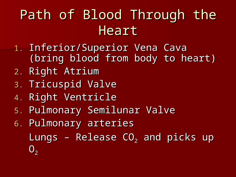
Path of Blood Through the Path of Blood Through the HeartHeart
1.1. Inferior/Superior Vena Cava (bring Inferior/Superior Vena Cava (bring blood from body to heart)blood from body to heart)
2.2. Right AtriumRight Atrium3.3. Tricuspid ValveTricuspid Valve4.4. Right VentricleRight Ventricle5.5. Pulmonary Semilunar ValvePulmonary Semilunar Valve6.6. Pulmonary arteriesPulmonary arteries
Lungs – Release COLungs – Release CO22 and picks and picks up Oup O22

Path of Blood Through the Path of Blood Through the HeartHeart
7.7. Pulmonary VeinsPulmonary Veins8.8. Left AtriumLeft Atrium9.9. Bicuspid ValveBicuspid Valve10.10. Left Ventricle (biggest part)Left Ventricle (biggest part)11.11. Aortic Semilunar ValveAortic Semilunar Valve12.12. AortaAorta13.13. ArteriesArteries14.14. Capillaries (release OCapillaries (release O22 to cells, pick up to cells, pick up
COCO22))15.15. Veins – Back to HeartVeins – Back to Heart
AGAINAGAIN

IV. Hip, Hip Hooray – Heart IV. Hip, Hip Hooray – Heart PhysiologyPhysiology
Atria collect bloodAtria collect blood Ventricles Ventricles
Discharge bloodDischarge blood Soooo…. Ventricles Soooo…. Ventricles
are the actual are the actual pump. When they pump. When they contract, blood contract, blood moves.moves.

A. Double Pump System A. Double Pump System
Right Side Right Side Pulmonary CircuitPulmonary CircuitReceives oxygen Receives oxygen
poor blood from poor blood from bodybody
Pumps to Lungs to Pumps to Lungs to pick up Oxygen pick up Oxygen and release and release carbon dioxidecarbon dioxide

A. Double Pump SystemA. Double Pump System
Left SideLeft Side Systemic CircuitSystemic Circuit Receives Oxygen Receives Oxygen
rich blood from rich blood from lungslungs
Pumps blood to Pumps blood to body cells to supply body cells to supply them with oxygen them with oxygen and pick up carbon and pick up carbon dioxidedioxide

B. ValvesB. Valves
1. Prevent Backwash1. Prevent Backwash 2. Heartbeat2. Heartbeat

B. Valves (cont)B. Valves (cont)
““lub”-bicuspid/tricuspid valve closing; lub”-bicuspid/tricuspid valve closing; longer and louder sound longer and louder sound
““dup”-Semilunars closing; shorter dup”-Semilunars closing; shorter and sharperand sharper

B. Arteries – Blood away from B. Arteries – Blood away from heartheart
Take blood away from the heartTake blood away from the heart No ValvesNo Valves High Pressure--thick walls with strong High Pressure--thick walls with strong
tunica media tunica media --strong and stretchy because close --strong and stretchy because close to the change in pressure (diastole) to the change in pressure (diastole) and close to the heart.and close to the heart.

C. Veins – Blood to heartC. Veins – Blood to heart
Take blood toward the heartTake blood toward the heart Valves to prevent backflow Valves to prevent backflow
because... because... Low pressureLow pressure Thin WallsThin Walls Far from the heart and Far from the heart and
change in pressure change in pressure

Mechanical heart valveMechanical heart valve

C. Intrinsic Conduction C. Intrinsic Conduction SystemSystem
Nodal SystemNodal SystemAtria beat at 60 beats/minAtria beat at 60 beats/minVentricles beat at 20-40 beats/minVentricles beat at 20-40 beats/minNodal system unifies it toNodal system unifies it to about 75 about 75
beats/minbeats/min
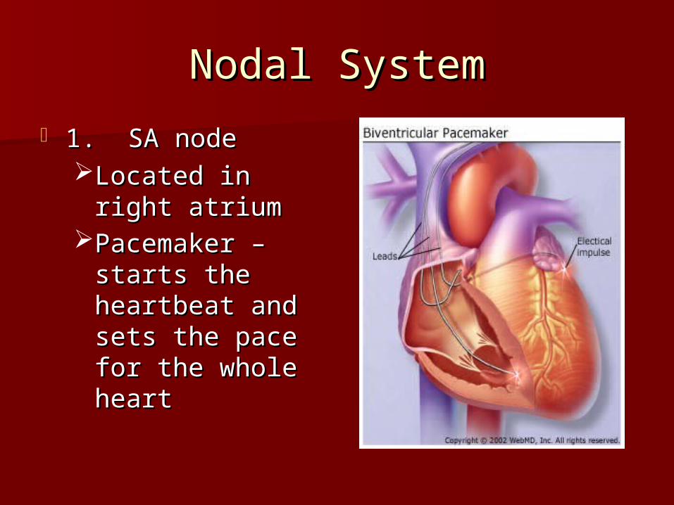
Nodal SystemNodal System
1. SA node 1. SA node Located in right Located in right
atriumatriumPacemaker – Pacemaker –
starts the starts the heartbeat and heartbeat and sets the pace for sets the pace for the whole heartthe whole heart

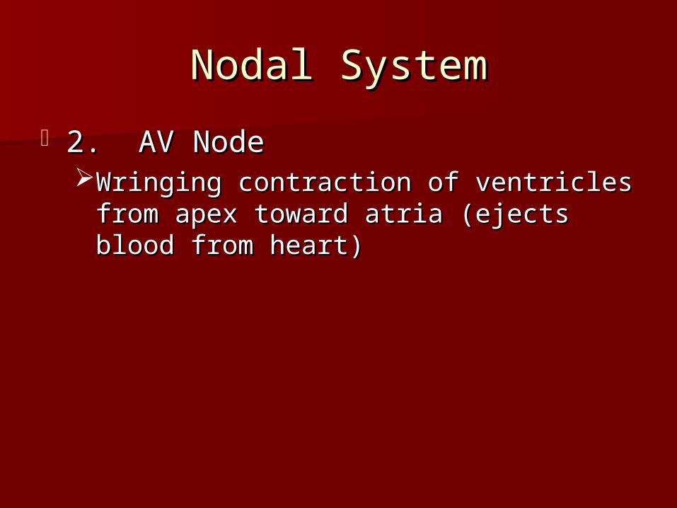
Nodal SystemNodal System
2. AV Node2. AV NodeWringing contraction of ventricles from Wringing contraction of ventricles from
apex toward atria (ejects blood from apex toward atria (ejects blood from heart)heart)

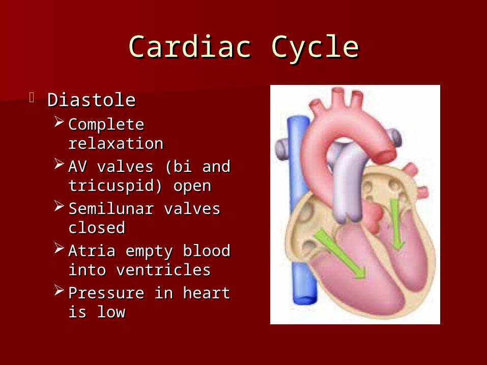
Cardiac CycleCardiac Cycle
DiastoleDiastole Complete relaxationComplete relaxation AV valves (bi and AV valves (bi and
tricuspid) opentricuspid) open Semilunar valves Semilunar valves
closedclosed Atria empty blood Atria empty blood
into ventriclesinto ventricles Pressure in heart is Pressure in heart is
lowlow

Cardiac CycleCardiac Cycle
SystoleSystole Bi/Tricuspid close, Bi/Tricuspid close,
Semilunar valves Semilunar valves openopen
Ventricles contractVentricles contract Blood rushes out of Blood rushes out of
heartheart Pressure in heart is Pressure in heart is
highhigh Atria are fillingAtria are filling

V. Blood VesselsV. Blood Vessels
Superhighway for bloodSuperhighway for blood Microscopic Anatomy of VesselsMicroscopic Anatomy of Vessels
Tunica interna (intima) – lines interior of Tunica interna (intima) – lines interior of vessels – made of endothelial tissuevessels – made of endothelial tissue
Tunica media –made of smooth muscle Tunica media –made of smooth muscle and elastic tissueand elastic tissue
Tunica externa – outermost layer – Tunica externa – outermost layer – protects the vesselsprotects the vessels


D. CapillariesD. Capillaries
Connect arteries to veinsConnect arteries to veins Very thin...Just tunica intimaVery thin...Just tunica intima Transparent, one-cell layer thickTransparent, one-cell layer thick GAS EXCHANGE TAKES PLACE HEREGAS EXCHANGE TAKES PLACE HERE

Flow of blood through Flow of blood through vesselsvessels
AortaAortaArteriesArteriesArteriolesArterioles
CapillariesCapillariesVenulesVenulesVeinsVeins
Vena CavaVena Cava

E. Blood Vessel PhysiologyE. Blood Vessel Physiology
1.1. Arterial PulseArterial Pulse
-Pressure wave created by the -Pressure wave created by the expansion and recoil of an artery that expansion and recoil of an artery that occurs with each beat of the left occurs with each beat of the left ventricle.ventricle.
Average is 70-76 beats per minuteAverage is 70-76 beats per minute Pulse points are listed in book. Take Pulse points are listed in book. Take
a look and try to find them on your a look and try to find them on your body. body.


2. Blood Pressure2. Blood Pressure
* Pressure the blood exerts against * Pressure the blood exerts against the inner walls of blood vessels.the inner walls of blood vessels.
* Force that keeps blood circulating * Force that keeps blood circulating between beatsbetween beats
* Pressure in arteries near the heart* Pressure in arteries near the heart Systolic: Pressure in arteries at peak Systolic: Pressure in arteries at peak
of ventricular contractionof ventricular contraction Diastolic: Pressure when ventricles Diastolic: Pressure when ventricles
are relaxingare relaxing


Procedure for taking blood Procedure for taking blood pressurepressure
1. Pump up to about 150 (exceed 1. Pump up to about 150 (exceed systolic). Stops blood flow.systolic). Stops blood flow.
2. Reduce pressure in cuff while 2. Reduce pressure in cuff while listening carefully.listening carefully.
3. When first soft tapping sounds are 3. When first soft tapping sounds are heard, SYSTOLICheard, SYSTOLIC
4. As pressure is reduced, sounds 4. As pressure is reduced, sounds get louder. When no sounds, record get louder. When no sounds, record DIASTOLICDIASTOLIC

3. What messes up blood 3. What messes up blood pressure?pressure?
* Friction in Blood Vessel (viscosity, * Friction in Blood Vessel (viscosity, atherosclerosis)atherosclerosis)

3. What messes up blood 3. What messes up blood pressure?pressure?
* Nervous System (narrows vessels)-* Nervous System (narrows vessels)-fear, exercise, blood lossfear, exercise, blood loss

3. What messes up blood 3. What messes up blood pressure?pressure?
* Kidneys (alter blood volume)* Kidneys (alter blood volume)
3. What messes up blood 3. What messes up blood pressure?pressure?
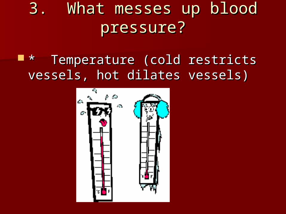
* Temperature (cold restricts * Temperature (cold restricts vessels, hot dilates vessels)vessels, hot dilates vessels)

3. What messes up blood 3. What messes up blood pressure?pressure?
* Chemicals (alcohol & histamines * Chemicals (alcohol & histamines dilate, nicotine constricts)dilate, nicotine constricts)

Cardiovascular SystemHeart/Lung Model Project
Your task is to construct a three Your task is to construct a three dimensional model of a human heart dimensional model of a human heart and lungs. You may use any kind of and lungs. You may use any kind of materials you choose; however, your materials you choose; however, your model must have all of the following model must have all of the following structures:structures:

1. Four heart chambers 1. Four heart chambers 2. The bicuspid and tricuspid valves in the 2. The bicuspid and tricuspid valves in the
correct locationscorrect locations 3. The aortic semilunar and pulmonary 3. The aortic semilunar and pulmonary
semilunar valves in the correct locationssemilunar valves in the correct locations 4. The septum4. The septum 5. The inferior vena cava in the correct 5. The inferior vena cava in the correct
locationlocation 6. The superior vena cava in the correct 6. The superior vena cava in the correct
locationlocation 7. The pulmonary arteries and veins in the 7. The pulmonary arteries and veins in the
correct locationscorrect locations 8. The aorta in the correct location.8. The aorta in the correct location. 9. Both lungs properly attached to the heart 9. Both lungs properly attached to the heart
with pulmonary arteries and veinswith pulmonary arteries and veins

ALL STRUCTURES MUST BE CLEARLY ALL STRUCTURES MUST BE CLEARLY AND CORRECTLY LABELED. Once AND CORRECTLY LABELED. Once your model is complete you must your model is complete you must meet with the teacher and explain meet with the teacher and explain how blood flows through the heart, how blood flows through the heart, lungs and body.lungs and body.
You will be allowed to make You will be allowed to make corrections until you meet all corrections until you meet all requirements listed above.requirements listed above.

Leaping Lymphatic SystemLeaping Lymphatic System
I. System AnatomyI. System Anatomy Lymphatic vesselsLymphatic vessels Lymph NodesLymph Nodes Lymph organs and tissuesLymph organs and tissues

II. System PhysiologyII. System Physiology
Returns lost excess tissue fluid Returns lost excess tissue fluid (lymph) to the blood(lymph) to the blood
Only flows toward the heartOnly flows toward the heart

III. Lymph NodesIII. Lymph Nodes Removes foreign material like bacteria and tumor Removes foreign material like bacteria and tumor
cellscells Produces lymphocytes (remember them?)Produces lymphocytes (remember them?) Macrophages engulf and destroy bacteria, virus, Macrophages engulf and destroy bacteria, virus,
and foreign substances as lymph is filtered and foreign substances as lymph is filtered through the nodes and before its returned to the through the nodes and before its returned to the blood.blood.
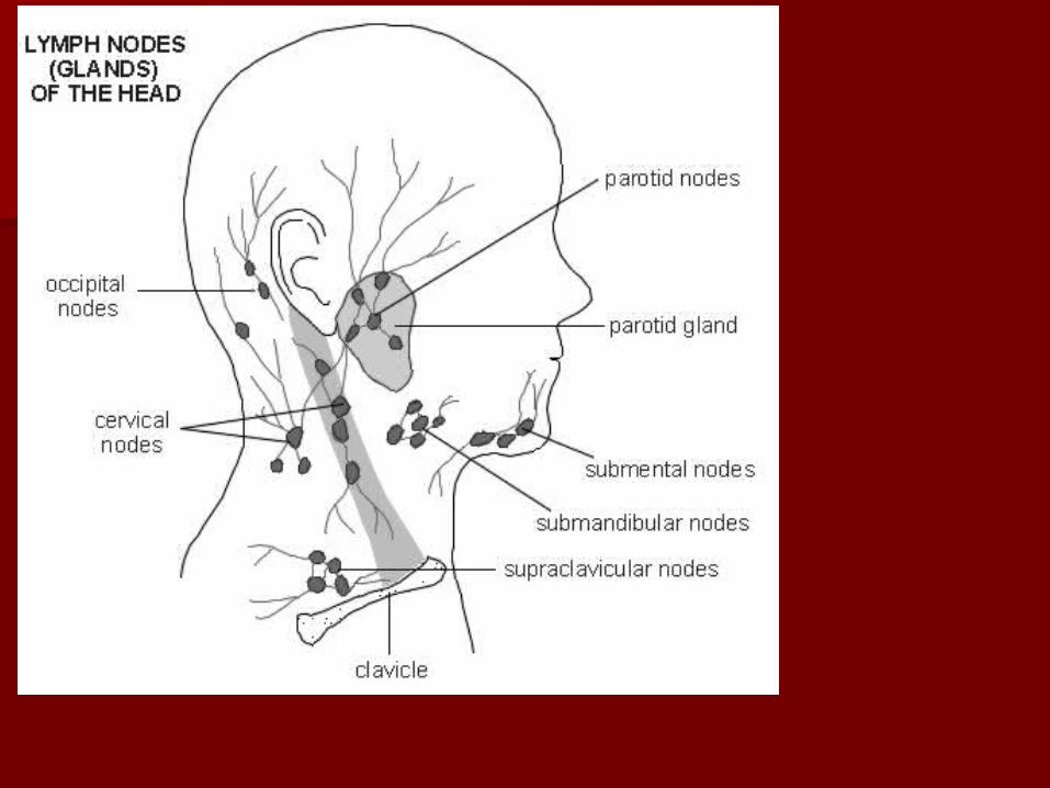
Kidney-shaped and are less than 1 inch. They are Kidney-shaped and are less than 1 inch. They are buried.buried.
Lymph nodes are working hard if they get Lymph nodes are working hard if they get inflammed and tender.inflammed and tender.
If large and not painful, often a sign of cancer.If large and not painful, often a sign of cancer.

IV. Lymph OrgansIV. Lymph Organs
A. SpleenA. Spleen Filters the blood of bacteria, virus, debrisFilters the blood of bacteria, virus, debris Destroy worn out RBCsDestroy worn out RBCs Recycle products to the liverRecycle products to the liver Stores plateletsStores platelets Blood resevoir—Blood resevoir—
empties duringempties during hemorrageshemorrages

IV. Lymph OrgansIV. Lymph Organs
B. Thymus - Programs the B. Thymus - Programs the lymphocytes (peaks during youth, lymphocytes (peaks during youth, then tapers)then tapers)

IV. Lymph OrgansIV. Lymph Organs
C. Tonsils - Trap and remove any C. Tonsils - Trap and remove any bacteria or foreign pathogensbacteria or foreign pathogens
IV. Lymph OrgansIV. Lymph Organs
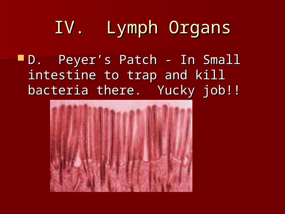
D. Peyer’s Patch - In Small intestine D. Peyer’s Patch - In Small intestine to trap and kill bacteria there. Yucky to trap and kill bacteria there. Yucky job!!job!!