Embed Size (px)
Citation preview

Blood VesselsBlood Vessels
11

Blood VesselsBlood VesselsThere are 100,000 miles of blood vessels.There are 100,000 miles of blood vessels.
That’s enough to circle the world 4 times!That’s enough to circle the world 4 times!
1/5 of the blood is in the lungs.1/5 of the blood is in the lungs.
The brain receives 1.5 pints of blood every The brain receives 1.5 pints of blood every minute.minute.
With the exception of cartilage (which is With the exception of cartilage (which is avascular), no cell is more than a few cell avascular), no cell is more than a few cell diameters away from a blood vessel, so they diameters away from a blood vessel, so they can get oxygen, nutrients, remove waste.can get oxygen, nutrients, remove waste.
22

Blood VesselsBlood VesselsArteries are the vessels that leave the heart. Arteries are the vessels that leave the heart. They get smaller and thinner and are then called They get smaller and thinner and are then called arterioles.arterioles.Arterioles get smaller and thinner until their Arterioles get smaller and thinner until their lumen is just one red blood cell in diameter. At lumen is just one red blood cell in diameter. At this point, they are called capillaries, and this is this point, they are called capillaries, and this is where the oxygen exchange takes place. where the oxygen exchange takes place. Capillaries take waste products away from the Capillaries take waste products away from the cells in the capillary bed and head back to the cells in the capillary bed and head back to the heart; then get larger and larger until they are heart; then get larger and larger until they are big enough to be called venules. big enough to be called venules. The venules get bigger and bigger, until they are The venules get bigger and bigger, until they are large enough to be called veins, and they return large enough to be called veins, and they return to the heart. to the heart.
33
Blood VesselsBlood VesselsFrom the heart the blood is pumped to the lungs to get From the heart the blood is pumped to the lungs to get more oxygen. more oxygen. They leave the heart, so they are arteries, but they are They leave the heart, so they are arteries, but they are blue. On their way to the lungs, they get smaller again blue. On their way to the lungs, they get smaller again until they are arterioles, then capillaries, then they get the until they are arterioles, then capillaries, then they get the oxygen from the lungs and drop off the waste products oxygen from the lungs and drop off the waste products (carbon dioxide). (carbon dioxide). Then they return to the heart, so they are now called Then they return to the heart, so they are now called venules, although they are red.venules, although they are red.Then they get larger until they are called veins, and the Then they get larger until they are called veins, and the blood returns to the heart to get pumped out to the body blood returns to the heart to get pumped out to the body again. again. All blood vessels (except the smallest) look similar.All blood vessels (except the smallest) look similar.
44

Structure of Arteries, Veins, and CapillariesStructure of Arteries, Veins, and Capillaries
Figure 19.1a55

Tunica intima Endothelium SubendotheliumTunica media Smooth muscles Elastic fibersTunica adventitia
Vasa vasorum
6

Structure of Blood VesselsStructure of Blood Vessels
Composed of three layers (tunics)Composed of three layers (tunics)– Tunica intimaTunica intima
ENDOTHELIUMENDOTHELIUM: simple squamous epithelium. : simple squamous epithelium. Allows for smooth flow of blood. We need for Allows for smooth flow of blood. We need for this layer to be smooth so platelets don’t catch this layer to be smooth so platelets don’t catch on it and start a blood clot. The endothelium is on it and start a blood clot. The endothelium is similar to endocardium.similar to endocardium.
SUBENDOTHELIUMSUBENDOTHELIUM: loose connective tissue.: loose connective tissue.
77

Structure of Blood VesselsStructure of Blood Vessels
Composed of three layers (tunics)Composed of three layers (tunics)– Tunica mediaTunica media
SMOOTH MUSCLE: SMOOTH MUSCLE: allows vasoconstriction. allows vasoconstriction. Allows blood to be directed to parts of body.Allows blood to be directed to parts of body.
ELASTIC FIBERSELASTIC FIBERS: within smooth muscles. : within smooth muscles. Allows for forced vasodilation during heart Allows for forced vasodilation during heart contraction.contraction.
88

Structure of Blood VesselsStructure of Blood Vessels
Composed of three layers (tunics)Composed of three layers (tunics)– TUNICA ADVENTITIA (TUNICA EXTERNA)TUNICA ADVENTITIA (TUNICA EXTERNA): :
dense fibrous connective tissue which thins dense fibrous connective tissue which thins out to loose connective tissue.out to loose connective tissue.
Protects the blood vessel (strong)Protects the blood vessel (strong)
Gives vessel strength for shapeGives vessel strength for shape
Anchors vessel to surrounding tissue; loosens with Anchors vessel to surrounding tissue; loosens with age.age.
Lumen – central blood-filled space of a Lumen – central blood-filled space of a vesselvessel
99
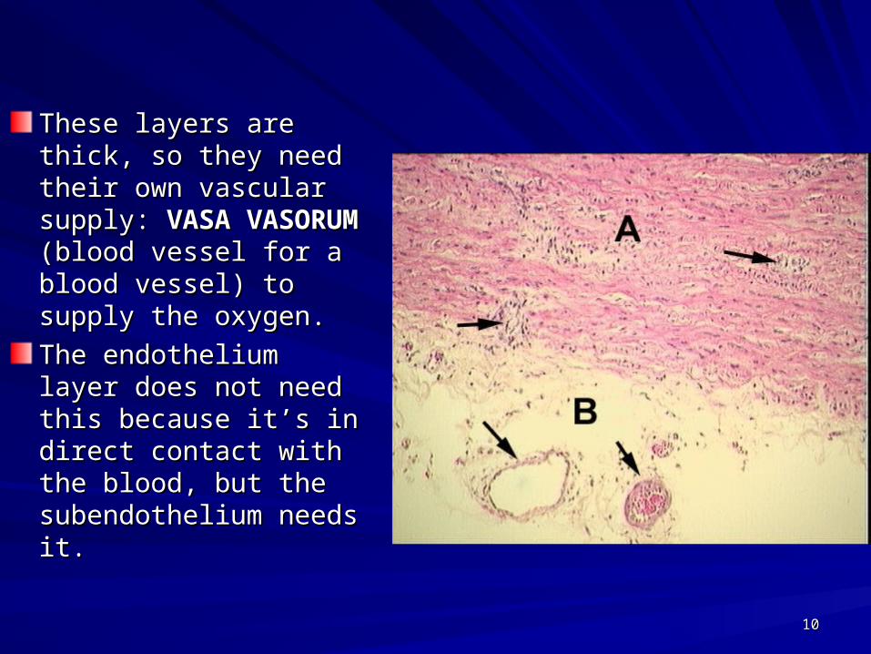
These layers are thick, These layers are thick, so they need their own so they need their own vascular supply: vascular supply: VASA VASA VASORUM VASORUM (blood (blood vessel for a blood vessel for a blood vessel) to supply the vessel) to supply the oxygen. oxygen.
The endothelium layer The endothelium layer does not need this does not need this because it’s in direct because it’s in direct contact with the blood, contact with the blood, but the subendothelium but the subendothelium needs it.needs it.
1010

Tunica intima Endothelium SubendotheliumTunica media Smooth muscles Elastic fibersTunica adventitia
Vaso vasorum
11

Structure of Arteries, Veins, and CapillariesStructure of Arteries, Veins, and Capillaries
Figure 19.1a1212

Types of Blood VesselsTypes of Blood Vessels
Arteries – carry blood away from the heartArteries – carry blood away from the heart– It does not matter if it is oxygenated or deoxy It does not matter if it is oxygenated or deoxy
blood. If it is leaving the heart, it is an artery.blood. If it is leaving the heart, it is an artery.
Veins – carry blood toward the heartVeins – carry blood toward the heartIt does not matter if it is oxygenated or deoxy blood. If It does not matter if it is oxygenated or deoxy blood. If it is entering the heart, it is a vein.it is entering the heart, it is a vein.
Capillaries – smallest blood vesselsCapillaries – smallest blood vessels– The site of exchange of molecules between The site of exchange of molecules between
blood and tissue fluidblood and tissue fluid
1313

ArteriesArteriesARTERIES ARTERIES carry blood carry blood away from the heart.away from the heart. Arteries have a smaller Arteries have a smaller lumen than veins of lumen than veins of similar size. similar size. The lumen of an artery is The lumen of an artery is more round than a veinmore round than a veinArterial walls are thicker Arterial walls are thicker than venous walls.than venous walls.Arteries have more Arteries have more elastin than veins.elastin than veins.Arteries have no valves Arteries have no valves because the blood because the blood pressure in arteries is pressure in arteries is high enough that there is high enough that there is no backflow of blood.no backflow of blood.
1414

ArteriesArteries
Two types of large arteries:
Elastic
Muscular
1515

Types of ArteriesTypes of Arteries
Elastic arteries – Elastic arteries – the largest arteries the largest arteries – Diameters range Diameters range
from 1 - 2.5 cm from 1 - 2.5 cm – Includes the aorta Includes the aorta
and its major and its major branchesbranches
– High elastin High elastin content dampens content dampens surge of blood surge of blood pressurepressure
Figure 19.2a1616

ELASTIC ARTERIESELASTIC ARTERIES
a.a. Largest, closest to heart. Largest, closest to heart.
b.b. Has to take the full force of the systolic Has to take the full force of the systolic contraction; compensates by expanding a contraction; compensates by expanding a lot. lot.
c.c. There of lots of elastic fibers in the tunica There of lots of elastic fibers in the tunica intima as well. intima as well.
d.d. Does blood flow during diastole? Yes; Does blood flow during diastole? Yes; elastic arteries return to original size, pumps elastic arteries return to original size, pumps blood. blood.
e.e. This is another pump besides the heart.This is another pump besides the heart.1717

Muscular ArteriesMuscular Arteries
Muscular Muscular (distributing) (distributing) arteriesarteries– Distal to elastic Distal to elastic
arteriesarteries– From 0.3 mm- 1 From 0.3 mm- 1
cm cm – Includes most of Includes most of
the named arteriesthe named arteries– Tunica media is Tunica media is
thickthick
Figure 19.2b1818

MUSCULAR ARTERIESMUSCULAR ARTERIES
a.a. Function is to distribute blood, and help Function is to distribute blood, and help control which regions of the body get blood. control which regions of the body get blood.
b.b. When you are exercising, you want the When you are exercising, you want the blood from the GI system to go to muscles.blood from the GI system to go to muscles.
c.c. When your hands are cold, your body is When your hands are cold, your body is using its blood for something more using its blood for something more important. Therefore, the vessels will important. Therefore, the vessels will constrict in the hands. constrict in the hands.
d.d. Dilation is just lack of constriction.Dilation is just lack of constriction.
1919
ARTERIOLESARTERIOLES
These are microscopic; they are the These are microscopic; they are the smallest type of artery. smallest type of artery.
Large ones look like muscular arteries.Large ones look like muscular arteries.
Small ones only have two layers: Small ones only have two layers: endothelium and tunica media. endothelium and tunica media.
One of the characteristics of an arteriole is One of the characteristics of an arteriole is that when it constricts, the lumen closes that when it constricts, the lumen closes completely.completely.
2020

Types of ArteriesTypes of Arteries
ArteriolesArterioles– Smallest Smallest
arteriesarteries– Diameters Diameters
range from 10 range from 10 µm to 0.3 mm µm to 0.3 mm
Figure 19.2c2121

AneurysmAneurysmA sac-like outpouching of an A sac-like outpouching of an arteryartery– Can rupture at any time; Can rupture at any time;
in aorta or brain can in aorta or brain can cause death within a few cause death within a few seconds. seconds.
– Symptoms: Swelling or Symptoms: Swelling or throbbing (asymptomatic throbbing (asymptomatic in brain)in brain)
Some common locations for Some common locations for aneurysms include:aneurysms include:– AortaAorta– Brain Brain – Leg Leg – Intestine (mesenteric Intestine (mesenteric
artery aneurysm) artery aneurysm) – Splenic artery Splenic artery
aneurysm (can form aneurysm (can form during pregnancy) during pregnancy)
2222

AneurysmAneurysm
Causes of an aneurysm:Causes of an aneurysm:– Defect in part of the artery wallDefect in part of the artery wall– High blood pressure (abdominal aortic High blood pressure (abdominal aortic
aneurysms)aneurysms)– Congenital (present at birth)Congenital (present at birth)
Usually not detected except by an Usually not detected except by an angiogram or ultrasound.angiogram or ultrasound.
Treatment: surgical repairTreatment: surgical repair
2323

MRI for Blood VesselsMRI for Blood Vessels
2424

StrokeStroke
2525

AneurysmAneurysm
2626

The Ovation Abdominal Stent Graft System
Aneurysms can be repaired through open surgery or less invasively with endograft repair using a stent graft otherwise known as an endograft.
Endografts feature a tube typically made of plastic material that is supported by a metal frame or stent. They are compressed into a delivery catheter, inserted into the femoral artery of the leg and then threaded into position in the weakened portion of the artery where they are released. Once released, the endograft expands against the wall of the aorta to redirect blood flow away from the aneurysm.
http://catalog.nucleusinc.com/generateexhibit.php?ID=68382&ExhibitKeywordsRaw=&TL=&A=2 2727

How to Recognize a Stroke How to Recognize a Stroke (“STROKE”)(“STROKE”)
S * Ask the individual to SMILE. S * Ask the individual to SMILE. T * Ask the person to TALK and SPEAK A SIMPLE T * Ask the person to TALK and SPEAK A SIMPLE SENTENCE (Coherently; i.e. It is sunny out today)SENTENCE (Coherently; i.e. It is sunny out today) R * Ask him or her to RAISE BOTH ARMS.R * Ask him or her to RAISE BOTH ARMS.O * Open the mouth and stick out the tongueO * Open the mouth and stick out the tongueK * Keep them comfortable and stillK * Keep them comfortable and stillE * Get EMERGENCY help (911)E * Get EMERGENCY help (911)
If one side of the body responds differently than the If one side of the body responds differently than the other side, or if they have trouble with the task, call other side, or if they have trouble with the task, call 911.911.
2828

Important:Important:
You don’t have enough blood to go around; you You don’t have enough blood to go around; you only have 5 liters for 100,000 miles of blood only have 5 liters for 100,000 miles of blood vessels. vessels. At any given time, most blood vessels will be At any given time, most blood vessels will be closed (except at lungs). closed (except at lungs). Are you using your legs now? When your legs Are you using your legs now? When your legs run low on oxygen, the vessels there will open run low on oxygen, the vessels there will open up again. up again. Are you using your brain now? I hope so! The Are you using your brain now? I hope so! The vessels there will be open. vessels there will be open. When your leg falls asleep, there is pressure on When your leg falls asleep, there is pressure on an artery which stops the blood flow. When the an artery which stops the blood flow. When the nerves are deprived of oxygen, they tingle.nerves are deprived of oxygen, they tingle.
2929

Some clinically significant arteries
Femoral artery: easy to find pulse, but susceptible to injury.
Circle of Willis: loop of arteries around pituitary and optic chiasma. Common area for stroke to cause blindness.
30

31

Circle of Circle of WillisWillis
3232

CapillariesCapillaries
Smallest blood vessels; they are found Smallest blood vessels; they are found everywhereeverywhereThese are the only sites of nutrient, gas These are the only sites of nutrient, gas exchange, and waste exchange in the exchange, and waste exchange in the cardiovascular system. cardiovascular system. – Diameter from 8–10 µmDiameter from 8–10 µm
Diameter is similar to an erythrocyteDiameter is similar to an erythrocyteRed blood cells pass through single fileRed blood cells pass through single fileThey only have an endothelium. They only have an endothelium.
3333

CapillariesCapillaries
Site-specific functions of capillariesSite-specific functions of capillariesIn the lungs – oxygen enters blood, carbon dioxide In the lungs – oxygen enters blood, carbon dioxide leavesleavesIn the small intestines – receive digested nutrientsIn the small intestines – receive digested nutrientsIn endocrine glands – pick up hormonesIn endocrine glands – pick up hormonesIn the kidneys – removal of nitrogenous wastesIn the kidneys – removal of nitrogenous wastes
3434

Capillary PermeabilityCapillary Permeability
Intercellular clefts – gaps of unjoined Intercellular clefts – gaps of unjoined membrane membrane – Small molecules can enter and exit Small molecules can enter and exit
Three types of capillariesThree types of capillaries– Continuous – most commonContinuous – most common– Fenestrated (“window”) – have poresFenestrated (“window”) – have pores– Discontinuous (Sinusoids) – have very large Discontinuous (Sinusoids) – have very large
gapsgaps
3535

Types of CapillariesTypes of Capillaries
CONTINUOUS CAPILLARIESCONTINUOUS CAPILLARIES
FENESTRATED CAPILLARIESFENESTRATED CAPILLARIES
DISCONTINUOUS CAPILLARIESDISCONTINUOUS CAPILLARIES
3636
CONTINUOUS CAPILLARIESCONTINUOUS CAPILLARIESAll capillaries are made of simple squamous All capillaries are made of simple squamous epithelium. epithelium.
Continuous capillaries are most common, found Continuous capillaries are most common, found in all organs of body. in all organs of body.
They have intracellular clefts, the function of They have intracellular clefts, the function of which is essential for plasma to leak out and which is essential for plasma to leak out and bathe each cell with extracellular fluid, which is bathe each cell with extracellular fluid, which is rich in oxygen and nutrients. rich in oxygen and nutrients.
Erythrocytes and platelets don’t fit through, but Erythrocytes and platelets don’t fit through, but leukocytes can squeeze through so they can leukocytes can squeeze through so they can enter and leave the blood vessels as needed. enter and leave the blood vessels as needed.
3737

Continuous CapillaryContinuous Capillary
Figure 19.4a3838

FENESTRATED CAPILLARIESFENESTRATED CAPILLARIES
These have a lot more leakage because These have a lot more leakage because there are more pores (holes). there are more pores (holes).
Found in areas where lots of fluids need to Found in areas where lots of fluids need to be moved back and forth (synovial be moved back and forth (synovial membranes, small intestine, CSF). membranes, small intestine, CSF).
3939

Fenestrated CapillaryFenestrated Capillary
Figure 19.4b4040

DISCONTINUOUS CAPILLARIES DISCONTINUOUS CAPILLARIES (sinusoidal capillaries) (sinusoidal capillaries)
These have very large gaps in the capillary. These have very large gaps in the capillary. Anything can go in and out here, including Anything can go in and out here, including erythrocytes. erythrocytes. These are found in red bone marrow, where These are found in red bone marrow, where RBCs are made, and they need to enter the RBCs are made, and they need to enter the circulation by way of the sinusoidal capillaries. circulation by way of the sinusoidal capillaries. These capillaries are also in the liver and These capillaries are also in the liver and spleen, where red blood cells are destroyed. spleen, where red blood cells are destroyed.
4141

SinusoidsSinusoids
Figure 19.4c4242

Capillary BedsCapillary Beds
Figure 19.3a4343

PRE-CAPILLARY SPHINCTERPRE-CAPILLARY SPHINCTER
A small muscle in front of each capillary, controls A small muscle in front of each capillary, controls the flow of blood to individual capillaries.the flow of blood to individual capillaries.
ARTERIOLESARTERIOLES direct the blood flow to the direct the blood flow to the specific specific tissuetissue. PRE-CAPILLARY . PRE-CAPILLARY SPHINCTERS direct the blood flow to specific SPHINCTERS direct the blood flow to specific cellscells. .
If one cell is starving, the capillary next to it will If one cell is starving, the capillary next to it will open. The sphincter opens and closes open. The sphincter opens and closes depending on the needs of individual cells.depending on the needs of individual cells.
4444

Capillary BedsCapillary Beds
Figure 19.3b4545

PRE-CAPILLARY SPHINCTERPRE-CAPILLARY SPHINCTER
There is not enough blood to go around, There is not enough blood to go around, so blood always flows only to those cells so blood always flows only to those cells and tissues that need it. and tissues that need it. They drop off nutrients, pick up CO2 and They drop off nutrients, pick up CO2 and other wastes. other wastes.
4646

VeinsVeins
Veins take blood TO the heart. Two types:Veins take blood TO the heart. Two types:– Venuole: from the capillary to the veinVenuole: from the capillary to the vein– Vein: takes blood to the heart.Vein: takes blood to the heart.
Thinner walls (less pressure here)Thinner walls (less pressure here)
Larger lumen (blood moves more slowly)Larger lumen (blood moves more slowly)
Skeletal muscle pushes on the vein to move the Skeletal muscle pushes on the vein to move the blood uphill. blood uphill.
Need valves in veins Need valves in veins
4747

Valves in VeinsValves in Veins
How does blood get uphill back to the heart? How does blood get uphill back to the heart? Veins need valves. Veins need valves.
Veins are the only BLOOD vessels that have Veins are the only BLOOD vessels that have valves (although LYMPH vessels also have valves (although LYMPH vessels also have valves). valves).
Valves in veins allow blood to move in only one Valves in veins allow blood to move in only one direction. What pushes the blood? The muscles direction. What pushes the blood? The muscles of the body constrict, squeezing the vessels. of the body constrict, squeezing the vessels. This is a type of blood pump.This is a type of blood pump.
4848

VeinsVeins
4949

BLOOD PUMPS
The heart Elastic arteries Muscles constricting the veins
50

Clinically Significant Veins
Greater Saphenous vein: used for coronary bypass; most likely becomes varicose.
Facial vein: “Danger triangle” infection spreads to meninges in brain.
Renal vein: oxygen poor, and contains the lowest concentration of nitrogen waste.
51

Veins that are rich in oxygen and nutrients
Pulmonary vein Umbilical vein Hepatic Portal vein
52

Fun Fact
Shivering increases your body heat by 18 fold.
Moderate walking only increases it by 3 fold.
53

Varicose VeinsVaricose Veins
The valves become incompetent:The valves become incompetent:
They can’t close all the way because too They can’t close all the way because too much fluid has built up in them and the much fluid has built up in them and the lumen has stretched too wide. lumen has stretched too wide.
They might be asymptomatic or they may They might be asymptomatic or they may be painful (phlebitis).be painful (phlebitis).
5454

Varicose VeinsVaricose Veins
5555

Telangiectasias(Spider veins)
Small dilated blood vessels (varicose veins) Small dilated blood vessels (varicose veins) near the surface of the skin or mucous near the surface of the skin or mucous membranes that blanch (turn colorless) with membranes that blanch (turn colorless) with direct pressure.direct pressure.
They can develop anywhere on the body but are commonly seen on the face around the nose, cheeks, and chin. They can also develop on the legs, specifically on the upper thigh, below the knee joint, and around the ankles.
5656

Telangiectasias
Telangiectasia in the legs is often related to the presence of venous hypertension within underlying varicose veins.
Age: The development of spider veins may occur at any age but usually occurs between 18 and 35 years, and peaks between 50 and 60 years.
Females are affected approximately four to one to males.
5757

Telangiectasias
Pregnancy is a key factor contributing to the formation of varicose and spider veins. The most important factor is circulating hormones that weaken vein walls. There's also a significant increase in the blood volume during pregnancy.
Varicose veins that form during pregnancy may spontaneously improve or even disappear a few months after delivery.
5858

Telangiectasias
Those who are involved with prolonged sitting or standing in their daily activities have an increased risk of developing varicose veins.
The weight of the blood continuously pressing against the closed valves causes them to fail, leading to vein distention.
5959

Other causes of spider veinsOther causes of spider veins
Acne rosaceaAcne rosacea
Sun or cold exposureSun or cold exposure
Trauma to skin such as contusions or surgical incisions.Trauma to skin such as contusions or surgical incisions.
Radiation exposure for the treatment of cancerRadiation exposure for the treatment of cancer
ChemotherapyChemotherapy
Chronic treatment with topical corticosteroidsChronic treatment with topical corticosteroids
6060

Spider VeinsSpider VeinsCause an unsightly appearance but are Cause an unsightly appearance but are not dangerous.not dangerous.
Injections of alcohol or saline into the vein Injections of alcohol or saline into the vein will sclerose them (scar them shut).will sclerose them (scar them shut).
A laser can also be used to do the same.A laser can also be used to do the same.
After treatment, macrophages will After treatment, macrophages will eventually phagocytize them and they will eventually phagocytize them and they will disappear.disappear.
6161

Spider VeinsSpider Veins
6262

Varicose Vein TreatmentVaricose Vein Treatment
Laser can be used to seal off the distal Laser can be used to seal off the distal end of the vein. It will close off.end of the vein. It will close off.
Sclerosing agents (alcohol or saline) Sclerosing agents (alcohol or saline) injected around the vein can be used to do injected around the vein can be used to do the same thing.the same thing.
Large painful veins can be surgically Large painful veins can be surgically removed (vein stripping)removed (vein stripping)
6363

Polidocanol
A sclerosant, an irritant injected to treat varicose veins. It causes fibrosis inside varicose veins, occluding the lumen of the vessel, and reducing the appearance of the varicosity.
The FDA has approved it for veins up to 3 mm in diameter.
It works by damaging the cell lining of blood vessels, causing them to close and eventually be replaced by other types of tissue.
6464
EdemaEdemaIf the veins are varicose for a long time, If the veins are varicose for a long time, plasma may leak out into the tissues, plasma may leak out into the tissues, causing edema. causing edema.
Edema means swelling anywhere in the Edema means swelling anywhere in the body (including from an injury or from body (including from an injury or from hanging your legs down too long like when hanging your legs down too long like when on an airplane), but it frequently occurs on an airplane), but it frequently occurs from incompetent veins in the legs.from incompetent veins in the legs.
6565

EdemaEdemaThere are two types of edema:There are two types of edema:– PittingPitting– Non-pittingNon-pitting
6666

Pitting EdemaPitting Edema
Pitting edema is when you can push your finger Pitting edema is when you can push your finger into the skin and it leaves behind your print into the skin and it leaves behind your print when you remove it.when you remove it.
This type is less serious; it tends to be better in This type is less serious; it tends to be better in the morning since the legs have been horizontal the morning since the legs have been horizontal all night.all night.
It will improve if a pressure bandage is applied.It will improve if a pressure bandage is applied.
6767

Pitting Edema Pitting Edema in the Footin the Foot
6868

Treatment for Pitting EdemaTreatment for Pitting EdemaAce wrapAce wrap– In the foot or leg always wrap from base of toes all the way In the foot or leg always wrap from base of toes all the way
to below the knee. Don’t leave a hole at the heel!to below the knee. Don’t leave a hole at the heel!– In the hand, always wrap from the base of the fingers to In the hand, always wrap from the base of the fingers to
right before the bend of the elbowright before the bend of the elbow
Support hose (don’t use the kind with the open heel; Support hose (don’t use the kind with the open heel; edema will push out of that area)edema will push out of that area)
Jobst Intermittent Compression Jobst Intermittent Compression – A machine is used to inflate air in a bag around the A machine is used to inflate air in a bag around the
leg. The air pressure is increased and decreased leg. The air pressure is increased and decreased every few minutes to milk the edema out. Patient every few minutes to milk the edema out. Patient goes in for therapy several times a week.goes in for therapy several times a week.
6969

Jobst Intermittent CompressionJobst Intermittent Compression
7070

Non-Pitting EdemaNon-Pitting Edema
Non-pitting edema is hardened tissue that Non-pitting edema is hardened tissue that does not leave your fingerprint.does not leave your fingerprint.
It is just as bad in the morning as it is at It is just as bad in the morning as it is at the end of the day.the end of the day.
This is more severe because it does not This is more severe because it does not go away easily.go away easily.
7171

Tip For Everyone!Tip For Everyone!
Buy your shoes at the end of the day when Buy your shoes at the end of the day when your feet are the most swollen.your feet are the most swollen.
Wear new shoes around the house for two Wear new shoes around the house for two hours to make sure they don’t hurt.hours to make sure they don’t hurt.
Diabetic people need to have someone Diabetic people need to have someone else examine their feet after wearing a else examine their feet after wearing a new pair of shoes for two hours. Check for new pair of shoes for two hours. Check for redness and blisters that they might not redness and blisters that they might not see or feel.see or feel.
7272
Venous Stasis UlcersVenous Stasis Ulcers
Might occur after the formation of varicose Might occur after the formation of varicose veins, when plasma has leaked out into the veins, when plasma has leaked out into the tissues, causing edema.tissues, causing edema.
Acid products from the blood plasma Acid products from the blood plasma (carbon dioxide, etc) can eventually erode (carbon dioxide, etc) can eventually erode all the way to the skin.all the way to the skin.
Common in diabetics.Common in diabetics.
Treatment must address sugar levels, vein Treatment must address sugar levels, vein problem, and the open wound.problem, and the open wound.
7373

Venous Venous Stasis Stasis UlcersUlcers
7474

PHLEBITISPHLEBITIS
Inflammation of a veinInflammation of a vein
Usually in the legs.Usually in the legs.
When phlebitis is associated with the When phlebitis is associated with the formation of blood clots (thrombosis), formation of blood clots (thrombosis), usually in the deep veins of the legs, the usually in the deep veins of the legs, the condition is called Deep Vein condition is called Deep Vein TThrombophlebitis (DVT)hrombophlebitis (DVT)..
7575

DEEP VEIN DEEP VEIN THROMBOPHLEBITISTHROMBOPHLEBITIS
Signs and SymptomsSigns and SymptomsRedness (erythema) and warmth with a Redness (erythema) and warmth with a temperature elevation of a degree or more temperature elevation of a degree or more above the baseline above the baseline Pain or burning along the length of the Pain or burning along the length of the vein vein Swelling (edema) Swelling (edema) Vein being hard, and cordlike Vein being hard, and cordlike Need ER if all symptoms are presentNeed ER if all symptoms are present
7676

DVTDVT
7777

Severe DVTSevere DVT
7878

Tissue Necrosis (gangrene)Tissue Necrosis (gangrene)
Necrosis = deadNecrosis = dead
Caused by infection, toxins, or traumaCaused by infection, toxins, or trauma
Almost always detrimental and can be Almost always detrimental and can be fatalfatal
7979

GangreneGangrene
Gangrene is a serious and potentially life-threatening Gangrene is a serious and potentially life-threatening condition that arises when a considerable mass of body condition that arises when a considerable mass of body tissue dies (necrosis).tissue dies (necrosis).
This may occur after an injury or infection, or in people This may occur after an injury or infection, or in people suffering from any chronic health problem affecting blood suffering from any chronic health problem affecting blood circulation.circulation.
The primary cause of gangrene is reduced blood supply The primary cause of gangrene is reduced blood supply to the affected tissues, which results in cell death.to the affected tissues, which results in cell death.
Diabetes and long-term smoking increase the risk of Diabetes and long-term smoking increase the risk of suffering from gangrenesuffering from gangrene
8080

Types of GangreneTypes of Gangrene
Dry gangreneDry gangrene
Wet gangreneWet gangrene
Gas gangreneGas gangrene
Necrotising fasciitisNecrotising fasciitis
8181
Dry GangreneDry Gangrene
Dry gangrene begins at the distal part of the limb Dry gangrene begins at the distal part of the limb due to ischemia, and often occurs in the toes due to ischemia, and often occurs in the toes and feet of elderly patients due to and feet of elderly patients due to arteriosclerosis. arteriosclerosis.
Dry gangrene is mainly due to arterial occlusion. Dry gangrene is mainly due to arterial occlusion. There is limited putrefaction and bacteria fail to There is limited putrefaction and bacteria fail to survive. survive.
Dry gangrene spreads slowly until it reaches the Dry gangrene spreads slowly until it reaches the point where the blood supply is adequate to point where the blood supply is adequate to keep tissue viable. keep tissue viable.
8282

Dry GangreneDry Gangrene
The affected part is dry, shrunken and dark reddish-The affected part is dry, shrunken and dark reddish-black, resembling mummified flesh. black, resembling mummified flesh.
The dark coloration is due to liberation of hemoglobin The dark coloration is due to liberation of hemoglobin from hemolyzed red blood cells, which is acted upon by from hemolyzed red blood cells, which is acted upon by hydrogen sulfide (H2S) produced by the bacteria, hydrogen sulfide (H2S) produced by the bacteria, resulting in formation of black iron sulfide that remains in resulting in formation of black iron sulfide that remains in the tissues.the tissues.
There is a line of separation where the blood supply There is a line of separation where the blood supply becomes adequate, and the gangrenous tissue falls off becomes adequate, and the gangrenous tissue falls off by itself if it is not removed surgically, also called by itself if it is not removed surgically, also called autoamputation.autoamputation.
8383

Dry GangreneDry Gangrene
8484

Wet GangreneWet Gangrene
Wet gangrene occurs in naturally moist tissue Wet gangrene occurs in naturally moist tissue and organs such as the mouth, bowel, lungs, and organs such as the mouth, bowel, lungs, cervix, and vulva.cervix, and vulva.
Bedsores occurring on body parts such as the Bedsores occurring on body parts such as the sacrum, buttocks, and heels are also sacrum, buttocks, and heels are also categorized as wet gangrene infections. categorized as wet gangrene infections.
It is characterized by numerous bacteria and has It is characterized by numerous bacteria and has a poor prognosis (compared to dry gangrene) a poor prognosis (compared to dry gangrene) due to septicemia (bacterial infection of the due to septicemia (bacterial infection of the bloodstream). bloodstream).
8585

Wet GangreneWet Gangrene
8686

Wet GangreneWet Gangrene
In wet gangrene, the tissue is infected by saprogenic In wet gangrene, the tissue is infected by saprogenic microorganisms (those that eat dead organic matter) microorganisms (those that eat dead organic matter) such as Clostridium perfringens or Bacillus fusiformis, such as Clostridium perfringens or Bacillus fusiformis, which cause tissue to swell and emit a fetid smell. which cause tissue to swell and emit a fetid smell.
Wet gangrene usually develops rapidly due to blockage Wet gangrene usually develops rapidly due to blockage of venous (mainly) and/or arterial blood flow. of venous (mainly) and/or arterial blood flow.
The affected part is saturated with stagnant blood, which The affected part is saturated with stagnant blood, which promotes the rapid growth of bacteria. promotes the rapid growth of bacteria.
The toxic products formed by bacteria are absorbed, The toxic products formed by bacteria are absorbed, causing systemic manifestation of septicemia and finally causing systemic manifestation of septicemia and finally death. death.
8787

Wet GangreneWet Gangrene
The affected part is edematous, soft, The affected part is edematous, soft, putrid, rotten and dark. putrid, rotten and dark.
The darkness in wet gangrene occurs due The darkness in wet gangrene occurs due to the same mechanism as in dry to the same mechanism as in dry gangrene. gangrene.
Wet gangrene is coagulative necrosis Wet gangrene is coagulative necrosis progressing to liquefactive necrosis progressing to liquefactive necrosis ((transformation of dead tissue into a liquid)..
8888

Gas GangreneGas GangreneThis is a bacterial infection that produces gas within tissues.
It is a deadly form of gangrene usually caused by Clostridium perfringens bacteria.
Infection spreads rapidly as the gases produced by bacteria expand and infiltrate healthy tissue in the vicinity.
Because of its ability to quickly spread to surrounding tissues, gas gangrene should be treated as a medical emergency.
8989

Gas GangreneGas Gangrene
9090

Gas GangreneGas Gangrene
These bacteria are mostly found in soil and enter the muscle through a wound and subsequently proliferate in necrotic tissue and secrete powerful toxins. These toxins destroy nearby tissue, generating gas at the same time.
Gas gangrene can cause necrosis, gas Gas gangrene can cause necrosis, gas production, and sepsis. production, and sepsis.
Progression to toxemia and shock is often very Progression to toxemia and shock is often very rapid.rapid.
9191

Necrotizing Fasciitis (flesh-eating disease)
This is a rare infection of the deeper layers of skin and This is a rare infection of the deeper layers of skin and subcutaneous tissues, easily spreading across the subcutaneous tissues, easily spreading across the fascial plane within the subcutaneous tissue.fascial plane within the subcutaneous tissue.
9292

Gangrene TreatmentGangrene Treatment
Debridement (laser or mechanical)Debridement (laser or mechanical)
AmputationAmputation
AntibioticsAntibiotics
Vascular surgeryVascular surgery
Maggot therapy Maggot therapy
Hyperbaric oxygen therapyHyperbaric oxygen therapy
9393

Peripheral Vascular Disease Peripheral Vascular Disease (PVD)(PVD)
Refers to the obstruction of large arteries, frequently in Refers to the obstruction of large arteries, frequently in the lower extremity. Usually caused from atherosclerosis the lower extremity. Usually caused from atherosclerosis (fatty plaques).(fatty plaques).
SymptomsSymptoms– Claudication: pain, weakness, numbness, or cramping Claudication: pain, weakness, numbness, or cramping
in muscles due to decreased blood flowin muscles due to decreased blood flow– Sores, wounds, or ulcers that heal slowly or not at allSores, wounds, or ulcers that heal slowly or not at all– Change in color (blueness or paleness) or temperature Change in color (blueness or paleness) or temperature
(coolness) when compared to the other limb(coolness) when compared to the other limb– Diminished hair and nail growth on affected limb and Diminished hair and nail growth on affected limb and
digits (shiny, hairless skin)digits (shiny, hairless skin)
9494

MigrainesMigrainesMigraines are severe headaches that cause Migraines are severe headaches that cause vomiting and photosensitivity (the person vomiting and photosensitivity (the person cannot tolerate any light).cannot tolerate any light).
They can be caused by several things, They can be caused by several things, including muscle spasms in the blood including muscle spasms in the blood vessels.vessels.
Caffeine can cause them, and so can caffeine Caffeine can cause them, and so can caffeine withdrawal.withdrawal.
Treatments may include medicines, botox Treatments may include medicines, botox injections, and magnesium infusions. injections, and magnesium infusions.
9595
VasculitisVasculitis
This is a group of disorders that are This is a group of disorders that are characterized by inflammatory destruction characterized by inflammatory destruction of blood vessels. Both arteries and veins of blood vessels. Both arteries and veins are affected. Lymphangitis is sometimes are affected. Lymphangitis is sometimes considered a type of vasculitis. Vasculitis is considered a type of vasculitis. Vasculitis is primarily due to leukocyte migration and primarily due to leukocyte migration and resultant damage.resultant damage.
Although both occur in vasculitis, Although both occur in vasculitis, inflammation of veins (phlebitis) or arteries inflammation of veins (phlebitis) or arteries (arteritis) on their own are separate entities.(arteritis) on their own are separate entities.
9696

VasculitisVasculitisBuerger's disease: vasculitis of the leg arteries and veins (gangrene).
Systemic Lupus Erythematosus (SLE)
9797

Buerger's disease
This is a non-atherosclerotic vascular disease also known as thromboangiitis obliterans (TAO), and is strongly associated with heavy tobacco use.
9898

Systemic Lupus Erythematosus
SLE is an autoimmune disease that can affect any part SLE is an autoimmune disease that can affect any part of the body.of the body.
SLE most often harms the heart, joints, skin, lungs, SLE most often harms the heart, joints, skin, lungs, blood vessels, liver, kidneys, and nervous system. blood vessels, liver, kidneys, and nervous system.
The course of the disease is unpredictable, with periods The course of the disease is unpredictable, with periods of illness (called flares) alternating with remissions. of illness (called flares) alternating with remissions.
The disease occurs nine times more often in women The disease occurs nine times more often in women than in men, especially in women in child-bearing years than in men, especially in women in child-bearing years ages 15 to 35, and is also more common in those of non-ages 15 to 35, and is also more common in those of non-European descent.European descent.
Typical skin manifestations are a butterfly rash on the face and photosensitivity.
9999

SLE Butterfly RashSLE Butterfly Rash
100100

Restless Legs Syndrome (Ekbom's Syndrome)
A condition in which a sense of uneasiness, restlessness, and itching, often accompanied by twitching and pain, is felt in the calves of the legs when sitting or lying down, especially in bed at night.
The cause is unknown: it may be inadequate circulation, peripheral neuropathy, deficiency of iron, vitamin B12, or folic acid, or a reaction to antipsychotic or antidepressant drugs.
101101

102