Embed Size (px)
Citation preview

Blood supply

Vascular Supply About 18% of the total blood volume in the body
circulates in the brain, which accounts for about 2% of the body weight.
The blood transports oxygen, nutrients, and other substances necessary for proper functioning of the brain tissues and carries away metabolites.
Loss of consciousness occurs in less than 15 seconds after blood flow to the brain has stopped, and irreparable damage to the brain tissue occurs within 5 minutes.
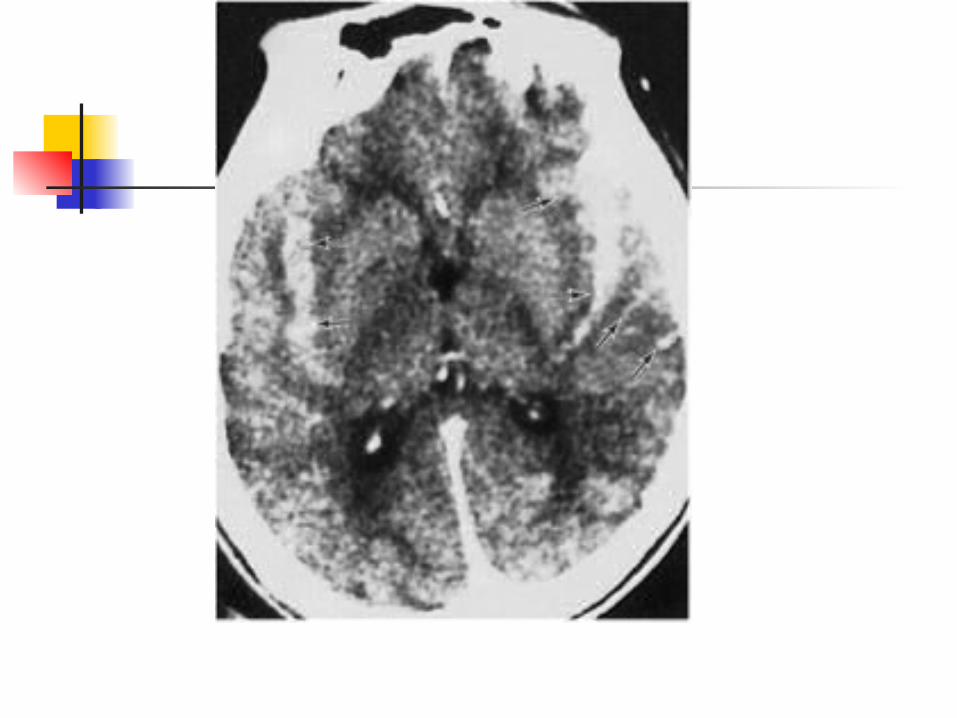
Cerebrovascular disease, or stroke, occurs as a result of vascular compromise or hemorrhage and is one of the most frequent sources of neurologic disability.
Nearly half of the admissions to many busy neurologic services are because of strokes.
Cerebrovascular disease is the third most common cause of death in industrialized societies.

BLOOD SUPPLY TO THE BRAIN
• High demand for oxygen and nutrients
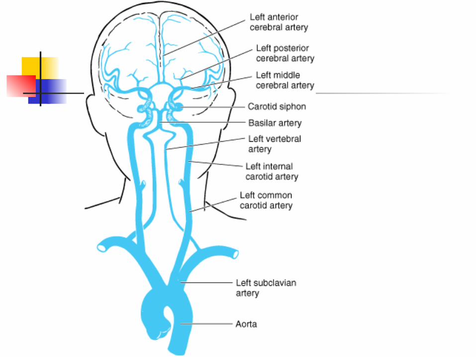
• Arterial blood through: internal carotid and vertebral arteries
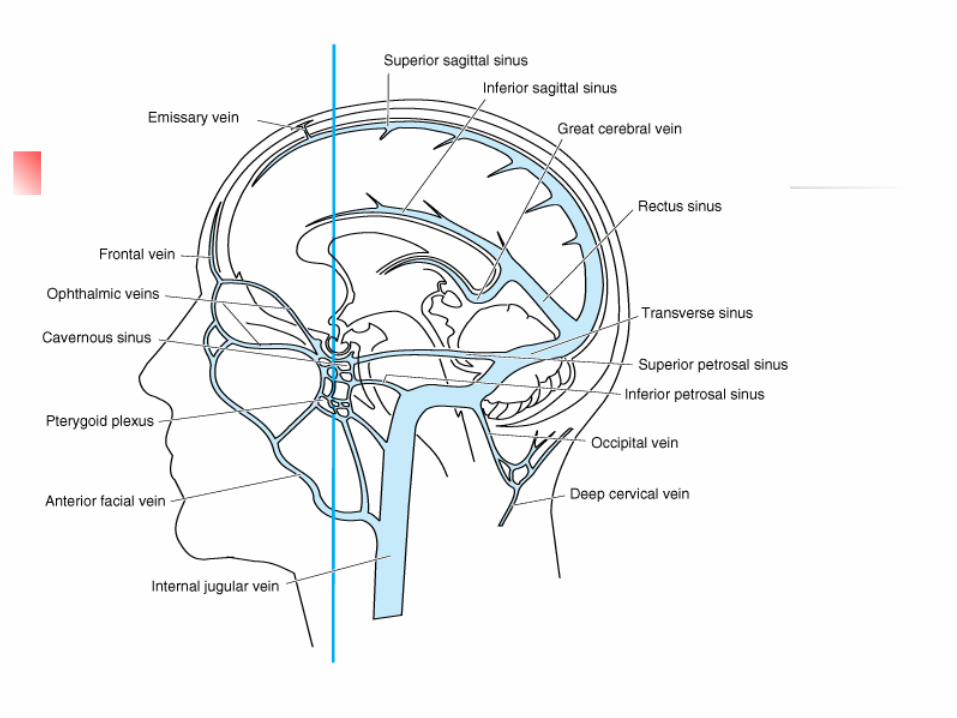
• Venous blood from brain in the internal
jugular veins
• Cerebrovascular accidents (CVA): stroke, shutting off blood supply to brain

Arterial supply The circle of Willis (after the English
neuroanatomist Sir Thomas Willis) is a confluence of vessels that gives rise to all of the major cerebral arteries.
Supplied by the paired internal carotid arteries and the basilar artery.
Contains a paired posterior communicating artery and an unpaired anterior communicating artery.
The circle of Willis shows many variations among individuals.
Occlusion of major cerebral arteries produces a characteristic clinical picture.


Why do individual differences in vascular supply matter?
What might they achieve? What problems might they give? How might they assist after
damage?


VENOUS DRAINAGE
The venous drainage of the brain and coverings includes: the veins of the brain itself, the dural venous sinuses, the dura's meningeal veins, and the diploic veins between the
tables of the skull.


CRANIAL MENINGES
• Dura mater: outer and inner layers• Arachnoid mater: epithelial layer andarachnoid trabeculae• Pia mater: sticks to brain surface• Pia and arachnoid = leptomeninges• Dural folds hold the brain in position -
Falx cerebriTentorium cerebelli
• Dural sinuses (veins located within the folds)


VENTRICLES OF THE BRAIN
• Lined with ependymal cells
• Lateral ventricle (each hemisphere)
• Third ventricle (diencephalon)
• Cerebral aqueduct (midbrain)
• Fourth ventricle: (btw pons and cerebellum, continuous with central canal of spinal cord)
• CSF flows within ventricles, central canal and
into subarachnoid space

Ventricles
CEREBROSPINAL FLUID
Surrounds and bathes the CNS
Functions:
1. Supporting of brain and spinal cord
2. Transport of nutrients, chemical
messengers, and waste products

THE FORMATION OF CSF
• Choroid plexus: contains specialised ependymal cells and capillaries (500ml/day), total volume: 150ml
• Choroid plexuses secrete CSF into ventricles
• Circulation: from choroid plexus to ventricles and central canal of spinal cord to subarachnoid space to sinuses

Circulation (cont.):
• CSF reaches subarachnoid space through two
lateral apertures and a single medial aperture in
the 4th ventricle
• Arachnoid granulations: penetrate dura
mater meningeal layer of venous sinuses, CSF
absorbed into the venous circulation
• Hydrocephalus: “water in the brain”