Embed Size (px)
Citation preview

Blood Groups – Duffy, and
MNSs Group Systems
Qun Lu, MD
Assistant Professor
Division of Transfusion Medicine
Department of Pathology and Laboratory Medicine
UCLA, School of Medicine
Los Angeles, California
2009-03-12

Duffy Blood Group System

History 1950: Mrs. Duffy, a multiply transfused hemophiliac
woman, developed an antibody not reacting with the known RBC antigens. Corresponding antigen was named after Mrs. Duffy
1951: Fyb antibody was described in a woman with 3 pregnancies.
1955: Majority of blacks tested Fy(a-b-)
1975: Fy(a-b-) RBCs were shown to resist infection by malaria organism Plasmodium vivax.
Later: more Duffy antigens (Fy3, Fy4, Fy5, Fy6) were discovered
ISBT: 008 for the Duffy Blood Group

Duffy Antigens
Most common: Fya and Fyb.
Present at 6 weeks of gestation, well developed at birth – anti-Fy can cause hemolytic disease of newborn
Duffy antigens can be destroyed by enzymes such as ficin, papain, bromelain, chymotrypsin, ZZAP
When compared to Rh or Kell antigens, Duffy antigens are not very immunogenic. So, anti-Fya or anti-Fyb is not common.
Fy (a-b-) is not Fy null, but homozygous for Fyb gene, they express Fyb antigen in other tissues, but not on RBCs → only will produce anti-Fya, not anti-Fyb.
Fy (a-b-) is negative for Fy6 antigen which is the receptor for P. vivax (Fy6 is + when Fya + or Fyb+)
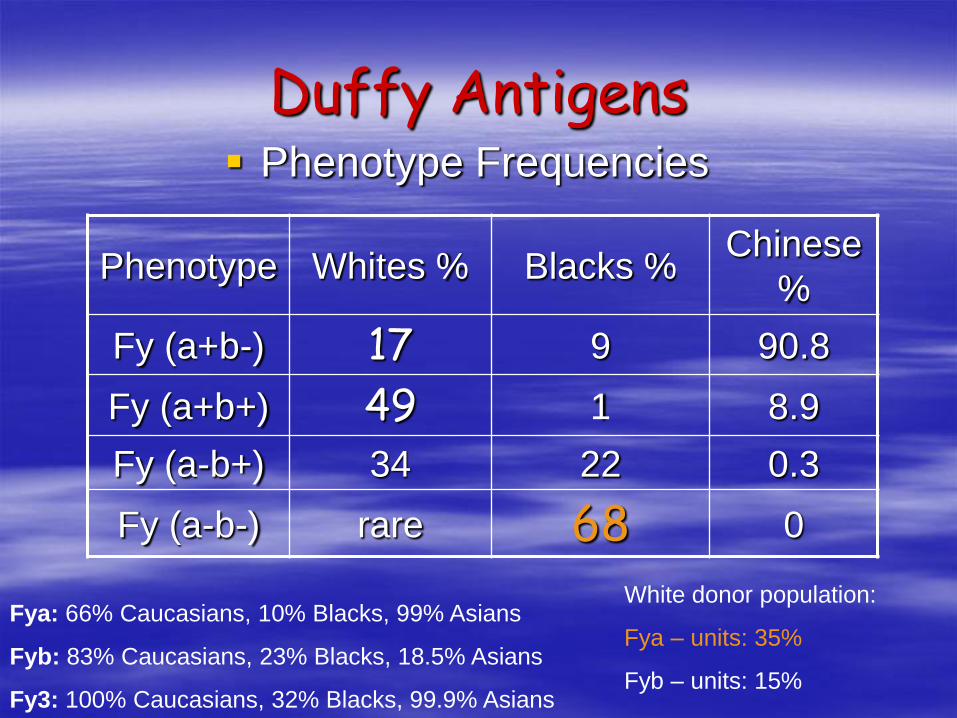
Duffy Antigens Phenotype Frequencies
Phenotype Whites % Blacks %Chinese
%
Fy (a+b-) 17 9 90.8
Fy (a+b+) 49 1 8.9
Fy (a-b+) 34 22 0.3
Fy (a-b-) rare 68 0
Fya: 66% Caucasians, 10% Blacks, 99% Asians
Fyb: 83% Caucasians, 23% Blacks, 18.5% Asians
Fy3: 100% Caucasians, 32% Blacks, 99.9% Asians
White donor population:
Fya – units: 35%
Fyb – units: 15%

Duffy Antigens
Biochemistry: glycoprotein, transmembrane (7
times, 3 extracellular loops)
Function: amino acid is similar to interleukin -8
receptor on WBCs, receptors of cytokines
Gene: chromosome 1q22-23. Rh gene also
located on chromsome 1, but not linked
Fyx gene: produced weak Fyb antigen – react with
some not all anti-Fyb, it can be typed as Fy(b-),
can cause confusion in paternity testing

Chromosome 1q22-23

REVIEW ARTICLE
Blood, Vol. 89 No. 9 (May 1), 1997: pp. 3077-3091
From Malaria to Chemokine Receptor: The Emerging Physiologic Role of the Duffy Blood Group Antigen
By Terence J. Hadley and Stephen C. Peiper
From the Departments of Medicine, Pathology, and Biochemistry, Henry Vogt Cancer Research Institute, James Graham Brown Cancer Center, University of Louisville; and the Department of Veterans Affairs Medical Center, Louisville, KY.

This thin film Giemsa stained micrograph reveals a mature Plasmodium vivax
trophozoite.
P. vivax trophozoites show amoeboid cytoplasm, large chromatin dots, and fine,
yellowish-brown pigment. RBCs are enlarged 1 1/2 - 2X, and may be distorted. If visible,
Schüffner's dots may appear finer than those seen in P. vivax.

Anti-Fya and Anti-Fyb
IgG, clinically significant, warm-reacting, exposure-requiring antibody
Transfusion reactions: acute or delayed hemolytic transfusions
HDN cases are usually mild
Anti-Fya is more common than anti-Fyb (Fya is more immunogenic than Fyb)
Anti-Fya or anti-Fyb do not react with enzyme treated RBCs (useful tech in ID)
Dosage effect is not as strong as anti-Jk
Antibody titers can fade over time, causing delayed hemolytic transfusion reaction
People with Fy (a-, b-) phenotype can make anti-Fy3 (anti-Fya & anti-Fyb reactivity)

Uncommon Duffy Antigen and
Antibody
Fy3: 1971 a case reported that anti-Fy3 was found in an Fy(a-b-) white. It reacted with all RBCs except Fy(a-b-) cells. Fy3 antigen is part of the Fya and Fyb antigen (one of the 3 extracelluar loops)
Fy4: 1973 a Fy(a+b+) black made anti-Fy4 reacted with all Fy(a-b-) blacks, Fy(a+b-), and Fy(a-b+) blacks, but not with Fy(a+b+) blacks and not with whites of any Duffy type. Fy(a-b-) blacks carry Fy4Fy4 antigens
Uncommon Duffy Antigen and
Antibody
Fy5: 1973 an Fy(a-b-) black child made anti-Fy5
which reacted with cell from a Fy(a-b-)Fy3-, but
did not react with Fy(a+) or Fy(b+) Rhnull red cells.
Fy5 antigen is the result of Duffy and Rh genes
Fy6: 1987, marine monoclonal antibody, much like
anti-Fy3, but destroyed by enzymes (Fy3, Fy4,
Fy5 antigens resist enzymes). Fy6 is the receptor
for P. vivax and present on all Fy + cells.
MNSs (002) Blood Group
System

MNSs Antigens
Total 46 antigens
Antigen Biochemistry:– Glycophorin A (GPA) forms the M, N antigens
– Glycophorin B (GPB) forms the S, s antigens and U antigen. U antigen is the common core of S, s antigens
– The glycophorins cross the RBC membrance once and have an external N-terminal and a intracellular C-terminal (linked to spectri skeleton)
– GPA (1 million copies) is much more abundant than GPB (200,000 copies)
Tight linkage between GPA and GPB
GPA is an erythroid marker and is the receptor for Plasmodium falciparum
M antigen is receptor of E. coli

AABB Technical Manual, 14th Edition, Page 320

MNSs Antigens
Destroyed by common enzymes (Papain, Ficin,
Bromelin, Pronase), but U antigen is resistant
Trypsin: M, N sensitive, S, s resistant
Alpha-chymotrypsin: M, N partialy sensitive, S, s
very sensitive
S-s-U- phenotype: 2% of Black American and a
higher propotion of Black African, due to deletion
of the GPB gene, will produce anti-S, s, U
S- units in White donors: 50%
S- units in White donors: 10%
MNSs Antibodies Anti-M, anti-N: IgM, insignificant, cold-reacting, naturally
occurring. Anti-M is common, but anti-N is rare
When anti-M, anti-N is reactive at 37C, antigen negative units should provided
Rare case of anti-M associated severe HDFN has been reported: – Lost 2 previous pregnancies, the third baby survived because of
intrauterine transfusion
– Fetus ruled out other causes of hemolysis
– M+ radio-labled RBCs were destroyed withing 3days after transfusion
– M- radio-labled RBCs survived 30days after transfusion to the mother (anti-M +).
Rare case of auto-anti-N causing fatal autoimmune hemolytic anemia
Anti-N is associate with hemodialysis, because N antigen is modified by formaldehyde in the dialysis machine.

MNSs Antibodies
Anti-S, anti-s, & anti-U: IgG, significant, warm-reacting, exposure-requiring
Whites – 100% S+, s+,1% of blacks-- S-, s-, and U- (make anti-U)
GP.Mur phenotype in Southeast Asia: hybrid gene of GPA and GPB produce an unusual amino acid sequence (antigen Mur = MNS10), immunogenic.
Anti-Mur is the most common antibody after anti-A and anti-B in Hong Kong and Taiwan

Gerbich System
Antigens:
– Located on Glycophorin C and D
– 8 antigens, 3 high prevalence (Ge 2, 3, 4), 5 low
prevalence
– Glycophorin C is the receptor for P. falciparum.
Antibodies
– IgG, react at AHG phase,
– Clinically insignificant, but anti-Ge3 has been
reported to cause HDFN, tend to manifest 2-4
wks after birth

RBC Antigens Functions
Duffy Receptor for chemokins,
plasmodium vivax
Kidd RBC urea transport (not kidney
urea transport)
Chido/Rodgers C4
Knops Receptor for C3/C4b
Colton, Coa, Cob RBC water transport
Cartwright, Yta, Ytb RBC AchE
Cromer CD55 = DAF (decay
accelerating factor)
MN, Gerbich antigen Receptor for p. falciparum