Embed Size (px)
Citation preview

The Proceeding of the 1st Congress of South East Asia Veterinary School Association IPB ICC, Bogor-Indonesia, July 20-22 2010
181
BLOOD FLOW CHARACTERISTICS ON ATRIA-VENTRICULAR VALVE AS ASSESSED BY PULSED WAVE DOPPLER ECHOCARDIOGRAPHY IN NORMAL MONGREL INDONESIAN DOG
Marina Wijayanti, Deni Noviana
Department of Clinic, Reproduction and Pathology
Faculty of Veterinary Medicine, Bogor Agricultural University (IPB) Jalan Agatis, Kampus IPB Dramaga, Bogor 16680, Jawa Barat, Indonesia
e-mail: [email protected]
Introduction
Indonesia Mongrel Dogs are dogs that have been known to exist in Indonesia but the strain of his offspring were not observed and recorded. Heart disease can be affected at all dog breeds including Indonesia mongrel dogs at a young age and adults. Echocardiography is selected as a tool to help diagnose heart disease because they have the advantage over other diagnostic tools. This diagnostic technique allows to obtain an accurate measure of the strength of contraction, the size of the heart chambers when the systolic and diastolic heart wall thickness, as well as evaluating the structure and valve abnormalities. Pulsed wave Doppler echocardiography (PWD) can even be used to determine the direction, velocity of blood flow or obstruction that occurs in the valves and blood vessels. The purpose of this study is to determine blood flow characteristics at the atria-ventricular valve i.e. mitral and tricuspid valves in normal Indonesia mongrel dog using transthoracic PWD echocardiography. Material and Methods
This study was performed using ultrasonography (USG) Sonoscape SSI-1000, convex type transducer with a small footprint, the frequency of transducer is 3.7-5 MHz, and electrocardiogram (ECG) Fukuda ME Cardisuny D300. Animals were observed in this study were nine adult male mongrel consisting of four males and five females. Dogs that are used have a range of ages 2-5 years and average weight 12.5 kg, where after a physical examination and electrocardiography showed good cardiac condition. Physical examination and data collection carried out on dogs in a conscious state and was not sedated. Echocardiography was performed by placing the animal's position right lateral recumbency for the examination of the Left apical Scanning Views (laps), in this position and trikuspidalis mitralis valve can be well observed. Seven parameters are observed by PWD echocardiography i.e. heart rate (HR), Velocity annulus (Va)/Epeak, Velocity ejection (Ve)/Apeak, Velocity Time Integral (VTi), Mean Pressure Gradient (MPG), Pulsatility Index (PI), and systole/diastole (S/D) (Penninck & d’Anjou 2008). Results and Discussions
Pulsed wave Doppler echocardiography examination results for heart rate indicated that heart rate measured at the mitral valve was 104 ± 20 bpm. This value is slightly higher than the tricuspid valve 101±13 bpm (P> 0.05). Adult dog has a heart rate of about 70-160 bpm, while a puppy has a heart rate up to 220 bpm (Nelson RW and Couto CG, 2008).
Annulus velocity (Va)/Epeak is a parameter measuring the flow velocity of blood flow passively-initial charging from the atria to the ventricle by 90% of blood volume due to the opening of atrio-ventricular valves (Penninck D, d'Anjou, 2008). Based on the results of PWD echocardiography examination showed that the mitral valve 106.75 ± 17.26 cm/s has a higher Va/Epeak than at the tricuspid valve 92.16±1.93 cm/s (P> 0.05).
Ejection velocity (Ve) Apeak is a parameter of blood flow velocity measurements derived from the strength of active atrial contraction. This type of contraction pushed approximately 10% of blood volume (Penninck D, d'Anjou, 2008). Apeak occurs when the ventricles are in the final period of diastole. In the ECG, Apeak is described as a P wave (atrial depolarization). Based on the results of PWD echocardiography, at mitral valve Apeak value 0.42 ± 0.03 cm/s was higher than the tricuspid valve 0.37±0.01 cm /s (P> 0.05). High peak velocity in the left ventricle was greatly influenced by the value of wall thickness of left ventricular at end-diastole.
Velocity Time Integral (VTI) is a parameter measuring the speed of blood flow entering the ventricles, the value is based on the comparison Epeak/ Apeak. VTI normal value is more than one. VTI calculation starts from an area of the base line up to the wave crest of Apeak and Epeak. The total area of these waves are affected by AT-acceleration time and DT-deceleration time. VTI measurement is useful to to measure the relative amount of blood into each ventricle (Penninck D, d'Anjou, 2008). Based on the results of PWD echocardiography examination showed that VTI of mitral valve 5.75 ± 2.19 cm/s have a higher value than the tricuspid valve 4.78±0.27 cm/s (P> 0.05).
P 2
1

The Proceeding of the 1st Congress of South East Asia Veterinary School Association IPB ICC, Bogor-Indonesia, July 20-22 2010
182
Mean Pressure Gradient (MPG) is useful to measure the obstruction that occurs in the valve. Based on the results, the value of mitral valve MPG 0.04 ± 0.01 mmHg have a higher value than the tricuspid valve 0.015± 0.005(P> 0.05).
Pulsatility index is used to describe the frequency or speed movement from one wave peak to the next wave peak during one cardiac cycle divided by the average time. Based on the results, shows that the mitral valve had PI 15,52±1,17 values higher than tricuspid valve 11,39±0,83 (P> 0.05).
Systole is the period in which the heart contracts and increase the pressure within the heart so that blood can be released into the systemic circulation and pulmonar. Period in which the heart relaxes and fill with blood is called diastole. Based on the results of PWD echocardiography shows that the mitral valve has a S/D value 251.5 ± 0.77 which was higher than the tricuspid valve 251.3±1.17 (P> 0.05). Ratio S/D at the mitral valve higher than in tricuspid valve can be connected with left ventricular function in charge of pumping blood throughout the body (Cunningham & Klein 2007).
Figure 1. Brightness mode and pulsed wave Doppler (PWD) echocardiography results of tricuspid valve.
Figure 2. Brightness mode and pulsed wave Doppler (PWD) echocardiography results of mitral valve.
References Cunningham JG, Klein BG. 2007. Textbook of Veterinary Physiology. 4th edition. : Saunders. Nelson RW, Couto CG. 2008. Small Animal Internal Medicine. 4th edition. USA : Mosby Inc. Penninck D, d’Anjou MA. 2008. Atlas of Small Animal Ultrasonography. 1st ed. : Blackwell Publishing.

The Proceeding of the 1st Congress of South East Asia Veterinary School Association IPB ICC, Bogor-Indonesia, July 20-22 2010
183
ANTIMICROBIAL SCREENING OF FOUR INDONESIAN MEDICINAL PLANT ACTIVITY AGAINST MYCOPLASMA GALLISEPTICUM
Min Rahminiwati1, Aulia Andi Mustika1, Soeripto2, Andriyanto1, Siti Sadiah1, Unang Patriana3
1Laboratorium of Pharmacology, Departement Of Anatomy, Physiology and Pharmacology, Faculty of
Veterinary Medicine, Bogor agricultural University, Jl. Agatis kampus IPB Darmaga-Bogor-16680 2Balitvet,
3BBPMSOH
Keywords: Mycoplasma gallisepticum, Complex Respiratory Disease (CRD), Sambiloto, Jahe (Ginger), Temulawak, Kencur
Introduction
Mycoplasma galliseticum is a pathogenic agent causing Complex Respiratory Disease (CRD) in chicken. The incident rate of CRD in the poultry farm especially in the layer chicken farm is so high that it gives a high risk on the economic achievement. Because of the microorganism resistance to several antibiotic, potency of antibiotic used in the management of the disease control has been decreased, these problems encourage to necessity discovering new potensial anti-mycoplasmic regiment. Several Indonesia herbal medicine namely Sambiloto (Andrographis paniculata [Burm. f. Nees], Jahe or Ginger (Zingiber officinale), Temulawak (Curcuma xhantorriza), and Kencur (Kaempferia galanga) have been reported for their antimicrobial activity against several microorganism. In spite of this, their antimicrobial effect on Mycoplasma gallisepticum has not been investigated yet. This experiment was carried out to study the potency of these extract on mycoplasma gallisepticum in vitro using disc diffusion method. Materials and Methods
Sambiloto was macerated with water for 24 hour, twice then concentrated by rotary evaporator. The rhizome of ginger, kencur and temulawak were blend and then pressed manually. The filtrate of rhizome and macerates of sambiloto was frozen and dried by freeze dryer. Antimicrobial susceptibility testing on extracts were carried out according to the standard method by Bauer et al. (1966).Three day prior to the test, Mycoplasma gallisepticum was grown in Mycoplasma broth (MB) that was made according to the method of Frey et al. (1968). The extracts were diluted with sterilized aquadest at 50, 10, 2.5 and 0.4 %. Sterilized disc paper impregnated with a series of diluted plant extract, enrofloxacyn 25 µg and erythromycin 25 µg put on the mycoplasma agar surface media.The plate was then incubated at 370C for 2 – 9 days. The inhibition zone found around the disc was measured and expressed in mm diameter.. Result and Disscusion
Ginger and Temulawak at concentration 50% inhibited mycoplasma growth with inhibitory zone is ±27.5 and ±19.5 mm respectively. At 10 % concentration, the inhibitory zone of ginger was 13.5 mm , enrofloxacyn 25 µg and erythromycin 25 µg are 26 and 14 mm respectivelly, but the inhibiton zone of erytromycine disappeared at day of 9. Diameter of inhibition zone reflect the potency of antimicrobial substance against mycoplasma. At the same consentration Ginger extract produce larger zone of inhibition in comparison with temulawak . More over at lower concentration than of Temulawak , Ginger still have activity against Mycoplasma . Thus, anti-mycoplasma of Ginger more better than of Temulawak and antimicrobial potency of Temulawak at 50 % is equal with enrofloxacyn 25 µg and at 10 % is equal with erythromycin 25 µg.
Temulawak, Kencur and Ginger were reported to have activity against pathogenic microorganism such as E.coli, Staphylococcus aureus, B.cereus dan L. Monocytogenes (Natta et al 2008). whereas antimicrobial activity exist in temulawak extract can also be fount againts Escherichia coli and Pseudomonas flouressens. The antimicrobial activity of medicinal plant mentioned above especially against E.coli suggests about its ability to kill mycoplasma. However the data obtained from this study does not fully support this hypothesis. Kencur and sambiloto did not have antimicrobial activity against mycoplasma although it was effective against E.coli. This may related to that contained substance in the extract. Antimicrobial active substances of androgaphis paniculata are found in the volatile oil consisting of monoterpena, diterpena and seskuiterpena. This substance may not exist in the extract because of the evaporatory method used in this experiment. Frozen and evaporation proses may damage the active substance.
P 2
2

The Proceeding of the 1st Congress of South East Asia Veterinary School Association IPB ICC, Bogor-Indonesia, July 20-22 2010
184
Conclusion According to this study, It was concluded that Ginger (Zingiber officinale) and Temulawak
(Curcuma xhantorriza) have potentially anti-microbial activity against Mycoplasma gallisepticum. The Antimicrobial activity of Ginger more powerful than antimicrobial activity of Temulawak. References Bauer , AM Kirby WVM , Serris JC and Turck ,M 1966. Antibiotic susceptibility testing by standardized
single disc methods. American Jornal of Clinical Patology 45;493-496. Frey,MC.,Hanson, RP, and Anderson DP. 1968 A medium for isolation of avian mycoplasma. Am.J. Vet.
Res. 29:2164-2171. Natta L, Orapin K, Krittika N, and Pantip B. 2008. Essential Oil From Five Zingiberaceae for Anti Food-
Borne Bacteria, International Food Research Journal 15(3).
The Proceeding of the 1st Congress of South East Asia Veterinary School Association IPB ICC, Bogor-Indonesia, July 20-22 2010
185
THE EFFECT OF SIPATAH-PATAH (CISSUS QUADRANGULARIS SALISB) EXTRACT ADMINISTRATION ON QUALITY OF BONE GROWTH IN NORMAL GROWING FEMALE RATS
Sabri M 1,2, Nurhidayat2, K. Sigit2, W. Manalu2, BP. Priosoeryanto3
1Laboratory of Anatomy, Faculty of Veterinary Medicine, Syiah Kuala University,
Banda Aceh, Indonesia . 2Departement of Anatomy, Physiology and Pharmacology, and 3Department of Clinic, Reproduction and
Pathology, Faculty of Veterinary Medicine, Bogor Agricultural University, Bogor 16680, Indonesia E-mail: [email protected]
Introduction
Osteoporosis is a global health problem having characteristic of microarchitecture damage of bone mass. Osteoporosis occurs due to the declining of estrogen secretion particularly in postmenopausal women. In addition to other factors, estrogen is essential in osteoblastogenesis in red marrow of myloid tissues (Smith 1993). Bone density during growth period is also one of the determining factors in osteoporosis occurrence during postmenopausal age (Karlson et al. 1995).
In India, Sri Lanka, and Malaysia Cissus qudrangularis Linn (Cq) is widely used to treat joint pain, syphilis, venereal disease, and osteoporosis (Shirwaikar 2003). In Aceh, Cissus quadrangularis Salisb (locally known as sipatah-patah) has long been used to treat bone fracture. Cissus quadrangularis was reported to contain calcium, phosphate, and phytoestrogens. Considering the composition and its emphirical efficacy in treating bone fracture, Cissus quadrangularis Salisb could be used as an antiosteoporosis, as was also reported for Indian Cissus quadrangularis (Shirwaikar 2003). This experiment was designed to study the efficacy of sipatah-patah extract (ESP) in improving the quality of bone microstructure of female rats during growing period.
Materials and Method
Twenty female Sprague Dawley rats at the age of 20 days were assigned into 5 groups: control group without ESP administration (NOV-0) and rats administered ESP daily at the age of 30 days (NOV-1), 60 days (NOV-2), 90 days (NOV-3), and 120 days (NOV-4) with dosage of 750 mg/kg body weight until the age of 180 days. Blood samples were taken every 30 days to analyze serum calcium and phosphate concentrations. At the age of 180 days, the experimental rats were sacrificed to measure and analyze bone growth and microstructure. Os tibia fibula slice was stained by using Hematoxylin Eosin (HE) method to observe osteoblast and osteoclast density, and Masson trichrome to observe trabeculae structure. Results and Discussions
The growth of rats administered ESP with various ranges of time showed higher body weights as compared to that of control group (NOV-0). The mean body weight of rats administered ESP for 150 days (NOV-1) was heavier than those of control group (by 26.84%), and other ESP treatments. This indicated that stages and duration of ESP administration significantly affect body weight gain.
The serum calcium concentrations of NOV-1 rats were higher (P <0.05) than those of control group. The highest serum calcium concentration was found in NOV-2 group, followed by NOV-3, and NOV-4 rats. Lower serum calcium concentration in NOV-1 rats was probably due to the higher calcium deposition in the bone (Manolagas 1995). Serum calcium concentrations of NOV-2 and NOV-3 rats were higher than those of NOV-0, NOV-1, and NOV-4 rats. These data indicated that serum calcium concentrations in the ESP treated rats were affected by the age and the duration of administration.
Serum phosphate concentrations were not affected (P> 0.05) by ESP administration. Intake of calcium, phosphate, and phytoestrogens in ESP was assumed to increase serum calcium and phosphate concentrations. The optimum serum calcium concentrations will support calcium deposition into the bone (Manolagas 1995).
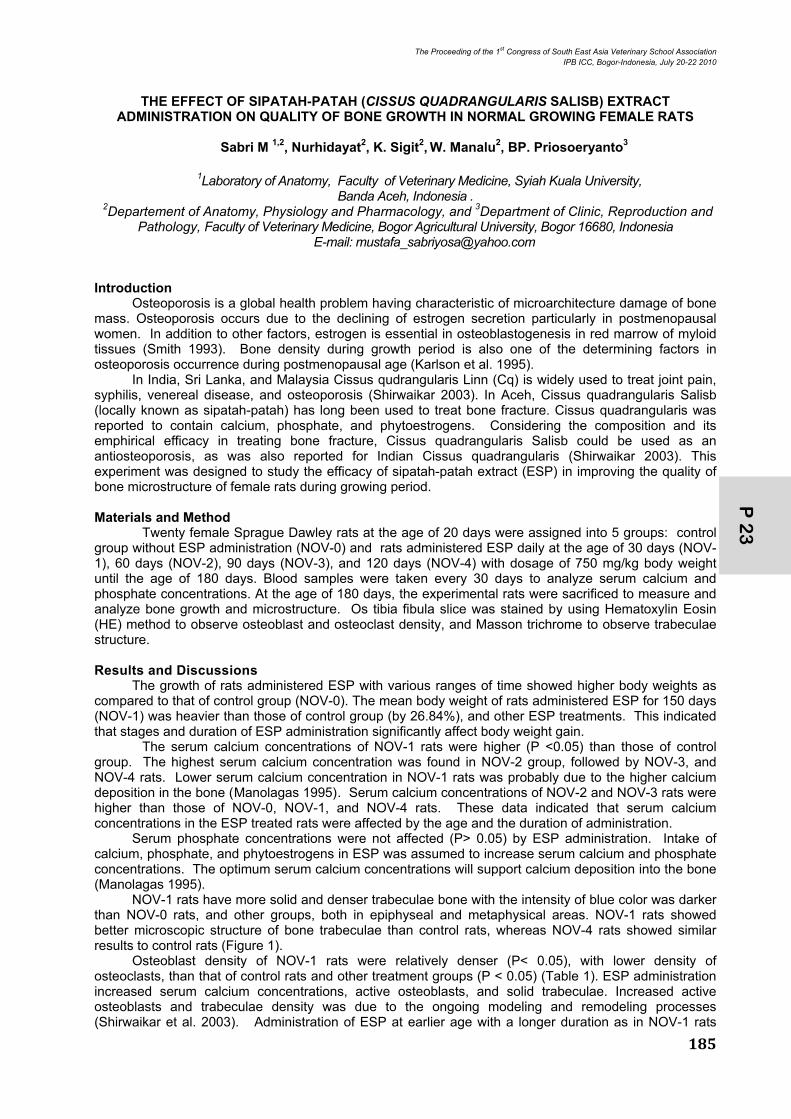
NOV-1 rats have more solid and denser trabeculae bone with the intensity of blue color was darker than NOV-0 rats, and other groups, both in epiphyseal and metaphysical areas. NOV-1 rats showed better microscopic structure of bone trabeculae than control rats, whereas NOV-4 rats showed similar results to control rats (Figure 1).
Osteoblast density of NOV-1 rats were relatively denser (P< 0.05), with lower density of osteoclasts, than that of control rats and other treatment groups (P < 0.05) (Table 1). ESP administration increased serum calcium concentrations, active osteoblasts, and solid trabeculae. Increased active osteoblasts and trabeculae density was due to the ongoing modeling and remodeling processes (Shirwaikar et al. 2003). Administration of ESP at earlier age with a longer duration as in NOV-1 rats
P 2
3
The Proceeding of the 1st Congress of South East Asia Veterinary School Association IPB ICC, Bogor-Indonesia, July 20-22 2010
186
significantly increased the osteoblast and trabeculae densities. Phytoestrogens in Cissus quadrangularis also play a role in increasing the density of osteoblasts (Jainu and Devi 2006) and stimulate collagen formation as a basic material of the bone trabeculae (Shirwaikar et al. 2003).
Based on this result, we concluded that the administration of sipatah-patah extract on normal female rats during growth period for 150, 120, 90, and 60 days improve bone microstructure and some physiological parameters.
A B C
Figure 1. non-ovariectomized rat trabeculae image on the day-180 A. contol rat. B. rats given ESP during 150 days showed solid trabeculae density. C. Rats given ESP during 60 days showed rare trabeculae density. Tb: trabeculae,
Se: epiphyseal plate
Table 1. Density of osteoblasts and osteoclasts in non-ovariectomized rats on day-180
Treatment Osteoblasts
Osteoclasts Active Pasif
NOV-0 34,58±8,05b 50,67±17,37a 6,99±3,35bc
NOV-1 38,92±8,09a 57,21±17,51a 2,50±2,96a
NOV-2 34,88±6,62b 47,63±8,69a 4,29±4,44b
NOV-3 20,25±8,99c 35,04±13,32b 5,58±2,60b
NOV-4 18,08±3,68c 34,96±6,38b 6,75±3,55bc
Note: NOV-0 = control rat; rats given ESP during 150 days (NOV-1), 120 days (NOV-2), 90 days (NOV-3), and 60 days (NOV-4).
References Jainu M, Vijaimohan K, Shyamala Devi CS. 2006. Protective effect of Cissus quadrangularis on
neutrophil mediated tissue injury induced by aspirin in rats. J. Ethnopharmacol 104:302–305. Karlson MK, Vergnauld P, Delmos PD and Obrant KJ. 1995. Indicates of bone formation in weight lifters.
Calcif. Tissue Int. 56 (3) 177-180. Manolagas SC. 2000. Bone marrow, cytokines and bone remodelling emerging insight into the
pathophysiology of osteoporosis. N Eng J Med 332 (21):115-137. Shirwaikar A, Khan S, Malini S. 2003. Antiosteoporotic effect of ethanol extract of Cissus quadrangularis
Linn on ovariectomized rat. J Ethnopharmacol 89: 245-250. Smith R. 1993. Bone physiology and the osteoporotic process. Resp Med 87 (Suppl A ). 3-7
Se
Tb Tb
Se
Tb Se

The Proceeding of the 1st Congress of South East Asia Veterinary School Association IPB ICC, Bogor-Indonesia, July 20-22 2010
187
BRUCELLOSIS ON CATTLE THAT WILL BE TRANSPORTED AT MERAK PORT BANTEN FROM JAVA TO SUMATRA
Rahmat Hidayata, Arum Kusnila Dewib, Eko Sugeng Pribadia, Fachriyan Hasmi Pasaribua
aDivision of Medical Microbiology, Department of Animal Disease and Veterinary Public Health Science,
Faculty of Veterinary Medicine, Bogor Agricultural University bAgricultural Quarantine Agency, Ministry of Agricultural of Republic Indonesia
Keywords: Brucellosis, cattle, RBT, CFT, I-ELISA
Introduction
Brucellosis is an infectious disease related to employment, such as milkman, laboratory workers, veterinarians, inseminator, breeders, breeding cattle and others. Course of the disease process caused by Brucella abortus in cattle starts with the entry of bacteria into the body through the mucous membrane of the eyes penetration, mucous membranes of reproductive tract, digestive tract, mouth, skin and respiratory tract (Hirsh et al. 2004 ). The main source of transmission of B. abortus in cattle is through the uterine fluid, placental tissue of fetus, colostrum and milk from cows patients (Quinn et al. 2006). B. abortus is a facultative intracellular (anerobic) that able to survive and thrive well in phagocytic cells (macrophages). B. abortus can also cause infertility, reproductive anomalies, death early pedet and miscarriage in sensitive livestock group. Materials and Methods
Research has been conducted in Faculty of Veterinary Medicine Bogor Agricultural University, BKP Class II Cilegon and BBALITVET use of RBT, CFT, I-ELISA and SDS-PAGE testing methods since January to October 2008. Serum obtained from cows originating from province of East Java, Central Java, Yogyakarta, West Java, Jakarta and Banten. The number of samples obtained in stages (multiple states), according to the frequency of expenditure and population per conveyance. The materials used were reagent RBT (BBALITVET), complement 10%, hemolysin (with dilution 1:100), hemolysin (with dilution 1:150), red blood cells of sheep 4%, coagulant of Na citrate (Sigma) 3.85%, NaCl (Oshaka) 0.95%, indirect I-ELISA Kits (SERELISA ® Mono Indirect antibody Brucella OCB), the positive control serum (BBALITVET), buffer perosidase substrate (PS) (SERELISA ® Mono Indirect antibody Brucella OCB), washing buffer (W) (SERELISA ® Mono Indirect antibody Brucella OCB), sample diluent (SD) (SERELISA ® Mono Indirect antibody Brucella OCB), the collecting solution and gel (stacking gel) 4% (Sigma Chemical), gel separator ( separating gel ) 12% (Sigma Chemical), running buffer (Promega) 2,76%, phosphat Buffer Saline PBS (Promega), and coomasie blue (Sigma Chemical) 0,1%. Results and Discussions
Results of a number of 173 serums inspection to B. abortus. Positive RBT is 0 sample. Positive CFT are 33 samples. Positive I-Elisa is 1sample (Table 1).
Table 1. Positive Results by RBT, CFT, I- ELISA testing methods
No. Origin Sample Methods
RBT (%) CFT (%) I-I-ELISA (%) 1 East Java 31 0 5 (16,1%) 0 2 Cetral Java and DIY 60 0 11 (18,3%) 0 3 West Java 72 0 15 (20,8%) 1 (1,4%) 4 Jakarta 4 0 0 0 5 Banten 6 0 2 (33,3%) 0 Total 173 0 33 1
Electrophoresis result obtained from cattle serum at group of character of protein Ig ( A and B) that
is protein ribbon between 225-175 kDa represent the IgG, 75-50 kDa represent the IgG which heavy BM that is IgG1, below 50 kDa showing the journey of infection Brucella, between 50-35 kDa represent the shares enchain the weight of IgG that is Fc and residing in between 25 kDa represent to enchain light of IgG that is Fab. Protein Ribbon below 25 kDa showing the natural infection reaction at animal and represent the reaktifitas from T-cell (Figure 1).
P 2
4

The Proceeding of the 1st Congress of South East Asia Veterinary School Association IPB ICC, Bogor-Indonesia, July 20-22 2010
188
Figure 1. SDS-Page Resuts from Positive Serum by CFT and ELISA
Parallel interpretation from RBT to CFT and I-Elisa to CFT own the meaning as a whole that is if one test of among from parallel is positive, hence result of the diagnostic test positive. Result of sensitifitas obtained high, hence low spesifisitas. Serial interpretation from RBT to CFT and I-Elisa to CFT own the meaning as a whole that is both of test from serial is positive, hence result of the diagnostic test positive. Result sensitifitas obtained low, hence high spesifisitas (Table 2).
Table 2. Serial and Parallel Interpretation RBT, CFT and ELISA
No RBT CFT Cattle 1 + + 0 2 + - 0 3 - + 33 4 - - 140
Sum up the Serum Samples 173
Sensitifitas Interpretation Paralel (P) 33/173 =19,10% Interpretation Serial (S) 0/173 = 0% Spesifisitas
(P) 140/173 = 80,92% (S) 173/173 = 100%
No I-I-ELISA CFT Cattle 1 + + 1 2 + - 0 3 - + 32 4 - - 140
Sum up the Serum Samples 173 Sensitifitas
(P) 33/173 =19,10% (S) 1/172 = 0,58%
Spesifisitas (P) 140/173 = 80,92% (S) 172/173 = 99,42%
Conclusion
The final result is method of diagnosa Brucellosis at 173 serum samples that is CFT more sensitive compared by RBT and ELISA method more specific from CFT. References Hirsh DC, Maclachlan NJ, Walker RL. 2004. Veterinary Microbiology. 2sd Ed Australia. Blackwell
publishing. Quinn PJ, Markey BK, Carter ME, Donnelly WJ, Leonard FC. 2006. Veterinary Microbiology and
Microbial Disease. Australia. Blackwell publishing.

The Proceeding of the 1st Congress of South East Asia Veterinary School Association IPB ICC, Bogor-Indonesia, July 20-22 2010
189
CLINICAL EVALUATION OF HYDROXYAPATITE-CHITOSAN (HA-C) AND HYDROXYAPATITE-TRICALCIUM PHOSPHATE (HA-TCP) BONE GRAFT
IN SHEEP’S BONE AS ANIMAL MODEL FOR HUMAN
Harry Soehartono, Gendis Aurum Paradisa, Riki Siswandi, M. Fakhrul Ulum, Gunanti
Division of Surgery and Radiology, Department of Clinic, Reproduction and Pathology, Faculty of Veterinary Medicine of Bogor Agricultural University,
Jl. Agatis Kampus IPB Darmaga-Bogor-16680.
Keywords: bone graft, sheep, hydroxyapatite, tricalsium phosphate, chitosan, bone healing, physical examination.
Introduction
Bone has its own formation cycle, known as 'remodeling'. Bone-making cells will form new bone using calcium and other minerals. In this study, Hydroxyapatite-tricalcium phosphate (HA-TCP) and Hydroxyapatite-Chitosan (HA-Chitosan) used as bone implant materials for bone regeneration and to compare the acceleration of bone healing viewed from clinical symptoms. Implant also give negative impacts such on the theory of the Bone Cement Implantation Syndrome (BCIS), there were some effects that can be generated from the use of bone cement implant such as reflex bradycardia, pulmonary hypertension, hypothermia, and others. The aim of this study was to examine the impacts of bone implantation viewed from clinical parameters.
Materials and Methods
Local sheep (Ovis aries) used in this study. Sheeps were divided into two groups, first group consist of three hydroxyapatite-chitosan (HA-chitosan) implanted sheeps and second group consist of three hydroxyapatite-tricalcium phosphate (HA-TCP) implanted sheeps. Next, sheeps were harvested at 30th, 60th, and 90th day post-surgery. Observations were carried out by clinical examination before and after surgery with clinical parameters and wound healing conducted every day. Results and Disscusion Temperature
Graph showed that implants administration of HA-Chitosan and HA-TCP were less affected on temperature. No side effects on body temperature such as direct effect of exothermic reaction of cement temperature, hypothermia and other side effects.
Pulsus/Heart rate
Graph showed that the implants administration of HA-Chitosan and HA-TCP were less affected on heart rate. No side effects on heart rate, such as reflex bradycardia, cardiac arrest, cardiac dysrhytmia and other side effects.
Figure 1. Body temperature.
P 2
5

The Proceeding of the 1st Congress of South East Asia Veterinary School Association IPB ICC, Bogor-Indonesia, July 20-22 2010
190
Respiration Rate Graph showed that the implants administration of HA-Chitosan and HA-TCP were less affected on breathing. No side effects on breathing such as pulmonary hypertension, bronchocontriction and other side effects.
Bone Healing Comparison between chitosan, TCP and normal bone (without treatment) viewed from the inflammation reaction in the implantation site:
Graph showed that HA-Chitosan and HA-TCP implanted bones have no different healing speed with control (normal) bone, so that the implants of HA-TCP and HA-Chitosan have no significant impacts in accelerating bone healing.
Conclusion
The combination of HA-Chitosan and HA-TCP can be accepted in body and have no effects on physiological conditions, but in its function to accelerate bone healing, it gives low results. Implant modifications and biophysical stimulation were necessary. Histopathologic and radiographic analysis also needed to examine the effectiveness of implants. References Kelly, W. R. 1984. Veterinary Clinical Diagnosis. Third Edition. London: Bailliera Tindall. Heath, E.&S.Olusanya. 1985. Anatomy and Physiology of Tropical Livestock. Intermediate Tropical
Agriculture Series. Longman London and New York.
Figure 3. Respiration rate.
Figure 4. Bone healing.

The Proceeding of the 1st Congress of South East Asia Veterinary School Association IPB ICC, Bogor-Indonesia, July 20-22 2010
191
PHYSICAL AND HEMATOLOGICAL PARAMETER OF MUSANG LUWAK (Paradoxurus hermaphroditus)
Sarmin
Faculty of Veterinary Medicine, Gadjah Mada University
Jl. Fauna 2 Karangmalang, Yogyakarta 55281.Email: [email protected] Introduction
Wildlife is one of natural resources whose contribution in the natural sustainability is great. Musang Luwak (Paradoxurus hermaphroditus) is a wildlife whose status is "vulnerable" or susceptible to extinction. Musang Luwak populations, particularly in Indonesia, are so few that research on the normal physiological status as an effort to control diseases in order to avoid extinction is needed. This research aims at to recognize the description of physical and hematological parameter in Musang Luwak. Materials and Methods
This research employs four Musang Luwak, consisting of two groups. The first is male (two animals) and the second is male (two animals) whose age are is around 2-4 months. Physical clinic check up and blood sample are done according to the schedule to fit the check up and sample taking in the clinic standard descriped by Baumgartner (1999). Results and Disscusion
The average number of pulse, breath, and temperature does not differ significantly P>0.05 for both male and female and what differs significantly is only their weight P >0.05 which is possibly caused by the difference in sex. Erithrocyte morphology of Musang Luwak does not have center, the periphery of the cells is dark and the center part is transparant, whose average diameter is 4.12 ±1.20 µm for male and 4.01 ±1.90 µm for female, the same as erithrocyte diameter of cat (Schalm, 1975). The average of the number of erithrocyte, PCV and MCV, hemoglobin do not differ significantly between the male and the female (P>0.05) as said by Jain (1986) while MCHC differs between the male and the female (P>0.05). The average of leukocyte, eosinophil, and monocyte does not differ between male and female, while neutrophil and limphocyte are influenced by sex (P<0.05). Those data fit Fowler’s report (1986).
Table 1. Physical examination
Group Sex Pulse/second Breathing/second Temperature/0C Weight/kg 1 Male
(n=2) 58.20±2.20 A 60.80 ± 5.90 A 38.20±0,70 A 1.40A
2 Female (n=2)
56.50 ± 2.50 A 63.00±2.00 A 36.70 ± 0.24 A 0.50B
Table 2. Haematology of Erythrocytes
Group Erythrocytes Hb (g/dl)
PCV(%)
MCV(fl) MCHC (%) Size (µm) Value
(milion/mm3
Male (n=2)
4.12 ±1.20 A 17.36± 3.74 A 7.65 ± 0.63 A 34.50 ± 2,12 A 20.15 ± 3.18 A 22.15 ± 0.49 A
Female (n=2)
4.01 ±1.90 A 7,91 ± 2.26 A 7.60± 1.69 A 28.50 ± 4.94 A 36.60 ± 4.24 A 26.55 ± 1.34 B
Table 3. Leukocytes and Differentials
Group Total leukocyte
(million/mm3) Neutrophil(milli
on /mm3) Eosinophil
(million/mm3) Limphocytes (million /mm3)
Monocytes (million /mm3)
Male (n=2) 6.25 ± 353.55A 17.50 ± 10.6A 0.50 ± 0.70 A 75.00 ± 14.14A
7.00± 2.82 A
Female (n=2)
5350.00 ± 0.00A 63.50 ± 4.94B 3.50 ± 2.12 A 29.00 ± 7.07B 4.00 ± 0.00 A
Conclussion
Sex determines the physiological indicator for Musang Luwak (Paradoxurus hermaphroditus) in term of weight and some blood components which are neutrophil and limphocyte as well as MCHC.
P 2
6

The Proceeding of the 1st Congress of South East Asia Veterinary School Association IPB ICC, Bogor-Indonesia, July 20-22 2010
192
Acknowledgement Thanks to Drh. Dwi Liliek Kusindarta, MP., Ph.D and drh. Woro Danur Wendo for the co-operation
in providing the animal for the test. My gratitude also goes to Lusi Jayatingrum, Apriani and Mitra S for the co-operation in the research. References Baumgartner, W. (1999). Klinische Propaedeutik der inneren Krankhelten und Hautkrankheiten der Haus-
und Heirntiere, 4. Auflage. Parey Buchverlag, Berlin. Pp. 27 – 354 Fowler, M.E. 1986. Zoo and Animal Medicine. 2 nd Ed. W.B. Saunders Company, Phyladelphia.Pp.822-
827. Jain, N.C. 1986. Schlam’s Clinical Pathology. 2 nd Ed. Lea and Febiger. Philadelphia. Pp. 350, 481, 518,
527. Schlam, O.W. Carrol, E.J. and Jain, N.C. 1975. Veterinary hematology, 3 th Ed. Lea and Febiger,
Philadelphia. Pp.87,411-420

The Proceeding of the 1st Congress of South East Asia Veterinary School Association IPB ICC, Bogor-Indonesia, July 20-22 2010
193
PREVIEW OF BLOOD GLUCOSE, CORTISOL AND PHYSICAL PARAMETERS IN BLIGON GOATS EXPERIENCING TRANSPORTATION
Sarmin1, Pudji Astuti1, C. Mona Airin1, Amelia Hana1, Asmarani Kusumawati2, Irkham Widiyono3,
Hera Maheswari4, Luthfiralda Sjahfirdi5, Mitra S6, Bashori6, Ariani Hasan6
1 Department of Physiology, Faculty of Veterinary Medicine, Gadjah Mada University, Yogyakarta. Jln.
Fauna 2 Karang Malang, Yogyakarta 55281, email: [email protected]
2 Department of Reproduction, Faculty of Veterinary Medicine, Gadjah Mada University 3Department of Internal Medicine, Faculty of Veterinary Medicine Gadjah Mada University
4 Department of Anatomy, Physiology and Pharmacology, Faculty of Veterinary Medicine, Bogor Agriculture University
5Department of Biology, Faculty of Natural Sciences, University of Indonesia. 6Faculty of Veterinary Medicine, Gadjah Mada University
Key words: Blood glucose, cortisol, Bligon Goats, physical parameter
Introduction
Transportation is a process that cannot be separated from production and marketing system of livestock that can cause stress on livestock. Livestock often experience stress from such activities that may disturb the normal physiological balance. Some factors that may cause stress on such livestock prior to transportation management, noise, vibration, new things, grouping, density, climatic factors, restrain, income and expenditure of time upon arrival, as well as feeding and drinking during transportation. However, the most frequent cause of major stress on the ranch is handling and transportation. Materials and Methods
This research was conducted using five bligon goats, male, 1.5 years with a body weight 24 to 26 kg. All of animals intensively maintained in cages, and performed a physical examination in the morning, noon and night. Transportation is then performed for 16 hours, starting from 6:00 pm on the first day and ends at 10:00 am on the second day. During the research blood sampling are carried out via jugular vein every four hours and when the goat was not transported, that is at 10:00 am until 2:00 pm. Blood is checked by using Vitros glucose with a sensitivity level of 20.0-625 mg / dl, the ability of intra-assay 0.5 mg / dl and extra assay, 1.5 mg / dl for the concentration of 86 mg / dl. Kortisol examined by ELISA. All data were processed using ANOVA and T-Test with significance level 0.05. Results and Disscusion
Transportation causes a change in average temperature, pulsus, weight and Capillary Refill Time (CRT). Time duration of transportation led to changes in average temperatures, breath, and body weight. The interaction between transport time and temperature, and breathing can be found significantly. The mean highest temperature occurred at 8 hours after transportation and continued during transport. The temperature difference that occurred in both groups experienced significant differences in the commencement of transportation. This shows that when the animal is mounted on the car, causing stress that resulted in increased metabolism and the effects are increased body temperature. Increased cortisol occurred in the fourth hour, indicating the goats high stress occurs first 4 hours of transport (Olvera and Lopez, 2004). Average glucose concentrations increased in the first 4 hours of transport, while other researchers reported two hours in ruminants (Hartmann, 1988), three hours in goat Spain (Kanan et al., 2000). Results also showed a correlation between glucose and cortisol, and glucose with the temperature according to Guyton (1996) because of gluconeogenesis in the liver during transport.
P 2
7

The Proceeding of the 1st Congress of South East Asia Veterinary School Association IPB ICC, Bogor-Indonesia, July 20-22 2010
194
Table 1. Mean physical and plasma chemistry (glucosa and cortisol) before and after transportation in different groups
Parameter Times -8 -4 0 4 8 12 16 4+ 8+
Temperature (0 C)
Treatment
37.78 ± 0.61A
38.98 ± 0.31A
39.48 ± 0.31 A
38.87 ± 0.34 A
38.88 ± 0.43 A
38.92 ± 0.35 A
38.78 ± 0.29 A
39.17 ± 0.23 A
38.88 ± 0.48 A
Control 37.90 ± 0.49A
38.96 ± 0.54 A
38.42 ± 0.52A
38.9 ± 0.53A
37.9 ± 0.809 A
38.79 ± 0.47 A
38.44 ± 0.36 A
38.98 ± 0.33 A
39.06 ± 0.27 A
Pulse (time/second)
Treatment
84.67 ± 5.32 A
98.67 ± 13.063 A
97.33 ± 20.34 A
105.87 ± 36.66 A
109.33 ± 15.52 A
110.00 ± 19.87 A
93.33 ± 12.816 A
141.33 ± 34.28 A
116.00 ± 16.19 A
Control 103.2 ± 19.67 A
108.80 ± 13.38A
96.00 ± 11.66 A
114.40 ± 18.67A
87.20 ± 10.35 A
107.2 0± 16.34 A
104.80 ± 16.59 A
114.40 ± 15.12 A
108.00± 22.09 A
Breathing (time/second)
Treatment
28.00 ± 3.58 A
31.33 ± 7.76 A
54.67 ± 16.72 A
72.53 ± 31.90 A
32.00 ± 9.12 A
59.33 ± 27.06 A
24 .00± 7.15 A
42.00 ± 12.06
40.00 ± 17.71 A
Control 28.00 ± 7.48 A
45.60 ± 15.38 A
51.20 ± 21.61 A
32.00 ± 4.00A
30.40 ± 7.79 A
33.60 ± 9.63 A
77.60 ± 3.58 A
55.20 ± 19.06
32.80 ± 15.34 A
CRT (seconds)
Treatment
1.25 ± 0.17 A
1.34 ± 0.19 A
1.96 ± 1.45 A
7.83 ± 15.37 A
1.70 ± 0.29 A
1.83 ± 0.11 A
1.57 ± 1.19 A
1.27 ± 0.13 A
1.57 ± 0.20 A
Control 1.402 ± 0.34 A
1.50 ± 0.31 A
1.36 ± 0.24 A
1.47 ± 0.21 A
1.31 ± 0.10 A
0.96 ± 0.16 A
1.35± 0.11
A 1.03± 0.31 A
1.51 ± 0.24 A
Body weight (kg)
Treatment
23.50 ± 1.64 A
23.67 ± 1.366 A
23.08 ± 1.28 A
27.04 ± 7.05 A
22.83 ± 1.17 A
22.50 ± 0.89 A
22 ± 1.414
A 23.75 ± 0.76
24.25 ± 1.78 A
Control 24.4 ± 1.516 A
25 ± 1.060 A
24.6 ± 1.516 A
26.3 ± 0.758B
24.6 ± 1.140 A
24.7 ± 0.836 A
25.2 ± 1.303 A
24.7 ± 1.823
25.6 ± 2.190 A
Blood glucose (mg/dl)
Treatment
- 58.4±5.13C
65±9.46C 117±55.08A
98.6±41.70BA
103±47.43A
102.6±30.90A
75±7.68BC
62.8±9.58C
Control - 49±10.72C 49±10.72C 73.2±6.38BC
70±3.03C 67±4C 51.6±6.7C 64±4.42C
61±7.71C
Cortisol (ng/ml)
Treatment
70.71±43.70C
66.23±33.77C
95.96±52.39BC
161.96±78.90A
123.69±57.06B
87.01±23.81BC
99.31±25.76BC
- -
Control 69.97±44.04C
78.34±53.97C
83.88±65.11C
87.23±52.70BC
77.65±42.74C
75.82±36.04C
67.38±35.05C
- -
Means with different supercripts are significantly different Conclusion
Transportation is carried out during 16 hours can have significant physical parameters, blood glucose and cortisol, which indicates the stress on the goats during transport progress References Guyton, A.C. and Hall, J.E.(2000) Textbook of Madical Physiology, 10th edition. Eds. A.C. Guyton and
J.E. Hall.W.B.Saunders Company, Philadelphia, USA, pp.253-262 Hartmann, H,. 1988. Criteres biochimiques et hematologiques du stress et leurs relations avec les
mecanismes de defense. Recueil de medicine veterinaire 164, 743-750 Kannan, G, T.H. Terrill, B. Kuoakuo, O.S. Gazalt, S, Gelaye, E.A. Amoah and S. Samake, 2000.
Transpotation of goat: Effect on physiological stress responses and liveweight loss. J. Anim. Sci., 78: 1450-1457
Olvera, Jorge Ramon Lopez. 2004. Capture, restraint and transport stress in shouthern chamois (Rupicapra pyrenaica). Bellaterra.

The Proceeding of the 1st Congress of South East Asia Veterinary School Association IPB ICC, Bogor-Indonesia, July 20-22 2010
195
DISTRIBUTION OF GLYCOCONJUGATES IN THE TESTIS OF THE CAVE SWIFLETS (Collocalia linchi)
Savitri Novelina, Aryani S Satyaningtijas, Srihadi Agungpriyono,
Heru Setijanto, Koeswinarning Sigit
Department of Anatomy, Physiology and Pharmacology, Faculty of Veterinary Medicine, Bogor Agricultural University, Bogor 16680,Indonesia
e-mail : [email protected]
Keywords: Lectin, glycoconjugate, Collocalia linchi, testis.
Introduction
Cave swiftlet or linchi swiftlet, Collocalia linchi, belongs to the swift family of Apodidae. The bird has economic importance as it is among the swift that produce edible nest for delicacy Chinese bird’s nest dishes as well as food supplement for human health. The nest is among potential export commodities of Indonesia.
There are two species of edible nest swiftlet that commonly found in Indonesia, Collocalia fuciphaga and Collocalia linchi. In contrast to C. fuciphaga, of the biological data available for C. linchi reproduction are very limited. The present study aimed to know the distribution and content of glycoconjugates in the testis in the cave swiftlets C. linchi, which may provide a better understanding on the spermatogenesis and testicular cycle of C. linchi. Materials and Methods
Four adult male cave swiftlet (C. linchi) were used in this study. The birds were sacrificed by cervical dislocation after an anesthesia using chloroform was given. The testis were exposed and observed macroscopically. Tissue samples of certain portions of the testis were taken out and fixed in Bouin fluid and processed routinely for embedding in paraffin. Paraffin sections were cut serially at 5 µm of thickness and stained with haematoxylin-eosin and seven biotinylated lectins: Con A, RCA, WGA, UEA, PNA, DBA, and SBA (Lectin Kit Vector Lab., USA), respectively. Results and Discussion
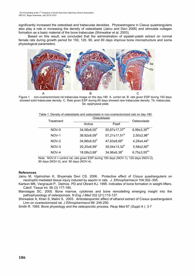
Lectins Con A, RCA, PNA, WGA, DBA, SBA and UEA were variously positive in the seminiferous tubules of C. linchi (Table 1). In the Sertoli cells RCA, PNA, WGA (Figure 1A), UEA and SBA were strongly positive and DBA was negative. These results suggested the presence of carbohydrates with galactose, acetylglucosamine, acetylgalactosamine, fucose and sialic acid sugar residues in the Sertoli cells. These glycoconjugates might associated with the function of Sertoli cells in the regulation of spermatogonium cells, peritubular cells and Leydig cells (Skinner 1991). In the spermatogonium cells, only Con A was positive with weak reaction. Moderate to strong reactions of Con A, RCA, UEA, PNA (Figure 1B), WGA, SBA (Figue 1C), and DBA were observed in the spermatocytes and spermatids. These might suggest that glicoconjugates with galactosa, acetylglucosamine, acetylgalactosamine, fucose and sialic acid sugar residues may play a role in the development of spermatocytes and spermatids. The staining pattern of the lectins observed in the testis of C. linchi might be related with the spermatogenesis and development of testis of the C. linchi.
Table 1. Staining intensities of biotinylated lectins in the testis of Collocalia linchi
Seminifeorus Lectins cells Con A RCA PNA WGA DBA SBA UEA
Sertoli + +++ + +++ - ++ +++ Spermatogonium + - ++ - - - -
Spermatocyte ++ ++ +++ +++ + +++ ++ Spermatid ++ ++ ++ ++ ++ ++ ++
-: negative; +:weak, ++: moderate, +++: strong
P 2
8
The Proceeding of the 1st Congress of South East Asia Veterinary School Association IPB ICC, Bogor-Indonesia, July 20-22 2010
196
Reference Skinner MK. 1991. Cell – Cell Interactions in The Testis. Endoc Rev 12 (1): 45 – 77.
Figure 1. The testis of the Collocalia linchishowing :WGA (A), PNA (B) and SBA (C) positive reactions in seminiferous tubules, WGA is positive in Sertoli cells and spermatocytes. PNA spermatogonium and spermatids and SBA is positive in spermatocyte and spermatozoa. DAB. Bar : 50 µm
A B
C

The Proceeding of the 1st Congress of South East Asia Veterinary School Association IPB ICC, Bogor-Indonesia, July 20-22 2010
197
TESTICULAR BIOPSY GUN NEEDLE AS AN ALTERNATIVE BIOPSY METHOD FOR THE ASSESSMENT OF SPERMATOGENESIS IN THE KIJANG MUNTJAK
(Muntiacus muntjak muntjak)
Sri Wahyuni1, Srihadi Agungpriyono2, I Ketut Mudite Adnyane2, Muhammad Agil3, Tuty Laswardi Yusuf3
1Laboratory of Anatomy, Faculty of Veterinary Medicine, Syiah Kuala University, Banda Aceh, Indonesia, 2Department of Anatomy, Physiology and Pharmacology-, 3Department of Clinic
Reproduction and Pathology , Faculty of Veterinary Medicine Bogor Agricultural University (IPB), Bogor 16680, Indonesia
E-mail: [email protected] Introduction
Kijang muntjak (Muntiacus muntjak muntjak) belongs to the family Cervidae. They are distributed in Java and South Sumatera and are protected by Indonesian Government. Reproductive biology of male kijang muntjak and its relation to antler growth has not been reported yet. Generally, there are three stages of antler growth in the male Cervids, hard antler, casting and velvet antler. In most of male cervids, the growing antler period has close relation with reproductive activity such those reported in Rusa Timor (Cervus timorensis). In rusa Timor stags, spermatogenesis is occurred only during the hard antler period (Handarini et al., 2004). On the contrary, male Formosan muntjak, spermatogenesis is still active although in velvet antler period. (Pei et al., 2009). Therefore, it is important to determine the reproductive activity in male kijang muntjak in Indonesia. Spermatogenesis can reflect the reproductive activity and can be investigated histologically in the testicular tissue obtained at each of the antler stageBiopsy procedures have been performed in the Eld’s deer stags (Cervus eldi thamin) (Monfort et al., 1993), and Rusa Timor stags (Cervus timorensis) (Handarini et al., 2004) using open biopsy method with testicular surgery which resulted in a longer recovery time of testis. In this present study, we carried out testicular biopsy using a gun needle, a simple method without testicular surgery. The objective of this work was to determine the effectiveness and advantages of gun needle method for the assessment of spermatogenesis in the kijang muntjak. Materials and methods Animals
Three adult and apparently healthy males kijang muntjak (2-4 years of age; 17-19 kg body weight) were individually housed in animal house measuring 2x1 m2, which has a connection to the outdoor enclosure. All of animals were maintained within visual and olfactory proximity to the female kijang muntjak. Testicular biopsy procedure
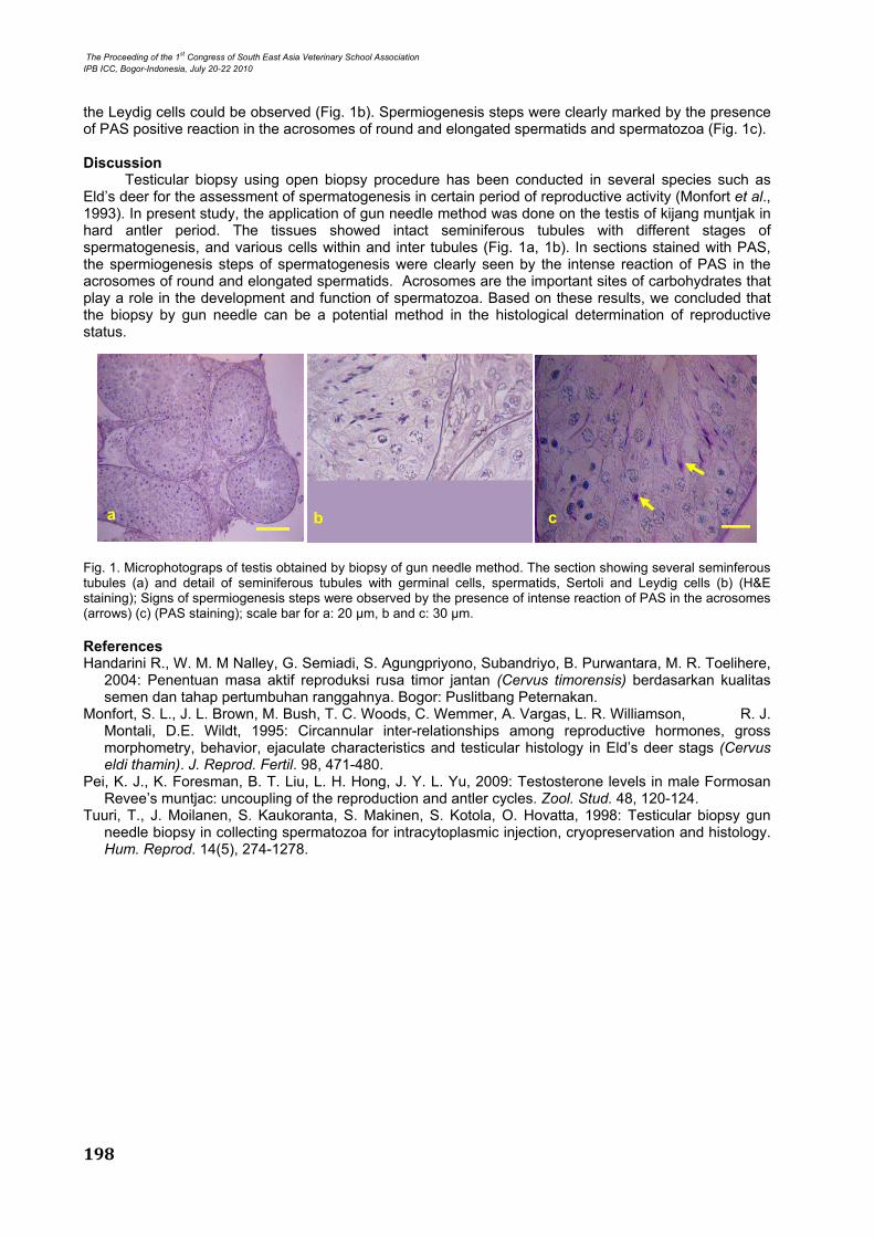
Prior the biopsy, each animal was immobilized using a combination of xylazine HCl and ketamin. The surface of scrotal skin was antisepticised using ethanol 70% and iodine. Biopsy method was performed as described previously by Tuuri et al. (1998). Biopsy was taken in dorsal area of right or left testis. A 14 gauge needle (Fine core ‘Toray’ 14 G, Japan) was inserted through scrotal skin to the testicular tissues (Fig. 1). Once needle was inserted into the testis, a strong negative pressure of biopsy gun was exerted to cut and obtained testicular tissue. After tissues aspiration, the needle was withdrawn slowly. The area of injection in the scrotal skin was treated by antibiotic ointment topically and the animal was given a long acting antibiotic. Histological preparation
Fragments of biopsy were immediately fixed in Bouin’s fixative for 24 h and transferred to ethanol 70%. The biopsy samples were processed by a routine histological procedure. Paraffin sections stained with haematoxylin and eosin (HE) and periodic acid Schiff (PAS). Results Recovery of testis
The recovery of injury caused by biopsy was 7-10 days. No inflammation sign neither haematomas appeared in scrotal skin. Histological evaluation
Histological evaluations showed 5-8 seminiferous tubules with different stages of the spermatogenesis in each of paraffin section (1a). Spermatogonia, spermatocytes and spermatids were distributed in adluminal of tubules, and also the Sertoli cells and perimyoid cells. In the interstitial area,
P 2
9
The Proceeding of the 1st Congress of South East Asia Veterinary School Association IPB ICC, Bogor-Indonesia, July 20-22 2010
198
the Leydig cells could be observed (Fig. 1b). Spermiogenesis steps were clearly marked by the presence of PAS positive reaction in the acrosomes of round and elongated spermatids and spermatozoa (Fig. 1c). Discussion
Testicular biopsy using open biopsy procedure has been conducted in several species such as Eld’s deer for the assessment of spermatogenesis in certain period of reproductive activity (Monfort et al., 1993). In present study, the application of gun needle method was done on the testis of kijang muntjak in hard antler period. The tissues showed intact seminiferous tubules with different stages of spermatogenesis, and various cells within and inter tubules (Fig. 1a, 1b). In sections stained with PAS, the spermiogenesis steps of spermatogenesis were clearly seen by the intense reaction of PAS in the acrosomes of round and elongated spermatids. Acrosomes are the important sites of carbohydrates that play a role in the development and function of spermatozoa. Based on these results, we concluded that the biopsy by gun needle can be a potential method in the histological determination of reproductive status. Fig. 1. Microphotograps of testis obtained by biopsy of gun needle method. The section showing several seminferous tubules (a) and detail of seminiferous tubules with germinal cells, spermatids, Sertoli and Leydig cells (b) (H&E staining); Signs of spermiogenesis steps were observed by the presence of intense reaction of PAS in the acrosomes (arrows) (c) (PAS staining); scale bar for a: 20 µm, b and c: 30 µm. References Handarini R., W. M. M Nalley, G. Semiadi, S. Agungpriyono, Subandriyo, B. Purwantara, M. R. Toelihere,
2004: Penentuan masa aktif reproduksi rusa timor jantan (Cervus timorensis) berdasarkan kualitas semen dan tahap pertumbuhan ranggahnya. Bogor: Puslitbang Peternakan.
Monfort, S. L., J. L. Brown, M. Bush, T. C. Woods, C. Wemmer, A. Vargas, L. R. Williamson, R. J. Montali, D.E. Wildt, 1995: Circannular inter-relationships among reproductive hormones, gross morphometry, behavior, ejaculate characteristics and testicular histology in Eld’s deer stags (Cervus eldi thamin). J. Reprod. Fertil. 98, 471-480.
Pei, K. J., K. Foresman, B. T. Liu, L. H. Hong, J. Y. L. Yu, 2009: Testosterone levels in male Formosan Revee’s muntjac: uncoupling of the reproduction and antler cycles. Zool. Stud. 48, 120-124.
Tuuri, T., J. Moilanen, S. Kaukoranta, S. Makinen, S. Kotola, O. Hovatta, 1998: Testicular biopsy gun needle biopsy in collecting spermatozoa for intracytoplasmic injection, cryopreservation and histology. Hum. Reprod. 14(5), 274-1278.
a b c

The Proceeding of the 1st Congress of South East Asia Veterinary School Association IPB ICC, Bogor-Indonesia, July 20-22 2010
199
IMMUNOHISTOCHEMICAL STUDY ON THE DEVELOPMENT OF FETAL GROWTH HORMONE AND PROLACTIN CELLS OF LONG-TAILED MACAQUE (Macaca fascicularis)
Supratikno1, Nurhidayat1, N. Kusumorini1, C Nisa’1, S. Novelina1, S. Wahyuni2
1 Department Anatomy, Physiology, and Pharmacology, Faculty of Veterinary Medicine, Bogor
Agricultural University, Bogor 16680, Indonesia 2 Faculty of Veterinary Medicine, Syiah Kuala University, Banda Aceh, Indonesia .
E-mail: [email protected]
Keywords: pituitary, growth hormone, prolactin, distribution.
Introduction
Numerous immunohistochemical investigations have been studied on the pituitary gland development of many species, but few data of the development of pituitary gland of long-tailed macaque have been reported. Anatomical aspect of this gland that interesting to be studied is growth hormone and prolactin cells especially on their onset and distribution.
Growth hormone (GH) and prolactin (PRL) cells derived from same precursor, under different transcription factor, those cells proliferate into specific cells. They proliferate precisely in time and location, and the abnormality during proliferation of those cells may cause seriously effect after birth. Actually GH and PRL have no important role during fetal life, but they induce of body growth and development of mammary gland at postnatal life.
The aim of this study are to investigate the development of GH and PRL cells of long-tailed macaque during fetal life especially to detect the time of cells producing growth hormone and prolactin in the pituitary and their distribution pattern during fetal life. Materials and Methods
Fetal pituitary glands of long-tailed macaque aged 70(F70), 85(F85), 100(F100), 120(F120), and 150(F150) days of gestation were used in this study. The fetuses were taken from pregnant monkeys with caesarean procedure. Samples were obtained from Primate Research Centre, Bogor Agricultural University. The research materials were taken under supervision of Animal Care and Use Committee (PSSP-IPB, 02-0030IR).
The pituitary glands were taken and fixed in paraformaldehyde 4%, then processed with standard procedure of paraffin embedding. The tissue blocks were serially sectioned at 10 µm in thickness sagittaly according to Sasaki et al. (1992). The sections of the pituitary were stained immunohisto-chemically using rabbit anti GH and mouse anti PRL serum (Gift NIDDK USA).
Results and Discussions
The GH immunoreactive (GH-ir) cells were firstly observed at F70, while the PRL immunoreactive cells (PRL-ir) at F85. At 70 days of gestation, GH-ir cells were distributed in the lateral portion and caudal anterior of Rathke’s lumen of pars distalis at low density and weak intensity (Table 1). The GH-ir cells distribution were continued spread to all part of pars distalis with exception of pars tuberalis. The density of GH-ir cells were increased gradually at F85 and reached highest density at F150. Those distribution patterns were resembled with GH-ir cells distribution of fetal pig pituitary (Sasaki et al. 1992) and human fetal pituitary (Bakker and Jaffe 1975). GH-ir cells were predominating on pars distalis. Simultaneously, the intensity of GH-ir cells were increased in the older fetuses and also reached highest intensity at F150 (Figure 1). The intensity GH-ir cells were correlated with synthesis and secretion of GH. In the fetal life, GH has relatively small contribution on fetal growth (Sasaki et al. 1992, Styne, 1997). It suggests that the GH-ir cells were producing GH but less in secretion.
Similarity, PRL-ir cells were also distributed in all areas of pars distalis. Weak intensity of PRL-ir were firstly observed in lateral and middle regions of gland, and in the caudal of anterior Rathke’s lumen of F85. The density and intensity of PRL-ir cells were increased with age of fetuses and reached highest level at F150 (Table 1). Distribution of PRL-ir cells were resembled with the GH-ir cells except in the pars tuberalis. The PRL-ir cells distribution were more to rostral (tuberalis) and anterior of Rathke’s lumen. In this region, precursor cells were differentiate into PRL cells stimulated by estrogen from pituitary portal vein, oxytocin and anti diuretic hormone from pars nervosa (Sasaki and Iwama 1988). The similar distribution pattern of GH-ir and PRL-ir cells might presumed of the presence of mammosomatotrop cells, those can produce both of GH and PRL.
P 3
0
The Proceeding of the 1st Congress of South East Asia Veterinary School Association IPB ICC, Bogor-Indonesia, July 20-22 2010
200
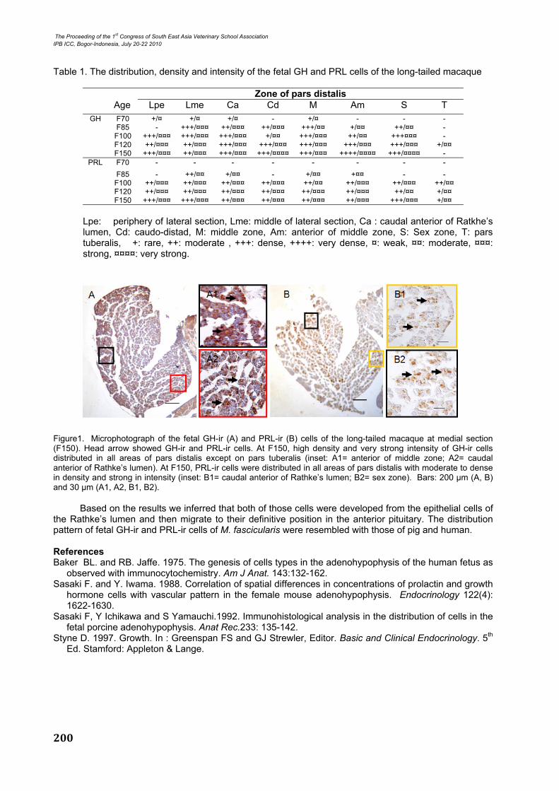
Table 1. The distribution, density and intensity of the fetal GH and PRL cells of the long-tailed macaque
Zone of pars distalis Age Lpe Lme Ca Cd M Am S T GH F70 +/¤ +/¤ +/¤ - +/¤ - - -
F85 - +++/¤¤¤ ++/¤¤¤ ++/¤¤¤ +++/¤¤ +/¤¤ ++/¤¤ - F100 +++/¤¤¤ +++/¤¤¤ +++/¤¤¤ +/¤¤ +++/¤¤¤ ++/¤¤ +++¤¤¤ - F120 ++/¤¤¤ ++/¤¤¤ +++/¤¤¤ +++/¤¤¤ +++/¤¤¤ +++/¤¤¤ +++/¤¤¤ +/¤¤ F150 +++/¤¤¤ ++/¤¤¤ +++/¤¤¤ +++/¤¤¤¤ +++/¤¤¤ ++++/¤¤¤¤ +++/¤¤¤¤ -
PRL F70 - - - - - - - - F85 - ++/¤¤ +/¤¤ - +/¤¤ +¤¤ - -
F100 ++/¤¤¤ ++/¤¤¤ ++/¤¤¤ ++/¤¤¤ ++/¤¤ ++/¤¤¤ ++/¤¤¤ ++/¤¤ F120 ++/¤¤¤ ++/¤¤¤ ++/¤¤¤ ++/¤¤¤ ++/¤¤¤ ++/¤¤¤ ++/¤¤ +/¤¤ F150 +++/¤¤¤ +++/¤¤¤ ++/¤¤¤ ++/¤¤¤ ++/¤¤¤ ++/¤¤¤ +++/¤¤¤ +/¤¤
Lpe: periphery of lateral section, Lme: middle of lateral section, Ca : caudal anterior of Ratkhe’s lumen, Cd: caudo-distad, M: middle zone, Am: anterior of middle zone, S: Sex zone, T: pars tuberalis, +: rare, ++: moderate , +++: dense, ++++: very dense, ¤: weak, ¤¤: moderate, ¤¤¤: strong, ¤¤¤¤: very strong.
Figure1. Microphotograph of the fetal GH-ir (A) and PRL-ir (B) cells of the long-tailed macaque at medial section (F150). Head arrow showed GH-ir and PRL-ir cells. At F150, high density and very strong intensity of GH-ir cells distributed in all areas of pars distalis except on pars tuberalis (inset: A1= anterior of middle zone; A2= caudal anterior of Rathke’s lumen). At F150, PRL-ir cells were distributed in all areas of pars distalis with moderate to dense in density and strong in intensity (inset: B1= caudal anterior of Rathke’s lumen; B2= sex zone). Bars: 200 µm (A, B) and 30 µm (A1, A2, B1, B2).
Based on the results we inferred that both of those cells were developed from the epithelial cells of
the Rathke’s lumen and then migrate to their definitive position in the anterior pituitary. The distribution pattern of fetal GH-ir and PRL-ir cells of M. fascicularis were resembled with those of pig and human.
References Baker BL. and RB. Jaffe. 1975. The genesis of cells types in the adenohypophysis of the human fetus as
observed with immunocytochemistry. Am J Anat. 143:132-162. Sasaki F. and Y. Iwama. 1988. Correlation of spatial differences in concentrations of prolactin and growth
hormone cells with vascular pattern in the female mouse adenohypophysis. Endocrinology 122(4): 1622-1630.
Sasaki F, Y Ichikawa and S Yamauchi.1992. Immunohistological analysis in the distribution of cells in the fetal porcine adenohypophysis. Anat Rec.233: 135-142.
Styne D. 1997. Growth. In : Greenspan FS and GJ Strewler, Editor. Basic and Clinical Endocrinology. 5th Ed. Stamford: Appleton & Lange.