Embed Size (px)
Citation preview
PowerPoint® Lecture Slides
prepared by
Karen Dunbar Kareiva
Ivy Tech Community College© Annie Leibovitz/Contact Press Images
Chapter 16
Blood
© 2017 Pearson Education, Inc.
Why This Matters
• Understanding the anatomy and physiology of
blood helps you to advise patients on activities
to prevent blood clots during hospital stays
© 2017 Pearson Education, Inc.

Video: Why This Matters
© 2017 Pearson Education, Inc.
16.1 Functions of Blood
• Blood is the life-sustaining transport vehicle of
the cardiovascular system
© 2017 Pearson Education, Inc.
Blood – Internal Transport System
16.1 Functions of Blood
• Functions include
– Transport
– Regulation
– Protection
© 2017 Pearson Education, Inc.
Transport
• Transport functions include:
– Delivering O2 and nutrients to body cells
– Transporting metabolic wastes to lungs and
kidneys for elimination
– Transporting hormones from endocrine
organs to target organs
© 2017 Pearson Education, Inc.

Regulation
• Regulation functions include:
– Maintaining body temperature by absorbing and
distributing heat
– Maintaining normal pH using buffers; alkaline
reserve of bicarbonate ions
– Maintaining adequate fluid volume in circulatory
system
© 2017 Pearson Education, Inc.

Protection
• Protection functions include:
– Preventing blood loss
• Plasma proteins and platelets in blood initiate clot
formation
– Preventing infection
• Agents of immunity are carried in blood
– Antibodies
– Complement proteins
– White blood cells
© 2017 Pearson Education, Inc.

16.2 Composition of Blood
• Blood is the only fluid tissue in body
• Type of connective tissue
– Matrix is nonliving fluid called plasma
– Cells are living blood cells called formed
elements
• Cells are suspended in plasma
• Formed elements
– Erythrocytes (red blood cells, or RBCs)
– Leukocytes (white blood cells, or WBCs)
– Platelets
© 2017 Pearson Education, Inc.

16.2 Composition of Blood
• Spun tube of blood yields three layers:
– Erythrocytes on bottom (~45% of whole blood)
• Hematocrit: percent of blood volume that is RBCs
– Normal values:
» Males: 47% ± 5%
» Females: 42% ± 5%
– WBCs and platelets in Buffy coat (< 1%)
• Thin, whitish layer between RBCs and plasma layers
– Plasma on top (~55%)
© 2017 Pearson Education, Inc.

Figure 16.1 The major components of whole blood.
© 2017 Pearson Education, Inc.
Withdraw blood
and place in tube.
1 2 Centrifuge the
blood sample.
Plasma
• 55% of whole blood
• Least dense component
Buffy coat
• Leukocytes and platelets
• <1% of whole blood
Erythrocytes
• 45% of whole blood(hematocrit)
• Most dense component
Formed
elements
Physical Characteristics and Volume
• Blood is a sticky, opaque fluid with metallic taste
• Color varies with O2 content
– High O2 levels show a scarlet red
– Low O2 levels show a dark red
• pH 7.35–7.45
• Makes up ~8% of body weight
• Average volume:
– Males: 5–6 L
– Females: 4–5 L
© 2017 Pearson Education, Inc.
Blood Plasma
• Blood plasma is straw-colored sticky fluid
– About 90% water
• Over 100 dissolved solutes
– Nutrients, gases, hormones, wastes, proteins,
inorganic ions
– Plasma proteins are most abundant solutes
• Remain in blood; not taken up by cells
• Proteins produced mostly by liver
• Albumin: makes up 60% of plasma proteins
– Functions as carrier of other molecules, as blood
buffer, and contributes to plasma osmotic pressure
© 2017 Pearson Education, Inc.
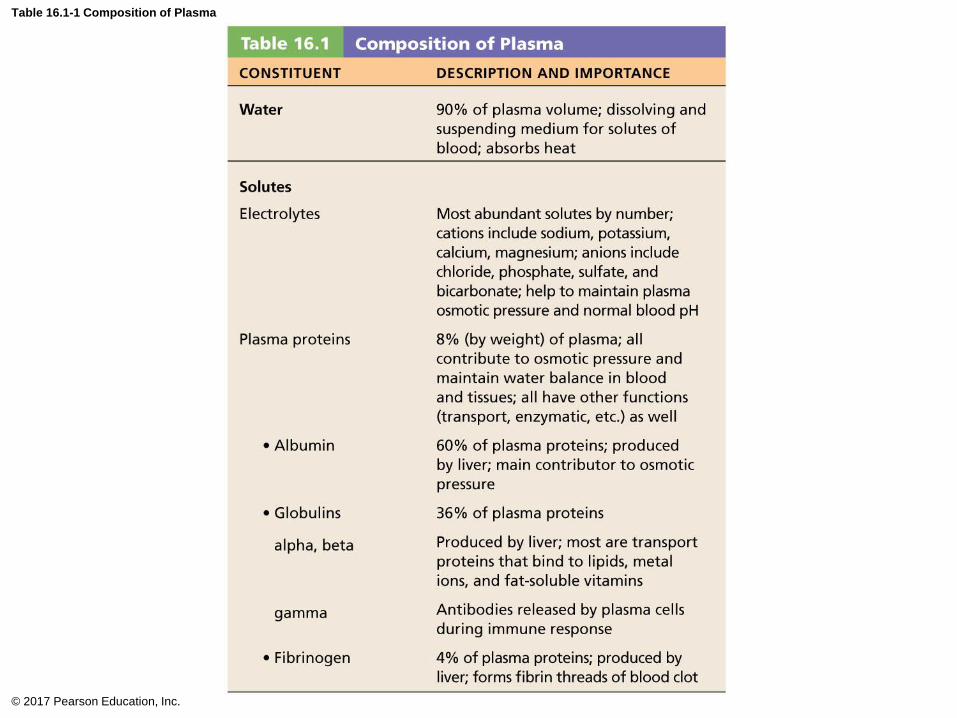
Table 16.1-1 Composition of Plasma
© 2017 Pearson Education, Inc.

Table 16.1-2 Composition of Plasma (continued)
© 2017 Pearson Education, Inc.
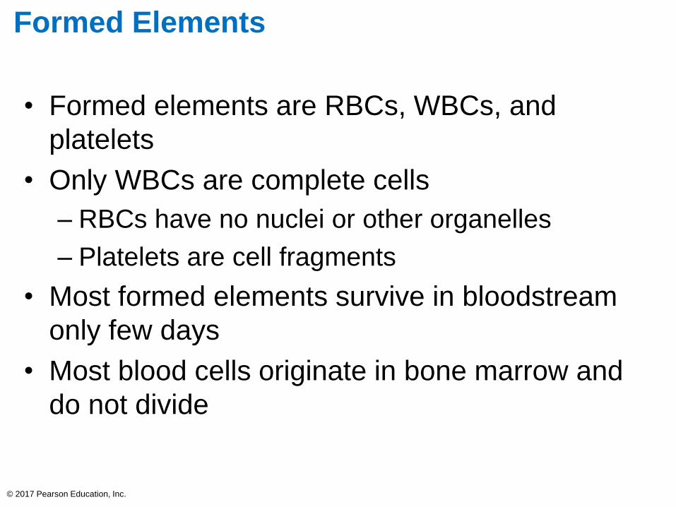
Formed Elements
• Formed elements are RBCs, WBCs, and
platelets
• Only WBCs are complete cells
– RBCs have no nuclei or other organelles
– Platelets are cell fragments
• Most formed elements survive in bloodstream
only few days
• Most blood cells originate in bone marrow and
do not divide
© 2017 Pearson Education, Inc.
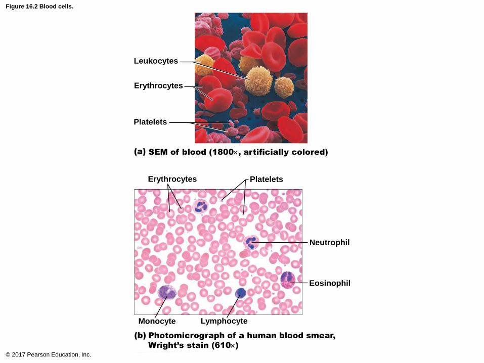
Figure 16.2 Blood cells.
© 2017 Pearson Education, Inc.
Leukocytes
Erythrocytes
Platelets
Platelets Erythrocytes
Neutrophil
Eosinophil
Monocyte Lymphocyte
Photomicrograph of a human blood smear,
Wright’s stain (610)
SEM of blood (1800, artificially colored)

Figure 16.2a Blood cells.
© 2017 Pearson Education, Inc.
Leukocytes
Erythrocytes
Platelets
SEM of blood (1800, artificially colored)

Figure 16.2b Blood cells.
© 2017 Pearson Education, Inc.
Platelets Erythrocytes
Neutrophil
Eosinophil
Monocyte Lymphocyte
Photomicrograph of a human blood smear,
Wright’s stain (610)
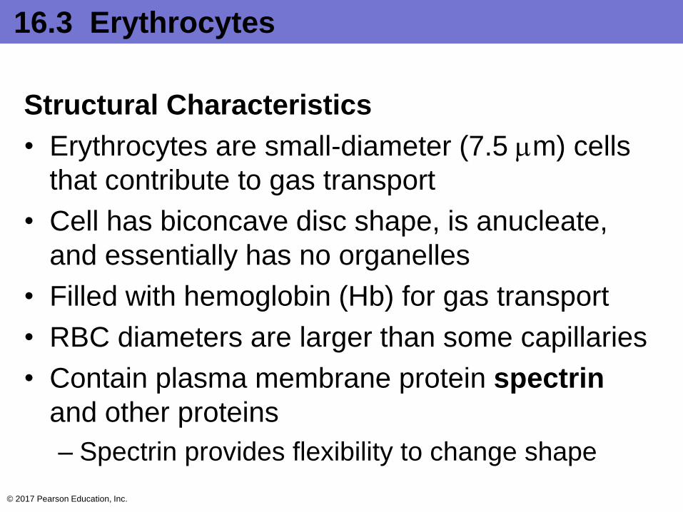
16.3 Erythrocytes
Structural Characteristics
• Erythrocytes are small-diameter (7.5 m) cells
that contribute to gas transport
• Cell has biconcave disc shape, is anucleate,
and essentially has no organelles
• Filled with hemoglobin (Hb) for gas transport
• RBC diameters are larger than some capillaries
• Contain plasma membrane protein spectrin
and other proteins
– Spectrin provides flexibility to change shape
© 2017 Pearson Education, Inc.
Structural Characteristics (cont.)
• Superb example of complementarity of structure
and function
• Three features make for efficient gas transport:
– Biconcave shape offers huge surface area
relative to volume for gas exchange
– Hemoglobin makes up 97% of cell volume (not
counting water)
– RBCs have no mitochondria
• ATP production is anaerobic, so they do not consume
O2 they transport
© 2017 Pearson Education, Inc.
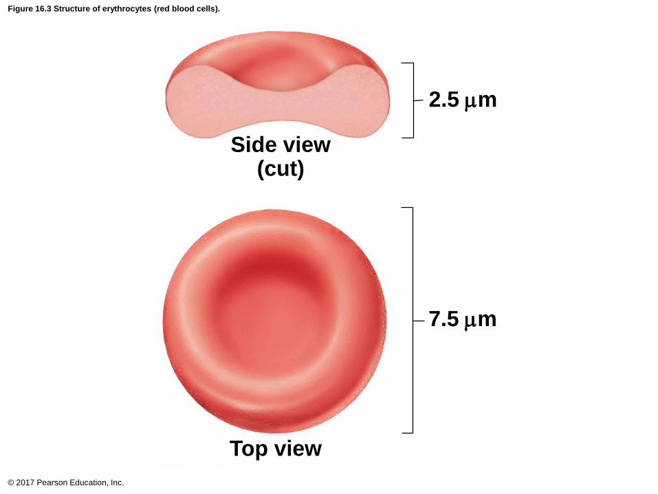
Figure 16.3 Structure of erythrocytes (red blood cells).
© 2017 Pearson Education, Inc.
Side view(cut)
Top view
7.5 m
2.5 m

Function of Erythrocytes
• RBCs are dedicated to respiratory gas transport
• Hemoglobin binds reversibly with oxygen
• Normal values: Males 13–18g/100ml;
Females: 12–16 g/100ml
• Hemoglobin consists of red heme pigment
bound to the protein globin
– Globin is composed of four polypeptide chains
• Two alpha and two beta chains
– A heme pigment is bonded to each globin chain
• Gives blood red color
• Each heme’s central iron atom binds one O2© 2017 Pearson Education, Inc.

Figure 16.4 Structure of hemoglobin.
© 2017 Pearson Education, Inc.
Hemoglobin consists of
globin (two alpha and two
beta polypeptide chains)
and four heme groups.
Iron-containing
heme pigment.
Heme
group Globin
chains
Globin
chains
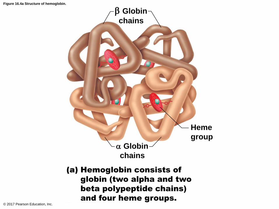
Figure 16.4a Structure of hemoglobin.
© 2017 Pearson Education, Inc.
Hemoglobin consists of
globin (two alpha and two
beta polypeptide chains)
and four heme groups.
Heme
group
Globin
chains
Globin
chains

Figure 16.4b Structure of hemoglobin.
© 2017 Pearson Education, Inc.
Iron-containing
heme pigment.
Function of Erythrocytes (cont.)
• Each Hb molecule can transport four O2
• Each RBC contains 250 million Hb molecules
• O2 loading in lungs
– Produces oxyhemoglobin (ruby red)
• O2 unloading in tissues
– Produces deoxyhemoglobin, or reduced
hemoglobin (dark red)
• CO2 loading in tissues
– 20% of CO2 in blood binds to Hb, producing
carbaminohemoglobin
© 2017 Pearson Education, Inc.
Production of Erythrocytes
• Hematopoiesis: formation of all blood cells
• Occurs in red bone marrow; composed of
reticular connective tissue and blood sinusoids
– In adult, found in axial skeleton, girdles, and
proximal epiphyses of humerus and femur
• Hematopoietic stem cells (hemocytoblasts)
– Stem cell that gives rise to all formed elements
– Hormones and growth factors push cell toward
specific pathway of blood cell development
– Committed cells cannot change
• New blood cells enter blood sinusoids © 2017 Pearson Education, Inc.

Production of Erythrocytes (cont.)
• Stages of erythropoiesis
– Erythropoiesis: process of formation of RBCs
that takes about 15 days
– Stages of transformations
1. Hematopoietic stem cell: transforms into myeloid
stem cell
2. Myeloid stem cell: transforms into proerythroblast
3. Proerythroblast: divides many times, transforming
into basophilic erythroblasts
4. Basophilic erythroblasts: synthesize many
ribosomes, which stain blue
© 2017 Pearson Education, Inc.
Production of Erythrocytes (cont.)
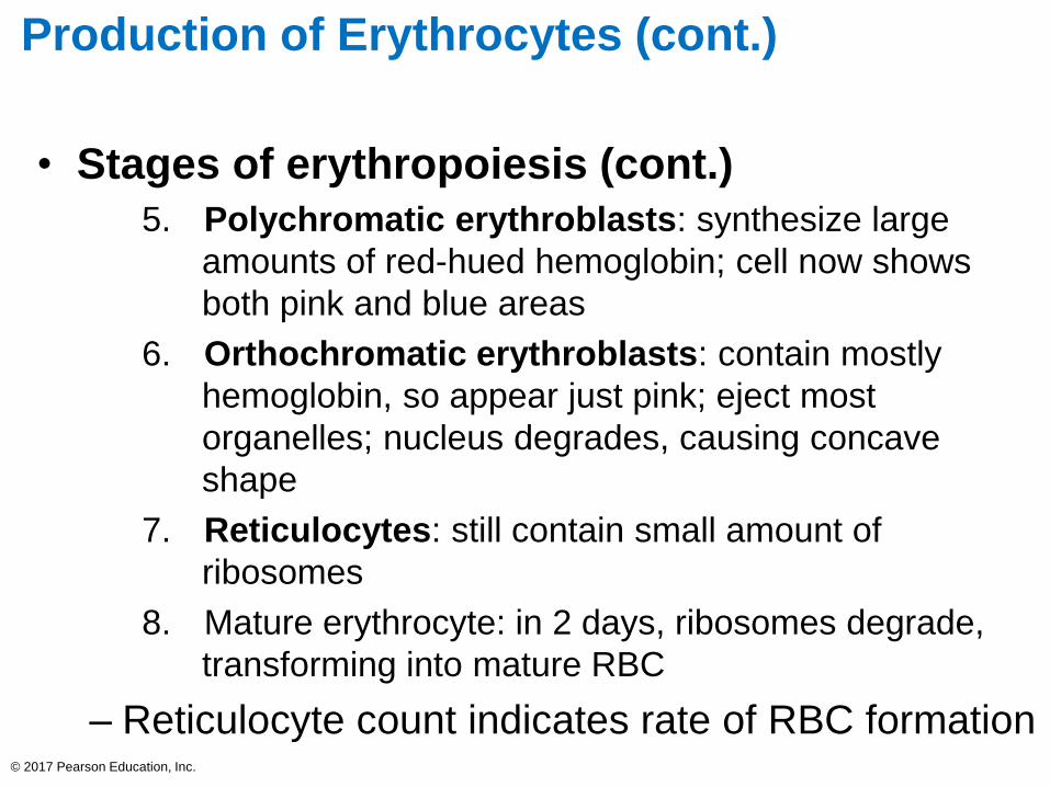
• Stages of erythropoiesis (cont.)
5. Polychromatic erythroblasts: synthesize large
amounts of red-hued hemoglobin; cell now shows
both pink and blue areas
6. Orthochromatic erythroblasts: contain mostly
hemoglobin, so appear just pink; eject most
organelles; nucleus degrades, causing concave
shape
7. Reticulocytes: still contain small amount of
ribosomes
8. Mature erythrocyte: in 2 days, ribosomes degrade,
transforming into mature RBC
– Reticulocyte count indicates rate of RBC formation© 2017 Pearson Education, Inc.

Figure 16.5 Erythropoiesis: formation of red blood cells.
© 2017 Pearson Education, Inc.
Hematopoietic stem
cell (hemocytoblast) ProerythroblastBasophilic
erythroblast
Polychromatic
erythroblastOrthochromatic
erythroblasts Reticulocyte Erythrocyte
Phase 1
Ribosome synthesis
Phase 2
Hemoglobin accumulation
Phase 3
Ejection of nucleus
Developmental pathwayCommitted cellStem cell
Regulation and Requirements of
Erythropoiesis
• Too few RBCs lead to tissue hypoxia
• Too many RBCs increase blood viscosity
• > 2 million RBCs are made per second
• Balance between RBC production and
destruction depends on:
– Hormonal controls
– Dietary requirements
© 2017 Pearson Education, Inc.
Regulation and Requirements of
Erythropoiesis (cont.)
• Hormonal control
– Erythropoietin (EPO): hormone that stimulates
formation of RBCs
• Always small amount of EPO in blood to maintain
basal rate
• Released by kidneys (some from liver) in response to
hypoxia
– At low O2 levels, oxygen-sensitive enzymes in kidney
cells cannot degrade hypoxia-inducible factor (HIF)
– HIF can accumulate, which triggers synthesis of EPO
© 2017 Pearson Education, Inc.

Regulation and Requirements of
Erythropoiesis (cont.)
• Hormonal control (cont.)
– Causes of hypoxia:
• Decreased RBC numbers due to hemorrhage or
increased destruction
• Insufficient hemoglobin per RBC (example: iron
deficiency)
• Reduced availability of O2 (example: high altitudes or
lung problems such as pneumonia)
© 2017 Pearson Education, Inc.

Regulation and Requirements of
Erythropoiesis (cont.)
• Hormonal control (cont.)
– Too many erythrocytes or high oxygen levels in
blood inhibit EPO production
– EPO causes erythrocytes to mature faster
• Testosterone enhances EPO production, resulting in
higher RBC counts in males
© 2017 Pearson Education, Inc.

Figure 16.6 Erythropoietin mechanism for regulating erythropoiesis.
© 2017 Pearson Education, Inc.
5
1
4
2
3
Homeostasis: Normal blood oxygen levels
Enhanced
erythropoiesis
increases RBC count.
Erythropoietin
stimulates red
bone marrow.
Kidney (and liver to
a smaller extent)
releases
erythropoietin.
O2-carrying
ability of blood
rises.
Hypoxia (inadequate
O2 delivery) due to
• Decreased
RBC count
• Decreased amount
of hemoglobin
• Decreased
availability of O2
Stimulus:
Slide 1
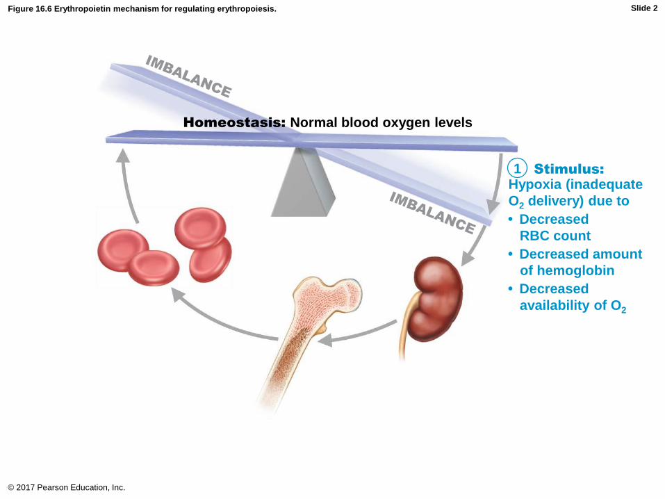
Figure 16.6 Erythropoietin mechanism for regulating erythropoiesis.
© 2017 Pearson Education, Inc.
1
Homeostasis: Normal blood oxygen levels
Hypoxia (inadequate
O2 delivery) due to
• Decreased
RBC count
• Decreased amount
of hemoglobin
• Decreased
availability of O2
Stimulus:
Slide 2
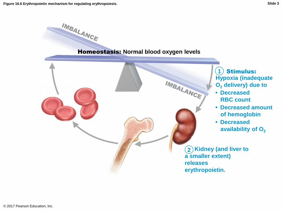
Figure 16.6 Erythropoietin mechanism for regulating erythropoiesis.
© 2017 Pearson Education, Inc.
1
2
Homeostasis: Normal blood oxygen levels
Kidney (and liver to
a smaller extent)
releases
erythropoietin.
Hypoxia (inadequate
O2 delivery) due to
• Decreased
RBC count
• Decreased amount
of hemoglobin
• Decreased
availability of O2
Stimulus:
Slide 3

Figure 16.6 Erythropoietin mechanism for regulating erythropoiesis.
© 2017 Pearson Education, Inc.
1
2
3
Homeostasis: Normal blood oxygen levels
Erythropoietin
stimulates red
bone marrow.
Kidney (and liver to
a smaller extent)
releases
erythropoietin.
Hypoxia (inadequate
O2 delivery) due to
• Decreased
RBC count
• Decreased amount
of hemoglobin
• Decreased
availability of O2
Stimulus:
Slide 4
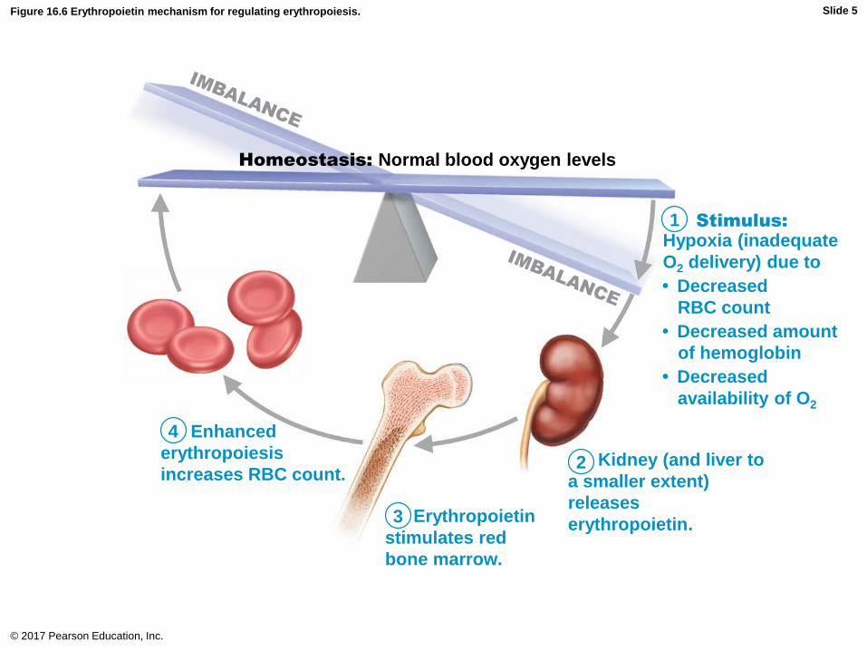
Figure 16.6 Erythropoietin mechanism for regulating erythropoiesis.
© 2017 Pearson Education, Inc.
1
4
2
3
Homeostasis: Normal blood oxygen levels
Enhanced
erythropoiesis
increases RBC count.
Erythropoietin
stimulates red
bone marrow.
Kidney (and liver to
a smaller extent)
releases
erythropoietin.
Hypoxia (inadequate
O2 delivery) due to
• Decreased
RBC count
• Decreased amount
of hemoglobin
• Decreased
availability of O2
Stimulus:
Slide 5
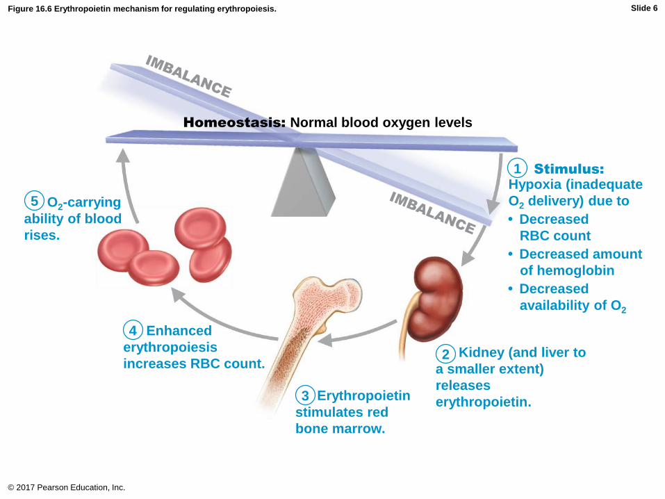
Figure 16.6 Erythropoietin mechanism for regulating erythropoiesis.
© 2017 Pearson Education, Inc.
5
1
4
2
3
Homeostasis: Normal blood oxygen levels
Enhanced
erythropoiesis
increases RBC count.
Erythropoietin
stimulates red
bone marrow.
Kidney (and liver to
a smaller extent)
releases
erythropoietin.
O2-carrying
ability of blood
rises.
Hypoxia (inadequate
O2 delivery) due to
• Decreased
RBC count
• Decreased amount
of hemoglobin
• Decreased
availability of O2
Stimulus:
Slide 6
Clinical – Homeostatic Imbalance 16.1
• Some athletes abuse artificial EPO
– Use of EPO increases hematocrit, which allows
athlete to increase stamina and performance
• Dangerous consequences:
– EPO can increase hematocrit from 45% up to
even 65%, with dehydration concentrating blood
even more
– Blood becomes like sludge and can cause
clotting, stroke, or heart failure
© 2017 Pearson Education, Inc.
Regulation and Requirements of
Erythropoiesis (cont.)
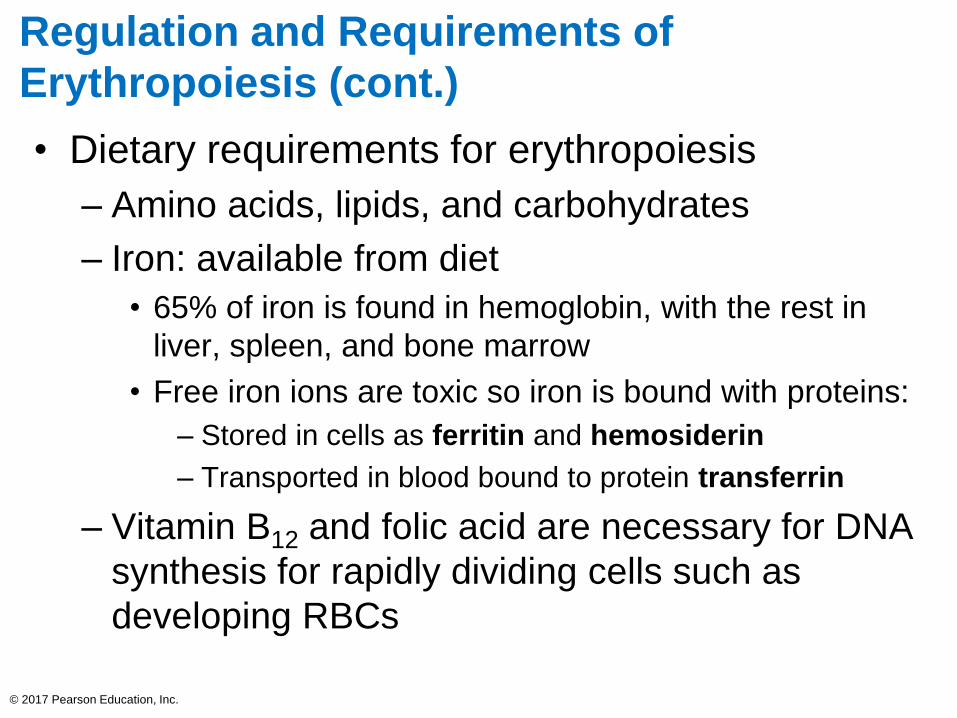
• Dietary requirements for erythropoiesis
– Amino acids, lipids, and carbohydrates
– Iron: available from diet
• 65% of iron is found in hemoglobin, with the rest in
liver, spleen, and bone marrow
• Free iron ions are toxic so iron is bound with proteins:
– Stored in cells as ferritin and hemosiderin
– Transported in blood bound to protein transferrin
– Vitamin B12 and folic acid are necessary for DNA
synthesis for rapidly dividing cells such as
developing RBCs
© 2017 Pearson Education, Inc.
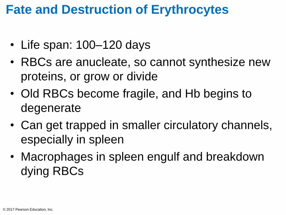
Fate and Destruction of Erythrocytes
• Life span: 100–120 days
• RBCs are anucleate, so cannot synthesize new
proteins, or grow or divide
• Old RBCs become fragile, and Hb begins to
degenerate
• Can get trapped in smaller circulatory channels,
especially in spleen
• Macrophages in spleen engulf and breakdown
dying RBCs
© 2017 Pearson Education, Inc.

• RBC breakdown: heme, iron, and globin are
separated
– Iron binds to ferridin or hemosiderin and is stored
for reuse
– Heme is degraded to yellow pigment bilirubin
• Liver secretes bilirubin (in bile) into intestines, where it
is degraded to pigment urobilinogen
– Urobilinogen is transformed into brown pigment
stercobilin that leaves body in feces
– Globin is metabolized into amino acids
• Released into circulation
© 2017 Pearson Education, Inc.
Fate and Destruction of Erythrocytes (cont.)
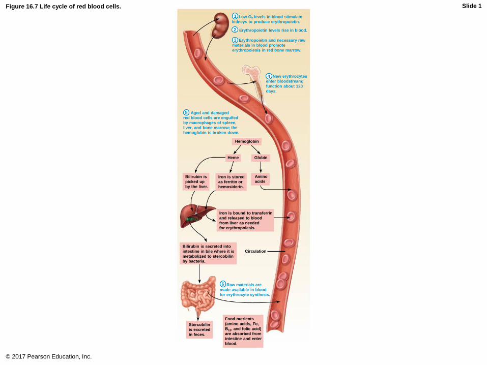
Figure 16.7 Life cycle of red blood cells.
© 2017 Pearson Education, Inc.
Hemoglobin
Heme Globin
Bilirubin is
picked up
by the liver.
Iron is stored
as ferritin or
hemosiderin.
Iron is bound to transferrin
and released to blood
from liver as needed
for erythropoiesis.
Bilirubin is secreted into
intestine in bile where it is
metabolized to stercobilin
by bacteria.
Circulation
Amino
acids
Stercobilin
is excreted
in feces.
Food nutrients
(amino acids, Fe,
B12, and folic acid)
are absorbed from
intestine and enter
blood.
5
4
3
2
1
6
Low O2 levels in blood stimulate
kidneys to produce erythropoietin.
Erythropoietin levels rise in blood.
Erythropoietin and necessary raw
materials in blood promote
erythropoiesis in red bone marrow.
New erythrocytes
enter bloodstream;
function about 120
days.
Aged and damaged
red blood cells are engulfed
by macrophages of spleen,
liver, and bone marrow; the
hemoglobin is broken down.
Raw materials are
made available in blood
for erythrocyte synthesis.
Slide 1
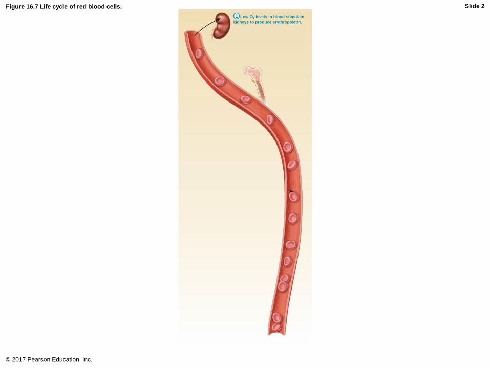
Figure 16.7 Life cycle of red blood cells.
© 2017 Pearson Education, Inc.
1 Low O2 levels in blood stimulate
kidneys to produce erythropoietin.
Slide 2
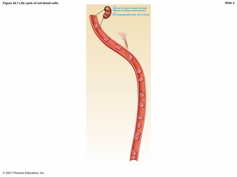
Figure 16.7 Life cycle of red blood cells.
© 2017 Pearson Education, Inc.
2
1 Low O2 levels in blood stimulate
kidneys to produce erythropoietin.
Erythropoietin levels rise in blood.
Slide 3

Figure 16.7 Life cycle of red blood cells.
© 2017 Pearson Education, Inc.
3
2
1 Low O2 levels in blood stimulate
kidneys to produce erythropoietin.
Erythropoietin levels rise in blood.
Erythropoietin and necessary raw
materials in blood promote
erythropoiesis in red bone marrow.
Slide 4

Figure 16.7 Life cycle of red blood cells.
© 2017 Pearson Education, Inc.
4
3
2
1 Low O2 levels in blood stimulate
kidneys to produce erythropoietin.
Erythropoietin levels rise in blood.
Erythropoietin and necessary raw
materials in blood promote
erythropoiesis in red bone marrow.
New erythrocytes
enter bloodstream;
function about 120
days.
Slide 5
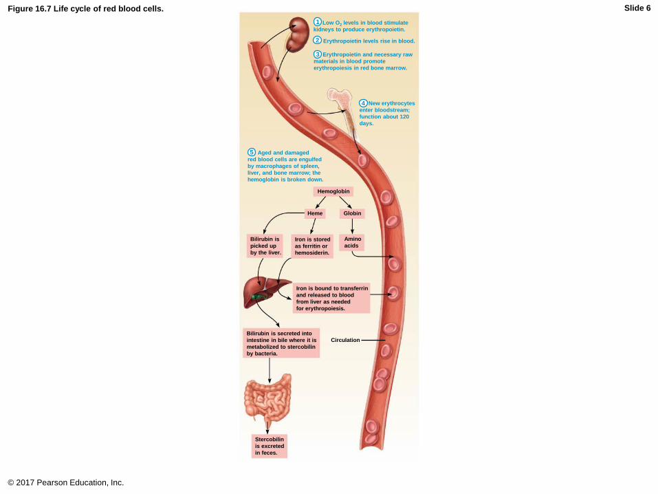
Figure 16.7 Life cycle of red blood cells.
© 2017 Pearson Education, Inc.
Hemoglobin
Heme Globin
Bilirubin is
picked up
by the liver.
Iron is stored
as ferritin or
hemosiderin.
Iron is bound to transferrin
and released to blood
from liver as needed
for erythropoiesis.
Bilirubin is secreted into
intestine in bile where it is
metabolized to stercobilin
by bacteria.
Circulation
Amino
acids
Stercobilin
is excreted
in feces.
5
4
3
2
1 Low O2 levels in blood stimulate
kidneys to produce erythropoietin.
Erythropoietin levels rise in blood.
Erythropoietin and necessary raw
materials in blood promote
erythropoiesis in red bone marrow.
New erythrocytes
enter bloodstream;
function about 120
days.
Aged and damaged
red blood cells are engulfed
by macrophages of spleen,
liver, and bone marrow; the
hemoglobin is broken down.
Slide 6

Figure 16.7 Life cycle of red blood cells.
© 2017 Pearson Education, Inc.
Hemoglobin
Heme Globin
Bilirubin is
picked up
by the liver.
Iron is stored
as ferritin or
hemosiderin.
Iron is bound to transferrin
and released to blood
from liver as needed
for erythropoiesis.
Bilirubin is secreted into
intestine in bile where it is
metabolized to stercobilin
by bacteria.
Circulation
Amino
acids
Stercobilin
is excreted
in feces.
Food nutrients
(amino acids, Fe,
B12, and folic acid)
are absorbed from
intestine and enter
blood.
5
4
3
2
1
6
Low O2 levels in blood stimulate
kidneys to produce erythropoietin.
Erythropoietin levels rise in blood.
Erythropoietin and necessary raw
materials in blood promote
erythropoiesis in red bone marrow.
New erythrocytes
enter bloodstream;
function about 120
days.
Aged and damaged
red blood cells are engulfed
by macrophages of spleen,
liver, and bone marrow; the
hemoglobin is broken down.
Raw materials are
made available in blood
for erythrocyte synthesis.
Slide 7
Erythrocyte Disorders
• Most erythrocyte disorders are classified as
either anemia or polycythemia
• Anemia
– Blood has abnormally low O2-carrying capacity
that is too low to support normal metabolism
– Sign of problem rather than disease itself
– Symptoms: fatigue, pallor, dyspnea, and chills
– Three groups based on cause
• Blood loss
• Not enough RBCs produced
• Too many RBCs being destroyed© 2017 Pearson Education, Inc.

Erythrocyte Disorders (cont.)
• Anemia (cont.)
– Blood loss
• Hemorrhagic anemia
– Rapid blood loss (example: severe wound)
– Treated by blood replacement
• Chronic hemorrhagic anemia
– Slight but persistent blood loss
» Example: hemorrhoids, bleeding ulcer
– Primary problem must be treated to stop blood loss
© 2017 Pearson Education, Inc.

Erythrocyte Disorders (cont.)
• Anemia (cont.)
– Not enough RBCs being produced
• Iron-deficiency anemia
– Can be caused by hemorrhagic anemia, but also by low
iron intake or impaired absorption
– RBCs produced are called microcytes
» Small, pale in color
» Cannot synthesize hemoglobin because there is a
lack of iron
– Treatment: iron supplements
© 2017 Pearson Education, Inc.

Erythrocyte Disorders (cont.)
• Anemia (cont.)
– Not enough RBCs being produced (cont.)
• Pernicious anemia
– Autoimmune disease that destroys stomach mucosa
that produces intrinsic factor
– Intrinsic factor needed to absorb B12
– B12 is needed to help RBCs divide
– Without B12 RBCs enlarge but cannot divide, resulting
in large macrocytes
– Treatment: B12 injections or nasal gel
– Can also be caused by low dietary intake of B12
» Can be a problem for vegetarians
© 2017 Pearson Education, Inc.
Erythrocyte Disorders (cont.)
• Anemia (cont.)
– Not enough RBCs being produced (cont.)
• Renal anemia
– Caused by lack of EPO
– Often accompanies renal disease
» Kidneys cannot produce enough EPO
– Treatment: synthetic EPO
© 2017 Pearson Education, Inc.

Erythrocyte Disorders (cont.)
• Anemia (cont.)
– Not enough RBCs being produced (cont.)
• Aplastic anemia
– Destruction or inhibition of red bone marrow
– Can be caused by drugs, chemicals, radiation,
or viruses
» Usually cause is unknown
– All formed element cell lines are affected
» Results in anemia as well as clotting and
immunity defects
– Treatment: short-term with transfusions, long-term
with transplanted stem cells
© 2017 Pearson Education, Inc.
Erythrocyte Disorders (cont.)
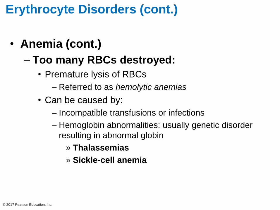
• Anemia (cont.)
– Too many RBCs destroyed:
• Premature lysis of RBCs
– Referred to as hemolytic anemias
• Can be caused by:
– Incompatible transfusions or infections
– Hemoglobin abnormalities: usually genetic disorder
resulting in abnormal globin
» Thalassemias
» Sickle-cell anemia
© 2017 Pearson Education, Inc.
Erythrocyte Disorders (cont.)
• Anemia (cont.)
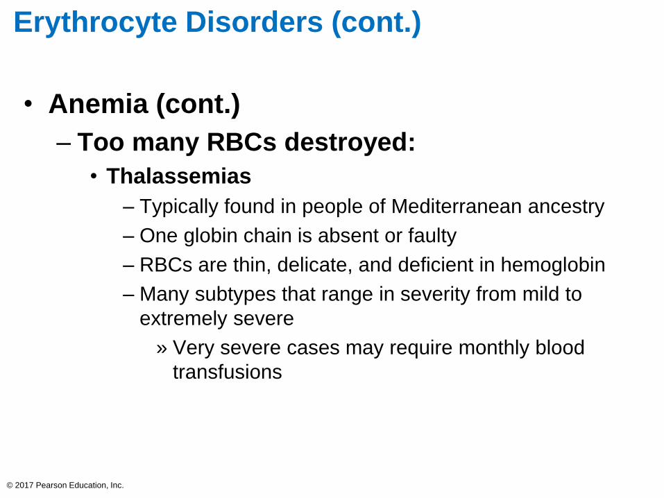
– Too many RBCs destroyed:
• Thalassemias
– Typically found in people of Mediterranean ancestry
– One globin chain is absent or faulty
– RBCs are thin, delicate, and deficient in hemoglobin
– Many subtypes that range in severity from mild to
extremely severe
» Very severe cases may require monthly blood
transfusions
© 2017 Pearson Education, Inc.
Erythrocyte Disorders (cont.)
• Anemia (cont.)
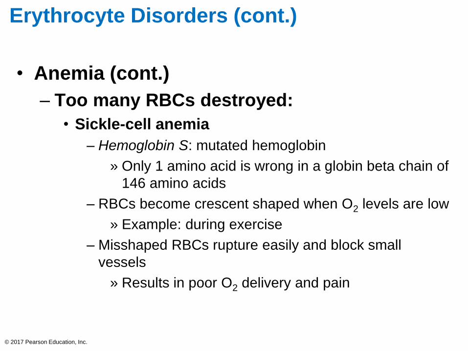
– Too many RBCs destroyed:
• Sickle-cell anemia
– Hemoglobin S: mutated hemoglobin
» Only 1 amino acid is wrong in a globin beta chain of
146 amino acids
– RBCs become crescent shaped when O2 levels are low
» Example: during exercise
– Misshaped RBCs rupture easily and block small
vessels
» Results in poor O2 delivery and pain
© 2017 Pearson Education, Inc.

Erythrocyte Disorders (cont.)
• Anemia (cont.)
– Too many RBCs destroyed:
• Sickle-cell anemia (cont.)
– Prevalent in black people of the African malarial belt
and their descendants
– Possible benefit: people with sickle cell do not contract
malaria
» Kills 1 million each year
» Individuals with two copies of Hb-S can develop
sickle-cell anemia
» Individuals with only one copy have milder disease
and better chance of surviving malaria
© 2017 Pearson Education, Inc.

Erythrocyte Disorders (cont.)
• Anemia (cont.)
– Too many RBCs destroyed:
• Sickle-cell anemia (cont.)
– Treatment: acute crisis treated with transfusions;
inhaled nitric oxide
– Prevention of sickling:
» Hydroxyurea induces formation of fetal hemoglobin
(which does not sickle)
» Stem cell transplants
» Gene therapy
» Nitric oxide for vasodilation
© 2017 Pearson Education, Inc.
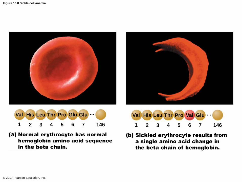
Figure 16.8 Sickle-cell anemia.
© 2017 Pearson Education, Inc.
Normal erythrocyte has normal
hemoglobin amino acid sequence
in the beta chain.
Sickled erythrocyte results from
a single amino acid change in
the beta chain of hemoglobin.
146 14676543217654321
ValVal His Leu Thr Pro Val GluHis Leu Thr Pro Glu Glu ... ...
Erythrocyte Disorders (cont.)
• Polycythemia
– Abnormal excess of RBCs; increases blood
viscosity, causing sluggish blood flow
– Polycythemia vera: Bone marrow cancer leading
to excess RBCs
• Hematocrit may go as high as 80%
• Treatment: therapeutic phlebotomy
– Secondary polycythemia: caused by low O2
levels (example: high altitude) or increased
EPO production
© 2017 Pearson Education, Inc.
Erythrocyte Disorders (cont.)
• Polycythemia (cont.)
– Blood doping: athletes remove, store, and
reinfuse RBCs before an event to increase O2
levels for stamina
© 2017 Pearson Education, Inc.
16.4 Leukocytes
General Structure and Functional
Characteristics
• Leukocytes, or WBCs, are only formed element
that is complete cell with nuclei and organelles
• Make up <1% of total blood volume
– 4800 to 10,800 WBCs per l blood
• Function in defense against disease
– Can leave capillaries via diapedesis
– Move through tissue spaces by amoeboid
motion and positive chemotaxis
© 2017 Pearson Education, Inc.

General Structure and Functional
Characteristics (cont.)
• Leukocytosis: WBC count over 11,000 per l
– Increase is a normal response to infection
• Leukocytes grouped into two major categories:
– Granulocytes: contain visible cytoplasmic
granules
– Agranulocytes: do not contain visible
cytoplasmic granules; two types:
• Mnemonic to remember decreasing abundance
in blood: Never let monkeys eat bananas
© 2017 Pearson Education, Inc.

Figure 16.9 Types and relative percentages of leukocytes in normal blood.
© 2017 Pearson Education, Inc.
Granulocytes
Differential
WBC count
(All total 4800–10,800/l)Formed
elements
(not drawnto scale)
Agranulocytes
Leukocytes
Platelets
Erythrocytes
Neutrophils (50–70%)
Eosinophils (2–4%)
Basophils (0.5–1%)
Lymphocytes (25–45%)
Monocytes (3–8%)

Granulocytes
• Granulocytes: three types
– Neutrophils, eosinophils, basophils
• Larger and shorter-lived than RBCs
• Contain lobed, rather than circular, nuclei
• Cytoplasmic granules stain specifically with
Wright’s stain
• All are phagocytic to some degree
© 2017 Pearson Education, Inc.

Granulocytes (cont.)
• Neutrophils
– Most numerous WBCs
• Account for 50–70% of WBCs
– About twice the size of RBCs
– Granules stain with both acid and basic dyes
– Granules contain either hydrolytic enzymes or
antimicrobial proteins, defensins
– Also called polymorphonuclear leukocytes
(PMNs or polys) because nucleus is lobular
• Cell has anywhere from three to six lobes
© 2017 Pearson Education, Inc.
Granulocytes (cont.)
• Neutrophils (cont.)
– Very phagocytic
• Referred to as “bacteria slayers”
• Kill microbes by process called respiratory burst
– Cell synthesizes potent oxidizing substances
(bleach or hydrogen peroxide)
– Defensin granules merge with phagosome
• Form “spears” that pierce holes in membrane of
ingested microbe
© 2017 Pearson Education, Inc.

Figure 16.10 Leukocytes.
© 2017 Pearson Education, Inc.
Granulocytes Agranulocytes
Neutrophil:
Multilobednucleus, palered and bluecytoplasmic granules
Eosinophil:
Bilobednucleus, redcytoplasmicgranules
Basophil:
Bilobednucleus,purplish-black cytoplasmicgranules
Lymphocyte(small): Largesphericalnucleus, thinrim of paleblue cytoplasm
Monocyte:
Kidney-shapednucleus,abundant paleblue cytoplasm

Figure 16.10a Leukocytes.
© 2017 Pearson Education, Inc.
Granulocytes
Neutrophil:
Multilobednucleus, palered and bluecytoplasmic granules
Granulocytes (cont.)
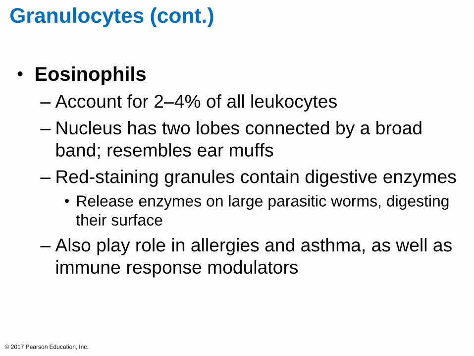
• Eosinophils
– Account for 2–4% of all leukocytes
– Nucleus has two lobes connected by a broad
band; resembles ear muffs
– Red-staining granules contain digestive enzymes
• Release enzymes on large parasitic worms, digesting
their surface
– Also play role in allergies and asthma, as well as
immune response modulators
© 2017 Pearson Education, Inc.

Figure 16.10b Leukocytes.
© 2017 Pearson Education, Inc.
Granulocytes
Eosinophil:
Bilobed
nucleus, red
cytoplasmic
granules

Granulocytes (cont.)
• Basophils
– Rarest WBCs, accounting for only 0.5–1% of
leukocytes
– Nucleus deep purple with one to two
constrictions
– Large, purplish black (basophilic) granules
contain histamine
• Histamine: inflammatory chemical that acts as
vasodilator and attracts WBCs to inflamed sites
– Are functionally similar to mast cells
© 2017 Pearson Education, Inc.
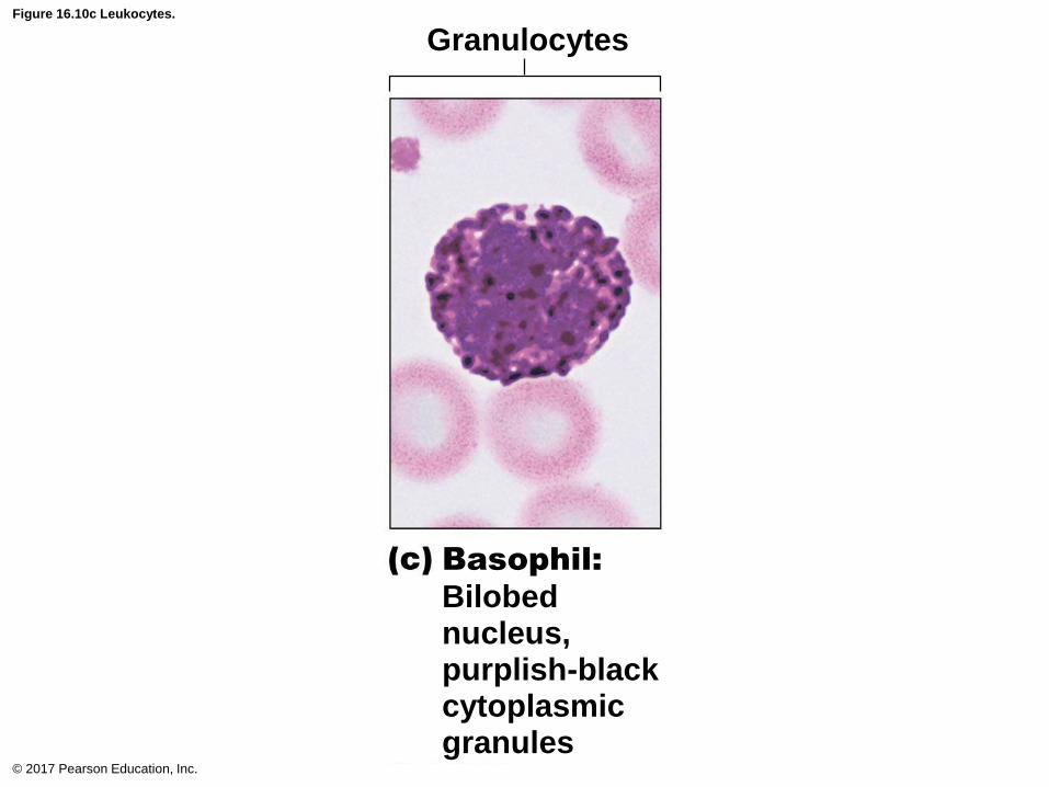
Figure 16.10c Leukocytes.
© 2017 Pearson Education, Inc.
Granulocytes
Basophil:
Bilobednucleus,purplish-black cytoplasmicgranules
Agranulocytes
• Agranulocytes lack visible cytoplasmic
granules
• Two types: lymphocytes and monocytes
• Both have spherical or kidney-shaped nuclei
© 2017 Pearson Education, Inc.
Agranulocytes (cont.)
• Lymphocytes– Second most numerous WBC, accounts for 25%
– Large, dark purple, circular nuclei with thin rim of
blue cytoplasm
– Mostly found in lymphoid tissue (example: lymph
nodes, spleen), but a few circulate in blood
– Crucial to immunity
– Two types of lymphocytes
• T lymphocytes (T cells) act against virus-infected
cells and tumor cells
• B lymphocytes (B cells) give rise to plasma cells,
which produce antibodies
© 2017 Pearson Education, Inc.
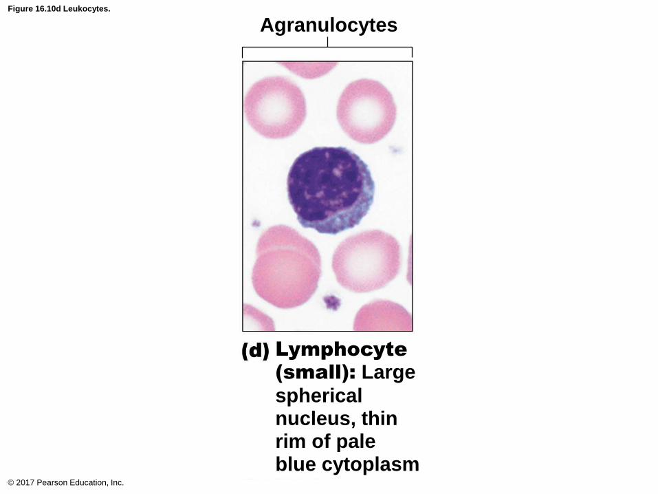
Figure 16.10d Leukocytes.
© 2017 Pearson Education, Inc.
Agranulocytes
Lymphocyte(small): Large
sphericalnucleus, thinrim of paleblue cytoplasm
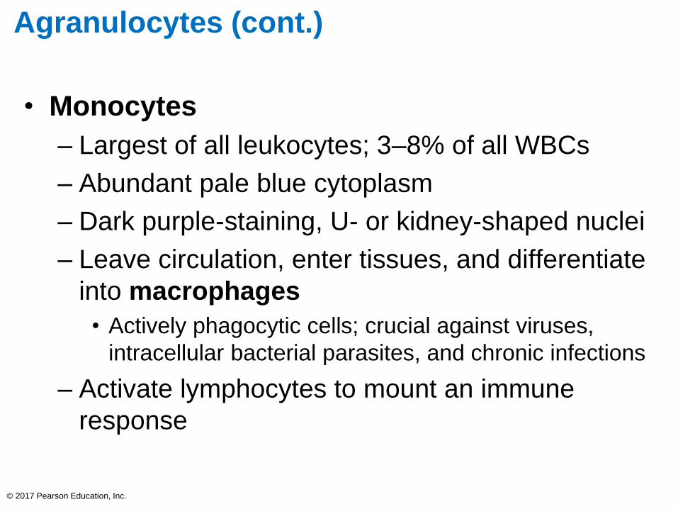
Agranulocytes (cont.)
• Monocytes
– Largest of all leukocytes; 3–8% of all WBCs
– Abundant pale blue cytoplasm
– Dark purple-staining, U- or kidney-shaped nuclei
– Leave circulation, enter tissues, and differentiate
into macrophages
• Actively phagocytic cells; crucial against viruses,
intracellular bacterial parasites, and chronic infections
– Activate lymphocytes to mount an immune
response
© 2017 Pearson Education, Inc.
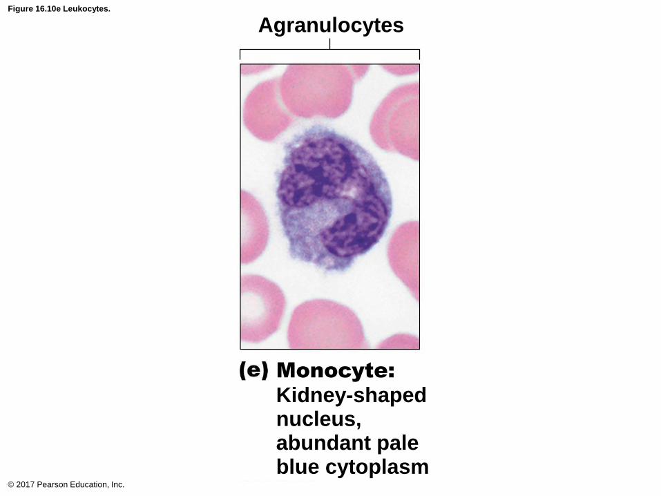
Figure 16.10e Leukocytes.
© 2017 Pearson Education, Inc.
Agranulocytes
Monocyte:
Kidney-shapednucleus,abundant paleblue cytoplasm
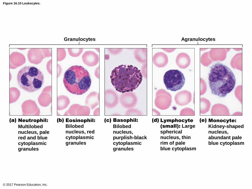
Figure 16.10 Leukocytes.
© 2017 Pearson Education, Inc.
Granulocytes Agranulocytes
Neutrophil:
Multilobednucleus, palered and bluecytoplasmic granules
Eosinophil:
Bilobednucleus, redcytoplasmicgranules
Basophil:
Bilobednucleus,purplish-black cytoplasmicgranules
Lymphocyte(small): Largesphericalnucleus, thinrim of paleblue cytoplasm
Monocyte:
Kidney-shapednucleus,abundant paleblue cytoplasm

Production and Life Span of Leukocytes
• Leukopoiesis: production of WBCs are
stimulated by two types of chemical messengers
from red bone marrow and mature WBCs
– Interleukins are numbered (e.g., IL-3, IL-5)
– Colony-stimulating factors (CSFs) are named
for WBC type they stimulate (e.g., granulocyte-
CSF stimulates granulocytes)
• All leukocytes originate from hemocytoblast
stem cell that branches into two pathways:
– Lymphoid stem cells produces lymphocytes
– Myeloid stem cells produce all other elements© 2017 Pearson Education, Inc.

Production and Life Span of Leukocytes
(cont.)
• Granulocyte production:
1. Myeloblasts: arise from myeloid line stem cells
2. Promyelocytes: accumulate lysosomes
3. Myelocytes: accumulate granules
4. Band cells: nuclei form curved arc
5. Mature granulocyte: nuclei become segmented
before being released in blood
• 10× more are stored in bone marrow than in blood
• 3× more WBCs are formed than RBCs, because
WBCs have a shorter life, cut short by fighting
microbes
© 2017 Pearson Education, Inc.
Production and Life Span of Leukocytes
(cont.)
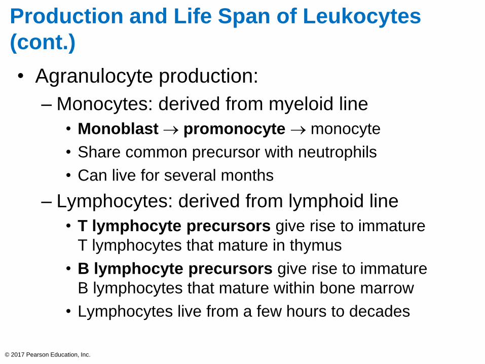
• Agranulocyte production:
– Monocytes: derived from myeloid line
• Monoblast → promonocyte → monocyte
• Share common precursor with neutrophils
• Can live for several months
– Lymphocytes: derived from lymphoid line
• T lymphocyte precursors give rise to immature
T lymphocytes that mature in thymus
• B lymphocyte precursors give rise to immature
B lymphocytes that mature within bone marrow
• Lymphocytes live from a few hours to decades
© 2017 Pearson Education, Inc.

Figure 16.11 Leukocyte formation.
© 2017 Pearson Education, Inc.
Myeloblast MonoblastMyeloblastMyeloblast
Myeloid stem cell Lymphoid stem cell
B lymphocyte
precursor
T lymphocyte
precursor
PromonocytePromyelocytePromyelocytePromyelocyte
Eosinophilic
myelocyte
Basophilic
myelocyteNeutrophilic
myelocyte
Eosinophilic
band cellsBasophilic
band cells
Neutrophilic
band cells
Eosinophils Basophils Neutrophils Monocytes B lymphocytes T lymphocytes
Effector T cellsPlasma cellsMacrophages (tissues)
Agranular
leukocytes
Granular
leukocytes
Developmental
pathway
Committed
cells
Hematopoietic stem cell
(hemocytoblast)
Stem cells
Some becomeSome becomeSome become

Clinical – Homeostatic Imbalance 16.2
• Many hematopoietic hormones (EPO and
CSFs) are used clinically
• Can stimulate bone marrow of cancer patients
receiving chemotherapy or stem cell transplants
• Also used to increase protective immune
responses of AIDS patients
© 2017 Pearson Education, Inc.

Leukocyte Disorders
• Overproduction of abnormal WBC: leukemias
and infectious mononucleosis
• Abnormally low WBC count: leukopenia
– Can be drug induced, particularly by anticancer
drugs or glucocorticoids
© 2017 Pearson Education, Inc.

Leukocyte Disorders (cont.)
• Leukemias
– Cancerous condition involving overproduction of
abnormal WBCs
• Usually involve clones of single abnormal cell
– Named according to abnormal WBC clone
involved
• Myeloid leukemia involves myeloblast descendants
• Lymphocytic leukemia involves lymphocytes
© 2017 Pearson Education, Inc.

Leukocyte Disorders (cont.)
• Leukemias (cont.)
– Acute (quickly advancing) leukemia derives from
stem cells
• Primarily affects children
– Chronic (slowly advancing) leukemia involves
proliferation of later cell stages
• More prevalent in older people
© 2017 Pearson Education, Inc.
Leukocyte Disorders (cont.)
• Leukemias (cont.)
– Without treatment, all leukemias are fatal
– Immature, nonfunctional WBCs flood
bloodstream
– Cancerous cells fill red bone marrow, crowding
out other cell lines
• Leads to anemia and bleeding
– Death is usually from internal hemorrhage or
overwhelming infections
– Treatments: irradiation, antileukemic drugs;
stem cell transplants
© 2017 Pearson Education, Inc.
Leukocyte Disorders (cont.)
• Infectious mononucleosis
– Highly contagious viral disease (“kissing disease”)
• Usually seen in young adults
– Caused by Epstein-Barr virus
– Results in high numbers of typical agranulocytes
• Involve lymphocytes that become enlarged
• Originally thought cells were monocytes, so disease
named mononucleosis
– Symptoms
• Tired, achy, chronic sore throat, low fever
– Runs course with rest in 4–6 weeks
© 2017 Pearson Education, Inc.
Platelets
• Cytoplasmic fragments of megakaryocytes
• Blue-staining outer region; purple granules
• Granules contain serotonin, Ca2+, enzymes,
ADP, and platelet-derived growth factor (PDGF)
– Act in clotting process
• Normal = 150,000– 400,000 platelets/ml of
blood
© 2017 Pearson Education, Inc.
16.5 Platelets
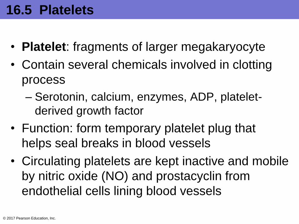
• Platelet: fragments of larger megakaryocyte
• Contain several chemicals involved in clotting
process
– Serotonin, calcium, enzymes, ADP, platelet-
derived growth factor
• Function: form temporary platelet plug that
helps seal breaks in blood vessels
• Circulating platelets are kept inactive and mobile
by nitric oxide (NO) and prostacyclin from
endothelial cells lining blood vessels
© 2017 Pearson Education, Inc.
16.5 Platelets
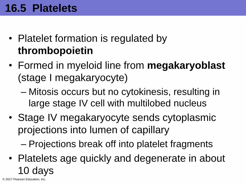
• Platelet formation is regulated by
thrombopoietin
• Formed in myeloid line from megakaryoblast
(stage I megakaryocyte)
– Mitosis occurs but no cytokinesis, resulting in
large stage IV cell with multilobed nucleus
• Stage IV megakaryocyte sends cytoplasmic
projections into lumen of capillary
– Projections break off into platelet fragments
• Platelets age quickly and degenerate in about
10 days© 2017 Pearson Education, Inc.

Figure 16.12 Formation of platelets.
© 2017 Pearson Education, Inc.
Stem cell Developmental pathway
Hematopoieticstem cell
(hemocytoblast)
Megakaryoblast(stage I
megakaryocyte)
Megakaryocyte(stage II/III)
Megakaryocyte(stage IV)
Platelets

Table 16.2-1 Summary of Formed Elements of the Blood
© 2017 Pearson Education, Inc.
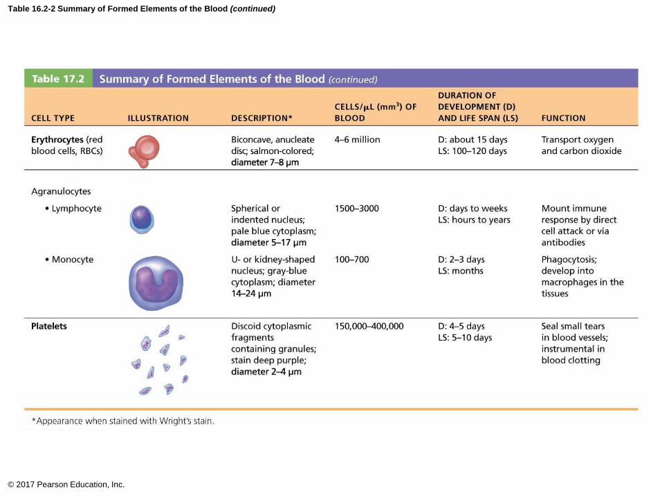
Table 16.2-2 Summary of Formed Elements of the Blood (continued)
© 2017 Pearson Education, Inc.
16.6 Hemostasis
• Hemostasis: fast series of reactions for
stoppage of bleeding
• Requires clotting factors and substances
released by platelets and injured tissues
• Three steps involved
Step 1: Vascular spasm
Step 2: Platelet plug formation
Step 3: Coagulation (blood clotting)
© 2017 Pearson Education, Inc.

Step 1: Vascular Spasm
• Vessel responds to injury with vasoconstriction
• Vascular spams are triggered by:
– Direct injury to vascular smooth muscle
– Chemicals released by endothelial cells and
platelets
– Pain reflexes
• Most effective in smaller blood vessels
• Can significantly reduce blood flow until other
mechanisms can kick in
© 2017 Pearson Education, Inc.
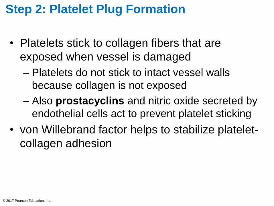
Step 2: Platelet Plug Formation
• Platelets stick to collagen fibers that are
exposed when vessel is damaged
– Platelets do not stick to intact vessel walls
because collagen is not exposed
– Also prostacyclins and nitric oxide secreted by
endothelial cells act to prevent platelet sticking
• von Willebrand factor helps to stabilize platelet-
collagen adhesion
© 2017 Pearson Education, Inc.
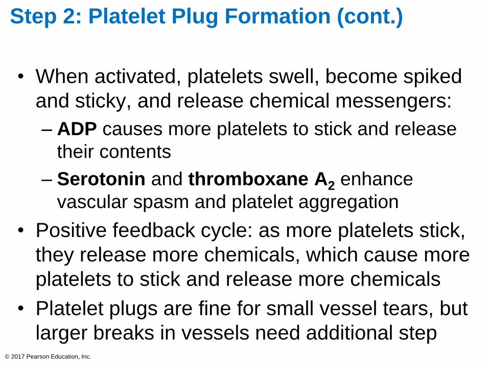
Step 2: Platelet Plug Formation (cont.)
• When activated, platelets swell, become spiked
and sticky, and release chemical messengers:
– ADP causes more platelets to stick and release
their contents
– Serotonin and thromboxane A2 enhance
vascular spasm and platelet aggregation
• Positive feedback cycle: as more platelets stick,
they release more chemicals, which cause more
platelets to stick and release more chemicals
• Platelet plugs are fine for small vessel tears, but
larger breaks in vessels need additional step© 2017 Pearson Education, Inc.
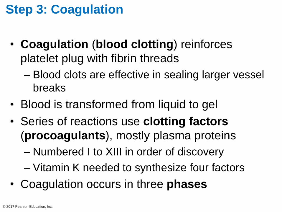
Step 3: Coagulation
• Coagulation (blood clotting) reinforces
platelet plug with fibrin threads
– Blood clots are effective in sealing larger vessel
breaks
• Blood is transformed from liquid to gel
• Series of reactions use clotting factors
(procoagulants), mostly plasma proteins
– Numbered I to XIII in order of discovery
– Vitamin K needed to synthesize four factors
• Coagulation occurs in three phases
© 2017 Pearson Education, Inc.
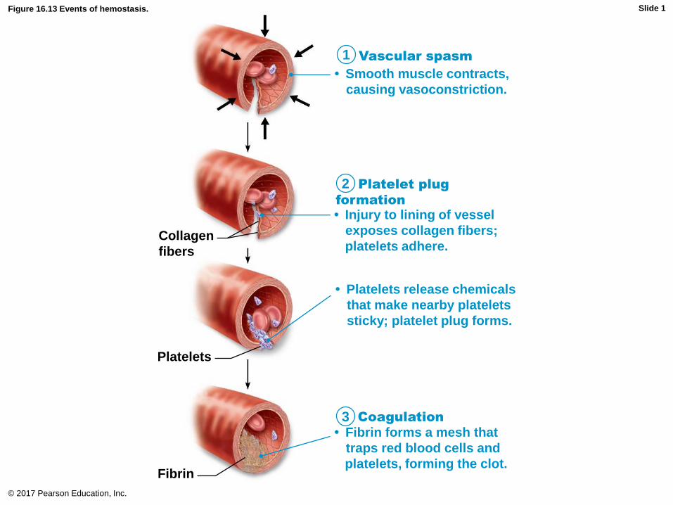
Figure 16.13 Events of hemostasis.
© 2017 Pearson Education, Inc.
1
2
3
Collagen
fibers
Platelets
Fibrin
• Platelets release chemicals
that make nearby platelets
sticky; platelet plug forms.
• Injury to lining of vessel
exposes collagen fibers;
platelets adhere.
• Fibrin forms a mesh that
traps red blood cells and
platelets, forming the clot.
Coagulation
Vascular spasm
Platelet plug
formation
• Smooth muscle contracts,
causing vasoconstriction.
Slide 1
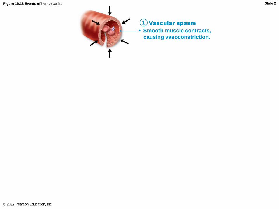
Figure 16.13 Events of hemostasis.
© 2017 Pearson Education, Inc.
1 Vascular spasm
• Smooth muscle contracts,
causing vasoconstriction.
Slide 2

Figure 16.13 Events of hemostasis.
© 2017 Pearson Education, Inc.
1
2
Collagen
fibers
Platelets
• Platelets release chemicals
that make nearby platelets
sticky; platelet plug forms.
• Injury to lining of vessel
exposes collagen fibers;
platelets adhere.
Vascular spasm
Platelet plug
formation
• Smooth muscle contracts,
causing vasoconstriction.
Slide 3

Figure 16.13 Events of hemostasis.
© 2017 Pearson Education, Inc.
1
2
3
Collagen
fibers
Platelets
Fibrin
• Platelets release chemicals
that make nearby platelets
sticky; platelet plug forms.
• Injury to lining of vessel
exposes collagen fibers;
platelets adhere.
• Fibrin forms a mesh that
traps red blood cells and
platelets, forming the clot.
Coagulation
Vascular spasm
Platelet plug
formation
• Smooth muscle contracts,
causing vasoconstriction.
Slide 4

Step 3: Coagulation (cont.)
• Phase 1: Two pathways to prothrombin
activator
– Initiated by either intrinsic or extrinsic pathway
(usually both)
• Triggered by tissue-damaging events
• Involves a series of procoagulants
• Each pathway cascades toward and ends with the
activation of factor X
– Factor X then complexes with Ca2+, PF3
(platelet factor 3), and factor V to form
prothrombin activator
© 2017 Pearson Education, Inc.

Step 3: Coagulation (cont.)
– Intrinsic pathway
• Called “intrinsic” because clotting factors are present
within the blood
• Triggered by negatively charged surfaces such as
activated platelets, collagen, or even glass of a test
tube
– Extrinsic pathway
• Called “extrinsic” because factors needed for clotting
are located outside blood
• Triggered by exposure to tissue factor (TF); also
called factor III
• Bypasses several steps of intrinsic pathway, so faster
pathway© 2017 Pearson Education, Inc.
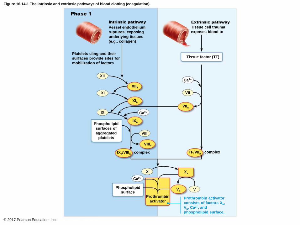
Figure 16.14-1 The intrinsic and extrinsic pathways of blood clotting (coagulation).
© 2017 Pearson Education, Inc.
XaX
Vessel endothelium
ruptures, exposing
underlying tissues
(e.g., collagen)
Tissue cell trauma
exposes blood to
Extrinsic pathwayIntrinsic pathway
Platelets cling and their
surfaces provide sites for
mobilization of factors
Tissue factor (TF)
Phospholipid
surfaces of
aggregated
platelets
complexcomplex
Phospholipid
surfaceProthrombin
activator
Phase 1
XII
XI
IX
XIIa
XIa
IXa
VIII
VIIIa
IXa/VIIIa
VIIa
VII
Ca2+
Ca2+
Ca2+
Va V
TF/VIIa
Prothrombin activator
consists of factors Xa,
Va, Ca2+, and
phospholipid surface.
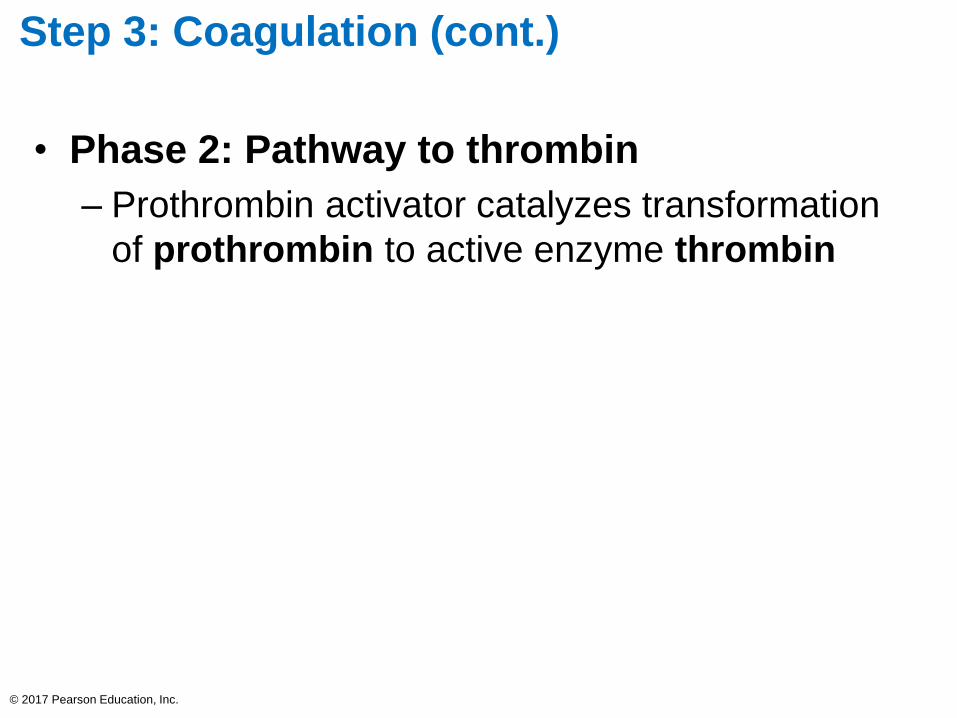
Step 3: Coagulation (cont.)
• Phase 2: Pathway to thrombin
– Prothrombin activator catalyzes transformation
of prothrombin to active enzyme thrombin
© 2017 Pearson Education, Inc.

Figure 16.14-2 The intrinsic and extrinsic pathways of blood clotting (coagulation).
© 2017 Pearson Education, Inc.
Phase 2
Phase 3
Prothrombin (II)
Thrombin (IIa)
Fibrinogen (I)
(soluble)Fibrin
(insolublepolymer)
Cross-linkedfibrin mesh
XIIIa
XIII
Ca2+
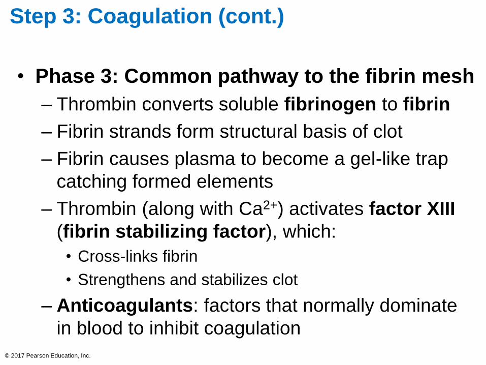
Step 3: Coagulation (cont.)
• Phase 3: Common pathway to the fibrin mesh
– Thrombin converts soluble fibrinogen to fibrin
– Fibrin strands form structural basis of clot
– Fibrin causes plasma to become a gel-like trap
catching formed elements
– Thrombin (along with Ca2+) activates factor XIII
(fibrin stabilizing factor), which:
• Cross-links fibrin
• Strengthens and stabilizes clot
– Anticoagulants: factors that normally dominate
in blood to inhibit coagulation
© 2017 Pearson Education, Inc.
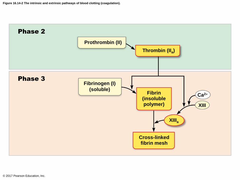
Figure 16.14-2 The intrinsic and extrinsic pathways of blood clotting (coagulation).
© 2017 Pearson Education, Inc.
Phase 2
Phase 3
Prothrombin (II)
Thrombin (IIa)
Fibrinogen (I)
(soluble)Fibrin
(insolublepolymer)
Cross-linkedfibrin mesh
XIIIa
XIII
Ca2+

Figure 16.14 The intrinsic and extrinsic pathways of blood clotting (coagulation).
© 2017 Pearson Education, Inc.
Phase 1
Phase 2
Phase 3
Platelets cling and their
surfaces provide sites for
mobilization of factors
Phospholipid
surfaces of
aggregated
platelets
Phospholipid
surfaceProthrombin
activator
Prothrombin (II)
Thrombin (IIa)
Fibrinogen (I)
(soluble)Fibrin
(insoluble
polymer)
Cross-linked
fibrin mesh
XIIIa
XIII
Ca2+
Prothrombin activator
consists of factors Xa,
Va, Ca2+, and
phospholipid surface.
Ca2+
Ca2+
complex
Vessel endothelium
ruptures, exposing
underlying tissues
(e.g., collagen)
Tissue cell trauma
exposes blood to
Extrinsic pathwayIntrinsic pathway
Ca2+
VII
VIIa
XII
XIIa
XI
IX
XIa
IXa
VIII
VIIIa
IXa/VIIIa complex
Tissue factor (TF)
TF/VIIa
Va V
X Xa
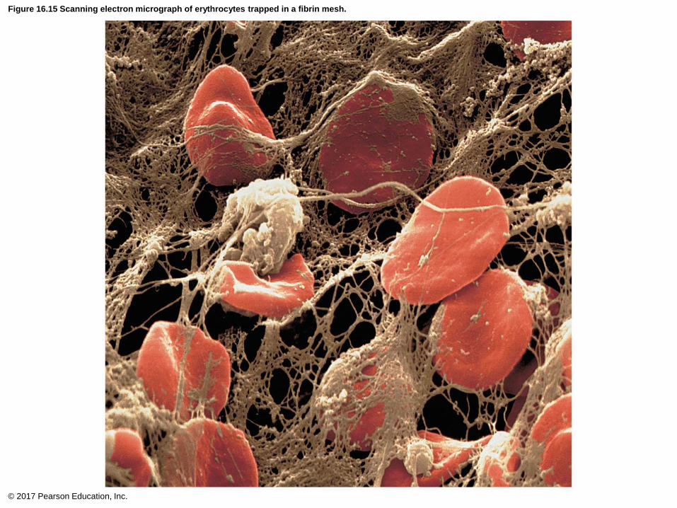
Figure 16.15 Scanning electron micrograph of erythrocytes trapped in a fibrin mesh.
© 2017 Pearson Education, Inc.

Table 16.3 Blood Clotting Factors (Procoagulants)
© 2017 Pearson Education, Inc.
Clot Retraction and Fibrinolysis
• Clot must be stabilized and removed when
damage has been repaired
• Clot retraction
– Actin and myosin in platelets contract within
30–60 minutes
– Contraction pulls on fibrin strands, squeezing
serum from clot
• Serum is plasma minus the clotting proteins
– Draws ruptured blood vessel edges together
© 2017 Pearson Education, Inc.
Clot Retraction and Fibrinolysis (cont.)
• Vessel is healing even as clot retraction occurs
• Platelet-derived growth factor (PDGF) is
released by platelets
– Stimulates division of smooth muscle cells and
fibroblasts to rebuild blood vessel wall
• Vascular endothelial growth factor (VEGF)
stimulates endothelial cells to multiply and
restore endothelial lining
© 2017 Pearson Education, Inc.
Clot Retraction and Fibrinolysis (cont.)
• Fibrinolysis
– Process whereby clots are removed after repair
is completed
– Begins within 2 days and continues for several
days until clot is dissolved
– Plasminogen, plasma protein that is trapped in
clot, is converted to plasmin, a fibrin-digesting
enzyme
• Tissue plasminogen activator (tPA), factor XII, and
thrombin all play a role in conversion process
© 2017 Pearson Education, Inc.

Factors Limiting Clot Growth or Formation
• Factors limiting normal clot growth
– Two mechanisms limit clot size
• Swift removal and dilution of clotting factors
• Inhibition of activated clotting factors
– Limited amount of thrombin is restricted to clot
by fibrin threads, preventing clot from getting
too big or escaping into bloodstream
• Antithrombin III inactivates any unbound thrombin
that escapes into bloodstream
• Heparin in basophil and mast cells inhibits thrombin
by enhancing antithrombin III
© 2017 Pearson Education, Inc.
Factors Limiting Clot Growth or Formation
(cont.)
• Factors preventing undesirable clotting
– Factors preventing platelet adhesion
• Smooth endothelium of blood vessels prevents
platelets from clinging
• Endothelial cells secrete antithrombic substances
such as nitric oxide and prostacyclin
• Vitamin E quinone, formed when vitamin E reacts
with oxygen, is a potent anticoagulant
© 2017 Pearson Education, Inc.
Disorders of Hemostasis
• Two major types of disorders
– Thromboembolic disorders: result in
undesirable clot formation
– Bleeding disorders: abnormalities that prevent
normal clot formation
• Disseminated intravascular coagulation
(DIC)
– Involves both types of disorders
© 2017 Pearson Education, Inc.
Disorders of Hemostasis (cont.)
• Thromboembolic conditions
– Thrombi and emboli
• Thrombus: clot that develops and persists in
unbroken blood vessel
– May block circulation, leading to tissue death
• Embolus: thrombus freely floating in bloodstream
• Embolism: embolus obstructing a vessel
Example: pulmonary or cerebral emboli
• Risk factors: atherosclerosis, inflammation, slowly
flowing blood or blood stasis from immobility
© 2017 Pearson Education, Inc.

Disorders of Hemostasis (cont.)
• Thromboembolic conditions (cont.)
– Anticoagulant drugs: used to prevent
undesirable clotting
• Aspirin: antiprostaglandin that inhibits
thromboxane A2
• Heparin: anticoagulant used clinically for pre-
and postoperative cardiac care
• Warfarin (Coumadin): used for people prone
to atrial fibrillation
– Interferes with action of vitamin K
• Dabigatran: directly inhibits thrombin
© 2017 Pearson Education, Inc.
Disorders of Hemostasis (cont.)
• Bleeding disorders
– Thrombocytopenia: deficient number of
circulating platelets
• Petechiae appear as a result of spontaneous,
widespread hemorrhage
• Due to suppression or destruction of red bone marrow
(examples: malignancy, radiation, or drugs)
• Platelet count <50,000/l is diagnostic
• Treatment: transfusion of concentrated platelets
© 2017 Pearson Education, Inc.
Disorders of Hemostasis (cont.)
• Bleeding disorders (cont.)
– Impaired liver function
• Inability to synthesize procoagulants (clotting factors)
• Causes include vitamin K deficiency, hepatitis, or
cirrhosis
• Liver disease can also prevent liver from producing
bile, which is needed to absorb fat and vitamin K
© 2017 Pearson Education, Inc.

Disorders of Hemostasis (cont.)
• Bleeding disorders (cont.)
– Hemophilia
• Includes several similar hereditary bleeding disorders
– Hemophilia A: most common type (77% of all cases)
due to factor VIII deficiency
– Hemophilia B: factor IX deficiency
– Hemophilia C: factor XI deficiency, milder
• Symptoms include prolonged bleeding, especially into
joint cavities
• Treatment: injections of genetically engineered
factors; has eliminated need for plasma transfusion
and risk of contracting hepatitis or HIV
© 2017 Pearson Education, Inc.
Disorders of Hemostasis (cont.)
• Disseminated intravascular coagulation (DIC)
– Involves both widespread clotting and severe
bleeding
• Widespread clotting occurs in intact blood vessels,
blocking blood flow
• Severe bleeding follows because residual blood is
unable to clot because clotting factors are being
depleted
– Can occur in septicemia, incompatible blood
transfusions, or complications in pregnancy
© 2017 Pearson Education, Inc.

16.7 Blood Transfusions
• Cardiovascular system minimizes effects of
blood loss by:
1. reducing volume of affected blood vessels
2. stepping up production of RBCs
• Body can compensate for only so much blood
loss
• Loss of 15–30% causes pallor and weakness
• Loss of more than 30% results in potentially
fatal severe shock
© 2017 Pearson Education, Inc.
Transfusing Red Blood Cells
• Whole-blood transfusions are used only when
blood loss is rapid and substantial
• Infusions of packed red blood cells, or PRBCs
(plasma and WBCs removed), are preferred to
restore oxygen-carrying capacity
• Blood banks usually separate donated blood into
components; shelf life of blood is about 35 days
• Human blood groups of donated blood must be
determined because transfusion reactions can be
fatal
– Blood typing determines groups
© 2017 Pearson Education, Inc.
Transfusing Red Blood Cells (cont.)
• Human blood groups
– RBC membranes bear different many antigens
• Antigen: anything perceived as foreign that can
generate an immune response
• RBC antigens are referred to as agglutinogens
because they promote agglutination
– Mismatched transfused blood is perceived as
foreign and may be agglutinated and destroyed
• Potentially fatal reaction
© 2017 Pearson Education, Inc.

Transfusing Red Blood Cells (cont.)
• Human blood groups (cont.)– Humans have at least 30 naturally occurring
RBC antigens
– Presence or absence of each antigen is used to
classify blood cells into different groups
– Some blood groups (MNS, Duffy, Kell, and
Lewis) are only weak agglutinogens• Not usually typed unless patient will need several
transfusions
– Antigens of ABO and Rh blood groups cause
most vigorous transfusion reactions; therefore,
they are major groups typed
© 2017 Pearson Education, Inc.

Transfusing Red Blood Cells (cont.)
– ABO blood groups
• Based on presence or absence of two agglutinogens
(A and B) on surface of RBCs
– Type A has only A agglutinogen
– Type B has only B agglutinogen
– Type AB has both A and B agglutinogens
– Type O has neither A nor B agglutinogens
• Blood may contain preformed anti-A or anti-B
antibodies (agglutinins)
– Act against transfused RBCs with ABO antigens not
present on recipient's RBCs
• Anti-A or anti-B form in blood at about 2 months of
age, reaching adult levels by 8–10 years of age
© 2017 Pearson Education, Inc.

Table 16.4 ABO Blood Groups
© 2017 Pearson Education, Inc.
Transfusing Red Blood Cells (cont.)
– Rh blood groups
• 52 named Rh agglutinogens (Rh factors)
• C, D, and E are most common
• Rh+ indicates presence of D antigen
– 85% Americans are Rh+
• Anti-Rh antibodies are not spontaneously formed in
Rh– individuals
– Anti-Rh antibodies form if Rh– individual receives Rh+
blood, or Rh– mom is carrying Rh+ fetus
• Second exposure to Rh+ blood will result in typical
transfusion reaction
© 2017 Pearson Education, Inc.
Clinical – Homeostatic Imbalance 16.3
• Hemolytic disease of newborn, also called
erythroblastosis fetalis only occurs in Rh– mom
with Rh+ fetus
• First pregnancy: Rh– mom exposed to Rh+ blood of
fetus during delivery; first baby born healthy, but
mother synthesizes anti-Rh antibodies
• Second pregnancy: Mom’s anti-Rh antibodies cross
placenta and destroy RBCs of Rh+ baby
• Baby treated with prebirth transfusions and
exchange transfusions after birth
• RhoGAM serum containing anti-Rh can prevent
Rh– mother from becoming sensitized© 2017 Pearson Education, Inc.

Transfusing Red Blood Cells (cont.)
• Transfusion reactions
– Occur if mismatched blood is infused
– Donor’s cells are attacked by recipient’s plasma
agglutinins
• Agglutinate and clog small vessels
• Rupture and release hemoglobin into bloodstream
– Result in:
• Diminished oxygen-carrying capacity
• Decreased blood flow beyond blocked vessel
• Hemoglobin in kidney tubules can lead to renal failure
© 2017 Pearson Education, Inc.
Transfusing Red Blood Cells (cont.)
• Transfusion reactions (cont.)
– Symptoms: fever, chills, low blood pressure,
rapid heartbeat, nausea, vomiting
– Treatment: preventing kidney damage with fluids
and diuretics to wash out hemoglobin
– Type O universal donor: no A or B antigens
– Type AB universal recipient: no anti-A or anti-B
antibodies
• Misleading as other agglutinogens that cause
transfusion reactions must also be considered
– Autologous transfusions: patient predonates
own blood that is stored and available if needed© 2017 Pearson Education, Inc.
Transfusing Red Blood Cells (cont.)
• Blood typing
– Donor blood is mixed with antibodies against
common agglutinogens
• If agglutinogen is present, clumping of RBCs will occur
– Blood is typed for ABO and for Rh factor in same
manner
– Cross matching: typing between specific donor
and specific recipient
• Mix recipient’s serum with donor RBCs
• Mix recipient’s RBCs with donor serum
© 2017 Pearson Education, Inc.

Figure 16.16 Blood typing of ABO blood types.
© 2017 Pearson Education, Inc.
Blood beingtested
Serum
Anti-A Anti-B
RBCs
Type AB (contains
agglutinogens A and B;agglutinates with bothsera)
Type A (containsagglutinogen A;agglutinates with anti-A)
Type B (containsagglutinogen B;agglutinates with anti-B)
Type O (contains noagglutinogens; does notagglutinate with eitherserum)
Restoring Blood Volume
• Death from shock may result from low blood
volume
• Volume must be replaced immediately with
– Normal saline or multiple-electrolyte solution
(Ringer’s solution) that mimics plasma electrolyte
composition
• Replacement of volume restores adequate
circulation but does not replace oxygen-carrying
capacities of RBCs
© 2017 Pearson Education, Inc.
16.8 Diagnostic Blood Tests
• Examination of blood can yield information on
persons health:
– Low hematocrit seen in cases of anemia
– Blood glucose tests check for diabetes
– Leukocytosis can signal infection
• Microscopic examination of blood can reveal
any variations in size or shape of RBCs
– Abnormal size, shape, or color could indicate
anemia
© 2017 Pearson Education, Inc.
16.8 Diagnostic Blood Tests
• Differential WBC count looks at relative
proportions of each WBC
– Increases in specific WBC can help with
diagnosis
• Prothrombin time and platelet counts assess
hemostasis
• CMP (comprehensive medical panel): blood
chemistry profile that checks various blood
chemical levels
– Abnormal results could indicate liver or kidney
disorders© 2017 Pearson Education, Inc.
16.8 Diagnostic Blood Tests
• Complete blood count (CBC) checks formed
elements, hematocrit, hemoglobin
© 2017 Pearson Education, Inc.