Embed Size (px)
Citation preview

Tansley review
Biotic interactions as drivers of algal origin and evolution
Juliet Brodie1*, Steven G. Ball
2, François-Yves Bouget
3, Cheong Xin Chan
4, Olivier De Clerck
5,
Mark Cock6, Claire Gachon
7, Arthur R. Grossman
8, Thomas Mock
9, John Raven
10, Mahasweta
Saha11, Alison G. Smith
12, Assaf Vardi
13, Hwan Su Yoon
14, Debashish Bhattacharya
15*
1 Natural History Museum, Department of Life Sciences, London SW7 5BD, United Kingdom
2 Université de Lille CNRS, UMR 8576 - UGSF- Unité de Glycobiologie Structurale et
Fonctionnelle, F 59000 Lille, France
3 University Pierre et Marie Curie, University of Paris VI, CNRS, Laboratoire d'Océanographie
Microbienne, Observatoire Océanologique, F-66650, Banyuls-sur-Mer, France
4 Institute for Molecular Bioscience and School of Chemistry and Molecular Biosciences, The
University of Queensland, Brisbane QLD 4072, Australia
5 Phycology Research Group, Ghent University, Krijgslaan 281, S8, 9000 Gent, Belgium
6 CNRS, Sorbonne Université, UPMC University Paris 06, Algal Genetics Group, UMR 8227,
Integrative Biology of Marine Models, Station Biologique de Roscoff, CS 90074, F-29688,
Roscoff, France, 2Bezhin Rosko, 29250, Santec, France
8 Department of Plant Biology, The Carnegie Institution for Science, Stanford, CA 94305, USA
9 School of Environmental Sciences, University of East Anglia, Norwich NR47TJ, United
Kingdom
10 Division of Plant Sciences, University of Dundee at the James Hutton Institute, Dundee DD2
5DA, United Kingdom
11 Helmholtz Center for Ocean Research, Kiel, 24105 Kiel, Germany
12 Department of Plant Sciences, University of Cambridge, Cambridge CB2 3EA, United
Kingdom
13 Department of Plant and Environmental Sciences, Weizmann Institute of Science, Rehovot,
76100, Israel
14 Department of Biological Sciences, Sungkyunkwan University, Suwon 440-746, Korea
Page 1 of 36 New Phytologist
This is the peer reviewed version of the following article: 'Biotic interactions as drivers of algal origin and evolution', New Phytologist, which has been published in final form at http://dx.doi.org/10.1111/nph.14760. This article may be used for non-commercial purposes in accordance with Wiley Terms and Conditions for Self-Archiving.

2
15 Department of Biochemistry and Microbiology, Rutgers University, New Brunswick, NJ
08901, USA
* These authors contributed equally to this work.
Author for correspondence: Debashish Bhattacharya
Tel: +1 848 932 6218
Email: [email protected]
Twitter handle: DebashB
Page 2 of 36New Phytologist

3
Summary
Biotic interactions underlie life’s diversity and are the lynchpin to understand its complexity and
resilience within an ecological niche. Algal biologists have embraced this paradigm, and studies
building on the explosive growth in omics and cell biology methods have facilitated in-depth
analysis of non-model organisms and communities from a variety of ecosystems. In turn, these
advances have enabled a major revision of our understanding of the origin and evolution of
photosynthesis in eukaryotes, bacterial-algal interactions, control of massive algal blooms in the
ocean, and the maintenance and degradation of coral reefs. Here we review some of the most
exciting developments in the field of algal biotic interactions and identify challenges for the next
generation of scientists. We foresee the development of an algal knowledgebase that integrates
ecosystem-wide omics data and the development of molecular tools/resources to perform
functional analyses of individuals in isolation and in populations. These assets will allow us to
move beyond mechanistic studies of a single species towards understanding the interactions
amongst algae and other organisms both in the laboratory and in the field.
Key words: algae, algal blooms, endosymbiosis, organellogenesis, genomics, holobiont,
symbiome, trophic interactions.
Page 3 of 36 New Phytologist

4
I. Introduction
Algae are key primary producers in aquatic environments and represent several emerging genetic
model systems (Armbrust et al., 2004; Hopes et al., 2016; Nymark et al., 2016). They also play
an increasingly important role in human nutrition (FAO, 2014). Algal photosynthesis provides
about one-half of the oxygen that we breathe, and their genomes reveal the story of a tangled past
that traverses the tree of life through the processes of endosymbiosis and horizontal gene transfer
(HGT) (Price et al., 2012; Cenci et al., 2017). Biotic interactions between algae and other
eukaryotes (e.g., (Worden et al., 2015) are extremely widespread in aquatic and terrestrial
ecosystems. The degree to which nature has experimented with these relationships is wide-
ranging, including interactions among organisms that maintain a few functional associations, to
those that have evolved a highly integrated suite of functions. In addition to the intracellular
interactions described below, algae also engage in extracellular/surface interactions in the
phycosphere, which is the ecologically and physiologically integrated neighborhood inhabited by
the alga (Bell & Mitchell, 1972). Epibiosis (surface colonization of one organism [the basibiont]
by other attached organisms [epibionts]) will not be covered in great detail here, but occurs on all
immersed surfaces in the aquatic environment, including those of micro and macroalgae, and is
of paramount importance in the marine environment (Wahl et al., 2012). Epibiotic interactions
(e.g., alga-alga, alga-bacterium, alga-virus [see below]) play key roles in nutrient acquisition and
recycling, metabolic flux, energy flow and developmental processes. In parallel with herbivory,
epibiosis represents one of the most important interactions that can determine the fate of an alga
and has been shown to shape entire marine communities (Korpinen et al., 2007).
In this review, we focus on research that has contributed some of the most exciting
insights concerning the ways in which biotic interactions shape algal evolution and physiology.
This perspective recognizes that “symbiomes” or “holobionts” are important targets of study to
elucidate the overall capacity of genomes to interact with the environment. Here symbiome
refers to co-localized and co-evolving (i.e., under selection) taxa comprising a given consortium,
whereas holobiont includes all physically associated taxa regardless of the nature of the biotic
interaction (Boucias et al., 2013; Bordenstein & Theis, 2015; Douglas & Werren, 2016; Tripp et
al., 2017). This revolution in understanding integrative ecosystem function has largely been
driven by the occurrence of technological advances in fields such as genomics, proteomics, and
cell biology. It is clear, however, that we are on the cusp of far greater advances, as the concept
Page 4 of 36New Phytologist
5
of the symbiome informs our experimental approaches. Below, we discuss prominent examples
of algal biotic interactions that have been selected to illustrate the importance of these
interactions in a broad range of contexts ranging from deep evolutionary time to processes of key
relevance in the current context of global climate change. The review will begin with a
discussion of the origin of photosynthetic organelles based on endosymbiosis, and will then look
at algal interactions in the coral symbioses and the threat that climate change imposes on this
association. Lastly we will examine the role of bacteria in algal biology, and the arms race
associated with alga-virus interactions.
II. Endosymbiosis
1. Complex biotic interactions explain plastid origin
1.1. Primary endosymbiosis in Archaeplastida
Algae originated as a consequence of primary plastid endosymbiosis, a process in which a
mitochondrion-containing, single-celled eukaryote engulfed and retained a cyanobacterium that
eventually became the photosynthetic organelle or plastid (Cavalier-Smith, 1982; Bhattacharya
et al., 2004). The product of this ca. 1.6 billion year old endosymbiotic event (Yoon et al., 2004)
eventually split into the three primary plastid lineages, the red algae, the glaucophyte algae, and
the green algae plus plants (together, the supergroup Archaeplastida) (Adl et al., 2012; Price et
al., 2012). Algae from these groups were themselves frequently engulfed by other protists,
giving rise to a rainbow of serially derived plastids distributed throughout the tree of life
(Palmer, 2003; Gould et al., 2008) (Fig. 1a). The process of primary plastid capture has
sometimes been depicted as a ‘hungry’ single-celled eukaryote engulfing a prokaryote followed
by the subsequent evolution of a functional organelle. This portrayal begs the obvious question:
if the process is so simple, then why has the event been so rare given that oceans and lakes are
replete with phagotrophic protists that have been feeding on prokaryote prey for hundreds of
millions of years? In fact, there are only two bona fide primary endosymbioses known that gave
rise to widespread organelles over the long history of eukaryotes; the event from which all
plastids originated, as explained above, and a prior event that led to the evolution of
mitochondria. Other more taxonomically limited cases of organelle origin are associated with the
photosynthetic amoeba lineage Paulinella (see below), the non-photosynthetic organelle of the
trypanosomatids (Kostygov et al., 2016; Morales et al., 2016), and nitrogen fixing spheroid
Page 5 of 36 New Phytologist

6
bodies in the rhopalodiacean diatoms (Nakayama et al., 2014; Zehr et al., 2016). The rarity of
primary endosymbiosis has fascinated scientists for many years and is usually attributed to the
extensive innovations required for organelle establishment. These include: a) events that lead to
the protection of the nascent endosymbiont from host digestion; b) tailoring of processes critical
for the exchange of metabolites between the endosymbiont and host cell (Facchinelli & Weber,
2011); c) the origin of an import system to move cytosolic proteins into the nascent organelle
(Schleiff & Becker, 2011); d) foreign gene acquisition through HGT and the integration of the
HGT-derived protein products into both host and newly developing organelle pathways
(Cavalier-Smith, 2002; Karkar et al., 2015); and e) movement of genes from the organelle to the
host nucleus to escape Muller’s ratchet, i.e., accumulation of mutations in non-recombining
genomes (Felsenstein, 1974). Processes that would exacerbate the impact of Muller’s ratchet and
make relocation of genes from the organelle to the nuclear genome more imperative are the
mutagenic effect of damaging reactive oxygen species (ROS) produced as a consequence of
photosynthesis in the organelle (van Creveld et al., 2015), and as yet unexplained processes
associated with greater damage of DNA in organelles than in their aerobic bacterial ancestors
(Raven, 2015). Explanations for why organelle genomes are retained include coordinated
synthesis of complexes assembled in the organelle, and the regulation of transcriptional and post-
transcriptional processes by the organelle redox state (van Creveld et al., 2015).
The most critical innovation listed above, is the first, namely how a captured bacterial
cell evades digestion by the host during the initial stages of plastid evolution. A potential answer
to this question comes from recent work exploring the evolution of mitochondria. Current
mitochondrial gene phylogenies indicate that this organelle originated from anciently diverged
environmental Rickettsiales-like pathogens with relatively large gene inventories (Wang & Wu,
2015; Ball et al., 2016c) whose descendants are now often found in association with protists
(Martijn et al., 2015). However, these taxa are distinct from the highly specialized animal
parasites with streamlined genomes, such as the typhus agent Rickettsia prowazekii
(Zomorodipour & Andersson, 1999), that were initially proposed as the alpha-proteobacterial
candidates based on limited data that was collected over ten years ago (Emelyanov, 2003). The
host of this mitochondrial endosymbiosis was likely to be a member of the recently discovered
archaeal ‘Asgard’ superphylum (including the Lokiarchaeota and Heimdallarchaeota), which is
the most closely related prokaryote to the eukaryote nuclear lineage (Spang et al., 2015;
Page 6 of 36New Phytologist

7
Zaremba-Niedzwiedzka et al., 2017). Therefore, the increasingly widely accepted view is that an
Asgard-like cell was infected by a relatively gene-rich Rickettsiales-like pathogen, thus laying
the foundation for mitochondrial endosymbiosis and eukaryogenesis. By virtue of their existing
ability to thrive in the intracellular environment, the ancestors of mitochondria were pre-adapted
to switch from pathogenesis to endosymbiosis. These cells had evolved efficient solutions to deal
with host innate immunity due to millions of years of coevolution with the Asgard lineage. These
findings suggest that, to become a successful proto-endosymbiont, the invading cell needs to
evade host defenses, which is more likely to be achieved by an intracellular pathogen adapted to
the cytosolic lifestyle (Ball et al., 2016b; Ball et al., 2016c; Cenci et al., 2017).
Application of this concept to the origin of plastids requires some modification because
extant cyanobacteria are not intracellular pathogens and lack the inherent capacity to evade host
defenses. We suggest two possible explanations for cyanobacterial survival. First, the
Archaeplastida host of this endosymbiosis may have developed mutations that reduced the
efficacy of its lytic/phagocytic functions. This provided the cyanobacterium sufficient residence
time within a host food vacuole to evolve a character(s) beneficial to the host (e.g., secretion of
fixed carbon or reduced nitrogen compounds), which allowed the establishment and spread of a
founder population. This scenario is more likely to have occurred in oligotrophic waters, which
lacked abundant prey. An alternative explanation is that the cyanobacterium was protected by a
third ‘player’ that could withstand host defenses. This latter idea receives support from the
finding that there are several dozen genes of chlamydial origin present in the nuclear genome of
algae and plants (Huang & Gogarten, 2007; Becker et al., 2008). Phylogenetic data suggest that
these genes are from environmental strains with relatively large genomes, such as those that
infect Acanthamoeba, and not the highly reduced human pathogens. In addition, many of the
products of these nucleus-encoded genes are plastid targeted and perform specialized functions
not associated with cyanobacteria (Huang & Gogarten, 2007; Moustafa et al., 2008). These
observations have led to the “ménage à trois” hypothesis (MATH) to explain the origin of
plastids. In this scenario a Chlamydiales ancestor evolved from a pathogenic to symbiotic
lifestyle, protecting the cyanobacterium in its inclusion vesicle (Ball et al., 2013; Cenci et al.,
2017). Although the MATH remains controversial due largely to issues associated with ‘deep
time’ gene phylogenies and the unresolved role of HGT in eukaryote evolution (Dagan et al.,
2013; Ball et al., 2016a), its complexity reflects well-established biotic interactions. As
Page 7 of 36 New Phytologist

8
illustrated in Fig. 1b, it predicts that an elementary body (chlamydial infectious particle) escapes
host defenses by remodeling the phagocytic membrane and by secreting chlamydial effector
proteins that enable bacterial specific metabolites of photosynthesis such as ADP-glucose to
enter the host cytosolic glycogen stores. Both glaucophytes and red algae store carbohydrates in
their cytosol suggesting that the glycogen/starch pool may have provided an opportunity to
buffer the unsynchronized demand and supply of carbon of the cyanobiont and its host. Several
observations support this idea: a) enzymes involved in manipulating host carbohydrate
metabolism are pathogen effectors secreted by the type-III secretion system (Gehre et al., 2016);
b) pathogenic Chlamydiae synthesize extracellular storage carbohydrates within parasitophorous
vacuoles using analogous nucleotide-sugars and nucleotide-sugar transporters (Gehre et al.,
2016); c) nucleotide-sugar transporters of host origin are evolutionary ancestors of plastid carbon
exporters in red and green algae, as well as in plastids of secondary or tertiary endosymbiotic
origin (Moog et al., 2015); and d) analysis of the tryptophan biosynthesis pathway in
Archaeplastida shows that one-half (4/8) of the genes encoding proteins in this pathway are
putatively of chlamydial origin, as are the E. coli tyr/mtr (tyrosine/tryptophan) transporter genes
(Cenci et al., 2016; Cenci et al., 2017).
Tryptophan starvation may have been a mechanism used by the host of the primary
plastid to combat chlamydial infection (Bonner et al., 2014). Tryptophan biosynthesis is by far
the most costly amino acid for cells to synthesize. In comparison to the eukaryotic host and the
cyanobiont, sensitivity of the chlamydial symbiont to tryptophan starvation would have been
exacerbated by the energy requirements for its synthesis (Bonner et al., 2014). This biotic
interaction would therefore have selected for movement of the chlamydial trp operon to the
cyanobacterial endosymbiont genome to ensure high levels of gene expression. Cenci et al.
(2016) posit that the chlamydial trp operon transfer occurred via conjugation during co-
localization of chlamydial and cyanobacterial cells in inclusion vesicles. At a later time, some trp
genes were moved to the Archaeplastida nuclear genome by endosymbiotic gene transfer (EGT)
(Martin & Herrmann, 1998) from the cyanobacterial plastid forerunner. The MATH is reinforced
not only by functional considerations, but also gene numbers. Chlamydiae HGTs are not
scattered randomly among the organisms of the tree of life, but rather, an outsize contribution
(ca. 30-50 genes, depending on the lineage being studied) is found when compared to other non-
cyanobacterial prokaryotic gene acquisitions in Archaeplastida nuclear genomes (Huang &
Page 8 of 36New Phytologist

9
Gogarten, 2007; Deschamps, 2014). In addition, analysis of the plastid proteome shows that
despite having >50-fold more proteobacterial than chlamydial sequences in current genome
databases (e.g., National Center for Biotechnology Information), Proteobacteria and Chlamydiae
genes represent the largest contribution to plastid functions (46 and 24 genes, respectively, in
Arabidopsis thaliana), with only 13 from alpha-Proteobacteria (Qiu et al., 2013). The MATH
provides a testable model that can be used to study the steps that led to plastid origin. Beyond its
specific predictions (Ball et al., 2013; Cenci et al., 2017), this theory highlights the complexity
of biotic interactions that underlie endosymbiosis. In the future, the aim should be to develop
systems in the laboratory to study the processes underlying endosymbiosis so that we can move
beyond trees and diagrams, to allow experimental elucidation of mechanisms underlying
organellogenesis.
1.2. Origin of the Paulinella chromatophore
The concept of multiple microbes contributing to plastid evolution in the Archaeplastida, may
also explain the maintenance and evolution of the plastid (termed the chromatophore) in
Paulinella chromatophora (Marin et al., 2005; Nowack et al., 2008; Yoon et al., 2009). In this
case, there is currently no evidence for chlamydial-facilitated organelle origin. However, over
200 bacterium-derived HGTs have been found in the nuclear genome of this species that
complement gene losses from the chromatophore genome. Specifically, many missing
components of critical endosymbiont pathways, such as for amino acid and peptidoglycan
biosynthesis and DNA replication, have been compensated for by the acquisition of a variety of
prokaryotic donor genes via HGT (Nowack et al., 2016). Access to these foreign genes was
likely facilitated by phagotrophic uptake of bacteria by the host amoeba, followed by HGT of
DNA to the amoeba nuclear genome. Once activated, nucleus-encoded gene products were
relocated to the chromatophore, possibly by trafficking through the secretory system, where they
could replace components of the pathways encoded on the chromatophore genome (Nowack &
Grossman, 2012). It should be stressed that a response to Muller’s ratchet acting on the
chromatophore genome (leading to genome reduction) in P. chromatophora is certainly
expected, but surprisingly, it is not primarily EGT and rerouting of host proteins in this relatively
‘young’ endosymbiosis (i.e., 90-140 million years old; Delaye et al., 2016) that facilitates this
process, but rather, repurposing of environmental DNA as a result of biotic interactions.
Page 9 of 36 New Phytologist

10
2. Complex biotic interactions explain the symbiosis between algae and corals
2.1. Maintenance of the symbiosis
Corals are the structural and trophic foundation of coral reefs, which support about 30% of all
described marine species (Wilkinson, 2004). Critically, reef-building corals are a symbiosis
between the coral animal per se and photosynthetic dinoflagellates in the genus Symbiodinium
(Figs. 2a, 2b3). Symbiodinium are also key algal symbionts in a wide range of coral reef animals,
including sea anemone, sponges, jellyfish, and clams. The coral-Symbiodinium association is one
of relaxed specificity: individual corals can harbor alternative and multiple symbiont types
simultaneously, and a Symbiodinium type may associate with a range of coral hosts (Silverstein
et al., 2012). Upon acquisition of Symbiodinium by host gastrodermal cells (within which the
algal cells reside), the Symbiodinium are physically separated from the cytoplasm by a host-
derived vacuole known as the symbiosome (Roth et al., 1988). Exposure to competent
Symbiodinium cells triggers an initial stress response in the coral Acropora digitifera, resulting in
transient suppression of protein synthesis and mitochondrial metabolism (Mohamed et al., 2016).
This finding supports the hypothesis that the symbiosome is a phagosome that has undergone
early arrest (Shinzato et al., 2011; Mohamed et al., 2016).
Coral reefs thrive in nutrient-poor waters. In return for shelter (e.g., from ultraviolet
radiation, predation), Symbiodinium photosynthesis may provide >90% of the fixed carbon
requirement (Muscatine & Porter, 1977) of the hosts. A critical limitation of photosynthesis is
access to dissolved inorganic carbon. Since they have no direct access to ambient seawater,
Symbiodinium cells depend on the host for delivery of inorganic carbon (Ci; CO2 or HCO3-) (see
Fig. 2c). When net photosynthesis takes place some Ci is generated via respiration, but in corals
the predominant Ci supply to photosynthesis is its accumulation within host tissue from external
sources (Shinzato et al., 2011). The concentration of Ci in the host tissue can be ~70-fold that of
seawater, which represents a steeper gradient than is observed for most organisms that use a
carbon concentrating mechanism (CCM) (Shinzato et al., 2011). Ci accumulation by the host
could also be related to the existence of an acidified space between the algal cell and the
symbiosome membrane, and the presence of an uncharacterized HCO3- transporter in the
symbiosome membrane and carbonic anhydrase activity in the acidified space. The host also
Page 10 of 36New Phytologist

11
appears to have an active role in regulating photosynthesis in the symbionts (Barott et al., 2015;
Bhattacharya et al., 2016).
The algae of the holobionts also accumulate Ci (Walker et al., 1980; Barott et al., 2015).
This is likely related to the fact that dinoflagellates such as Symbiodinium have Form II ribulose-
1,5-bisphosphate carboxylase-oxygenase (RuBisCO), which shows low CO2:O2 selectivity and,
probably, a low affinity for CO2 (Leggat et al., 2000). Other symbioses have different roles for
the animal in Ci supply to Symbiodinium; e.g., in tridacnid giant clams, where the symbionts are
extracellular, the haemolymph is the immediate source of Ci. The Ci concentration in the
haemolymph in the light is lower than that in seawater (Muscatine & Porter, 1977), thus there is
no evidence for accumulation of Ci to higher concentrations than in seawater during influx
through the gill epithelium. In this case, the accumulation of Ci by the photobiont presumably
plays an even more vital role in algal primary production.
The symbiotic relationship between Symbiodinium and its coral hosts determines not only
the rate of coral-reef growth (calcium carbonate deposition), but also how corals respond to
environmental stress (Voolstra et al., 2015). A modest episodic period of increased temperature
of the ocean surface (e.g., a few days at 1-2°C above the mean summer minimum) can set off a
cascade of photoinhibition, the decoupling of carbon flow between the symbiont and host
(breakdown of symbiosis), oxidative damage, and physical loss of symbiont cells (Wooldridge,
2013). This process, known as coral bleaching, leaves the coral host at risk for starvation,
disease, and death unless the symbiosis is soon re-established (Hoegh-Guldberg, 1999). In this
way, algae are essential for survival and maintenance of coral reef ecosystems. The impact of
current environmental change on the health of the symbiotic association is particularly alarming,
especially in recent years. For example, >90% of the 911 reefs surveyed in 2015-2016 at the
Great Barrier Reef (the world’s largest continuous reef system) showed signs of severe bleaching
(Albright et al., 2016).
2.2. Omics perspective on the coral symbiosis
The cellular and molecular processes of symbiosis that are actively being explored include
recognition, capture of the symbiont in the symbiosome, proliferation of symbionts in host tissue,
loss of symbionts from the host tissue, and metabolic exchange and nutrient trafficking between
Symbiodinium and the host across multiple membranes. There is much to be learned about these
Page 11 of 36 New Phytologist

12
topics from a broader genomic and molecular evolutionary perspective. Symbiodinium is
classified into nine clades based on phylogenetic markers, although they represent a highly
divergent group of dinoflagellate species (LaJeunesse et al., 2005; Wham & LaJeunesse, 2016)
and may include >100 species capable of forming symbiotic associations with corals. Hurdles in
studying genomic and molecular aspects of the coral system include difficulties associated with
the establishment of axenic cultures of the various Symbiodinium types, their slow growth, and
the size and complexity of their genomes. The dinoflagellate nuclear genome can be massive, up
to 250 Gbp in size (LaJeunesse et al., 2005; Lin, 2011), and exhibits unusual features including
non-canonical nucleotides, atypical intron-exon splice signals (Lin, 2011), and RNAs that are
trans-spliced (Zhang et al., 2007). RNA editing of transcripts has been described in
mitochondrial and plastid genomes (Lin, 2011; Jackson & Waller, 2013; Mungpakdee et al.,
2014), whilst the plastid genome is comprised of distinct DNA minicircles, each containing a
gene, a few genes, or in some cases, no genes (Zhang et al., 1999; Howe et al., 2008). The
nuclear genomic features are set against a backdrop of gene or genome-fragment duplications,
and abundant non-coding repetitive elements (McEwan et al., 2008; Shoguchi et al., 2013).
Three genome sequences of Symbiodinium from distinct clades were recently published
(Shoguchi et al., 2013; Lin et al., 2015; Aranda et al., 2016), and their estimated genome sizes of
1.1-1.5 Gbp are smaller than the earlier estimates of 3-5 Gbp (LaJeunesse et al., 2005). In
addition, these genomes share little sequence similarity; i.e., <1% of total sequenced reads from
S. kawagutii mapped onto the genome assembly of S. minutum, and vice versa (Lin et al., 2015).
These results indicate a high level of genome divergence among distinct Symbiodinium clades.
Further complexity in corals comes from a three-way functional complementarity
between the coral host, the dinoflagellate, and the associated microbiome of bacteria and viruses
(Ziegler et al., 2017). For example, the incomplete cysteine biosynthesis pathway in the coral
Acropora digitifera (Shinzato et al., 2011; Shinzato et al., 2014) is compensated for by
Symbiodinium (Shoguchi et al., 2013; Lin et al., 2015), whereas bacteria likely play a key role in
regulating the availability of nitrogen to the coral host and algae and in resistance to thermal
stress (Radecker et al., 2015; Ziegler et al., 2017). Nevertheless, given the diversity of
Symbiodinium species, a ‘one-reference-genome-fits-all’ assumption will not be possible for
studying the coral-dinoflagellate symbiosis and interactions, and additional genome data from
the different species types/clades will be necessary. An effective approach would be to integrate
Page 12 of 36New Phytologist

13
multi-omics data from the coral and the associated Symbiodinium and microbiome; i.e. the
holobiont (Bordenstein & Theis, 2015), to tease apart the individual contributions of each
component in sustaining a healthy holobiont. Availability of additional data from free-living
dinoflagellates will help address key questions including the evolutionary events and functional
innovations that lead to the transition from a free-living to a symbiotic lifestyle. At the same
time, tractable lab model systems are being developed (Shapiro et al., 2016) that will enable the
study of cellular mechanisms that underlie the response to elevated temperature and pathogens.
Findings from such studies will inform strategies for conservation of and risk mitigation for reef
ecosystems.
III. Biotic interactions within the phycosphere
3.1 Alga-bacterium biotic interactions
Interactions between algae and bacteria are likely to be universal in the environment. Many
notable examples are species specific, such as the green seaweed Ulva mutabilis, which relies on
different bacterial strains for successful morphogenesis (Spoerner et al., 2012). In the laboratory,
rather than forming the typical blade- or tube-like morphology, axenic gametes of U. mutabilis
develop into callus-like aggregates of undifferentiated cells with abnormal cell walls. These
findings suggest the existence of chemical signaling between bacteria and the alga, and
potentially, complementarity of metabolic pathways. Similar interactions have also been found
between the bacterium Sulfitobacter pseudonitzschiae and the diatom Pseudo-nitzschia
multiseries (Amin et al., 2015), and bacteria have been shown to facilitate acclimation of the
brown seaweed Ectocarpus siliculosis to a freshwater environment (Dittami et al., 2016).
Another striking example of this phenomenon is the ‘Jekyll-and-Hyde’ (named by the authors)
relationship between the roseobacter Phaeobacter gallaeciensis, a biofilm forming symbiont of
the bloom-forming haptophyte alga Emiliania huxleyi (Seyedsayamdost et al., 2011). Under
normal growth conditions P. gallaeciensis secretes antibiotics and growth phytohormones (e.g.
the auxin indole-3-acetic acid) that appear to benefit the alga. However, as the algal population
ages, the bacteria shift their small molecule biosynthesis pathways to the production of
algaecides, and act as an E. huxleyi pathogen (Seyedsayamdost et al., 2011; Segev et al., 2016).
A different type of biotic interaction involves capture and ‘farming’ of the cryptophyte alga
Page 13 of 36 New Phytologist

14
Teleaulax amphioxeia by its host ciliate, Mesodinium rubrum, to extract nutrients from the
captured, intact alga (Qiu et al., 2016).
More general interactions are seen with bacteria that play a key role in providing
micronutrients to algae. Examples are essential organic compounds such as thiamine (vitamin
B1) and cobalamin (vitamin B12). These compounds are required as enzyme cofactors, but many
phytoplankton species are unable to synthesize them. Only prokaryotes (and only then, a subset
of both Eubacteria and Archaea) can synthesize cobalamin de novo (Warren et al., 2002), and
levels free in the aquatic environment are generally too low to support algal growth (Sañudo-
Wilhelmy et al., 2012). Direct provision of the vitamin from bacteria to algae has been
demonstrated in the laboratory (Croft et al., 2005; Wagner-Döbler et al., 2010; Kazamia &
Smith, 2014; Durham et al., 2015) and evidence that similar exchanges occur in the natural
environment comes from correlations observed between the presence of B12-producing bacteria
and algal blooms (Gobler et al., 2007; Bertrand et al., 2015). There is still specificity in this
interaction, however, demonstrated by the fact that, whilst cyanobacteria are B12 producers, they
make a variant known as pseudocobalamin which is considerably less bioavailable to eukaryotic
algae than cobalamin, the variant produced by many heterotrophic bacteria (Helliwell et al.,
2016). Thus provision of photosynthate from the algae may provide the signal to attract and
retain cobalamin-producers within the phycosphere. As well as B12, recent studies have
demonstrated that bacteria can also provide either thiamine (vitamin B1) or its precursors to
phytoplankton (McRose et al., 2014; Paerl et al., 2015), and because these organic
micronutrients are often limiting in the ocean (Sañudo-Wilhelmy et al., 2012), thiamine-
producing bacteria could potentially regulate phytoplankton blooms.
The specificity and extent of such algal-bacterial interactions in the natural environment
remain to be determined however. One exciting development that will enable better
understanding of the diverse and multifaceted ways in which algal cells interact with their biotic
and abiotic environments is the explosion of metagenomics and metatranscriptomics information
that is being produced by projects such as the TARA Oceans Expedition (Bork et al., 2015).
Current analyses of the ‘interactome’ in the photic zone have revealed novel partnerships and
unexpected factors controlling community structure (Lima-Mendez et al., 2015). Together with
mechanistic examinations of algal physiology and biochemistry in laboratory conditions (e.g.
(Durham et al., 2015), these omics-enabled analyses will fundamentally change our views of
Page 14 of 36New Phytologist

15
how algae sense and survive in the current world, and how resilient they may be to fluctuating
conditions wrought by climate change.
3.2. Host-virus arms race during algal blooms
3.2.1. Viral control of algal blooms
Many algal species exhibit the phenomenon of ‘blooms’, for example ‘red tides’, where there is a
massive increase in cell numbers over a short period, frequently as a result of changing
environmental conditions, such as agricultural run-off or ocean upwelling. In some cases, these
can pose threats to human health (so-called harmful algal blooms; HABs) due to the toxins that
are produced by the algae and/or associated bacteria (Petitpas et al., 2014). Such blooms are
ephemeral events of exceptionally high primary productivity that regulate the flux of nutrients
and metabolites across aquatic food webs. These large-scale events also contribute to global net
primary production, one-half of which is provided by oceanic phytoplankton (Behrenfeld et al.,
2006). Several key biotic interactions can control the extent and fate of phytoplankton blooms in
the ocean, among them top-down regulation by grazers, interactions with algicidal bacteria, and
viral infection (Bidle, 2015). Viruses play a key role in this process because they infect many
marine algal species, such as the major ‘brown tide’ alga Aureococcus anophagefferens
(Moniruzzaman et al., 2016), resulting in cessation of phytoplankton blooms. Viruses are the
most abundant biological entities in the marine environment and are considered to be major
ecological, evolutionary and biogeochemical drivers of marine microbial life (Suttle, 2007).
Moreover, they enhance the diversity and composition of the microbial communities by
facilitating HGT among their hosts.
Recent reports have highlighted a novel inventory of auxiliary metabolic genes found in
the genomes of marine viruses that were previously thought to be restricted to the genomes of
their hosts (Enav et al., 2014; Rosenwasser et al., 2016) with functions including photosynthesis,
the pentose phosphate pathway, phosphate regulation, sulfur metabolism, polysaccharide
synthesis, sphingolipid metabolism, and DNA/RNA processing. These genes can expand
metabolic capabilities within the infected phototrophs and affect the flux of metabolites and
infochemicals to the phycosphere. Viruses infecting terrestrial plants are typically small RNA
viruses that encode a few genes and therefore their life cycle is tightly integrated with and
Page 15 of 36 New Phytologist

16
dependent on the cellular processes of their host plants (Roossinck, 1997). In contrast, viruses
that infect eukaryotic algae can have a high burst size (i.e., number of viruses released from each
infected cell), and have genomes of 160 to 560 kbp that encode up to 600 proteins (Wilson et al.,
2009). Thus, these viruses require substantial resources such as fatty acids, amino acids,
nucleotides, and energy to facilitate replication and assembly. Nevertheless, there is still no
fundamental understanding of how such large viruses rewire the metabolism of their
photosynthetic host to support their unique life cycle.
Although the ecological importance of host-virus interactions is well recognized, the
ability to assess their functional/ecological impact is limited to current approaches that focus
mainly on quantification of viral abundance, gene content and diversity (Brum & Sullivan,
2015). Developing laboratory-based model systems for ecologically relevant algal-virus
interactions, coupled with a molecular toolbox and genomic and post-genomics resources have
deepened our mechanistic understanding of these interactions and their ecological impact (Fig. 3)
(Read et al., 2013).
3.2.2. Emiliania huxleyi-EhV- an important host-pathogen model system
The cosmopolitan coccolithophore E. huxleyi is a unicellular alga that forms massive oceanic
blooms covering thousands of square kilometers (Tyrrell & Merico, 2004). The intricate calcite
exoskeleton of E. huxleyi accounts for approximately one third of total marine CaCO3 production
(Monteiro et al., 2016). E. huxleyi is also a major producer of dimethyl sulfide (DMS), a
bioactive gas with a significant climate-regulating role that enhances cloud formation
(Alcolombri et al., 2015). Therefore, biotic interactions that regulate the fate of these blooms
play a profound role in determining atmospheric conditions and nutrient cycling in the ocean.
Annual E. huxleyi spring blooms are frequently terminated by infection with a specific large
dsDNA virus (EhV) (Schroeder et al., 2002) that belongs to the Coccolithoviruses group within
the monophyletic Phycodnaviridae, a family of nucleocytoplasmic large DNA viruses. This
model host-virus interaction spans more than 10 orders of spatial magnitude, from the individual
cell (~10-6 m) to mesoscale oceanic eddies (~10
5 m) (Lehahn et al., 2014). The system is
physiologically well characterized and has the great advantage of a wealth of genomic
information from the alga (Read et al., 2013) and from specific viral strains with different
degrees of susceptibility to viral infection. Genome analysis of EhV revealed a cluster of putative
Page 16 of 36New Phytologist

17
sphingolipid biosynthetic genes (Wilson et al., 2005). Production of glycosphingolipids is
strongly induced during viral infection. These lipids are major constituents of EhV membranes
and can induce host programmed cell death (PCD) during lytic infection in cultures and during
natural blooms (Vardi et al., 2012). Indeed, during lytic infection, EhV triggers hallmark PCD
responses, including production of ROS (Vardi et al., 2012; Sheyn et al., 2016), induction of
caspase activity, metacaspase expression and compromised membrane integrity (Bidle et al.,
2007). Viral infection also induced remodeling of the host antioxidant gene network and redox
metabolism through co-induction of glutathione and H2O2 synthesis, both essential for successful
viral replication (Sheyn et al., 2016). Viral infection “engineers” sphingolipid metabolism of the
host by causing down-regulation of host sphingolipid biosynthesis genes while the viral genes
are highly up-regulated (Rosenwasser et al., 2014), resulting in altered substrate specificity of
serine palmitoyl-CoA transferase activity (Ziv et al., 2016). The viral enzymes have different
substrate specificities from those of the host and regulate the production of virus-specific
glycosphingolipids composed of unusual hydroxylated C17 sphingoid-bases (t17:0) (Ziv et al.,
2016). These virus-specific sphingolipids are essential for assembly and infectivity by the virion.
Combined transcriptomic and metabolomic analyses over the course of an E. huxleyi viral
infection revealed major, rapid transcriptome remodeling that elicited elevated de novo fatty acid
synthesis to support viral assembly and a high demand for viral internal lipid membranes
(Rosenwasser et al., 2014). Remodeling of lipid metabolism was mediated by accumulation of
distinct lipid droplets containing highly saturated triacylglycerols (TAGs) (Malitsky et al., 2016).
Stored TAGs may serve as energy and lipid reservoirs that are catabolized for viral assembly
during later stages of infection.
These approaches, which involved rigorous quantification of the rewired metabolism
during algal-virus interactions have provided fundamental insights into the strategies employed
during their biochemical “arms race”. Identification of specific metabolites synthesized during
these interactions may yield biomarkers for sensitive detection of active viral infection in the
marine environment (Vardi et al., 2009).
IV. Future prospects
As described in this review, there has been significant progress in studies of the algal symbiome
that stress the primacy of biotic factors in algal growth and productivity in the environment.
Page 17 of 36 New Phytologist

18
These analyses have provided significant mechanistic insight into emerging systems across the
algal tree of life. Nevertheless, there still remain many gaps in our knowledge and approaches.
For example most studies at the functional level have focused on “pairs” such as
bacteria/microalgae or viruses/microalgae, whereas these are likely to be much more complex in
the natural environment. Similarly, whilst studies of microbial communities during annually
reoccurring phytoplankton blooms provide clues about microalgae/bacteria interactions at the
community level and in relation to changing environmental conditions, including those driven by
global change (e.g. (Needham & Fuhrman, 2016), few address specific interactions. This is
important because short-term fluctuations of environmental parameters (e.g., diurnal
fluctuations) may be buffered by biotic interactions and are therefore invisible to the investigator,
which would lead to the conclusion that they are not important, even though they might have an
impact over a longer time scale. Furthermore, most studies do not look beyond correlations based
on co-occurrence networks, which, while providing useful preliminary data on who interacts
with whom, do not provide insights into the biological processes that orchestrate these
interactions.
To tackle these challenges, future studies should include detailed biochemical analyses of
metabolites both in environmental samples in situ and under controlled laboratory conditions,
using either natural or synthetic communities. The combined analyses of natural and synthetic
communities and the use of microbial mutants that impact specific pathways will help determine
activities associated with ecosystem function. Genome editing applied to model microalgae and
bacteria in combination with biochemical analyses of processes that govern their interactions will
provide a step change in understanding how significant these interactions are in relation to
abiotic drivers of biological diversity such as temperature, nutrients, seasonality and solar
irradiance. By studying communities across global-scale environmental gradients such as
coastal/open sea, surface/deep ocean, or polar/tropics, it should be possible to identify
commonalities between taxonomically distinct, yet functionally equivalent communities.
Finally, metagenomic data is of vital importance to this field, but need to be combined
with functional studies. We are now presented with an overwhelming amount of genomics and
meta data and the time has come to start ferreting out the biological ‘meaning’ of this
information using algal model systems, genetic tools, and functional genomics to understand
gene function and cellular mechanism and connect these insights with in-depth studies of
Page 18 of 36New Phytologist

19
physiology, metabolism, and life cycle phenotypes. Adding the new dimension of single cell
analysis is another emerging area that will likely fundamentally chmage how we interpret algal
diversity, behavior, and acclimation strategies. With these integrative approaches, we may even
be able to provide key insights into how global change not only impacts the diversity of specific
taxa but the complex interacting communities of species in the ocean that underpin marine
ecosystem services responsible for the health and well-being of human societies.
Acknowledgements
This manuscript is an outcome of a symposium hosted in June 2016 by The Royal Society
entitled, “Into the genome: advances in the world of algal genomics,” in Chicheley Hall,
Buckinghamshire, United Kingdom. The symposium organizers, J.B. and D.B. are grateful to the
Royal Society for supporting this event. The University of Dundee is a registered Scottish
charity, No 015096. We thank Dr. Jean-Baptiste Raina at the University of Technology, Sydney,
Australia who generously provided the Symbiodinium images.
References
Adl SM, Simpson AG, Lane CE, Lukeš J, Bass D, Bowser SS, Brown MW, Burki F, Dunthorn M, Hampl V et al. 2012. The revised classification of eukaryotes. Journal of
Eukaryotic Microbiology 59: 429-493.
Albright R, Anthony KRN, Baird M, Beeden R, Byrne M, Collier C, Dove S, Fabricius K, Hoegh-Guldberg O, Kelly RP et al. 2016. Ocean acidification: linking science to
management solutions using the Great Barrier Reef as a case study. Journal of
Environmental Management 182: 641-650.
Alcolombri U, Ben-Dor S, Feldmesser E, Levin Y, Tawfik DS, Vardi A. 2015. Identification
of the algal dimethyl sulfide-releasing enzyme: A missing link in the marine sulfur cycle.
Science 348: 1466-1469.
Amin SA, Hmelo LR, van Tol HM, Durham BP, Carlson LT, Heal KR, Morales RL, Berthiaume CT, Parker MS, Djunaedi B et al. 2015. Interaction and signalling
between a cosmopolitan phytoplankton and associated bacteria. Nature 522: 98-101.
Aranda M, Li Y, Liew YJ, Baumgarten S, Simakov O, Wilson MC, Piel J, Ashoor H, Bougouffa S, Bajic VB et al. 2016. Genomes of coral dinoflagellate symbionts highlight
evolutionary adaptations conducive to a symbiotic lifestyle. Scientific Reports 6: 39734.
Page 19 of 36 New Phytologist

20
Armbrust EV, Berges JA, Bowler C, Green BR, Martinez D, Putnam NH, Zhou S, Allen AE, Apt KE, Bechner M et al. 2004. The genome of the diatom Thalassiosira
pseudonana: ecology, evolution, and metabolism. Science 306: 79-86.
Ball SG, Bhattacharya D, Qiu H, Weber AP. 2016a. Commentary: Plastid establishment did
not require a chlamydial partner. Frontiers in Cellular and Infection Microbiology 6: 43.
Ball SG, Bhattacharya D, Weber AP. 2016b. Infection and the first eukaryotes - response.
Science 352: 1065-1066.
Ball SG, Bhattacharya D, Weber AP. 2016c. Pathogen to powerhouse. Science 351: 659-660.
Ball SG, Subtil A, Bhattacharya D, Moustafa A, Weber AP, Gehre L, Colleoni C, Arias MC, Cenci U, Dauvillee D. 2013. Metabolic effectors secreted by bacterial pathogens:
essential facilitators of plastid endosymbiosis? Plant Cell 25: 7-21.
Barott KL, Venn AA, Perez SO, Tambutte S, Tresguerres M. 2015. Coral host cells acidify
symbiotic algal microenvironment to promote photosynthesis. Proceedings of the
National Academy of Sciences of the United States of America 112: 607-612.
Becker B, Hoef-Emden K, Melkonian M. 2008. Chlamydial genes shed light on the evolution
of photoautotrophic eukaryotes. BMC Evolutionary Biology 8: 203.
Behrenfeld MJ, O'Malley RT, Siegel DA, McClain CR, Sarmiento JL, Feldman GC, Milligan AJ, Falkowski PG, Letelier RM, Boss ES. 2006. Climate-driven trends in
contemporary ocean productivity. Nature 444: 752-755.
Bell W, Mitchell R. 1972. Chemotactic and growth responses of marine bacteria to algal
extracellular products. Biological Bulletin 143: 265-277.
Bertrand EM, McCrow JP, Moustafa A, Zheng H, McQuaid JB, Delmont TO, Post AF, Sipler RE, Spackeen JL, Xu K et al. 2015. Phytoplankton-bacterial interactions mediate
micronutrient colimitation at the coastal Antarctic sea ice edge. Proceedings of the
National Academy of Sciences of the United States of America 112: 9938-9943.
Bhattacharya D, Agrawal S, Aranda M, Baumgarten S, Belcaid M, Drake JL, Erwin D, Forêt S, Gates RD, Gruber DF et al. 2016. Comparative genomics explains the
evolutionary success of reef-forming corals. eLife 5: e13288.
Bhattacharya D, Yoon HS, Hackett JD. 2004. Photosynthetic eukaryotes unite: endosymbiosis
connects the dots. BioEssays 26: 50-60.
Bidle KD. 2015. The molecular ecophysiology of programmed cell death in marine
phytoplankton. Annual Review of Marine Science 7: 341-375.
Bidle KD, Haramaty L, Barcelos e Ramos J, Falkowski P. 2007. Viral activation and
recruitment of metacaspases in the unicellular coccolithophore, Emiliania huxleyi.
Page 20 of 36New Phytologist

21
Proceedings of the National Academy of Sciences of the United States of America 104:
6049-6054.
Bonner CA, Byrne GI, Jensen RA. 2014. Chlamydia exploit the mammalian tryptophan-
depletion defense strategy as a counter-defensive cue to trigger a survival state of
persistence. Frontiers in Cellular and Infection Microbiology 4: 17.
Bordenstein SR, Theis KR. 2015. Host biology in light of the microbiome: ten principles of
holobionts and hologenomes. PLoS Biology 13: e1002226.
Bork P, Bowler C, de Vargas C, Gorsky G, Karsenti E, Wincker P. 2015. Tara Oceans
studies plankton at planetary scale. Science 348: 873.
Boucias DG, Kariithi HM, Bourtzis K, Schneider DI, Kelley K, Miller WJ, Parker AG, Abd-Alla AM. 2013. Transgenerational transmission of the Glossina pallidipes
hytrosavirus depends on the presence of a functional symbiome. PLoS ONE 8: e61150.
Brum JR, Sullivan MB. 2015. Rising to the challenge: accelerated pace of discovery transforms
marine virology. Nature Reviews Microbiology 13: 147-159.
Cavalier-Smith T. 1982. The origins of plastids. Biological Journal of the Linnean Society 17:
289-306.
Cavalier-Smith T. 2002. The phagotrophic origin of eukaryotes and phylogenetic classification
of protozoa. International Journal of Systematic and Evolutionary Microbiology 52: 297-
354.
Cenci U, Bhattacharya D, Weber AP, Colleoni C, Subtil A, Ball SG. 2017. Biotic host-
pathogen interactions as major drivers of plastid endosymbiosis. Trends in Plant Science
22: 316-328.
Cenci U, Ducatez M, Kadouche D, Colleoni C, Ball SG. 2016. Was the chlamydial adaptative
strategy to tryptophan starvation an early determinant of plastid endosymbiosis?
Frontiers in Cellular and Infection Microbiology 6: 67.
Croft MT, Lawrence AD, Raux-Deery E, Warren MJ, Smith AG. 2005. Algae acquire
vitamin B12 through a symbiotic relationship with bacteria. Nature 438: 90-93.
Dagan T, Roettger M, Stucken K, Landan G, Koch R, Major P, Gould SB, Goremykin VV, Rippka R, Tandeau de Marsac N et al. 2013. Genomes of Stigonematalean
cyanobacteria (subsection V) and the evolution of oxygenic photosynthesis from
prokaryotes to plastids. Genome Biology and Evolution 5: 31-44.
Delaye L, Valadez-Cano C, Pérez-Zamorano B 2016. How really ancient is Paulinella
chromatophora? PLoS Currents Tree of Life
doi:10.1371/currents.tol.e68a099364bb1a1e129a17b4e06b0c6b.
Page 21 of 36 New Phytologist

22
Deschamps P. 2014. Primary endosymbiosis: have cyanobacteria and Chlamydiae ever been
roommates? Acta Societatis Botanicorum Poloniae 83: 291-302.
Dittami SM, Duboscq-Bidot L, Perennou M, Gobet A, Corre E, Boyen C, Tonon T. 2016. Host-microbe interactions as a driver of acclimation to salinity gradients in brown algal
cultures. ISME Journal 10: 51-63.
Douglas AE, Werren JH. 2016. Holes in the hologenome: why host-microbe symbioses are not
holobionts. MBio 7: e02099.
Durham BP, Sharma S, Luo H, Smith CB, Amin SA, Bender SJ, Dearth SP, Van Mooy BA, Campagna SR, Kujawinski EB et al. 2015. Cryptic carbon and sulfur cycling between
surface ocean plankton. Proceedings of the National Academy of Sciences of the United
States of America 112: 453-457.
Emelyanov VV. 2003. Mitochondrial connection to the origin of the eukaryotic cell. European
Journal of Biochemistry 270: 1599-1618.
Enav H, Mandel-Gutfreund Y, Béjà O. 2014. Comparative metagenomic analyses reveal viral-
induced shifts of host metabolism towards nucleotide biosynthesis. Microbiome 2: 9.
Facchinelli F, Weber AP. 2011. The metabolite transporters of the plastid envelope: an update.
Frontiers in Plant Science 2: 50.
FAO. 2014. The State of World Fisheries and Aquaculture. Rome: Food and Agriculture
Organization of the United Nations.
Felsenstein J. 1974. The evolutionary advantage of recombination. Genetics 78: 737-756.
Gehre L, Gorgette O, Perrinet S, Prevost MC, Ducatez M, Giebel AM, Nelson DE, Ball SG, Subtil A. 2016. Sequestration of host metabolism by an intracellular pathogen. eLife 5:
e12552.
Gobler CJ, Norman C, Panzeca C, Taylor GT, Sañudo-Wilhelmy SA. 2007. Effect of B-
vitamins (B1, B12) and inorganic nutrients on algal bloom dynamics in a coastal
ecosystem. Aquatic Microbial Ecology 49: 181-194.
Gould SB, Waller RF, McFadden GI. 2008. Plastid evolution. Annual Review of Plant Biology
59: 491-517.
Helliwell KE, Lawrence AD, Holzer A, Kudahl UJ, Sasso S, Krautler B, Scanlan DJ, Warren MJ, Smith AG. 2016. Cyanobacteria and eukaryotic algae use different
chemical variants of vitamin B12. Current Biology 26: 999-1008.
Hoegh-Guldberg O. 1999. Climate change, coral bleaching and the future of the world's coral
reefs. Marine and Freshwater Research 50: 839-866.
Page 22 of 36New Phytologist

23
Hopes A, Nekrasov V, Kamoun S, Mock T. 2016. Editing of the urease gene by CRISPR-Cas
in the diatom Thalassiosira pseudonana. Plant Methods 12: 49.
Howe CJ, Nisbet RE, Barbrook AC. 2008. The remarkable chloroplast genome of
dinoflagellates. Journal of Experimental Botany 59: 1035-1045.
Huang JL, Gogarten JP. 2007. Did an ancient chlamydial endosymbiosis facilitate the
establishment of primary plastids? Genome Biology 8: R99.
Jackson CJ, Waller RF. 2013. A widespread and unusual RNA trans-splicing type in
dinoflagellate mitochondria. PLoS ONE 8: e56777.
Karkar S, Facchinelli F, Price DC, Weber AP, Bhattacharya D. 2015. Metabolic connectivity
as a driver of host and endosymbiont integration. Proceedings of the National Academy
of Sciences of the United States of America 112: 10208-10215.
Kazamia E, Smith AG. 2014. Assessing the environmental sustainability of biofuels. Trends in
Plant Science 19: 615-618.
Korpinen S, Honkanen T, Vesakoski O, Hemmi A, Koivikko R, Loponen J, Jormalainen V. 2007. Macroalgal communities face the challenge of changing biotic interactions: review
with focus on the Baltic Sea. Ambio 36: 203-211.
Kostygov AY, Dobáková E, Grybchuk-Ieremenko A, Váhala D, Maslov DA, Votýpka J, Lukeš J, Yurchenko V. 2016. Novel trypanosomatid-bacterium association: evolution of
endosymbiosis in action. MBio 7: e01985.
LaJeunesse TC, Lambert G, Andersen RA, Coffroth MA, Galbraith DW. 2005. Symbiodinium (Pyrrhophyta) genome sizes (DNA content) are smallest among
dinoflagellates. Journal of Phycology 41: 880-886.
Leggat W, Rees TA, Yellowlees D. 2000. Meeting the photosynthetic demand for inorganic
carbon in an alga-invertebrate association: preferential use of CO2 by symbionts in the
giant clam Tridacna gigas. Proceedings of the Royal Society B: Biological Sciences 267:
523-529.
Lehahn Y, Koren I, Schatz D, Frada M, Sheyn U, Boss E, Efrati S, Rudich Y, Trainic M, Sharoni S et al. 2014. Decoupling physical from biological processes to assess the
impact of viruses on a mesoscale algal bloom. Current Biology 24: 2041-2046.
Lima-Mendez G, Faust K, Henry N, Decelle J, Colin S, Carcillo F, Chaffron S, Ignacio-Espinosa JC, Roux S, Vincent F et al. 2015. Ocean plankton. Determinants of
community structure in the global plankton interactome. Science 348: 1262073.
Lin S. 2011. Genomic understanding of dinoflagellates. Research in Microbiology 162: 551-569.
Page 23 of 36 New Phytologist

24
Lin S, Cheng S, Song B, Zhong X, Lin X, Li W, Li L, Zhang Y, Zhang H, Ji Z et al. 2015. The Symbiodinium kawagutii genome illuminates dinoflagellate gene expression and
coral symbiosis. Science 350: 691-694.
Malitsky S, Ziv C, Rosenwasser S, Zheng S, Schatz D, Porat Z, Ben-Dor S, Aharoni A, Vardi A. 2016. Viral infection of the marine alga Emiliania huxleyi triggers lipidome
remodeling and induces the production of highly saturated triacylglycerol. New
Phytologist 210: 88-96.
Marin B, Nowack ECM, Melkonian M. 2005. A plastid in the making: evidence for a second
primary endosymbiosis. Protist 156: 425-432.
Martijn J, Schulz F, Zaremba-Niedzwiedzka K, Viklund J, Stepanauskas R, Andersson SG, Horn M, Guy L, Ettema TJ. 2015. Single-cell genomics of a rare environmental
alphaproteobacterium provides unique insights into Rickettsiaceae evolution. ISME
Journal 9: 2373-2385.
Martin W, Herrmann RG. 1998. Gene transfer from organelles to the nucleus: how much,
what happens, and why? Plant Physiology 118: 9-17.
McEwan M, Humayun R, Slamovits CH, Keeling PJ. 2008. Nuclear genome sequence survey
of the dinoflagellate Heterocapsa triquetra. Journal of Eukaryotic Microbiology 55: 530-
535.
McRose D, Guo J, Monier A, Sudek S, Wilken S, Yan S, Mock T, Archibald JM, Begley TP, Reyes-Prieto A et al. 2014. Alternatives to vitamin B1 uptake revealed with
discovery of riboswitches in multiple marine eukaryotic lineages. ISME Journal 8: 2517-
2529.
Mohamed AR, Cumbo V, Harii S, Shinzato C, Chan CX, Ragan MA, Bourne DG, Willis BL, Ball EE, Satoh N et al. 2016. The transcriptomic response of the coral Acropora
digitifera to a competent Symbiodinium strain: the symbiosome as an arrested early
phagosome. Molecular Ecology 25: 3127-3141.
Moniruzzaman M, Gann ER, LeCleir GR, Kang Y, Gobler CJ, Wilhelm SW. 2016. Diversity and dynamics of algal Megaviridae members during a harmful brown tide
caused by the pelagophyte, Aureococcus anophagefferens. FEMS Microbiology Ecology
92: fiw058.
Monteiro FM, Bach LT, Brownlee C, Bown P, Rickaby RE, Poulton AJ, Tyrrell T, Beaufort L, Dutkiewicz S, Gibbs S et al. 2016. Why marine phytoplankton calcify.
Science Advances 2: e1501822.
Moog D, Rensing SA, Archibald JM, Maier UG, Ullrich KK. 2015. Localization and
evolution of putative triose phosphate translocators in the diatom Phaeodactylum
tricornutum. Genome Biology and Evolution 7: 2955-2969.
Page 24 of 36New Phytologist

25
Morales J, Kokkori S, Weidauer D, Chapman J, Goltsman E, Rokhsar D, Grossman AR, Nowack EC. 2016. Development of a toolbox to dissect host-endosymbiont interactions
and protein trafficking in the trypanosomatid Angomonas deanei. BMC Evolutionary
Biology 16: 247.
Moustafa A, Reyes-Prieto A, Bhattacharya D. 2008. Chlamydiae has contributed at least 55
genes to Plantae with predominantly plastid functions. PLoS ONE 3: e2205.
Mungpakdee S, Shinzato C, Takeuchi T, Kawashima T, Koyanagi R, Hisata K, Tanaka M, Goto H, Fujie M, Lin S et al. 2014. Massive gene transfer and extensive RNA editing of
a symbiotic dinoflagellate plastid genome. Genome Biology and Evolution 6: 1408-1422.
Muscatine L, Porter JW. 1977. Reef corals: mutualistic symbioses adapted to nutrient-poor
environments. Bioscience 27: 454-460.
Nakayama T, Kamikawa R, Tanifuji G, Kashiyama Y, Ohkouchi N, Archibald JM, Inagaki Y. 2014. Complete genome of a nonphotosynthetic cyanobacterium in a diatom
reveals recent adaptations to an intracellular lifestyle. Proceedings of the National
Academy of Sciences of the United States of America 111: 11407-11412.
Needham DM, Fuhrman JA. 2016. Pronounced daily succession of phytoplankton, archaea and
bacteria following a spring bloom. Nature Microbiology 1: 16005.
Nowack EC, Grossman AR. 2012. Trafficking of protein into the recently established
photosynthetic organelles of Paulinella chromatophora. Proceedings of the National
Academy of Sciences of the United States of America 109: 5340-5345.
Nowack ECM, Melkonian M, Glockner G. 2008. Chromatophore genome sequence of
Paulinella sheds light on acquisition of photosynthesis by eukaryotes. Current Biology
18: 410-418.
Nowack ECM, Price DC, Bhattacharya D, Singer A, Melkonian M, Grossman AR. 2016. Gene transfers from diverse bacteria compensate for reductive genome evolution in the
chromatophore of Paulinella chromatophora. Proceedings of the National Academy of
Sciences of the United States of America 113: 12214-12219.
Nymark M, Sharma AK, Sparstad T, Bones AM, Winge P. 2016. A CRISPR/Cas9 system
adapted for gene editing in marine algae. Scientific Reports 6: 24951.
Paerl RW, Bertrand EM, Allen AE, Palenik B, Azam F. 2015. Vitamin B1 ecophysiology of
marine picoeukaryotic algae: strain-specific differences and a new role for bacteria in
vitamin cycling. Limnology and Oceanography 60: 215-228.
Palmer JD. 2003. The symbiotic birth and spread of plastids: how many times and whodunit?
Journal of Phycology 39: 4-11.
Petitpas CM, Turner JT, Deeds JR, Keafer BA, McGillicuddy DJ, Jr., Milligan PJ, Shue V, White KD, Anderson DM. 2014. PSP toxin levels and plankton community composition
Page 25 of 36 New Phytologist

26
and abundance in size-fractionated vertical profiles during spring/summer blooms of the
toxic dinoflagellate Alexandrium fundyense in the Gulf of Maine and on Georges Bank,
2007, 2008, and 2010: 2. Plankton community composition and abundance. Deep Sea
Research Part II: Topical Studies in Oceanogrpahy 103: 350-367.
Price DC, Chan CX, Yoon HS, Yang EC, Qiu H, Weber AP, Schwacke R, Gross J, Blouin NA, Lane C et al. 2012. Cyanophora paradoxa genome elucidates origin of
photosynthesis in algae and plants. Science 335: 843-847.
Qiu D, Huang L, Lin S. 2016. Cryptophyte farming by symbiotic ciliate host detected in situ.
Proceedings of the National Academy of Sciences of the United States of America 113:
12208-12213.
Qiu H, Price DC, Weber APM, Facchinelli F, Yoon HS, Bhattacharya D. 2013. Assessing
the bacterial contribution to the plastid proteome. Trends in Plant Science 18: 680-687.
Radecker N, Pogoreutz C, Voolstra CR, Wiedenmann J, Wild C. 2015. Nitrogen cycling in
corals: the key to understanding holobiont functioning? Trends in Microbiology 23: 490-
497.
Raven JA. 2015. Implications of mutation of organelle genomes for organelle function and
evolution. Journal of Experimental Botany 66: 5639-5650.
Read BA, Kegel J, Klute MJ, Kuo A, Lefebvre SC, Maumus F, Mayer C, Miller J, Monier A, Salamov A et al. 2013. Pan genome of the phytoplankton Emiliania underpins its
global distribution. Nature 499: 209-213.
Roossinck MJ. 1997. Mechanisms of plant virus evolution. Annual Review of Phytopathology
35: 191-209.
Rosenwasser S, Mausz MA, Schatz D, Sheyn U, Malitsky S, Aharoni A, Weinstock E, Tzfadia O, Ben-Dor S, Feldmesser E et al. 2014. Rewiring host lipid metabolism by
large viruses determines the fate of Emiliania huxleyi, a bloom-forming alga in the ocean.
Plant Cell 26: 2689-2707.
Rosenwasser S, Ziv C, Creveld SG, Vardi A. 2016. Virocell metabolism: metabolic
innovations during host-virus interactions in the ocean. Trends in Microbiology 24: 821-
832.
Roth E, Jeon K, Stacey G 1988. Homology in endosymbiotic systems: the term 'symbiosome'.
In: Palacious R, Verma DPS eds. Molecular Genetics of Plant-Microbe Interactions. St
Paul, MN: American Phytopathology Society, 220-225.
Sañudo-Wilhelmy SA, Cutter LS, Durazo R, Smail EA, Gomez-Consarnau L, Webb EA, Prokopenko MG, Berelson WM, Karl DM. 2012. Multiple B-vitamin depletion in
large areas of the coastal ocean. Proceedings of the National Academy of Sciences of the
United States of America 109: 14041-14045.
Page 26 of 36New Phytologist

27
Schleiff E, Becker T. 2011. Common ground for protein translocation: access control for
mitochondria and chloroplasts. Nature Reviews Molecular Cell Biology 12: 48-59.
Schroeder DC, Oke J, Malin G, Wilson WH. 2002. Coccolithovirus (Phycodnaviridae):
characterisation of a new large dsDNA algal virus that infects Emiliana huxleyi. Archives
of Virology 147: 1685-1698.
Segev E, Wyche TP, Kim KH, Petersen J, Ellebrandt C, Vlamakis H, Barteneva N, Paulson JN, Chai L, Clardy J et al. 2016. Dynamic metabolic exchange governs a marine algal-
bacterial interaction. eLife 5.
Seyedsayamdost MR, Case RJ, Kolter R, Clardy J. 2011. The Jekyll-and-Hyde chemistry of
Phaeobacter gallaeciensis. Nature Chemistry 3: 331-335.
Shapiro OH, Kramarsky-Winter E, Gavish AR, Stocker R, Vardi A. 2016. A coral-on-a-chip
microfluidic platform enabling live-imaging microscopy of reef-building corals. Nature
Communications 7: 10860.
Sheyn U, Rosenwasser S, Ben-Dor S, Porat Z, Vardi A. 2016. Modulation of host ROS
metabolism is essential for viral infection of a bloom-forming coccolithophore in the
ocean. ISME Journal 10: 1742-1754.
Shinzato C, Mungpakdee S, Satoh N, Shoguchi E. 2014. A genomic approach to coral-
dinoflagellate symbiosis: studies of Acropora digitifera and Symbiodinium minutum.
Frontiers in Microbiology 5: 336.
Shinzato C, Shoguchi E, Kawashima T, Hamada M, Hisata K, Tanaka M, Fujie M, Fujiwara M, Koyanagi R, Ikuta T et al. 2011. Using the Acropora digitifera genome to
understand coral responses to environmental change. Nature 476: 320-323.
Shoguchi E, Shinzato C, Kawashima T, Gyoja F, Mungpakdee S, Koyanagi R, Takeuchi T, Hisata K, Tanaka M, Fujiwara M et al. 2013. Draft assembly of the Symbiodinium
minutum nuclear genome reveals dinoflagellate gene structure. Current Biology 23: 1399-
1408.
Silverstein RN, Correa AM, Baker AC. 2012. Specificity is rarely absolute in coral-algal
symbiosis: implications for coral response to climate change. Proceedings of the Royal
Society B: Biological Sciences 279: 2609-2618.
Spang A, Saw JH, Jorgensen SL, Zaremba-Niedzwiedzka K, Martijn J, Lind AE, van Eijk R, Schleper C, Guy L, Ettema TJ. 2015. Complex archaea that bridge the gap between
prokaryotes and eukaryotes. Nature 521: 173-179.
Spoerner M, Wichard T, Bachhuber T, Stratmann J, Oertel W. 2012. Growth and thallus
morphogenesis of Ulva mutabilis (Chlorophyta) depends on a combination of two
bacterial species excreting regulatory factors. Journal of Phycology 48: 1433-1447.
Page 27 of 36 New Phytologist

28
Suttle CA. 2007. Marine viruses—major players in the global ecosystem. Nature Reviews
Microbiology 5: 801-812.
Tripp EA, Zhang N, Schneider H, Huang Y, Mueller GM, Hu Z, Haggblom M, Bhattacharya D 2017. Reshaping Darwin's tree: impact of the symbiome. Trends in
Ecology & Evolution doi:10.1016/j.tree.2017.05.002.
Tyrrell T, Merico A 2004. Emiliania huxleyi: bloom observations and the conditions that induce
them. In: Thierstein HR, Young JR eds. Coccolithophores: From Molecular Processes to
Global Impact. Berlin, Heidelberg: Springer, 75-97.
van Creveld SG, Rosenwasser S, Schatz D, Koren I, Vardi A. 2015. Early perturbation in
mitochondria redox homeostasis in response to environmental stress predicts cell fate in
diatoms. ISME Journal 9: 385-395.
Vardi A, Haramaty L, Van Mooy BA, Fredricks HF, Kimmance SA, Larsen A, Bidle KD. 2012. Host-virus dynamics and subcellular controls of cell fate in a natural
coccolithophore population. Proceedings of the National Academy of Sciences of the
United States of America 109: 19327-19332.
Vardi A, Van Mooy BA, Fredricks HF, Popendorf KJ, Ossolinski JE, Haramaty L, Bidle KD. 2009. Viral glycosphingolipids induce lytic infection and cell death in marine
phytoplankton. Science 326: 861-865.
Voolstra C, Miller D, Ragan M, Hoffmann A, Hoegh-Guldberg O, Bourne D, Ball E, Ying H, Foret S, Takahashi S et al. 2015. The ReFuGe 2020 Consortium—using “omics”
approaches to explore the adaptability and resilience of coral holobionts to environmental
change. Frontiers in Marine Science 2: 68.
Wagner-Döbler I, Ballhausen B, Berger M, Brinkhoff T, Buchholz I, Bunk B, Cypionka H, Daniel R, Drepper T, Gerdts G et al. 2010. The complete genome sequence of the algal
symbiont Dinoroseobacter shibae: a hitchhiker's guide to life in the sea. ISME Journal 4:
61-77.
Wahl M, Goecke F, Labes A, Dobretsov S, Weinberger F. 2012. The second skin: ecological
role of epibiotic biofilms on marine organisms. Frontiers in Microbiology 3: 292.
Walker NA, Smith FA, Cathers IR. 1980. Bicarbonate assimilation by fresh-water charophytes
and higher plants: I. Membrane transport of bicarbonate ions is not proven. The Journal
of Membrane Biology 57: 51-58.
Wang Z, Wu M. 2015. An integrated phylogenomic approach toward pinpointing the origin of
mitochondria. Scientific Reports 5: 7949.
Warren MJ, Raux E, Schubert HL, Escalante-Semerena JC. 2002. The biosynthesis of
adenosylcobalamin (vitamin B12). Natural Product Reports 19: 390-412.
Page 28 of 36New Phytologist

29
Wham DC, LaJeunesse TC. 2016. Symbiodinium population genetics: testing for species
boundaries and analysing samples with mixed genotypes. Molecular Ecology 25: 2699-
2712.
Wilkinson C. 2004. Status of Coral Reefs of the World: 2004. Townsville, QLD: Australian
Institute of Marine Science.
Wilson WH, Schroeder DC, Allen MJ, Holden MT, Parkhill J, Barrell BG, Churcher C, Hamlin N, Mungall K, Norbertczak H et al. 2005. Complete genome sequence and
lytic phase transcription profile of a Coccolithovirus. Science 309: 1090-1092.
Wilson WH, Van Etten JL, Allen MJ. 2009. The Phycodnaviridae: the story of how tiny giants
rule the world. Current Topics in Microbiology and Immunology 328: 1-42.
Wooldridge SA. 2013. Breakdown of the coral-algae symbiosis: towards formalising a linkage
between warm-water bleaching thresholds and the growth rate of the intracellular
zooxanthellae. Biogeosciences 10: 1647-1658.
Worden AZ, Follows MJ, Giovannoni SJ, Wilken S, Zimmerman AE, Keeling PJ. 2015. Rethinking the marine carbon cycle: factoring in the multifarious lifestyles of microbes.
Science 347: 1257594.
Yoon HS, Hackett JD, Ciniglia C, Pinto G, Bhattacharya D. 2004. A molecular timeline for
the origin of photosynthetic eukaryotes. Molecular Biology and Evolution 21: 809-818.
Yoon HS, Nakayama T, Reyes-Prieto A, Andersen RA, Boo SM, Ishida K, Bhattacharya D. 2009. A single origin of the photosynthetic organelle in different Paulinella lineages.
BMC Evolutionary Biology 9: 98.
Zaremba-Niedzwiedzka K, Caceres EF, Saw JH, Bäckström D, Juzokaite L, Vancaester E, Seitz KW, Anantharaman K, Starnawski P, Kjeldsen KU et al. 2017. Asgard archaea
illuminate the origin of eukaryotic cellular complexity. Nature 541: 353-358.
Zehr JP, Shilova IN, Farnelid HM, Muñoz-Marin MC, Turk-Kubo KA. 2016. Unusual
marine unicellular symbiosis with the nitrogen-fixing cyanobacterium UCYN-A. Nature
Microbiology 2: 16214.
Zhang H, Hou Y, Miranda L, Campbell DA, Sturm NR, Gaasterland T, Lin S. 2007. Spliced leader RNA trans-splicing in dinoflagellates. Proceedings of the National
Academy of Sciences of the United States of America 104: 4618-4623.
Zhang Z, Green BR, Cavalier-Smith T. 1999. Single gene circles in dinoflagellate chloroplast
genomes. Nature 400: 155-159.
Ziegler M, Seneca FO, Yum LK, Palumbi SR, Voolstra CR. 2017. Bacterial community
dynamics are linked to patterns of coral heat tolerance. Nature Communications 8: 14213.
Page 29 of 36 New Phytologist

30
Ziv C, Malitsky S, Othman A, Ben-Dor S, Wei Y, Zheng S, Aharoni A, Hornemann T, Vardi A. 2016. Viral serine palmitoyltransferase induces metabolic switch in
sphingolipid biosynthesis and is required for infection of a marine alga. Proceedings of
the National Academy of Sciences of the United States of America 113: E1907-E1916.
Zomorodipour A, Andersson SG. 1999. Obligate intracellular parasites: Rickettsia prowazekii
and Chlamydia trachomatis. FEBS Letters 452: 11-15.
Page 30 of 36New Phytologist
31
Figure legends
Fig. 1 Evolutionary history of algae. a) Schematic tree of eukaryotes showing the polyphyletic
origins of algae. Plastids derived through primary cyanobacterial endosymbiosis are shown in
blue text and when derived through secondary or higher order endosymbioses are shown in
brown text. The major clades are often referred to as supergroups, except the orphan algae that
do not yet have a stable position in molecular phylogenies. b) The ménage à trois hypothesis
(MATH) for primary plastid origin in the Archaeplastida ancestor. The MATH proposes that
environmental Chlamydiales played a direct role in plastid endosymbiosis vis-à-vis a tripartite
relationship between the host, captured cyanobacterium, and a chlamydial symbiont (Ball et al.,
2013; Ball et al., 2016a). Under this view, a chlamydial infectious particle (EB: elementary
body, black circle) enters a host cell together with a cyanobacterium (turquoise circle). The EB
remodels the phagocytic membrane into a chlamydia-controlled inclusion, thereby escaping host
defenses. The EB differentiates into reticulate bodies (RBs; pink circles) that attach to the
inclusion and secrete chlamydial effector proteins corresponding to glycogen metabolism
enzymes into both the inclusion and the host cytosol. The cyanobacterium recruits chlamydial
transporters through conjugation with Chlamydiae that allow the export of glucose-6-phosphate
(G6P) through the UhpC transporter (yellow circle in the cyanobacterial cell envelopes). The
G6P feeds glycogen synthesis within the inclusion through the ADP-G dependent chlamydial
pathway of glycogen metabolism. Excess ADP-G in the inclusion exits through a host derived
NST (nucleotide sugar transporter, red circle) and is incorporated into the host glycogen pool.
This hypothetical sequence of steps is believed to have led to the establishment of the long-term
symbiosis.
Fig. 2 Dinoflagellate symbionts in corals. a) Acropora millepora and b) A. tenuis showing
tentacles associated with individual coral polyps and tissue color (with individual cells visible in
A. tenuis) associated with a high abundance of Symbiodinium within the coral gastrodermis tissue
layers (photo credits: Jean-Baptiste Raina). c) Metabolic exchange and nutrient trafficking
between the coral animal and its Symbiodinium symbionts and extracellular microbes.
Fig. 3 Genome-enabled technologies can define distinct metabolic state of life cycle states during
host-pathogen/symbiont interactions. The metabolic state of diverse life cycle states during host-
Page 31 of 36 New Phytologist

32
pathogen/symbiont interactions can be defined by combined high-throughput approaches
including transcriptomics, proteomics, and metabolomics. Dimension reduction analyses such as
principal component analysis (PCA) can be used to define the specific metabolic states of the
algal host, its pathogen or free-living symbiont, and the unique metabolism of infected algae.
Such characterization provides metabolic fingerprints that can serve as novel biomarkers to
assess host-pathogen interactions in the marine environment, and to unravel the strategies
employed during biochemical “arms race” of host-pathogen co-evolution. Figure modified from
Rosenwasser et al. (2016).
Page 32 of 36New Phytologist

Fig. 1 Evolutionary history of algae. a) Schematic tree of eukaryotes showing the polyphyletic origins of algae. Plastids derived through primary cyanobacterial endosymbiosis are shown in blue text and when
derived through secondary or higher order endosymbioses are shown in brown text. The major clades are often referred to as supergroups, except the orphan algae that do not yet have a stable position in molecular phylogenies. b) The ménage à trois hypothesis (MATH) for primary plastid origin in the
Archaeplastida ancestor. The MATH proposes that environmental Chlamydiales played a direct role in plastid endosymbiosis vis-à-vis a tripartite relationship between the host, captured cyanobacterium, and a
chlamydial symbiont (Ball et al., 2013; Ball et al., 2016a). Under this view, a chlamydial infectious particle (EB: elementary body, black circle) enters a host cell together with a cyanobacterium (turquoise circle). The
EB remodels the phagocytic membrane into a chlamydia-controlled inclusion, thereby escaping host defenses. The EB differentiates into reticulate bodies (RBs; pink circles) that attach to the inclusion and
secrete chlamydial effector proteins corresponding to glycogen metabolism enzymes into both the inclusion and the host cytosol. The cyanobacterium recruits chlamydial transporters through conjugation with
Page 33 of 36 New Phytologist
Chlamydiae that allow the export of glucose-6-phosphate (G6P) through the UhpC transporter (yellow circle in the cyanobacterial cell envelopes). The G6P feeds glycogen synthesis within the inclusion through the
ADP-G dependent chlamydial pathway of glycogen metabolism. Excess ADP-G in the inclusion exits through a host derived NST (nucleotide sugar transporter, red circle) and is incorporated into the host glycogen pool. This hypothetical sequence of steps is believed to have led to the establishment of the long-term symbiosis.
112x135mm (300 x 300 DPI)
Page 34 of 36New Phytologist

Fig. 2 Dinoflagellate symbionts in corals. a) Acropora millepora and b) A. tenuis showing tentacles associated with individual coral polyps and tissue color (with individual cells visible in A. tenuis) associated with a high abundance of Symbiodinium within the coral gastrodermis tissue layers (photo credits: Jean-
Baptiste Raina). c) Metabolic exchange and nutrient trafficking between the coral animal and its Symbiodinium symbionts and extracellular microbes.
61x22mm (300 x 300 DPI)
Page 35 of 36 New Phytologist
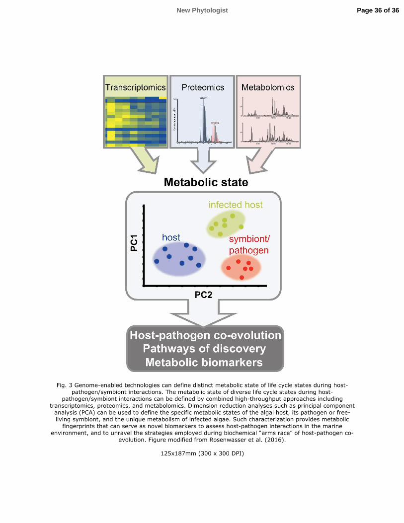
Fig. 3 Genome-enabled technologies can define distinct metabolic state of life cycle states during host-pathogen/symbiont interactions. The metabolic state of diverse life cycle states during host-
pathogen/symbiont interactions can be defined by combined high-throughput approaches including
transcriptomics, proteomics, and metabolomics. Dimension reduction analyses such as principal component analysis (PCA) can be used to define the specific metabolic states of the algal host, its pathogen or free-living symbiont, and the unique metabolism of infected algae. Such characterization provides metabolic fingerprints that can serve as novel biomarkers to assess host-pathogen interactions in the marine
environment, and to unravel the strategies employed during biochemical “arms race” of host-pathogen co-evolution. Figure modified from Rosenwasser et al. (2016).
125x187mm (300 x 300 DPI)
Page 36 of 36New Phytologist