Embed Size (px)
Citation preview

BIOTECH PL A Z A
Table of contents
MISSION STATEMENT 2–3
INSTITUTE OVERVIEW 4–5
INTRODUCTION – TARGET AREAS 6–7
HEALTH AND DISEASE 10–15
NEURODEGENERATION 18–25
SMALL MOLECULE PHARMA 28–39
CELL AND PROTEIN TECHNOLOGIES 42–57
BIG DATA 60–67
PLANTS AND FOOD 70–79

32
BI is Finland’s flagship institute for molecular
biosciences, home to more than 250 researchers from
all over the world. Our scientists are at the international
forefront of research in genomics, structural biology,
bioinformatics, molecular neuroscience, cell and
developmental biology and plant sciences. BI’s main
focus is basic science, but we also seek to commercialize
our research findings, for society’s benefit.
This prospectus is the launch document for Biotech
Plaza. It is aimed at potential investors, academic
and industrial partners, funding agencies, business
developers, end-users and any other types of
stakeholder, whom we invite to join with us to turn our
dreams into reality.
Howy JacobsInstitute Director
Here we set out a range of projects in progress, with strong
commercial potential. Some are broad, others narrowly
focused. Some are already close to being realized, whilst
others are long-term and aspirational. Some are aimed at
well-defined niche markets, whereas others seek to establish
technologies to tackle globally important problems.
All of them have arisen directly from BI’s discovery science.
But to develop them will require different kinds of support
and expertise than our scientists can provide on their own.
Biotech Plaza brings together a wide spectrum of advisors,
actors and financing tools to accomplish this task, with a
strong emphasis on the global marketplace. Meanwhile, our
research teams continue to replenish the discovery pipeline.
Troy FaithfullDevelopment Manager, Biotech Plaza

54
GLOBALBI is Finland’s flagship institute for molecular biosciences, home to
more than 250 researchers from all over the world. With a culture
of excellence and a strong track-record of success, it nurtures
discovery, technology, careers and businesses.
CUTTING-EDGEOur scientists are at the international forefront of research
in genomics, structural biology, bioinformatics, molecular
neuroscience, cell and developmental biology and plant sciences.
Our main focus is basic science but, in addition, we vigorously
pursue commercialization of our research findings.
TECHNOLOGYCurrent projects with major potential impact include a revolutionary
technology for treating killer diseases caused by defects in the cell’s
energy system; new drugs to reverse the nerve damage that causes
neurodegenerative diseases, and protein engineering tools based
on the splicing activities of inteins.
INNOVATIONBI’s future innovation potential includes studies of pathogen
resistance in cereal crops, structural analyses of complex viruses
like measles, targeting RNA and RNA metabolism as therapies for
cancer and neurodegeneration, unraveling the genetic circuitry
controlling organ formation, computational tools to interpret the
massive data sets emerging from genome analysis, and screens for
molecules capable of blocking substrate-specific protein secretion.
INVESTMENTBI seeks partnerships from the private sector, in order to develop
and commercialize its inventions. World-class discovery science,
combined with Finland’s transparent business environment and
highly skilled workforce, are ingredients for commercial success.
OPPORTUNITYInvestors and supporters can become stakeholders in BI as a
whole, in specific projects or in startups to be established jointly.
Contact us to explore possibilities.
BI
in numbers
established 1989
7 bioscience
technologies established in
Finland
10startups
>250doctoral theses
25research grants
in excess of $1 million
~300publications in
top-ranked international
journals
145applied patents
bi.helsinki.fiAN INDEPENDENT INSTITUTE OF THE UNIVERSITY OF HELSINKI
5
Contact us to explore
possibilities at
biotechplaza.org

76
HEALTH AND DISEASEMany of the world’s most intractable
diseases arise from disrupted intracellular
communication or homeostasis. Our
scientists are working on novel technologies
to alleviate damage arising from deranged
cell signaling, focusing on key regulatory
pathways affecting proteotoxicity in the
endoplasmic reticulum, oxidative stress in
mitochondria, and interactions between
the cell nucleus and cytoplasm. BI aims to
produce breakthrough treatments for a huge
spectrum of pathological states, ranging
from septic shock, to inherited heart disease,
diabetes and cancer.
NEURODEGENERATIONAge-associated neurodegenerative diseases
are currently incurable, and impose a
massive societal and economic burden, as
well as individual suffering on a colossal scale.
Many labs around the world are working
to understand the underlying pathological
mechanisms as a route to therapy. We have
prioritized ‘out-of-the-box’ approaches,
based on novel strategies to activate
natural, neuroprotective, stress-handling
mechanisms. We believe our new concepts,
presented here, will bring concrete and
dramatic benefits to the treatment of stroke,
movement disorders and dementia within
the next decade.
SMALL MOLECULE PHARMAThe search for new and better drugs for
use in the personalized therapies of the
future is one of the major drivers of today’s
pharmaceutical sector. This involves not just
the identification of more specific targets but
the implementation of ‘designer’ screening
platforms. Biotech Plaza features BI’s ongoing
work to create more specific anticancer
drugs, immune modulators, antibiotics,
and agents directed at new targets in
neurodegenerative and viral diseases. This
builds on our strong profile in structural and
cellular biology.
Biotech Plaza showcases the innovative technologies arising from BI’s scientific work at the frontiers of biological knowledge. We are looking for partnerships and financing to turn our ideas into products and services that will have major societal impact.
CELL AND PROTEIN TECHNOLOGIESFuture medicine will be based upon strategies
for tissue repair and regeneration, and the
delivery of exquisitely targeted therapeutic
drugs and proteins with minimal side-effects.
Biotech Plaza presents a wide spectrum of
new technologies and discovery science to
enable these goals, ranging from new systems
to engineer proteins and drug-conjugates, to
the elucidation of the molecular mechanisms
whereby stem cells stay young and retain
regenerative capacity. We are also developing
services for customized gene expression
and manipulation to serve the research
community, and novel reporter systems to
profile pathological disturbances in cellular
growth status and in subcellular protein
localization.
BIG DATAThe new science of genomics has been made
possible not only by the orders-of-magnitude
advances in DNA sequencing and the equally
astonishing decrease in its costs, but also
by the development of data-handling and
pattern-recognition tools that enable us to
make sense of the vast amounts of data
from biological and clinical specimens that
DNA sequencing generates. BI’s research
teams are at the forefront of developing and
applying computational methods to make
sense of big data emerging from genomic
analyses, and using it to predict the safety
of nanomaterials, the hazards of post-
operative infections or to serve the needs
of conservation biology, environmental
management and agriculture.
PLANTS AND FOODModern genetics opens up enormous
possibilities for altering the properties of
crop plants, to address the major global
challenges of our time: climate change,
resource depletion, pollution, and hunger.
We are focusing on improved pest-resistance
in cereal crops, enhancing the rate of carbon
capture in sustainable forests, modifying
trees so as to produce more suitable raw
materials for the chemical and biofuel
industries, and a more efficient way to
engineer targeted gene modifications in
common crop plants.
Target areas of Research Projects

9
Health & Disease
THE ALTERNATIVE OXIDASE AOX – A REVOLUTIONARY TOOL FOR WIDE-SPECTRUM THERAPY | Howy Jacobs
MANF AS A NOVEL THERAPEUTIC TARGET FOR THE TREATMENT OF DIABETES | Maria Lindahl
MITOCHONDRIAL MEMBRANE PROTEINS – STRUCTURE AND FUNCTION IN HEALTH AND SICKNESS | Vivek Sharma
1110
Howy Jacobs
The Alternative Oxidase AOX – a revolutionary tool for wide-spectrum therapyWe are developing a revolutionary therapy for diseases caused by malfunction of the mitochondria, the energy plants of our
cells. We take advantage of a natural system found in lower organisms for resisting mitochondrial stresses. By deploying
this system in humans, we hope to treat a wide spectrum of currently incurable and fatal diseases, whether caused by
environmental poisons, inherited mutations, metabolic overload or damage due to ‘wear and tear’ during aging.
Mitochondria recover the primary chemical
energy released from the breakdown of
food molecules in forms that the cell can
use. This enables vital processes such as
muscle contraction, electrical conduction
between nerve cells and hormone secretion.
Mitochondrial dysfunction causes many
fatal conditions, including rare, multi-system
diseases of infancy, as well as degenerative
disorders of old age. Major killers such as
heart attack and stroke result from the
inability of mitochondria to shut down and
power up properly, when the oxygen supply
is transiently interrupted. The inflammatory
processes leading to chronic lung diseases
are initiated by specific poisons in toxic
smoke that disable the mitochondria and
prevent the efficient clearance of particulates.
And fatal multiple organ failure in sepsis has
been attributed to mitochondrial shutdown.
Our novel, therapeutic approach employs
the alternative oxidase, AOX. AOX by-passes
the major energy supply pathway when
it is blocked, maintaining cellular functions,
preventing the build-up of toxic oxygen
radicals, minimizing the disruption of normal
metabolism and preventing cell death. We
have shown that transgenic rodents, flies or
mammalian cells endowed with the AOX gene
transplanted from a marine invertebrate are
protected from many pathological insults that
affect mitochondria in humans, including:
• inherited mitochondrial mutations causing
fatal infantile disease
• the toxicity of cyanide or the cocktail of
poisons in vehicle exhaust or cigarette smoke
• multiple organ failure due to bacterial sepsis
• a key indicator of physiological stress
following cardiac infarct, and
• neurodegeneration seen in models of
Parkinson’s and Alzheimer’s diseases
AOX thus has immense and broad potential
in medicine, ranging from a treatment
for rare diseases to the protection of
populations at risk from biohazardous
agents. Our strategy is to supply AOX (using
viral vectors, mRNA-mimetics or as protein
via cell-permeating peptides) to combat
mitochondrial damage.
Our strategy is to supply AOX … to combat mitochondrial damage
10
Our immediate aim is to develop partnerships with others in the field of genetic therapy.
We will develop ways of delivering AOX safely and effectively to affected tissues and organs,
starting with life-threatening conditions where we have solid data from animal trials.
End-users of our technology will initially be in the pharmaceutical sector, but it should also be
applicable to protection of military personnel, civilian first-responders and populations at risk
from terrorism or pollution.

1312
Maria Lindahl
MANF as a novel therapeutic target for the treatment of diabetesWe have identified a new growth factor for pancreatic insulin-producing β-cells. In the future, it could be used to regenerate
the lost β-cell mass in diabetic patients. This would restore normal metabolic control and avoid the severe, life-threatening
complications resulting from diabetes.
Collaborator: Mart Saarma
Diabetes mellitus is a group of metabolic
disorders characterized by hyperglycemia, in
turn caused by the inability of the endocrine
pancreas to maintain sufficient levels of
circulating insulin. Type 1 diabetes (T1D)
results from the autoimmune destruction
of insulin-producing β-cells, leading to total
insulin deficiency, whereas type 2 diabetes
(T2D) develops when the β-cells are no longer
able to respond to an increased insulin
demand due to insulin resistance. Today
more than 380 million people are affected
by the disease, and its prevalence is rapidly
increasing.
Current medications, insulin and anti-diabetic
drugs, are able only to alleviate diabetic
symptoms. Their administration does not
faithfully mirror the physiological response of
β-cells and does not prevent the devastating
micro- and macrovascular complications
of the disease. The reasons for β-cell
destruction both in T1D and T2D are still
unclear, but increasing evidence implicates
endoplasmic reticulum (ER) stress as a key
mechanism, accompanied by prolonged
activation of the signaling pathway linked to
the unfolded protein response (UPR). In turn,
this arrests protein translation and activates
protein refolding and degradation.
Thus, one of the main strategies for
improving diabetes therapy is to define
and validate novel approaches to protect β
-cells from stress, as well as activate their
regeneration. Various nutrients, hormones
and growth factors are known to affect β -cell
proliferation, but their use as therapeutic
factors has remained minimal, because of
lack of specificity. Therefore there is a need
for new and more specific targets for pre-
clinical screening and eventual clinical testing.
Mesencephalic astrocyte-derived
neurotrophic factor (MANF) was originally
identified as a secreted trophic factor for
dopaminergic neurons in vitro. MANF and
its homologue CDNF, discovered by our
group, can protect and repair neurons
and cardiomyocytes in animal disease
models. The protective role of MANF has
been suggested to depend on its ability to
rescue cells from ER stress. To understand
its physiological role in vivo, we generated
MANF knockout (KO) mice that developed
severe insulin-dependent diabetes due to
progressive loss of pancreatic β-cells. One
of the underlying mechanisms was chronic
ER stress. We found that recombinant MANF
protein enhanced β-cell proliferation in
cultured adult mouse islets, whilst pancreatic
MANF overexpression in a mouse T1D model
increased β -cell proliferation and prevented
cell death.
MANF is therefore a promising new
therapeutic candidate for β -cell protection
and regeneration. Our ongoing studies
combine MANF transgenic mouse lines and
animal models for both T1D and T2D, as
well as cell-lines and organ culture. Other
potential approaches will also be investigated
for using MANF as a regenerative protein for
β-cells in diabetes.
12
Background photo: Jari Rossi and Ömer Acar
PhD Student: Tatiana Danilova

1514
Vivek Sharma
Mitochondrial membrane proteins – structure and function in health and sicknessThe main focus of our research is to understand how enzymes involved in ATP generation (respiratory enzymes) function at a
molecular level, and how point mutations cause them to malfunction, giving rise to a range of serious disorders. For this purpose,
we apply state-of-the-art computational methods on the available structural data, and explore spatial and temporal scales that are
not easily reachable within the current experimental framework. Overall, our aim is to shed light on the molecular mechanisms of
enzyme action, and use this knowledge to design possible remedies to combat pathological dysfunction.
Life on earth is powered by ATP, which
is produced primarily in mitochondria
as a result of electron transfer reactions
that are coupled to proton translocation
across the inner-mitochondrial membrane.
These elementary reactions (electron and
proton transfers) involve charged species,
and are efficiently performed by enzymes
embedded in a low-dielectric medium, i.e.
the membrane. Malfunction in one or many
intermediate steps of the catalytic cycle of
these enzymes leads to various metabolic,
neurodegenerative and mitochondrial
disorders. Moreover, under environmental
stress conditions these enzymes produce
excess reactive oxygen species (ROS), which
are harmful to other cellular components, and
lead to various mitochondrial disorders. It is
of the utmost importance to understand how
these ROS are produced, and how that can be
contained to normal physiological levels.
We have performed large-scale atomistic
molecular dynamics simulations and high-
level quantum chemical calculations on
respiratory complex I, which is the first entry
point for electrons and one of the largest
enzymes in the respiratory chain. Our results
show that both electrostatics and long-range
conformational transitions play a key role in
enzyme function, coupling the redox reaction
to proton pumping as far as 200 Å away from
the former. Our work provides one of the first
molecular insights into complex I function,
and moves a step closer to identifying
the underlying causes of mitochondrial
dysfunction.
By performing long timescale fully-atomistic
classical molecular dynamics simulations on
complex IV of the respiratory chain, we have
shown how the enzyme manages to pump
protons with high efficiency (~90%). This is
achieved by microscopic gating elements,
such as conformational dynamics of amino
acid side chains and mobile water molecules.
Here, our work strengthens the view that
evolution has led to optimal nanostructures
that very efficiently convert energy from one
form to another.
We envisage two key areas where our
research will have profound impacts; a)
mitochondrial medicine, and b) bioenergy. By
studying molecular structures of intermediate
states in the catalytic cycle of mitochondrial
enzymes the underlying molecular causes of
diseases can be identified. Second, a deeper
understanding of the molecular mechanism
of respiratory enzymes has enormous
potential in the design and development of
biofuel cells of high efficiency.
A deeper understanding of the molecular mechanism of respiratory enzymes has enormous potential in the design and development of biofuel cells of high efficiency.
15

17
Neurodegeneration
REJUVENATING THE BRAIN: NEW STRATEGIES FOR NEUROREGENERATION | Mikko Airavaara and Andrii Domanskyi
A NOVEL APPROACH TO TREATING PARKINSON’S DISEASE | Jaan-Olle Andressoo
TARGETING RNA TOXICITY IN DEGENERATIVE DISORDERS | Susana Garcia
SYNAPTIC ADHESION MOLECULES - MAKING NEURONAL CONNECTIONS
TO COMBAT COGNITIVE DISORDERS | Tommi Kajander

1918
Mikko Airavaara & Andrii Domanskyi
Rejuvenating the brain: new strategies for neuroregenerationA viable strategy to treat neurodegeneration requires a toolbox for restoring neuronal networks in the damaged or aging brain. Using
a unique set of in vivo and in vitro models and tools, we are developing a broad experimental approach to achieving this goal, focusing
on two of the commonest such disorders, ischemic stroke and Parkinson’s disease. We use cutting-edge in vivo rodent models, combined
with virus- and CRISPR-Cas9-mediated transgenesis, plus surgical and toxicological procedures, to evaluate novel drugs, neurotrophic
factors, and non-coding RNAs with neuroprotective and neurorestorative potential. Using cultured primary cells isolated from embryonic
and adult brain, we also investigate pathways regulating proliferation, migration and differentiation of neural stem cells and astrocytes,
in order to assess the ways these cells could be used for rejuvenation of degenerating neuronal networks.
Aging increases the incidence of
neurodegenerative diseases and stroke,
leading to severe deficits in life quality.
Our ultimate goal is to increase the
quality of life for the elderly, by preventing
neurodegeneration and promoting neuronal
regeneration. Neuronal loss is caused
by various stressors and by the aging
process itself. Current drug therapies
for neurodegeneration are still based on
alleviating symptoms. The major challenge is
to develop safe, curative therapies, based on
enhancing the brain’s endogenous self-repair
mechanisms. Neurotrophic factors such as
Cerebral Dopamine Neurotrophic Factor
(CDNF), Mesencephalic Astrocyte-derived
Neurotrophic Factor (MANF) and Glial cell
line-Derived Neurotrophic Factor (GDNF)
and its family members promote survival of
neurons during development and disease. We
are working with these factors, studying their
interplay, mechanism of action, regulation
by microRNAs and their target genes, in
protecting neurons against stress. These will
lead to novel treatment strategies to combat
neurodegeneration.
We have established novel rodent models
where ischemic injury is restricted to the
cortex, enabling studies of functional recovery,
brain adaptations, neuronal rearrangements
and transdifferentiation of non-neuronal cells.
For Parkinson’s disease, we utilize toxin-based
(6-OHDA, MPTP, lactacystin), α-synuclein
fibril-induced or genetically engineered
rodent models. Applying a broad array of
behavioral tests we have demonstrated the
neuroprotective potential of CDNF and MANF
in these models, and have recently found
that stimulation of the microRNA biogenesis
pathway promotes survival of cultured
dopaminergic neurons. We are currently
testing the efficacy of selected microRNAs and
drugs, used in combination with neurotrophic
factors, in protecting neurons in vivo. Drug therapies for neurodegeneration are still based on alleviating symptoms and the major challenge we have is to develop disease modifying therapies.
WE AIM TO USE OUR
MODELS AND TOOLS:
• to identify new
neuroprotective drugs and
microRNAs, plus their target
genes and pathways, and
evaluate their activity in
promoting neuronal survival.
• to alleviate endoplasmic
reticulum stress as a way
of treating protein-folding
diseases in the brain.
• to mobilize transcription
factors, neurotrophic factors
and microRNAs to stimulate
endogenous self-repair
mechanisms based on
neuroprogenitor cells and
reactive astrocytes.
We are looking for
pharmaceutical, academic,
governmental and non-profit
organizations as partners
to support our mission
in developing efficient
treatments for devastating
neurodegenerative diseases.
19

2120
Our main approach is to target regulatory
areas of genes, called 3´ UTRs. For proof
of concept, we have studied the 3´ UTRs of
two neurotrophic factors, GDNF and BDNF,
that are already known to protect against
Parkinson’s disease (PD). The development
and maintenance of the brain involves
precisely controlled chemoattraction, elicited
by molecules such as neurotrophic factors
(NTFs), that are secreted by the brain’s own
cells. Because NTFs strongly promote the
survival and function of adult neurons, they
have been proposed for the treatment of
neurodegenerative conditions, such as PD.
Intracranial delivery of GDNF has already
been and currently is in clinical trials for the
treatment of Parkinson’s disease. However,
this approach has adverse side-effects and
variable efficacy, hindering its clinical use.
In our new approach, GDNF is not applied
ectopically, avoiding the massive sprouting of
dopaminergic fibers towards the site of GDNF
delivery, with unknown consequences with
respect to side effects, treatment efficacy
and behavior, that has been observed
in previous studies in both experimental
animals and humans. Our results, recently
published in PLoS Genetics (Kumar et al,
2015), imply that measures that promote
elevation of endogenous GDNF levels may
have clinical potential in the treatment of PD.
Our next-generation results, based on this
concept, take us one step closer to defining
a novel, viable treatment strategy for PD [for
reasons of confidentiality, we are able to
disclose further details only under separate
agreement].
We are now looking for investment funding
and/or commercial collaborations for testing
endogenous GDNF/BDNF elevation as a
therapeutic strategy, and for extending the
proof of concept so as to generate better
disease models for validation of novel
treatments.
Jaan-Olle Andressoo
A novel approach to treating Parkinson’s diseaseLittle is known of the therapeutic and scientific potential of elevating normal gene function at native sites of action,
which is hypothetically enormous. Our main objective is to develop novel tools that will enable upregulation of the
expression of specific genes in their native context, aiming to employ these concepts in therapy.
Our main approach is to target regulatory areas of genes, called 3’UTRs.
JAAN-OLLE ANDRESSOO, PHD
Principle Investigator
Institute of Biotechnology
P.O. Box 56, Viikinkaari 5D
FI-00014 University of Helsinki, Finland
Tel. +358-2941 59394
GSM +358 503581213
biocenter.helsinki.fi/bi/andressoo
20 Photomicrograph by M.Sc. Kert Mätlik

2322
RNA toxicity results from mutations in genes
where a small region of just a few repeated
nucleotide-pairs is inappropriately amplified
in a repetitive manner. This leads not only
to loss of gene function but also, when
expressed, to toxic transcripts containing
the expanded repeats. Expanded repeat
RNA toxicity is increasingly recognized as
a cause of human degenerative disorders,
from Amyotrophic Lateral Sclerosis (ALS) to
Myotonic Dystrophies (DM), with a wide range
of symptoms that include muscle wasting,
weakness, cataracts, heart conduction
defects and insulin resistance. These
disorders are predicted to share mechanisms
that lead to cellular dysfunction and disease.
Currently they are managed only by early
detection and palliative treatment, costing
several billion dollars annually but with little
return in terms of improved quality of life.
Our research focuses on DM, affecting
at least 1 in 8000 people worldwide, and
constituting a paradigm for RNA toxicity in
disease pathogenesis. We have developed
an animal model of DM, using the worm C.
elegans, providing an opportunity for high-
throughput small-molecule screening to
identify compounds that rescue locomotor
function, and thus hold therapeutic potential
in DM and similar diseases caused by
RNA repeat toxicity. Hits should include
modulators of alternative splicing, RNA
clearance, and other cellular processes.
Pre-clinical, early stage drug discovery with
a whole-animal model offers the advantages
of “fast-fail”, since compounds that are
inherently toxic at the whole-animal level
will be immediately rejected. Compared with
traditional drug-screening platforms, our
approach is faster, more economic, and can
be automated with high-throughput scaling,
key components for the rapid identification of
new therapeutics. We have recently identified
a previously unknown genetic suppressor of
RNA toxicity, the nonsense-mediated mRNA
decay (NMD) pathway, which is conserved in
humans, allowing for rapid translation into
human cell-lines for further validation, with a
high expectation of success.
RNA toxicity has recently been implicated
as an additional pathological mechanism in
a subset of disorders previously believed to
result only from protein repeats, and our C.
elegans model system has detected common
modifiers of DM and one well-studied such
disorder, Huntington’s disease. This widens
the potential of our system in uncovering
modifiers of toxicity, common to a broader
range of neurodegenerative disorders.
The worldwide market for DM-related
therapies is estimated at approximately
$2 billion, but there are currently no
approved drugs. Thus, our approach
has high commercialization potential. A
provisional patent has been filed on our C.
elegans model and we are in the process of
establishing it as a platform to screen small
molecule libraries including FDA approved
compounds. In the second phase, we will
license leads to established pharmaceutical
companies that have expressed interest
in developing DM-targeted therapeutics,
reinvesting licensing revenue into additional
screens and disease-model development.
We are looking for partners interested
in investing in the identification of these
candidate molecules for commercialization.
Susana Garcia
Targeting RNA toxicity in degenerative disorders
Our research focuses on Myotonic Dystrophies, an RNA-based disorder affecting at least 1 in 8000 people worldwide.
Myotonic Dystrophy (DM) worms exhibiting one of the proteins disrupted in DM, tagged with red fluorescence - the splicing factor MBNL-1.
The toxic repeat-bearing RNAs expressed in DM worms can be detected as green fluorescence.
23

2524
Tommi Kajander
Synaptic adhesion molecules - making neuronal connections to combat cognitive disorders
Neurons form a vast network of connections
that underlie all brain functions. Synapses,
where one neuron connects with another,
depend on specific adhesion proteins.
They are required for synaptic assembly,
maturation and stability, and thus play a
central role in the formation and function of
the brain. Moreover, dysfunction of synaptic
adhesion molecules is linked to specific
cognitive impairments such as schizophrenia,
autism and bipolar disorder.
Our broad scientific goal is to dissect the
molecular details of how these proteins
interact, how they are regulated and
expressed, and what are the results of
their inhibition in vitro. This way we hope to
understand the formation and dynamics
of synaptic structures and decipher how
synaptic adhesion proteins are involved in
disease. A longer-term goal is to translate
this knowledge in to clinical use, by creating
tools to treat and eventually cure and prevent
cognitive disorders, which represent a huge
societal burden.
Synaptic adhesion molecules are able to
induce synapse formation by binding to their
receptors on the target cells, where synaptic
differentiation then begins. This involves the
accumulation of different adhesion proteins
and expression of ion channels on the
post-synaptic side, mediated by intracellular
organizer proteins such as PSD-95 or GRIP.
We currently focus on studying the molecular
structures and interactions of the synaptic
leucine rich repeat (LRR) adhesion molecules
and their pre-synaptic ligand-adhesion
receptors, protein tyrosine phosphatase
receptors and neurexins.
We are working to establish several
applications for recombinant synaptic
adhesion molecules in basic research,
together with a wide network of
collaborators. For instance, we are using
these proteins to screen for new ligands
and functions, by affinity purification and
proteomics. Using adhesion protein-
patterned surfaces, we will study their
effects on synapse formation and neuronal
connectivity in cell culture. Starting from cells
derived from patients, where dysfunction
of synaptic adhesion receptors is the
underlying cause of disease, we will use the
technology of induced pluripotent stem-cells
(iPSC) to build disease models that could
be used in future as a platform for drug
screening or for testing other personalized
therapeutic strategies. Finally, we plan to
conduct small-molecule screens to identify
compounds able to enhance or inhibit
synapse formation, specifically targeting the
activity of the LRR adhesion proteins. For all
of these endeavours, we are eager to identify
industrial and academic partners, as well as
funders.
24

27
Small Molecule Pharma
UNDERSTANDING MITOCHONDRIAL PROTEIN SYNTHESIS FOR BETTER TARGETING OF THERAPEUTICS | Brendan Battersby
NOVEL TARGETS TO COMBAT RNA VIRUSES | Sarah Butcher
LEUKOCYTE BETA2-INTEGRINS IN HEALTH AND DISEASE | Susanna Fagerholm
BMH-21, A UNIQUE ANTICANCER MOLECULE TARGETING POL I | Marikki Laiho
TWINFILIN AND CYCLASE-ASSOCIATED PROTEIN INHIBITORS FOR PREVENTING
CANCER INVASION AND CHEMORESISTANCE | Pekka Lappalainen
DISCOVERY OF SUBSTRATE-SELECTIVE SMALL MOLECULE PROTEOSTASIS MODULATORS | Ville Paavilainen

2928
The goal of my research programme is
to understand how biological circuits are
established in the cell for the synthesis of
the distinct subset of 13 proteins required
for aerobic energy metabolism. These
proteins are synthesised in a unique cellular
compartment, the mitochondria, which acts a
metabolic hub and energy generator for the
cell. We focus on discovering the molecular
mechanisms at key regulatory nodes in the
pathways of mitochondrial protein synthesis,
and the response to acute and chronic
disruptions, both of pharmacological and of
genetic origin.
Protein synthesis in mitochondria is
molecularly similar to that in bacteria. Since
various classes of antibiotics target protein
synthesis, a potentially toxic side-effect of
antibiotic usage is disruption of mitochondrial
protein synthesis, which can have profound
effects on human health. Identifying the
mitochondrial targets of antimicrobials is
therefore crucial to enhancing the specificity
of these molecules and reducing their toxicity
to humans.
We have established that the 13
mitochondrial DNA-encoded proteins are
synthesised in excess of demand so that at
least 80% of newly synthesised mitochondrial
proteins are degraded, thus minimizing the
errors that arise naturally during protein
synthesis and folding. The cell requires a
tightly regulated quality-control pathway
to ensure that this labile pool of potentially
harmful proteins is degraded and does not
over accumulate. My current research has
shown how genetic and pharmacological
disruptions to this protein turnover pathway
can be deleterious to the cell and is the
underlying molecular defect in a number of
inherited human diseases. Typically, these
quality control pathways are also upregulated
in cancer cells, where they may become
essential for cell survival.
Fundamental research in my laboratory is
thus generating a molecular understanding
of how acute and chronic disruption to the
quality control of mitochondrial protein
synthesis is linked to cellular function. This
paradigm will provide insight into how
mitochondrial homeostasis is disturbed
by different types of pharmacological and
genetic insults. On the one hand, this will
enable better targeted therapies to limit
such damage, which can be applied, for
example, in neurodegenerative disorders.
On the other, it can also be exploited as a
novel cytotoxic strategy, leading to more
precise treatments for cancer. Finally, it will
inform the development of next-generation
antibiotics, free from damaging side-effects.
Brendan Battersby
Understanding mitochondrial protein synthesis for better targeting of therapeutics
29
Mouse (skin cell) fibroblast stained for mitochondria (red) and alpha-tubulin (green). Mitochondria move throughout the cell on the microtubule cytoskeleton that is composed of alpha and beta tubulin. Image by Paula Marttinen

3130
Sarah Butcher
Novel targets to combat RNA virusesThough non-enveloped icosahedrally-symmetric ssRNA viruses are one of the largest groups of viral pathogens, their assembly
mechanisms are still poorly understood. The control of ssRNA viruses by vaccination is unlikely to ever be possible for all but a small
subset of viruses, so innovative routes to antiviral therapy are urgently required.
Our group has identified sequence- and structure-specific nucleotide motifs within the genome of a human picornavirus that play a
key role in virus assembly. Referred to as “packaging signals”, due to their cooperative interaction with the key structural viral proteins
(capsid proteins), these motifs overlap untranslated and coding regions. Viral assembly is therefore in competition with other functions
of the genome. Disrupting these high affinity packaging signal-protein contacts has deleterious consequences for capsid assembly, and
thus presents a novel antiviral drug target. For a given single-stranded RNA (ssRNA) virus species, the packaging signals share certain
common features that increase the number of target sites per virus per drug, and show a lowered genetic variation between strains.
Drugs targeting such sites will be highly disruptive to ssRNA virus assembly and will not be easily defeated by the development of
resistance. Thus, our strategy offers potential new therapies against clinically relevant viruses such as poliovirus, hepatitis C (significant
chronic infection), and rhinovirus (causing the common cold: economic costs annually in the tens of billions of dollars).
31
We have established a wide range of
experimental and theoretical tools for
the identification and characterization
of packaging signal function(s) in capsid
assembly, revealing a previously unsuspected
principle in the assembly of single stranded
RNA viruses. The implications have the
potential to transform our understanding of
the fundamental biology of these systems,
including the mechanisms of infection and
evolution.
The patent we have applied for relates to
targets for anti-viral agents that either mimic
or bind to packaging signals of RNA viruses
that function in viral capsid formation. It
describes pharmaceutical and plant viral
control compositions for use in the treatment
of viral infections; methods to treat viral
infections; and five methods to screen for
packaging signals in viral RNA genomes.
A second approach we employ aims to identify
the key interactions between pathogenic RNA
viruses and the human host, so as to describe
the molecular pathogenesis of the infections,
and identify novel targets for antiviral agents
and diagnostics. This approach combines a
systematic analysis of all molecular reactions
(interactome), with structure-function studies
on emerging viruses.
With an appropriate pharma partner, our
existing targets plus our capability to produce
additional targets could be harnessed
to deliver a powerful new range of anti-
viral treatments. We thus require further
investment for drug screening, mammalian
animal models and 3D cell culture, to progress
this technology to phase-one drug trials.
Image courtesy of Pasi Laurinmäki

3332
The immune system is of fundamental importance
for human survival as it functions to protect us from
disease. However, it also poses a significant threat to
the individual, as immune cell-mediated diseases such
as autoimmunity and allergy are becoming increasingly
common. We are interested in leukocyte beta2-integrins,
important cell surface receptors in leukocytes which
regulate both adhesion and signaling in immune cells.
Integrin-mediated cell adhesion, migration and signaling
is crucial for proper immune system function. Studying
these processes is therefore fundamental for our
understanding of immunity. Importantly, integrins are
also recognized therapeutic targets. Our group studies
the in vivo roles and regulation of leukocyte beta2-
integrins both in the healthy and the dysfunctional
immune system, using novel animal models. In addition,
we aim to elucidate the role of these receptors
in immune-related diseases such as allergy and
autoimmunity, and investigate their possible targeting
to modify immune responses in diseases such as cancer.
Susanna Fagerholm
Leukocyte beta2-integrins in health and disease
SPECIFIC PROJECTS:
Integrins and SLE. Certain integrin genetic variants are strongly associated
with systemic lupus erythematosus (SLE), an autoimmune disease with
a significant genetic component. By studying these variants and their
signaling capabilities in vitro and in vivo, we aim to reveal novel therapeutic
targets for autoimmune disease. The cell- and animal models we have
already produced, or aim to establish, will also be used in future drug-
development programs aimed at creating more specific therapies for
autoimmune disease, in a personalized medicine approach.
Integrins and cancer. We have recently revealed that integrins work as
a “brake” in certain immune cells, restricting their signaling, activation
and migration. By targeting integrins and/or their signaling pathways, we
therefore aim to modify immune cell behavior to optimize treatment of
disorders with an immune-related component, such as cancer.
32

3534
Marikki Laiho
BMH-21, a unique anticancer molecule targeting Pol ICurrent studies on cancer pathway alterations are focused mostly on cancer genome abnormalities, their impact on the cancer
phenotype, and how they can be targeted therapeutically. However, cancer cells also become dependent on specific cellular metabolic
pathways, which include the need for an increased rate of protein production. This ultimately depends on the synthesis of rRNAs,
governed by RNA polymerase I (Pol I), and on the program of ribosome biogenesis. The Pol I transcriptional machinery is highly
responsive to oncogenic stimuli and commonly deregulated in cancer. Yet, this program has not been viewed as a clinically relevant
target and very few attempts have been made to manipulate it.
Pol I has traditionally been considered to
have a housekeeping function that cannot be
interfered with, and thus to be undruggable.
No markers have been developed to assess
Pol I transcription rates that would support
clinical decision-making, and therapeutic
development towards Pol I is thus still in
its infancy. In response to the critical need
to develop Pol I-targeting drugs, and to
conduct proof-of-principle studies, we have
introduced a novel small-molecule compound
(BMH-21), that targets Pol I activity. This
represents a new therapeutic candidate in
cancer, with huge potential impact.
BMH-21, a unique
pyridoquinazolinecarboxamide, was
discovered in our high-throughput screen
for anticancer small-molecules. We have
demonstrated that it acts by blocking
Pol I transcription and brings about the
destruction of the catalytic subunit of the Pol
I complex. It has broad anticancer activity as
tested in the NCI60 cancer cell panel, with a
potent GI50 of 0.16 µM. BMH-21 is cell and
tissue permeable, orally bioavailable, and
has low toxicity in normal cells compared to
cancer cells. We have conducted structure-
activity relationship analyses of BMH-21
by developing over 40 analogs, and so far
identified several equipotent derivatives. The
physicochemical characteristics of the active
analogs define the activity of the molecule
in a tightly defined chemical space. BMH-21
demonstrates promising activity in preclinical
mouse models, with significant inhibition of
tumor growth in melanoma, prostate and
colon carcinoma models.
Given that Pol I deregulation occurs at very
high frequency in cancers, we predict that a
Pol I targeting approach could have a major
impact in many cancer types. To support this,
we are conducting mechanistic studies on
Pol I complex assembly and stability, aiming
to identify factors that mediate deregulated
Pol I transcription in different cancers. Via
chemogenomic profiling of BMH-21 activity,
we are attempting to identify factors that
sensitize cancer cells to Pol I inhibition. At the
same time, we are proceeding with target-
based chemical biology screens to identify
new Pol I inhibitors.
Our long-term goals are to develop Pol I
targeting, using BMH-21-like molecules,
as cancer therapies. Studying its activity in
combination with other cancer intervention
strategies should reveal further opportunities.
We have patents issued and pending on
BMH-21 and its derivatives in the US and
Europe. The work is a collaboration between
my labs in Helsinki and Johns Hopkins
Universities, with intellectual property jointly
owned, but managed by the Johns Hopkins
University Technology Transfer Office.
35

3736
Pekka Lappalainen
Twinfilin and cyclase-associated protein inhibitors for preventing cancer invasion and chemoresistanceWe use a combination of genetics, cell biology, and biochemistry to study the role of the cytoskeleton in cancer. Our main
aim is to elucidate the structural and biochemical basis of the cancer-associated functions of two key cytoskeletal proteins,
twinfilin-1 and cyclase-associated protein (CAP), which promote cancer cell invasion and chemoresistance.
Coordinated building and break down of the
actin cytoskeleton gives the cell the ability
to undertake many fundamental cellular
processes such as migration, morphogenesis,
adhesion, and cytokinesis. Defects in its
organization and dynamics are therefore
central in cancer. Progression to malignancy
involves the dysregulation of multiple actin-
driven processes. Consequently, several
actin-binding proteins have been directly
linked to cancer invasion and metastasis,
notably twinfilin-1 and CAP.
Twinfilins are highly conserved proteins that
regulate cytoskeletal dynamics in a variety
of cell-types. CAP is a large multifunctional
protein which promotes actin recycling.
Elevated expression levels of CAP and
twinfilin-1 are associated with many
cancers, whilst CAP depletion inhibits the
proliferation and invasion of breast-cancer
cells. These molecules have therefore
emerged as promising new targets for cancer
chemotherapy.
Our studies have revealed the molecular
mechanisms whereby twinfilins control actin
filament assembly. The mammalian non-
muscle twinfilin isoform, twinfilin-1, has been
linked to progression and chemoresistance
of breast cancer and lymphomas. Its
suppression delays lymphoma progression
in mice, and extends animal survival
following chemotherapy. Furthermore, it was
identified as the major functional target of
the regulatory micro-RNA miRNA30c, which is
already used as a prognostic marker in breast
cancer. Twinfilin-1 depletion dramatically
inhibits invasion and spread of breast-cancer
cells and makes them more sensitive to
chemotherapy agents.
These studies allow us to design screens to
identify specific twinfilin-1 and CAP inhibitors
from small compound libraries. These will
serve as useful proof-of-principle compounds
for developing new drugs for use in cancer
chemotherapy.
37

3938
Secreted and membrane proteins constitute approximately 30% of the human proteome and many are important drug targets. Yet, their targeting with small-molecule therapeutics has remained a daunting task.
Ville Paavilainen
Discovery of substrate-selective small molecule proteostasis modulatorsWe are investigating the fundamental question of how protein molecules are correctly targeted to their final cellular locations or
to the extracellular environment. This targeting is achieved by a host of cellular factors that recognize distinct sequence features in
newly-made proteins and deliver them to their ultimate functional destinations. We focus on dissecting and functionally classifying
the chemical information contained in these protein-targeting ‘zip codes’ as well as identifying the cellular machineries responsible
for protein transport to correct locations. Many diseases are associated with dysregulated protein transport and secretion.
Therefore, we are exploring the protein targeting zip code as a novel druggable motif amenable to small-molecule modulation.
About 30% of all proteins are secreted
outside of the cells or integrated into
cell membranes. Secreted and integral
membrane proteins enable cells to exchange
information and respond to their specific
environments. They act as signaling receptors
and enable transport of molecules across
membranes. Most importantly, they represent
the majority of current drug targets.
Our work in this area has two goals. First,
we aim to understand the biological roles
of the protein secretion apparatus and
how its function is altered in disease
states. We aim to functionally identify and
classify information contained in secretory
zip codes and build accurate tools for
predicting secretory protein function from
genomic sequence data. Such tools will
allow prediction of secretory protein loss-
of-function by scanning disease genome
sequences for point mutations. Secondly, we
are exploring the possibility of preventing the
production of specific, mislocalized proteins
associated with disease, by discovering potent
small-molecules with the ability to prevent
their biosynthesis. In this work, we explore
naturally occurring small molecules that have
evolved to target the particular branches
of the protein homeostasis machinery with
highly specific mechanisms of action.
We have thus far demonstrated the ability
of diverse natural product small molecules
to selectively prevent the formation of only
a subset of secreted proteins, and have
evidence that these compounds target
specific recognition elements of the secretion
machinery. We are currently working on
identifying new drug-like natural products and
natural product-inspired small molecules that
can prevent biogenesis of different subsets
of secreted or membrane proteins. We have
established a novel technology platform
for differentiating between functional and
non-functional secretory zip codes and are
using this system to collect vast amounts of
functional sequence data, which will allow
development of different sequence prediction
algorithms. In addition to allowing accurate
prediction of secretory protein function from
genomic data, this method will also act as
a powerful tool for identifying and profiling
mechanistically unique secretory protein
inhibitors with ability to prevent the creation
of different disease-associated proteins.
We envision that new substrate-selective
proteostasis modulators can serve not only
as powerful molecular probes, but also as
lead molecules for the development of future
therapeutics. These compounds would
have much higher selectivity than current
inhibitors of components of the proteostasis
machinery, such as the proteasome or
cellular chaperones.
39

41
Cell & Protein Technologies
CRANIOFACIAL REGENERATION: FROM EVO-DEVO TO THERAPEUTIC APPLICATIONS | Nicolas Di-Poi
“GO IIST” | Hideo Iwai
STEM CELLS AND AGING | Pekka Katajisto
DNA DREAM LAB – EFFORTLESS REALIZATION OF DREAM CONSTRUCTS | Konstantin Kogan
DECLINE OF THE DENTURE: USING STEM CELLS TO REGROW OUR TEETH | Frederic Michon
NOVEL PEPTIDES AND DRUGS TO DIRECT DIFFERENTIATION OF STEM CELLS | Osamu Shimmi
ACTIN-REGULATED TRANSCRIPTION IN CANCER AND LAMINOPATHIES | Maria Vartiainen
PROFILING CELLULAR QUIESCENCE TO DEVELOP NOVEL ANTI-CANCER DRUGS | Norman Zielke

4342
Understanding the key signaling pathways
of craniofacial tissue regeneration
in reptiles will allow us to design
preclinical tests of therapeutic
strategies in mammalian
models and human cell/
organ culture leading
to ground-breaking
clinical applications.
43
Nicolas Di-Poi
Craniofacial regeneration: from Evo-Devo to therapeutic applicationsDevelopmental & Stem Cell Biology has become a crucial field for the understanding of tissue regeneration and the implementation
of regenerative medicine. The ability to repair or regenerate tissue is a fundamental property present in all multicellular organisms,
but there is tremendous diversity in how this process occurs within vertebrates. In particular, adult mammals (including conventional
experimental models such as mice and rats) have limited regenerative capacities compared to other model organisms such as reptiles.
These offer one of the best examples of regeneration in vertebrates. Hence, studying such taxa is a priority for improving our general
understanding of tissue development, patterning and regeneration. Importantly, it should help identify new targets and/or new
therapeutic approaches for regenerative medicine.
Our laboratory is investigating the molecular
genetic basis of the development, evolution
and regeneration of different craniofacial
tissues (tooth, retina, brain) with unique
regenerative capacity in non-classical reptilian
models (lizards and snakes). Surprisingly,
in contrast to the long-lasting interest in
craniofacial development in mammals, and
the numerous studies resulting from it,
relatively little is known about the embryonic
development and adult regeneration of these
tissues in other vertebrate classes. Our focus
on craniofacial research is motivated by the
fact that craniofacial diseases and disorders
account for a considerable and increasing
portion of health problems worldwide. In
addition, craniofacial organs are perfect
targets for evolutionary and ecological
studies, because of their excellent fossil
record, and because they show high levels
of morphological variation and adaptive
innovation. This is of crucial importance in
understanding tissue development from an
evolutionary perspective, which will be needed
to promote regenerative capacity in humans.
Our research projects use a multi-disciplinary
approach that deploys state-of-the-art,
methods combining molecular embryology,
genetics, 3D imaging, phylogenomics,
morphometrics, and theoretical modeling.
The extensive analysis of reptilian models
helps us to understand why and how
regeneration in mammals is absent or
limited. In addition, by revealing the
molecular and cellular principles underlying
regeneration, they identify new targets and
approaches for the treatment of common
human disorders such as neurodegenerative,
retinal, and dental degenerative diseases.
Our systematic approach, combining
Evolutionary Developmental Biology (Evo-
Devo) with Regenerative/Stem Cell Biology is
unique not only in Finland, but worldwide.
Our recent data already indicate that reptile
studies carry enormous potential and high
relevance to common human diseases, since
reptilian tissue structural and functional
circuitry, neuronal and epithelial cell types, as
well as signaling pathways are exceptionally
well conserved (see, e.g., Di-Poi et al., Science
Advances 2016; Milinkovitch, Di-Poi et al.,
Science 2013; Di-Poi et al., Nature 2010).

4544
Hideo Iwai
“GO IIST”
The IIST system, standing for Ion Inducible Self-cleavage tag, is a new technology for protein
purification and ligation, with wide applications.
Every year, the number of peptide-, protein- and antibody-based drugs entering clinical
trials is steadily increasing. There is a pressing demand for more cost-efficient and effective
approaches to the production of peptide-based conjugates for this market.
The IIST system, based on patented technology developed by Dr Hideo Iwai in BI, University
of Helsinki, is a novel and cost-effective way of achieving this goal. Based on natural or
engineered protein-segments with self-cleavage activity, IIST can support many uses both
in basic research and in industry.
In a conventional approach to protein purification and conjugation, a tagged protein of
interest has to be treated with expensive proteases following the affinity-capture step.
These proteases also need to be removed in a final recovery process. IIST eliminates the
need for expensive proteases, by making use of the ion-inducible self-cleavage activity of
the IIST domain, located between the protein of interest and the tag. The final recovery
step needs only a re-use of the affinity column, since the only thing that needs to be
removed from the final product is the tagged IIST domain.
Applications include not only the purification of proteins and peptides, but also the
conjugation of protein and peptide fragments, peptide cyclization and amidation, and
antibody conjugation to drugs or immunotoxins. The conjugation step is enabled by IIST
because of its inherent protein-splicing activity.
The global market for protein therapeutics, including monoclonal antibodies, is expected to
reach $165 billion by 2019. To bring this new technology to market needs the involvement
of one or more commercialization experts or an ‘entrepreneur-in-residence’ to strengthen
the research team, plus the interest of industrial collaborators and potential investors.
The global market for protein therapeutics, including monoclonal antibodies, is expected to reach $165 billion by 2019.
44

4746
Pekka Katajísto
Stem cells and agingYoung stem cells renew tissues constantly,
but old stem cells can no-longer support
functional tissue renewal at the required
rate. The resulting decline manifests as aging.
Stem cells fail at their regenerative task
due to the damage they have accumulated.
We investigate the mechanisms that have
evolved to minimize damage accumulation in
stem cells, and how such mechanisms could
provide points for intervention in aging-related
diseases and even the aging process itself.
One possible way for nature to reduce
damage in a stem-cell lineage would be to
selectively apportion damaged components
away from the new stem cell during
asymmetric cell division. We have discovered
that stem cells do, indeed, harbour distinct
populations of intracellular organelles
of different ages, and that certain old
organelles, for example the mitochondria,
are asymmetrically segregated at stem-
cell division, and become enriched in
the differentiating daughter cell. In other
words, this novel mechanism potentially
ensures that stem cells retain young and ‘fit’
organelles, whilst purging themselves of old
and possibly damaged, cellular components,
which are selectively partitioned into the
differentiating cell lineage.
We have thus established a new scientific
paradigm of age-selective organelle
segregation. At the same time we have
developed a unique set of research tools to
label subcellular components in an age-
specific manner, and then follow their fate
inside live and dividing stem cells. Together,
this concept and toolkit enable us to address
important research questions that are
inaccessible to anyone else. Our current work
aims to determine how stem cells recognize
the age of their organelles, and how age-
selective segregation is regulated.
Once we have identified the molecular
mechanisms underlying this phenomenon,
we will address how to induce or modify
age-selective organelle partition, with the
following aims in view:
• clear away damaged components from
stem cells after harmful events
• rejuvenate old stem cells, so as to promote
renewal of old tissues
• reduce the risk of aging-related diseases
and, by combining these goals
• slow down the aging process itself
Our group is keen to attract funders,
investors and industrial partners to help us
turn our discoveries into viable technologies
to limit, delay and eventually reverse aging.
46

4948
DNA Dream Lab
Konstantin Kogan
DNA Dream Lab – effortless realization of dream constructsDNA Dream Lab (DDL) is a new initiative at BI that aims to serve life-science users, initially in the Helsinki area, but with the
potential to serve the whole of Finland and beyond.
DDL is a one-stop-shop facility for plasmid DNA. It is widely acknowledged that most genetic manipulations revolve
around plasmid DNA, since fast and accurate molecular cloning is at the core of almost any life-science project nowadays.
Our mission is to help both basic and industrial researchers in two major directions: consultation and implementation.
48
Plasmid DNA consultation services include
realization of customer needs by searching
for and designing optimal solutions, with
emphasis on the experiment where these
plasmids are going to be used. Simplicity,
price, and time are the criteria for selecting
the most suitable strategy. Plasmid DNA
experts perform the search using a number
of external service providers (e.g. gene
synthesis companies, plasmid depositories,
local and international cDNA collections)
as well as DDL’s own comprehensive and
constantly growing local depository. Our
deep knowledge and expertise in structural
and molecular biology and protein folding,
coupled with years of experience in
plasmid construction, form a solid basis for
high-quality design of plasmid DNA. Our
awareness of downstream applications and
our ability to obtain any required number
of plasmids of almost any complexity allows
for the specific design of constructs to
increase the success of our client’s further
experiments. This greatly impacts how the
experimental set-up is planned by a lab
researcher using our services.
Plasmid DNA implementation services
complements the plasmid DNA consultation
part. Here we use state of the art methods
in DNA cloning, most of which are sequence
and ligation independent (e.g. SLIC, RF,
Gibson Assembly, LCR, USER). To make the
service complete we also securely store
DNA collections of our customers, having
backups in three different locations, stored
in three different forms. Depending on the
design, we outsource some of our activities
to leading companies in gene synthesis, oligo
manufacturing, and sequencing.
Being a not-for-profit, academic organization
we are completely transparent in our
activities whether it is in design, production
or management, making use of open-source
technologies or outsourcing to licensed
providers at cost. We accept students to
implement their own cloning under our
supervision using our materials, protocols,
and instrumentation. Upon agreement with
researchers, we disseminate the plasmids
as well as all the available information; yet
keep private unpublished plasmids, or
those having patents pending approval.
We implement custom-made information
technology solutions to allow immediate
access, search, and retrieval of any publicly
shared item fast, easily, and cheaply, due to
concise and proper annotation of every single
plasmid, and automated storage systems.
We deliver ready-to-use plasmid DNA for any
experiment in any required amount or purity
to any place in the world.
We are looking for appropriate funding and
partnerships to expand and develop our
services.

5150
Frederic Michon
Decline of the denture: Using stem cells to regrow our teethHistorically, an important goal of developmental biology has been to obtain the knowledge needed to achieve the regeneration of tissues
and organs. Understanding developmental mechanisms, and the underlying networks of regulatory genes, is instrumental for future
tissue and organ bioengineering. We also need to understand the properties of the stem and progenitor cells and the cues that induce
their differentiation. Recent progress in stem-cell research has led to realistic bioengineering of tissues such as bone and cartilage.
However, to regenerate whole organs composed of several cell lineages is far more challenging and needs a deeper understanding of
organogenesis and the roles of different classes of stem cells.
Several organs in vertebrates develop as
appendages of the outmost layer of the
embryo, called the ectoderm. These include
hair, feathers, scales, nails, teeth, cornea,
lachrymal and mammary glands. Their
development shares similar molecular and
morphological mechanisms, leading to the
formation of functional organs. While some
ectodermal organs are heavily studied, less
is known about dental stem cells. The human
tooth is not a renewing or cycling organ.
However, stem cells have been identified in
the dental pulp and periodontal ligament,
which can give rise to hard dental tissues.
These stem cells are not able to generate
whole new teeth: this will undoubtedly
require us to understand their biology in
greater detail. Our current research, started
in Prof. Irma Thesleff’s lab, focuses on the
continuously growing front teeth of mice.
Their renewal is fueled by two specialized
stem-cell niches located at the base of the
tooth, providing an excellent model to study
dental stem-cell biology.
We aim to elucidate the genetic basis of dental
tissue identity, and the origin and lineages of
dental stem cells. As part of a pan-European
research collaboration, our goal is to establish
a clinically applicable tooth bioengineering
protocol. To achieve this, we need to find
out what determines the fate of dental stem
cells, and the genetic networks involved in
their maintenance and differentiation into
specific cell lineages. This will enable us to
design in vitro differentiation protocols that
are the crucial first step towards the dream
of dental bioengineering. Our main focus is
on the specification of the dental epithelial
tissues, responsible for the hard enamel
(tooth crown), and tooth attachment to the
jaw. Our current work is showing the crucial
role of the dental Sox2-expressing cells in the
neo-formation of teeth, ex and in vivo.
The anticipated results will help us
understand how dental stem-cells determine
tissue identity, and may be applicable to
other ectodermal organs. Even though dental
aberrations can nowadays be treated, the
loss of teeth still has a dramatic impact on
human health and self-esteem, and requires
a wide spectrum of expensive medical
interventions over the lifetime of a patient.
Our research will lead to improved clinical
treatment and prevention of dental defects,
in particular loss of teeth.
51
Photo courtesy of Jukka Jernvall

5352
Osamu Shimmi
Novel peptides and drugs to direct differentiation of stem cellsMembers of the Bone Morphogenetic Protein (BMP) family have been associated with many pathologies, including vascular diseases,
obesity, diabetes and cancer. Several BMPs have been shown to be clinically useful, and BMP2 and BMP7 have been approved for
treatment of spinal fusion, fracture healing and dental tissue engineering. The BMP2:BMP6 heterodimer has also been proposed for
use in vitro to promote the differentiation of embryonic stem cells (ESCs).
Implantation of BMPs together with
autologous stem cells has emerged as a
promising technique in tissue engineering,
aiming at the regeneration of various body
parts. However, large-scale production of
BMP ligands remains costly due to multiple
preparation and purification processes,
and clinically effective doses of such
recombinant BMPs are much higher than
their physiological levels. To overcome these
issues, development of a novel, “superBMP”
agonist is desired. One such way is to develop
ligands with improved bioactive properties
through increased solubility, stability and
receptor affinity.
Due to the fact that BMPs play crucial
roles in many different contexts in animal
development, their protein sequences in
mammals are highly conserved. We study
Drosophila BMPs to understand the ways
in which they have become functionally
optimized during evolution. The Drosophila
BMP5-8-like protein Scw is in a unique
position since it is exclusively expressed in
the early embryo and is evolutionarily highly
derived. Scw is required for generating peak
levels of the BMP gradient in Drosophila
embryos. Thus, its protein sequence appears
to have evolved to provide maximal biological
activity. We have also shown that post-
translational modifications of Scw modulate
BMP signaling activity both in vitro and in vivo.
We postulate that tolerance for structural
changes of BMPs is dependent on a range of
developmental and evolutionary constraints.
By employing optimized sequences through
natural selection of novel peptides, the
biological properties of BMP ligands can be
improved. Combined with our knowledge
of how evolution has shaped Scw, we aim
to develop a superBMP, as an efficient
and specific differentiation factor for stem
cells. Such a product should be applicable
for differentiation of stem cells into
chondrocytes, osteoblasts or cardiomyocytes.
This will involve not only optimization of the
peptide sequence, but also post-translational
modifications that can have a profound
effect on its potency, specificity, stability and
solubility.
We are looking for partner organizantions
to help us implement the various technical
steps in our project, enabling us to produce
and evaluate BMP variants based on our
predictions. These approaches will create and
develop novel products for commercialization
in the regenerative medicine sector.
53

5554
Maria Vartiainen
Actin-regulated transcription in cancer and laminopathiesActin operates in the cell’s cytoplasm as a key part of the cytoskeleton, which provides the cell its shape and enables movement. The
actin cytoskeleton is therefore critical for cell migration, which is a pre-requisite for cancer metastasis. Thus, it is not surprising that it is
deregulated not only in many cancers, but also other diseases, which show disturbed tissue architecture. In recent years, we discovered
that actin plays an important second role in cellular processes, inside the cell nucleus as well, where it influences gene expression. This
provides a mechanism whereby actin could contribute to disease development and progression independently of its direct effects on cell
structure and motility. Our data also supports the involvement of nuclear actin in laminopathies. These are a group of genetic disorders
caused by mutations in genes encoding proteins of the nuclear lamina, the fibrillar structure responsible for the internal organization of
the nucleus. Laminopathies display a large variety of clinical symptoms, such as skeletal and cardiac muscular dystrophy, lipodystrophy,
diabetes, and premature aging, progeria. One conceptual puzzle with laminopathies has always been the fact that, whist they mainly
affect specific tissues, the underlying genes are expressed everywhere in the body.
NUCLEAR ACTIN AND CANCER
The hallmark of cancer cells is unregulated
growth. However, most cancer deaths are
not due to the primary tumor, but rather
the spread of cancer cells, metastasis, to
other organs. In normal cells, cell growth
and migration are tightly regulated by many
signaling pathways and transcriptional
programs. Nuclear actin has been shown
specifically to regulate two transcriptional
programs critical for cancer progression:
the growth-regulating Hippo-pathway and
metastasis-regulating MKL1-SRF pathway. Our
preliminary data reveals extensive connections
between these two programs, and we are
examining how this crosstalk influences
cancer progression. By high-throughput
approaches, we aim to identify common
players and small molecules targeting both
pathways. This will not only shed light on the
basic mechanisms of cancer progression. It
will identify novel targets and molecules for
diagnosis and, importantly, a new generation
of more effective anti-cancer drugs.
NUCLEAR ACTIN AND LAMINOPATHIES
In collaboration with researchers from the
US, we demonstrated a novel mechanism
that could provide insights into the
cause of the cardiac phenotype in many
laminopathies. We showed that two
components of the nuclear lamina, lamin
A/C and emerin, modulate nuclear actin
and thus regulate gene expression through
the MKL1-SRF pathway. This regulation is
disrupted in laminopathies. Subsequently we
discovered a physical association between
the transcription coactivator MKL1 and
components of the nuclear lamina, and we
are currently analyzing their relevance for
MKL1-SRF-regulated gene expression. Here
too, high-throughput screening will help us
identify novel regulators and small molecule
targeting the pathway, and test their ability
to correct impaired MKL1–SRF signaling in
laminopathic cells. These approaches should
enable us to ameliorate the drastic cardiac
pathology associated with laminopathies.
PERSPECTIVE: We seek major funding to
support our high-throughput screening
pipeline, aiming to identify compounds to test
further in cell and animal models of cancer
progression and laminopathy, ahead of
clinical trials.
55

5756
Norman Zielke
Profiling cellular quiescence to develop novel anti-cancer drugsThe tissues and organs of multi-cellular organisms are comprised of specialized cell types, which originate from stem cells.
Stem-cell pools are typically heterogeneous in regard to many cellular processes, including transcription, cell signaling,
proliferation status, and susceptibility to apoptosis. Superficially minor differences between cells can have a profound impact
on cell fate. Heterogeneity is also found in most tumors, resulting in differential sensitivities to cytotoxic or anti-proliferative
drugs. Our work aims to develop and exploit tools that can discriminate between superficially similar cells that differ in key
properties such as proliferation status and drug susceptibility.
Understanding the characteristics of
quiescent stem cells has been hampered
by the rarity of these cells in many tissues.
To bypass this limitation, we are currently
developing a biosensor that facilitates
detection and purification of quiescent cells.
The isolated cells can be further analyzed by
various omics methods, thereby providing
novel insights into the transcriptome,
and the epigenetic status of quiescent
cells. In addition, this reporter system can
aid screening for novel, more effective
anti-cancer drugs. Many conventional
chemotherapeutics target key regulators of
cell proliferation. However, a large fraction
of the targeted cell-cycle genes are down-
regulated in quiescent cells, which may
explain why certain cells in a tumor are
resistant to drug treatment. Therefore, we
plan to use our biosensor-based system to
screen for compounds specifically targeting
quiescent cells, or aimed at releasing them
from quiescence to make them susceptible to
conventional treatments.
FUCCI (fluorescent ubiquitination-based cell
cycle indicator) biosensors are a promising
approach for studying cell proliferation at
single-cell resolution. By assigning specific
fluorescent labels to different cell cycle
stages, it provides a reliable readout for cell
proliferation that can be applied to live-cell
imaging or high-content screening. Combined
with flow cytometry it enables detailed
molecular characterization of isolated single
cells. We have recently extended this concept
to the fruit fly Drosophila melanogaster,
where it can be applied in conjunction with
the powerful genetic toolkit available in this
model organism. Since its introduction in
2014 Fly-FUCCI has proven to be a reliable
method for monitoring the proliferation
status of stem cells e.g. in the posterior
signaling center (PSC) of the Drosophila lymph
gland or the intestinal stem cells (ISCs) in
the regenerating midgut. We are currently
developing a next-generation FUCCI sensor
that allows robust detection of quiescent cells
in flies and mammalian cell culture.
We aim to develop this technology further
in two directions: first, we plan to use it to
assess how variability of the proliferation
status affects cell-fate decisions. This will
help us to understand how abnormalities in
cell-fate programming in stem cells can lead
to cancer or degenerative disease. Second,
it will support high-content screening in 2D
and 3D cell culture, as well as small model
organisms such as Drosophila, for drugs or
druggable targets that switch cells into or out
of quiescence, or target them specifically for
destruction.
Micrograph of an eye imaginal disc derived from the fruit fly Drosophila melanogaster, in which their characteristic
cell cycle pattern was visualized with the Fly-FUCCI (fluorescent ubiquitination-based cell cycle indicator) system
(Zielke et al. 2014). Cells residing in G1 phase were marked in green; cells in S phase were labeled red and cells in
G2 phase were labeled yellow. Image courtesy of N. Zielke and H. Lorenz (ZMBH – Imaging Facility).

59
Big Data
TOOLS AND SERVICES FOR ‘BESPOKE’ GENOME ANALYSIS | Petri Auvinen
PREDICTING SAFE AND NOVEL USES OF NANOMATERIALS | Dario Greco
BIOINFORMATICS SOLUTIONS FOR PRECISION MEDICINE | Liisa Holm
WASABI – WEB-BASED VISUALIZATION AND ANALYSIS OF COMPARATIVE SEQUENCE DATA | Ari Löytynoja

6160
Petri Auvinen
Tools and services for ‘bespoke’ genome analysis
Genome analysis requires a lot more than
just DNA sequencing, even though efficient
and accurate high-throughput sequencing
is a necessary starting point. Our group
combines this ‘bedrock’ service with
comprehensive bioinformatic analyses, using
tools also developed in-house for key steps
in the process, such as sequence alignment,
annotation and protein-function prediction.
Throughout the work, we establish and
maintain close interactions with the teams of
biologists behind the various projects that we
undertake.
We have created our facility primarily to
implement the assembly, annotation, and
functional and evolutionary analysis of
large eukaryotic genomes, building on early
experience as part of a wide collaboration to
determine the genome sequence of barley.
The first two examples of species whose
genomes have been fully analysed by our
group are the Glanville fritillary butterfly
(Melitaea cinxia; Nymphalidae) and the
silver birch (Betula pendula). Based on these
successes we are now involved in a number
of other genome projects, notably the ringed
seal, where our focus is the endangered
population unique to Lake Saimaa, and the
strawberry. In addition, we have developed
extensive expertise in metagenomics.
This has been applied to the gut and skin
microbiome in relation to disease, to food
spoilage, and to the analysis of environmental
samples, such as the microbial communities
in deep crystalline rocks.
This varied portfolio has enabled us to
build up not only a state-of-the-art toolkit
for genome analysis, but also the expert
knowledge required to interface and team-
up with a broad spectrum of biologists, so
as to extract information from genomes in
the ways most relevant to specific scientific
questions and real-world applications. This
has led to biologically significant findings. In
the case of the Glanville fritillary butterfly,
comparative genome analysis revealed a
thus-far unique evolutionary feature in the
Lepidotera, not seen in other groups of
animals such as mammals, whereby the
order of genes along the chromosomes
seems to be stable extending back at least
140 million years. The silver birch genome, on
the other hand, has been studied because of
its economic importance in boreal forestry.
Combined with appropriate field work, the
genome sequence opens up the possibility
of identifying the molecular basis of growth
rate regulation, wood quality and pest-
resistance. In the case of the Saimaa seal, the
emphasis is on conservation biology, and on
the bottleneck effect on speciation in a large
mammal. By correlating patterns of genomic
variation with specific biological traits we
hope to understand the molecular basis of
adaptation to locally changing environments
in a species under extreme threat.
Metagenomics is becoming an increasingly
important tool for detecting environmental
changes before they become irreversible.
It will also have an increasingly important
predictive role in medicine, as we learn more
about the interaction between microbial
communities and ‘host’ metabolism and
disease processes.
In the future, the expertise we have acquired
enables us to contemplate extending beyond
purely academic collaborations and launching
‘bespoke’ genome analysis as a commercial
service directly offered to animal or plant
breeders, wildlife conservation agencies,
healthcare providers and diverse other
organizations around the world. Thus, we are
in the early phase of a search for investors
and developers to identify the most relevant
markets and create a viable business out
of what hitherto has been curiosity-driven
research.
60

6362
Dario Greco
Predicting safe and novel uses of nanomaterialsNanoparticles are molecules with at least one dimension smaller than 100 nm. Nanotechnology is categorized as one of the ‘Key
Enabling Technologies’ in the EU’s current Research and Innovation program, Horizon 2020. The industry already employs more than
300,000 people in Europe, and the global market value of nanotechnology-enabled products is estimated at $2 trillion in 2015. These
products are already present in our everyday life and new Engineered NanoMaterials (ENM) are continuously produced. However, the
same physical, chemical, magnetic and electrical properties that make ENM extremely valuable, namely their high reactivity, are also
regarded as a potential threat to human health and the environment. The possible risks associated with ENM are a major impediment
to their adoption worldwide. Moreover, current safety testing of ENM is much too slow and laborious. This even applies to novel,
controlled and safe applications of existing ENM in the field of nanomedicine. Thus, there is a tremendous need to accelerate
hazard assessment and to predict the effects of ENM. On the other hand, some ENM effects could provide a great opportunity, for
instance, in the contexts of innovative therapeutic protocols. Hence, it is essential to understand nano-bio interactions in depth and at
multiple levels, in order to maximize ENM usability as well as safety.
The focus of our research group is
predictive and systems nanotoxicology. We
use cutting-edge genomic technologies,
artificial intelligence methods and systems
biology approaches to formulate predictive
computational models of ENM toxicity and
biomedical usability. OMICs technologies
allow us to thoroughly characterize the
effects on human cells of exposure to ENM.
Novel bioinformatics methods are then
developed to integrate the experimentally
derived big data-sets, with the aim of
characterizing the mode of action of ENM
and revealing relevant biomarkers for use in
future regulatory applications. The respiratory
system is the main exposure route for
humans, especially in work environments,
and is thus the context for one of our most
important current research projects.
Our group is currently completing the
development of the first such computational
tool, INSIdE nano (Integrated Network of
Systems bIology Effects of NANOmaterials),
allowing systematic contextualization of
the mode of ENM action with the respect
to interactions with other chemicals,
ability to cause diseases, and prospective
combinatorial use with drugs. Thus, we
aim not only to predict hazards, but also to
enhance existing therapeutic strategies.
Our long-term goals are (1) to develop
procedures for rapid, large-scale hazard
assessment of ENM; (2) to formulate
predictive algorithms capable of indicating
the potential adverse effects of ENM, based
just on the knowledge of their intrinsic
chemico-physical properties; (3) to produce
computational tools for the systematic
prediction of ENM uses in broader biomedical
contexts.
Screenshot from our tool INSIdE nano tool depicting the network of interactions that Parkinson’s disease (PD, purple) have with chemicals (eg. the known dopaminergic neuron toxin MPTP, green), drugs (eg. Levodopa the preferred drug for the treatment of PD, red) and nanomaterials (blue). INSIdE nano is helping scientists and regulators to formulate hypotheses concerning the involvement of nanomaterials in pathogenesis of specific types of diseases (as in this example), or it could otherwise suggest a possible use of nanomaterials as a treatment for diseases.

6564
Liisa Holm
Bioinformatics solutions for precision medicine
Post-operative complications caused
by microbial pathogens in the wound
occur in approximately 2% of all surgical
interventions. In the US, they claim about
8,200 lives annually, and their societal cost is
estimated at $1.6 billion.
One of the most essential steps in the
diagnosis and management of surgical site
infections and other healthcare-associated
infections is the prompt, accurate, and
reliable identification of the etiological
agents. To date, however, pathogen
detection from clinical samples has relied
heavily on culture-based approaches and
phylogenetic marker gene identification that
are narrow in scope and unreliable for the
characterization of many pathogens.
These limitations in conventional diagnostics
techniques may compromise patient health
and delay the initiation of the correct
antimicrobial therapy. The consequences
are a matter of life and death for individual
patients, including the danger of long-term
disability and decreased life quality. At the
societal level, the deficiencies in pathogen
recognition have contributed markedly to
the favoring of untargeted, broad-spectrum
antibiotics in infection control, an extremely
detrimental approach in the era of increasing
antimicrobial resistance. To facilitate more
appropriate treatment and to minimize
the unnecessary use of broad-spectrum
antibiotics, it is imperative to develop novel
techniques for identification of infectious
pathogens and their virulence and antibiotic
resistance determinants.
Our project aims to advance our
understanding of the etiology of surgical
infections and develop cost-effective tools
for early recognition and customized
treatment. As a pilot project in personalized
medicine, it combines top expertise in
microbiology, computational biology, surgery
and clinical wound management. We employ
cutting-edge DNA sequencing, healthcare
informatics, and bioinformatics algorithms
to define the relationship between infections
and the microbiota residing in surgical sites.
Our group coordinates and implements the
computational aspects of the project. Our
expertise is in developing bioinformatics
analysis pipelines that can annotate big
data sets, as generated by metagenomics
(typically hundreds of gigabytes) in a clinically
relevant time frame. This is accomplished
using in-house algorithms deployed in
superfast sequence database search servers.
Our approach belongs to a new generation
of algorithms which is orders of magnitude
faster than BLAST, the industry standard.
Importantly, these algorithms scale better
because search time is independent of
database size, enabling the processing
of larger quantities of data, against
exponentially growing databases, with the
same computer power.
We have tested our computational pipeline
on test samples from patients and spiked
controls, and shown that it identifies the
potentially infectious agents.
The project is a wide collaboration between
BI, clinical and non-clinical departments, and
hospital services. As a spin-off it will establish
novel molecular tools and a tailored pipeline
for analysing microbial community genomics
data. It thus has substantial potential for
improving the quality and cost-effectiveness
of healthcare, and should be of interest to a
wide and global market.
The Overview
SURGICAL SITE INFECTION INFECTION INFORMATION
• Wound biopsies
• Swab Samples
• Patient information
METATRANSCRIPTOMICS SAMPLE PROCESSING
• Homogenization
• Extraction of total RNA
• DNAse I digestion
• Bead-based enrichment of bacterial RNA
RNA-SEQ LIBRARY PREPARATION
• Ribosome depletion
• Fragmentation
• Adapter ligation
HIGH-THROUGHPUT SEQUENCING DATA GENERATION
DATA AND SPECIMEN COLLECTION FUNCTIONAL ANALYSIS
• Species/strains
• Antibiotic resistance
• Virulence factors
SECONDARY ANALYSIS
• Feature counting
• ORF mapping
• ORF calling
PRIMARY ANALYSIS
• rRNA removal
• Human mRNA removal
• Quality trimming
• Adapter removal
Infected surgical siteUninfected surgical siteBurn wound

6766
Ari Löytynoja
Wasabi – Web-based visualization and analysis of comparative sequence data
Wasabi, developed by our team in BI, is a web-based
environment for analysis and visualization of comparative
sequence data. The Wasabi analysis environment is versatile
and adapts to diverse tasks in molecular genetics: for
example, the phylogenetic profiling of human mutations,
comparative analyses of microbial protein fragments,
amplicon analyses of marker genes or epidemiological studies
of pathogens. Wasabi runs inside a web browser and does
not require any installation: it can be immediately accessed
and implemented, via wasabiapp.org. The Wasabi visualization
library is a self-contained package for web-based display of
sequence data. Wasabi windows can be embedded in web-
pages (see figure) and in web-applications, and allow live,
browsable views of underlying data and fast data manipulation
(translation, filtering, text search), performed locally within the
browser. A prototype of embedded Wasabi visualization for
scientific publishing is shown at wasabiapp.org/journal.
Wasabi represents a specific output from our work, serving
the academic community. As such, the Wasabi environment
is based on open-source software and wasabiapp.org is
available to all as a free service. However, for users requiring
strict confidentiality or having special needs, custom services
can also be provided.
67
These can include:
• in-house installation of Wasabi software
with user training and support
• extension of the Wasabi environment
with custom analysis tools
• construction of reference data sets
(e.g. microbial protein families,
pathogen marker genes)
• embedded data browser,
e.g. for scientific journals or
sequence databases
Wasabi data visualization is currently
integrated in the Ensembl genome
browser (ensembl.org). Our aspiration
is to develop these extended services
on a commercial basis, serving diverse
business communities, such as publishers,
pharmaceutical companies, healthcare
providers or public agencies.
For further details, see the videos and
our publication describing Wasabi at
wasabiapp.org/about.
Live, browsable Wasabi window embedded on a web-page, showing cetacean-specific changes in the haptoglobin gene, potentially preventing hypoxia-induced cell damage as an adaptation to deep diving.

69
Plants & Food
MOLECULAR ENGINEERING THE FOREST: RATE AND QUALITY OF WOOD FORMATION | Yrjö Helariutta
WARMING TREES TO COMBAT GLOBAL WARMING | Howy Jacobs
REGULATING GENE EXPRESSION AND HORMONAL SIGNALING TO BOOST
BIOMASS PRODUCTION IN TREES | Ari-Pekka Mähönen
A TOOL FOR IMPROVING THE EFFICIENCY AND TARGETING OF GENE MODIFICATIONS IN PLANTS | Alan Schulman
A ROBUST RESISTANCE GENE FOR YELLOW (STRIPE) RUST | Alan Schulman

7170
Yrjö Helariutta
Molecular engineering the forest: rate and quality of wood formationThe aim of our research is to understand molecular mechanisms controlling plant vascular development, and use this knowledge to
develop ways of enhancing economically useful traits in forest trees. The plant vasculature, which comprises the conducting tissues of
phloem and xylem, provides structural support to the plant and enables the transport of water and sugars between shoot and root.
Wood, the secondary xylem of plant stems, represents most of the carbohydrate biomass of the plant, and its composition is under
genetic control. Our research thus has immense potential for the forest industry: the better we understand the molecular mechanisms
controlling wood formation and composition, the easier it is to apply this knowledge. The molecular regulators that we have identified
represent optimal target genes for tree breeding and forest biotechnology.
In our labs, we explore the molecular basis of
vascular development in two complementary
model systems: herbaceous Arabidopsis
thaliana and woody forest trees. Despite
its small size, Arabidopsis produces wood
and has emerged as a versatile model for
plant vascular development. Based on our
Arabidopsis research, we have identified a
wealth of novel regulators of wood production.
Using both transgenic and non-transgenic
gene editing methods, we can translate
this knowledge to modify the analogous
developmental processes in a tree trunk.
Besides boosting biomass production in
forest trees, we aim to optimize its properties
for various industrial purposes. Our current
focus is to modify lignocellulosic plant
biomass by adding callose to cellulose-rich
cell walls. Both callose and cellulose are
glucose polysaccharides, but with strikingly
different structures: in contrast to cellulose,
callose does not form recalcitrant microfibril
structures. By modifying the function
of a callose synthase enzyme, we have
already engineered transgenic plants for
overproduction of callose. Callose-enriched
wood should require less energy to liberate
simple sugars from cell walls and thus
significantly reduce the costs of industrial
processes that use the released sugars, for
example, to yield biofuels.
PERSPECTIVE: Our ultimate aim is to
engineer forest trees, so as to optimize
biomass production to yield high-value
products in the biorefineries of the future.
70

7372
Howy Jacobs
Warming trees to combat global warmingThe Boreal Forest is one of the world’s major carbon sinks. Accelerating its growth or extending its range would contribute
significantly to decreasing net CO2 emissions. When combined with sustainable forestry, it could result in increased
production of renewable fuels and many other products that today are sourced primarily from fossil materials.
We are working on a strategy to address
the challenge of accelerating tree growth,
by taking advantage of a natural system
present in all plants, the alternative oxidase,
AOX. AOX buffers stress and overload in the
mitochondrial energy system, by serving as
a by-pass that conducts electrons released
by biological oxidations directly to oxygen.
In so doing, it releases energy as heat,
rather than as ATP. This pathway is used
naturally by some plants to generate heat,
for example, by arum lilies to volatilize insect
attractants. AOX activity is induced under
extreme cold in many plants, and may play
a role in resistance to freezing. However,
in the model plant species Arabidopsis,
engineering AOX over-expression has already
been shown to promote growth of the plant
even at the more temperate condition of 15
°C. In effect, some of the energy captured
from sunlight by photosynthesis is being
‘wasted’ to produce heat. But the net effect
on biomass production is strongly positive,
because of the temperature-dependence of
the chemistry of biosynthesis.
Extending this concept to tree species
means that we can realistically contemplate
engineering trees to over-express AOX,
thus generating heat that will promote their
growth at sub-optimal temperatures, such
as those encountered globally across the
far north, where the majority of the world’s
remaining forests are located.
Previous studies have shown that the growth
of the major tree species in the Boreal Forest,
notably Scots Pine, Norway Spruce and Silver
Birch, is strongly influenced by temperature
during the growth season, and this effect
is most pronounced at environmental
temperatures encountered near the northern
limits of the range. Ironically, global warming
itself has already had a demonstrable effect
on tree growth in Finland. The advent of
gene-editing technology now makes it
theoretically feasible to engineer AOX over-
expression in commercial trees, promoting
greatly accelerated growth, increased carbon
capture and, in the longer term, increased
production of renewable raw materials.
Our research group is now looking for
investors and commercial partners
to accelerate this project, developing
and licensing this technology for use in
commercial forestry.
73
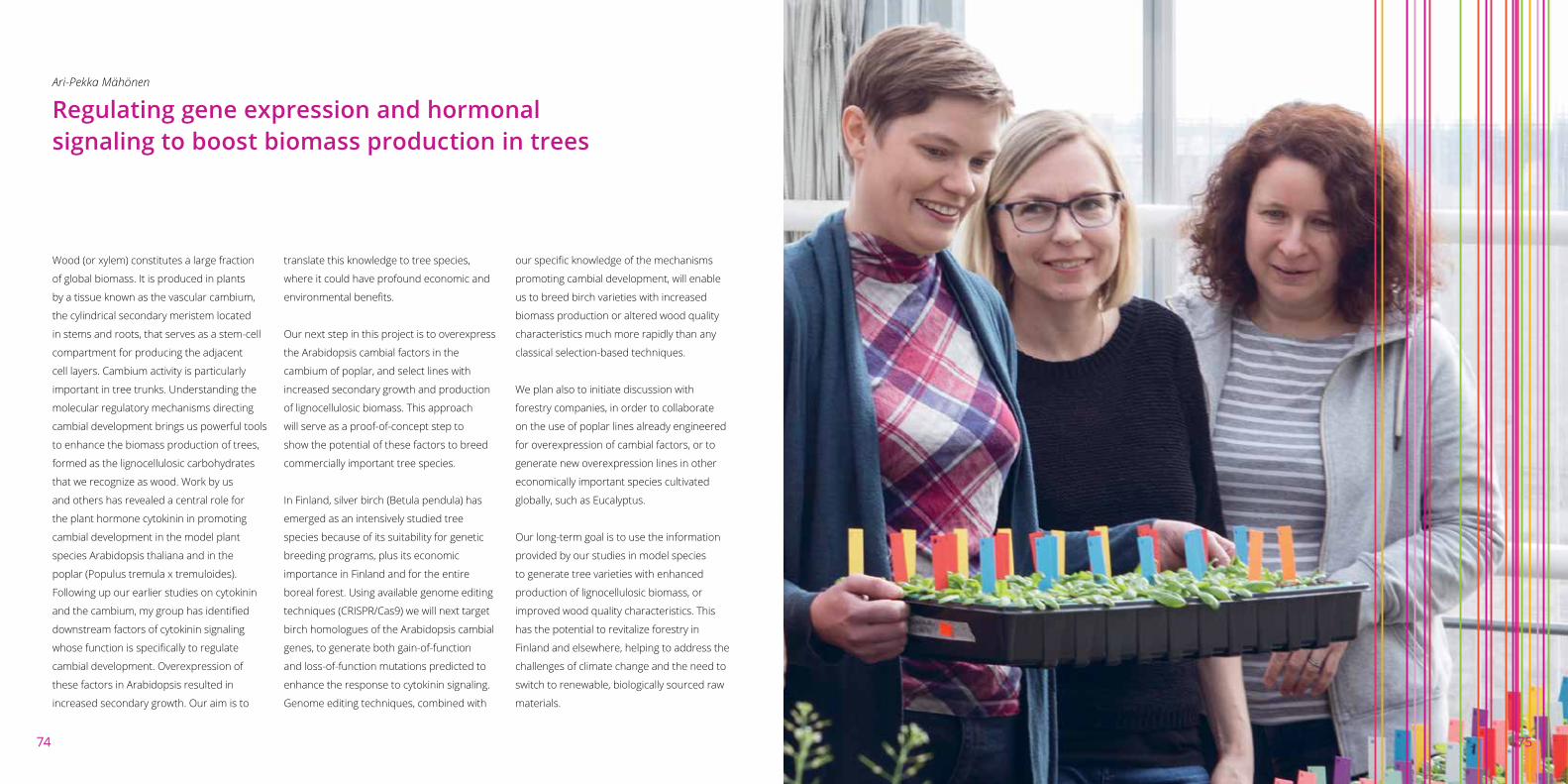
7574
Ari-Pekka Mähönen
Regulating gene expression and hormonal signaling to boost biomass production in trees
Wood (or xylem) constitutes a large fraction
of global biomass. It is produced in plants
by a tissue known as the vascular cambium,
the cylindrical secondary meristem located
in stems and roots, that serves as a stem-cell
compartment for producing the adjacent
cell layers. Cambium activity is particularly
important in tree trunks. Understanding the
molecular regulatory mechanisms directing
cambial development brings us powerful tools
to enhance the biomass production of trees,
formed as the lignocellulosic carbohydrates
that we recognize as wood. Work by us
and others has revealed a central role for
the plant hormone cytokinin in promoting
cambial development in the model plant
species Arabidopsis thaliana and in the
poplar (Populus tremula x tremuloides).
Following up our earlier studies on cytokinin
and the cambium, my group has identified
downstream factors of cytokinin signaling
whose function is specifically to regulate
cambial development. Overexpression of
these factors in Arabidopsis resulted in
increased secondary growth. Our aim is to
translate this knowledge to tree species,
where it could have profound economic and
environmental benefits.
Our next step in this project is to overexpress
the Arabidopsis cambial factors in the
cambium of poplar, and select lines with
increased secondary growth and production
of lignocellulosic biomass. This approach
will serve as a proof-of-concept step to
show the potential of these factors to breed
commercially important tree species.
In Finland, silver birch (Betula pendula) has
emerged as an intensively studied tree
species because of its suitability for genetic
breeding programs, plus its economic
importance in Finland and for the entire
boreal forest. Using available genome editing
techniques (CRISPR/Cas9) we will next target
birch homologues of the Arabidopsis cambial
genes, to generate both gain-of-function
and loss-of-function mutations predicted to
enhance the response to cytokinin signaling.
Genome editing techniques, combined with
our specific knowledge of the mechanisms
promoting cambial development, will enable
us to breed birch varieties with increased
biomass production or altered wood quality
characteristics much more rapidly than any
classical selection-based techniques.
We plan also to initiate discussion with
forestry companies, in order to collaborate
on the use of poplar lines already engineered
for overexpression of cambial factors, or to
generate new overexpression lines in other
economically important species cultivated
globally, such as Eucalyptus.
Our long-term goal is to use the information
provided by our studies in model species
to generate tree varieties with enhanced
production of lignocellulosic biomass, or
improved wood quality characteristics. This
has the potential to revitalize forestry in
Finland and elsewhere, helping to address the
challenges of climate change and the need to
switch to renewable, biologically sourced raw
materials.
75

7776
Alan Schulman
A tool for improving the efficiency and targeting of gene modifications in plants
Production of transgenic plants for
agriculture is increasingly important
worldwide; 182 million hectares of GM
crops were planted in 2014, with an annual
increase of 3-4 %. Soybean (79% GM), maize
(32%), cotton (70%) and rapeseed (24%)
have the greatest proportion of total area
in GM varieties. However, whilst wheat and
barley are both in the world’s top four cereal
crops, no GM barley or wheat is grown. One
limitation is that only a few varieties are
transformable; frequencies above 25% are
seldom achieved in barley, and are far lower
in wheat. Another, more general problem is
the lack of a targeting mechanism to direct
the transgene to a particular site in the
genome. With current methods, a transgene
may be integrated anywhere. Given the
high proportion of heterochromatin in
barley, wheat and other large genomes,
most insertions occur in areas with poor
expression. As a result, many transgenic lines
must be produced in order to recover just
one with suitable expression levels. There
is also the issue of silencing due to position
effects, which can occur even if transgene
expression is initially satisfactory.
A solution to this problem is provided by
retrotransposons and retroviruses, nature’s
genetic engineers, which insert their own
genomes into the DNA of the cell as part of
their natural life cycle. Retrotransposons are
abundant, non-infectious components of all
plant genomes, carrying out their transposition
using an enzyme called integrase. We have
studied the retrotransposon BARE, found in
both barley and wheat, as well as in related
cereals, for over 25 years, accruing a large
body of knowledge of its life-cycle and
integrase function.
We propose to develop BARE integrase as a
commercially viable transformation vector for
the targeting of transgenes to specific locations
in cereal genomes. Although the integrase
normally inserts its substrate semi-randomly
throughout the genome, specificity can be
achieved by tethering the integrase to another
protein with DNA or protein-binding capacity.
For example, linking integrase to a particular
transcription factor will lead to integration
wherever in the genome that transcription
factor binds. Proof-of-concept has been
achieved using retroviral (e.g. HIV-1) integrases
to create targeted integrations for gene
therapy. Investment is now needed to develop
tethered integrases and transformation
complexes for cereal genome modification,
extending this proof-of-concept to BARE.
The resulting technology would offer many
benefits. It could be used as a proprietary
system for in-house transformation, offered
as a service for plant breeding companies,
or licensed further. It could in principle be
tailored for targeted integration of transgenes
in any crop species where BARE integrase
will function. Because integrases are highly
conserved, this is likely to cover a wide
species spectrum, certainly including the
Triticeae (wheat, barley, rye, timothy) and
Pooideae (e.g. oats).
Competing technologies are problematic.
Homologous recombination for specific
targeting is possible, but is extremely inefficient
and needs selection. Specific removal of
genes has been reported with the piggyBac
insect transposon. The CRISPR/Cas9 system
is becoming widely used for site-specific
mutagenesis, but still depends on homologous
recombination for targeted insertion. Thus,
tethered BARE integrase should have a secure
place in a potentially vast market.
76

7978
Alan Schulman
A robust resistance gene for yellow (stripe) rust
Wheat is the world’s second most important
grain crop, planted on 219 million Ha
and yielding 716 million tons worldwide
(FAOSTAT, 2013). Wheat supply and demand
has seesawed over recent years, with eight
seasons of surpluses and 12 of deficits
over the last two decades. As witnessed
by the North African food riots of 2008,
political stability is closely linked to wheat
prices, which are in turn affected by wheat
production. The yellow (or stripe) rust disease
is today one of the most signficant threats to
wheat production. It is caused by the fungus
Puccinia striiformis f. sp. tritici, the spores
of which can be wind-dispersed over long
distances, even thousands of kilometers.
While the best strategy to overcome
the disease is to breed resistant wheat,
the pathogen has repeatedly developed
immunity against available resistance genes
by evolving increased virulence.
Wild emmer wheat, Triticum dicoccoides,
is an important source for novel yellow-
rust resistance (Yr) genes, which provide a
potential solution. Yr15, a major gene located
on chromosome 1BS of T. dicoccoides, was
previously reported to confer resistance to a
broad spectrum of stripe-rust isolates, at both
seedling and adult plant stages. Introgressions
of Yr15 into cultivated T. aestivum (bread
wheat) and T. durum (pasta wheat) have been
widely used since the 1980s, but remain
problematic, due largely to associated “linkage
drag” of undesirable flanking genes.
We have been working on a long-term project
to clone Yr15, using a variety of genetic and
genomic methods. We currently have a
candidate gene identified and under testing
for verification. The candidate gene is not a
typical NBS-LRR type resistance gene, which
explains why the resistance conferred by Yr15
has, unusually, not been broken in the 30
years since it was first used. Final confirmation
of the Yr15 gene identity will require a
combination of knockout and gain-of-function
strategies. Knowledge of the molecular details
of the resistance gene (sequence, control
of expression, natural variation) will enable:
1) direct transfer of the gene without the
problem of linkage drag; 2) editing of the gene
for introducing new variants to overcome
any emerging resistance; 3) potential design
of new resistance genes for other crops and
fungal diseases or direct use of Yr15 in barley
and related cereals.
The confirmed Yr15 gene can be made
available on a commercial basis for in-house
or licensed use by plant breeding companies.
Its benefits will include the development not
only of improved disease-resistant bread-
and durum-wheat varieties, but of fungal
pathogen-resistant strains of other crops.
The Yr15 gene has no current competition,
as it is the only gene that confers sustained
resistance to all known yellow-rust isolates.
Other resistance genes tend to lose
effectiveness over time, as the fungal strains
evolve. Thus, Yr15 can provide a unique and
robust answer to a major scourge of world
agriculture.
79


![Lithium-Ion battery SOC estimation...defines the cell’s internal resistance and the cell’s dynamics [14]. Hysteresis is a part of cell behavior and exhibits the dependency of](https://img.dokumen.tips/doc/110x75/5ebb5ced0c2acc01ef418f19/lithium-ion-battery-soc-estimation-deines-the-cellas-internal-resistance.jpg)