Embed Size (px)
Citation preview

Post-print de Current Chemical Biology 2014, 8, 10-16
Biosynthetic Pathways to Glycosidase Inhibitors
Fernando Gomollón-Bel, Ignacio Delso, Tomás Tejero and Pedro Merino*
Laboratorio de Síntesis Asimétrica. Departamento de Síntesis y Estructura de Biomoléculas. Instituto de
Síntesis Química y Catálisis Homogénea (ISQCH). Facultad de Ciencias. Universidad de Zaragoza-
CSIC. Campus San Francisco. 50009 Zaragoza, Aragón. Spain
ABSTRACT
Glycosidase inhibitors are important compounds that can interfere with several
biosynthetic processes including N-linked glycosylation and the biosynthesis of several
glycoproteins. Understanding the biogenesis of naturally occurring glycosidase
inhibitors would be a crucial step towards the chemical synthesis of analogues of
choice. This review focuses on the current knowledge regarding the biosynthesis of a
series of polyhydroxylated saturated nitrogen heterocycles including nojirimycin and
swainsonine among others, with a potent biological activity as inhibitors of glycosidases
and transglycosidases.
KEYWORDS
Biosynthesis / Glycosidase Inhibitors / Nojirimycin / Deoxynojirimycin / Swainsonine

Post-print de Current Chemical Biology 2014, 8, 10-16
1. Introduction
Glycosydase inhibitors are a rapidly growing family of molecules mostly
consisting of polyhydroxylated mono- and bicyclic saturated nitrogen heterocycles
commonly referred to as iminosugars (Figure 1).[1-3] Compounds like nojirimycin 1, 1-
deoxynojirimycin 2, DMDP 3, castanospermine 4 or swainsonine 5 and their derivatives
play crucial roles in the biological activities of some pharmaceutically important
compounds. [4-6]
Figure 1. Glycosidase inhibitors
Nojirimycin 1 was originally isolated from cultures of several strands of
Streptomyces [7,8] and Bacillus[9] and 1-deoxynojirimycin was isolated from plants of
genus Morus.[10] Compounds 1 and 2 inhibit various glycosidases having important
effects on the biosynthesis of membrane and secretory glycoproteins.[11] DMDP 3 can
be isolated from the cyanobacterial genus Cylindrospermum and it is capable of
effectively inhibiting digestive glycosidases.[12] The 6-deoxyderivative 4 has been
isolated from Angylocalyx pynaertii and in contrast to other polyhydroxylated
pyrrolidines it was found to be unique in inhibiting β–mannosidase.[13]
Castanospermine 5 was first isolated from the seeds of Castanosperma australe[14] and
it has demonstrated antiviral activity.[15,16] It is also known that castanospermine
interfere with the metabolism of glycogen[17] and it inhibits several glycosidases.[18]
Swainsonine 6 was first isolated from swainsona in Australia but it is also present in
numerous plants and fungi. Compound 6 has antitumoral activity[19,20] although some
clinical trials were discouraging.[21] Other natural pyrrolidine alkaloids like alexine 7,
australine 8 and casuarine 9 have also been isolated from plants and
microorganisms.[22]

Post-print de Current Chemical Biology 2014, 8, 10-16
Calystegines were found in the medicinal plant Atropa belladonna and consist of
a nortropane skeleton with three or four hydroxyl groups (Figure 2). There are up to 14
different structures of natural occurring calystegines isolated from a variety of vascular
plants. They cannot be found in fungi or microorganisms. The chemistry and biology of
calystegines including chemotaxonomy, biological activity and some insights on the
biogenesis in the context of co-occurrence with tropane alkaloids have been compiled
by Dräger in an excellent review[23] and a chapter book,[24] covering literature from
1998 to middle 2003 and up to 2007, respectively. Since then there has not been
relevant communications in the topic; so, in this review calystegines will not be treated
and for previous work the reader is directed to the above mentioned reviews.
Figure 2. Calystegines
The interest in glycosidase inhibitors has increased enormously during the past
two decades and the number of synthetic approaches to their preparation is extremely
extensive and growing at a rapid rate.[25,26] One reason for this huge synthetic activity
is the great variety of biological activity against different enzymes that can be found
depending on the absolute configuration of the stereogenic centers bearing the hydroxyl
groups.
The mechanism of the various existing glycosidases is known to proceed
through oxocarbenium-like transition structures[27,28] and it is well-accepted that
inhibition of typical glycosidase inhibitors occur because such sugar mimics resemble
the structural features of the transition state.[29] Different configurations as well as
conformational restrictions in inhibitors help to a better recognition by the enzyme
contributing to a higher inhibition activity.[30]
In the large group of glycosidase inhibitors, synthetic studies have already been
highlighted in several classes,[31-33] e.g. for pyrrolidines,[34,35] piperidines,[36,37]
bicyclic compounds such as indolizidines[38] and pyrrolizidines[39,40] and imino
disaccharides.[41,42] However, only a little is known about biosynthetic routes towards
that sort of compounds. It can be expected that the knowledge of the biosynthetic

Post-print de Current Chemical Biology 2014, 8, 10-16
pathways can be used to manipulate the metabolite pattern of involved microorganisms,
directing the fermentation process to produce desired metabolites.
In this review, we summarize the current knowledge regarding the biosynthesis
of naturally occurring glycosidase inhibitors. Semisynthetic studies on the structural and
pharmaceutical properties of these compounds have been extensively reported and will
not be covered here. Similarly, for detailed information of natural occurrence of
discussed molecules and precise synthetic approaches the reader is referred to previous
reviews. On the other hand, experimental evidences supporting the existence of various
routes in microorganisms will be discussed.
2. Biosynthesis of monocyclic compounds. Piperidines and pyrrolidines
Nojririmycin 1 has been isolated from a variety of microorganisms including S.
roseochromogenes, S. lavendulae, S. nojiriensis and S. subrutilus. The last one, when
grown on a glucose-containing soyabean medium produces both 1-
deoxymannojirimycin and 1-deoxynojirimycin. Experiments with deuterated glucose
showed incorporation of deuterium at C-6 in both alkaloids indicating that the first step
in the biosynthesis of both iminosugars is the isomerization of glucose to fructose.
Accordingly, it is proposed mannojirimycin 17 as the first iminosugar to be formed.[43]
In fact, when 6,6-[2H2]-glucose was employed, NMR analysis of deuterium
labellled 1-deoxynojirimycin 2 showed that only the equatorial proton at C-1 had been
replaced by deuterium, in agreement with the oxidation of the primary hydroxyl group
at C-6 in fructose to give 13 with the loss of one hydrogen atom. The introduction of the
amino group is certainly unknown and three ways are possible through derivatives 14-
16, all of them being possible precursors of mannojirimycin 17.[44] Elimination of
water from 17 and further reduction afforded the observed 1-deoxymannojirimycin 19.
This route was confirmed by using 5-[2H]-glucose in the fermentation. Under such
conditions deuterium was only incorporated at C-2 in mannojirimycin 17. The 1-
deoxynojirimycin 2 obtained in this experiment did not show incorporation of
deuterium. This is in agreement with the hypothesis that either mannojirimycin 17 or its
1-deoxy derivative 19 are precursors in the biosynthetic scheme, since loss of hydrogen
isotope from C-2 would be expected upon epimerization of 17 or 19. Further
experiments with deuterated substrates deomonstrated that epimerization occurs
predominantly at nojirimycin level, i.e. between 17 and 1. It had been reported that 1-
deoxynojirimycin 2 could be epimerized to 1-deoxymannojirimycin 19 by a strain of

Post-print de Current Chemical Biology 2014, 8, 10-16
Agrobacterium sp. trough an oxidation to a cyclic ketocompound followed by reduction
to give the epimeric derivative. However, by considering this hypothesis it is difficult to
justify the presence of nojirimycin 1.
Scheme 1. Biosynthesis of mannojirimycin 17, nojirimycin 1 and their deoxy
derivatives 19 and 2 from D-glucose in Streptomyces subrutilus
Similar experiments carried out with Bacillus subtilis var niger only produced 1-
deoxynojirimycin 2 and no traces of 1-deoximannojirimycin 19 were found.[45] Also in
this case, labeling studies demonstrated that glucose is the precursor of 1-
deoxynojirimycin 2. Additional enzyme assays and labeling studies supported that both
mannojirimycin 17 and nojirimycin1 are intermediates in the biosynthesis of 1-
deoxynojirimycin 2.
Recently, the complete genome sequence of Bacillus amiloliquefaciens has been
determined[46] and a gene cluster that initiates the biosynthesis of 2 in such
microorganism has been identified and provided further evidence for the pathway
illustrated in Scheme 1.[47] Additionally, three enzymes involved in the first steps of

Post-print de Current Chemical Biology 2014, 8, 10-16
the biosynthesis have also been identified. Noteworthy, the same gene cluster has been
found in Bacillus atrophaeus[48] as well as in Bacillus subtilis,[49] known producers of
1-deoxynojirimycin 1.
The biosynthetic route to 1-deoxynojirimycin 2 is, however, different for higher plants
as demonstrated by Shibano and co-workers.[50] These authors studied the biosynthesis
of 2 by using 1-[13
C]-glucose in the higher plant Commelia communis. While a
significant 13
C enrichment was observed at C-6 for compound 2 obtained in
microorganisms, in the case of that being produced in plants the 13
C enrichment was
located at C-1. These experiments resulted in the proposal outlined in Scheme 2.
According to this hypothesis C-1/C-5 cyclization is produced in the original glucose
molecule without any type of inversion. Additional support was provided by the fact
that the same 13
C enrichment was observed in fructose obtained from administration of
1-[13
C]-glucose.
Scheme 2. Biosynthesis of nojirimycin 1 and1-deoxynojirimycin 2 from D-glucose in
Commelia communis.
When imination is produced on fructose, formed by isomerization of glucose, the same
process led to DMDP 3 (Scheme 3). Indeed, compound 3 is obtained from 1-[13
C]-
glucose under the same conditions employed for the preparation of 1-deoxynojirimycin
2.[50]
Post-print de Current Chemical Biology 2014, 8, 10-16
Scheme 3. Biosynthesis of DMDP 3 from D-glucose in Commelia communis.
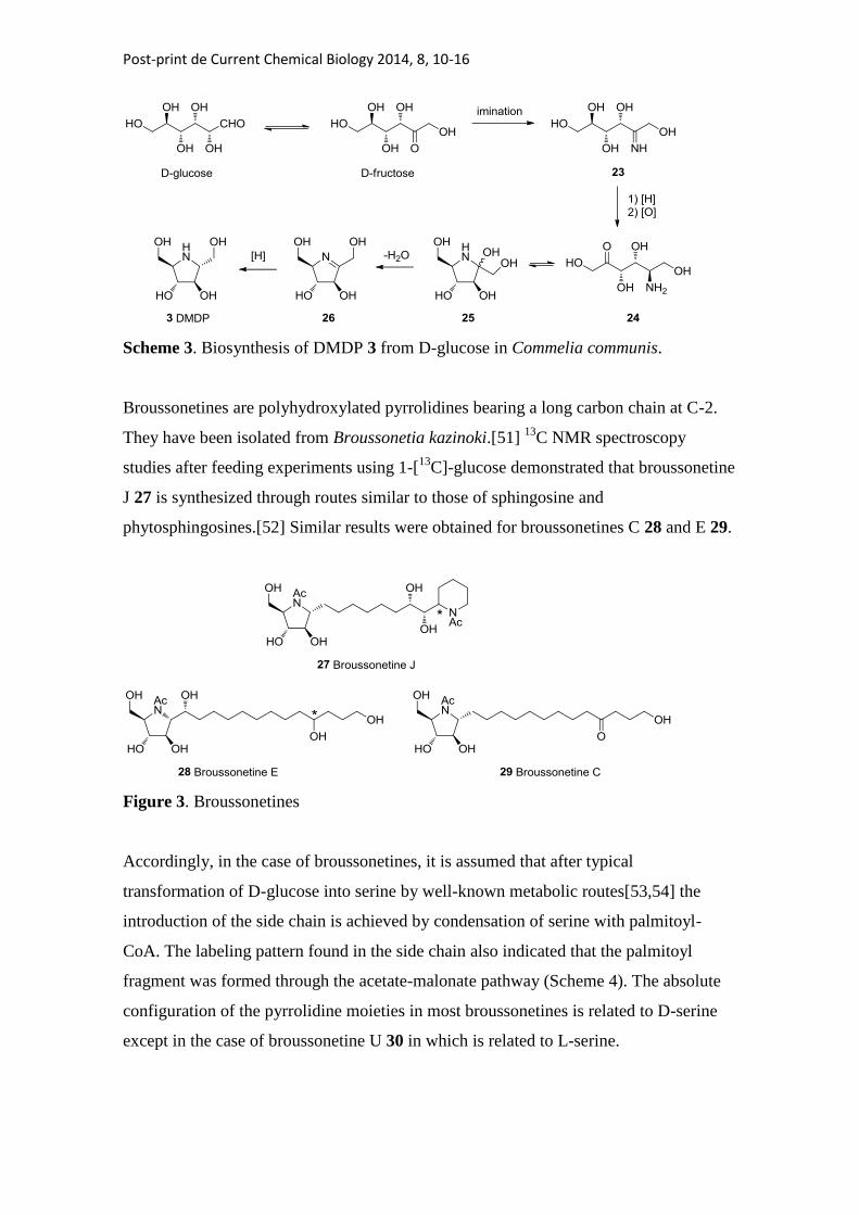
Broussonetines are polyhydroxylated pyrrolidines bearing a long carbon chain at C-2.
They have been isolated from Broussonetia kazinoki.[51] 13
C NMR spectroscopy
studies after feeding experiments using 1-[13
C]-glucose demonstrated that broussonetine
J 27 is synthesized through routes similar to those of sphingosine and
phytosphingosines.[52] Similar results were obtained for broussonetines C 28 and E 29.
Figure 3. Broussonetines
Accordingly, in the case of broussonetines, it is assumed that after typical
transformation of D-glucose into serine by well-known metabolic routes[53,54] the
introduction of the side chain is achieved by condensation of serine with palmitoyl-
CoA. The labeling pattern found in the side chain also indicated that the palmitoyl
fragment was formed through the acetate-malonate pathway (Scheme 4). The absolute
configuration of the pyrrolidine moieties in most broussonetines is related to D-serine
except in the case of broussonetine U 30 in which is related to L-serine.

Post-print de Current Chemical Biology 2014, 8, 10-16
Scheme 4. Biosynthesis of broussonetine J from D-glucose in Broussonetia kazinoki.
3. Biosynthesis of bicyclic compounds. Indolizidines.
The biosynthesis of the piperidine nucleus of bicyclic iminosugars starts with the
production of pipecolic acid, which is the precursor of several compounds such as
swainsonine or slaframine. Pipecolic acid was found to be a product of lysine
catabolism in animals, microorganisms and plants. Grobbelaar and Steward established
the transformation oflysine into pipecolic acid through route A (Scheme 5) by using
labeled lysine in bean plants Phaseolus vulgaris.[55] According to their findings the
nitrogen of the pipecolic acid should be supplied by the α-amino group of the lysine.
Scheme 5. Biosynthesis of pipecolic acid from lysine.

Post-print de Current Chemical Biology 2014, 8, 10-16
On the other hand, Gupta and Spenser demonstrated that in rats the conversion
of lysine into pipecolic acid proceeds via ε-amino-α-ketocapric acid 35.[56] This
mechanism indicated that the nitrogen atom of the pipecolic acid should be supplied by
the ε-amino group of lysine (Scheme 5, route B). Similar experiments carried out with
Neurospora crassa and Phaseolus vulgaris afforded identical results being in conflict
with previous findings. Further studies in animals and plants demonstrated that both
routes A and B, illustrated in Scheme 5, distinguishable at the loss of a particular amino
group of the lysine, are possible. In fact, there are several experimental evidences
supporting the existence of various routes in microorganisms, as well as specific
enzymes involved in some steps. This topic has been reviewed elsewhere[57] and the
reader is referred to that publication for more details concerning the biosynthesis of
pipecolic acid.
The biosynthesis of slaframine 42 has been studied in Rhizoctonia leguminicola
a fungus that causes black spot disease of red clover.[58] By using radiolabelled 1-
[14
C]-lysine and 6-[14
C]-lysine it has been demonstrated their incorporation into
slaframine.[59] Moreover, that incorporation was efficiently blocked by adding
pipecolic acid, thus indicating that pipecolic acid is an intermediate in the prcess of
biogenesis. The same authors also studied the origin of pipecolic acid in Rhizoctonia
leguminicola and verified the biosynthetic pathway illustrated in Scheme 5.[60] Further
experiments with radiolabelled pipecolic acid showed incorporation of radioactivity to
slaframine in the expected positions.
The origin of the pyrrolidine ring of slaframine was also investigated[61] and it was
found to be formed from malonic acid and acetic acid. Spectrometric analysis of
radiolabelled/deuterated compounds indicated that the methyl carbon of acetate is joined
to the carboxyl carbon of the pipecolate. These results suggest the formation of
intermediate 38 by acylation of malonate with pipecolic acid (Scheme 6). Furthermore,
preparation of deuterated 40 allowed to identify this compound as an advanced
intermediate in the biogenesis of slaframine[62] and to propose 1-
oxooctahydroindolizine 39 as the intermediate precursors of 40. In addition to slframine
42, swainsonine 6 was also isolated from Rhizoctonia leguminicola.[63] By employing
perdeutero pipecolic acid it was demonstrated that both slaframine and swainsonine
have common precursors in their biogenesis.[64]

Post-print de Current Chemical Biology 2014, 8, 10-16
Scheme 6. Biogenesis of slaframine from lysine in Rhizoctonia leguminicola
.
In the case of swainsonine, compound 39 is reduced by the other face providing
43. Oxidation at C-8a of this intermediate should be postulated, probably through an
iminium ion, with subsequent reduction by the appropriate face to provide the R
configuration of swainsonine 6 (Scheme 6). Further experiments with deuterated
compounds allowed to corroborate that hypothesis.[65]
The biosynthesis of swainsonine 6 has also been studied in plants. In particular,
studies carried out with Astragalux oxyphysus showed that swainsonine 6 is
biosynthesized in that plant by a very similar pathway (if not identical) to that in the
fungus by incorporating pipecolic acid into the swainsonine skeleton.[66] On the other
hand, it has been observed that the plant does not produce slaframine 42; neither does it
produce intermediates 40 and 41.
The role of both pipecolic acid and malonic acid in the biosynthesis of
swainsonine 6 has also been pointed out by stimulating production of such alkaloid by
transformed root cultures of Swainsona galegifolia.[67]
4. Conclusions
Polyhydroxylated saturated nitrogen heterocyles provided a variety of biosynthetic
challenges. Up to now, several aspects related to their biogenesis have been revealed.
However, there is still much work to do. Further investigations are still required to
clarify the biosynthetic enzymes involved in the catalytic processes. Such enzymes

Post-print de Current Chemical Biology 2014, 8, 10-16
should be of a great value for enzymatic approaches to iminosugars and their
biomimetic synthesis. There are experimental evidences indicating that different
biosynthetic mechanisms operate on diverse microorganisms (for instance Bacillus
subtilis vs. Streptomyces subrutilus for 1-deoxynojirimycin) or higher plants. Very
recently, a gene cluster has been identified providing evidence of the biosynthetic
pathways. In this regard, the catalytic mechanism of individual domains should continue
to be probed by using standard mutagenesis techniques with studies involving purified
enzymes. By acquiring this knowledge it will be possible to design new analogues by
developing more sophisticated and improved strains. The fields of synthetic organic
chemistry and biochemistry will be united by employing new bioorganic tools for
further chemical elaboration of new compounds of pharmaceutical interest.
Acknowledgments
This study was supported by the Ministerio de Ciencia e Innovacion (MICINN) and
FEDER Program (Madrid, Spain, project CTQ2010-19606) and the Gobierno de
Aragon (Zaragoza, Spain, Bioorganic Chemistry Group. E-10). F.G.-B. thanks CSIC for
a JAE predoctoral grant.
References
[1] Nash RJ, Asano N, Watson AA. In: Pelletier SW, Ed. Alkaloids: Chemical and
Biological perspectives. Oxford: Elsevier 1996; 345-76.
[2] Nishimura Y. In: Atta-ur-Rahman, Ed. Studies in natural products chemistry.
Oxford: Elsevier 1999; 495-583.
[3] Asano N. Glycosidase inhibitors: update and perspectives on practical use.
Glycobiology 2003; 13: 93R-104R
[4] Stütz, AE, Ed. Iminosugars as glycosidase inhibitors. Nojirimycin and beyond
Weinheim: Wiley-VCH 1999
[5] Compain P. Martin OR, Eds. Iminosugars. From synthesis to therapeutic
applications. Chichester: John Wiley & sons 2007

Post-print de Current Chemical Biology 2014, 8, 10-16
[6] Winchester BG. Iminosugars: from botanical curiosities to licensed drugs.
Tetrahedron: Asymmetry 2009; 20: 645-51
[7] Ishida N, Kumagai K, Niida T, Tsuruoka T, Yumoto H. Nojirimycin, a new
antibiotic. II. Isolation, characterization and biological activity. J. Antibiot. Ser. A,
1967; 20: 66-71.
[8] Inouye S, Tsuruoka T, Ito T, Niida T. Structure and synthesis of nojirimycin.
Tetrahedron 1968; 24; 2125-44
[9] Schmidt DD, Frommer W, Muller L, Truscheit E. Glucosidase inhibitors from
Bacilli. Naturwissenschaften 1979; 66: 584-5.
[10] Yagi, M.; Kouno, T.; Aoyagi, Y.; Murai, H. The structure of moranoline, a
piperidine alkaloid from Morus species. Nippon Nogei Kagaku Kaishi 1976; 50: 571-2.
[11] Peyrieras N, Bause E, Legler G, Vasilov L, Claesson L, Peterson P, Ploegh H.
Effects of the glucosidase inhibitors nojirimycin and deoxynojirimycin on the
biosynthesis of membrane and secretory glycoproteins. EMBO J 1983; 2: 823-32.
[12] Juttner F, Wessel HP. Isolation of di(hydroxymethyl)dihydroxypyrrioldine from
the cyanobacterial genus Cyclindrospermum. J Phycol 2003; 39: 26-32
[13] Molyneux RJ, PanYT, H.; Tropea JE, Elbein AD, Lawyer CH, Hughes DJ, Fleet
GWJ. 2-hydroxymethyl-3,4-dihydroxy-6-methyl-`yrroline (6-deoxy-DMDP), an
alkaloid beta-mannosidase inhibitor from seeds of angylocalyz pynaertii. J Nat Prod
1993; 56: 1356-1364
[14] Hohenschutz LD, Bell AE, Jewess PJ, Leworthy DP, Pryce RJ, Arnold E, Clardy
J. Castanospermine, a 1,6,7,8-tetrahydroxyoctahydroindolizine alkaloid, from seeds of
Castanospermum australe Phytochemistry 1981; 20: 811-4.

Post-print de Current Chemical Biology 2014, 8, 10-16
[15] Whitby K, Pierson TC, Geiss B, Lane K, Engle M, Zhou Y, Doms RW,
Diamond MS. Castanospermine, a potent inhibitor of dengue virus infection in vitro and
in vivo. J Virol. 2005; 79: 8698-706.
[16] Walker BD, Kowalski M, Goh WC, Kozarsky K, Krieger M, Rosen C,
Rohrschneider L, Haseltine WA, Sodroski J. Inhibition of human immunodeficiency
virus syncytium formation and virus replication by castanospermine. PNAS 1987; 84:
8120-4.
[17] Saul R, Ghidoni JJ, Molyneaux RJ, Elbein AD. Castanospermine inhibits alpha-
glucosidase activities and alters glycogen distribution in animals. PNAS 1985; 82: 93-
97.
[18] Saul R, Chambers JP, Molyneaux RJ, Elbein AD. Castanospermine, a
tetrahydroxylated alkaloid that inhibits β-glucosidase and β-glucocerebrosidase. Arch
Biochem Biophys 1983; 221: 593-5.
[19] Sun JY, Zhu MZ, Wang SW, Miao S, Xie YH, Wang JB. Inhibition of the
growth of human gastric carcinoma in vivo and in vitro by swainsonine. Phytomedicine:
Int J Phytother Phytopharm 2007; 14: 353-9.
[20] Sun YJ, Yang H, Miao S, Li JP, Wang SW, Zhu MZ, Xie YH, Wang JB, Liu Z,
Yang Q. Suppressive effects of swainsonine on C6 glioma cell in vitro and in vivo.
Phytomedicine: Int J Phytother Phytopharm 2009; 16: 1070-4.
[21] Shaheen PE, Stadler W, Elson P, Knox J, Winquist E, Bukowski RM. Phase II
study of the efficacy and safety of oral GD0039 in patients with locally advanced or
metastasic renal cell carcinoma. Investig New Drugs 2005; 23: 577-81.
[22] For an excellent and comprehensive review on the natural occurrence of a great
variety of sugar-mimic glycosidase inhibitors see: Asano N, Nash RJ, Moylneux RJ,
Fleet GWJ. Sugar-mimic glycosidase inhibitors: natural occurrence, biological activity

Post-print de Current Chemical Biology 2014, 8, 10-16
and prospects for therapeutic application. Tetrahedron: Asymmetry 2000; 11: 1645-
1680.
[23] Dräger B. Chemistry and biology of calystegines. Nat Prod Rep 2004; 21: 211-
23.
[24] Biastoff S, Dräger B. In: Cordell GA. Ed, The alkaloids. Chemistry and Biology.
New York: Academic Press 2007; 49-102.
[25] Compain P, Chagnault V, Martin OR. Tactics and strategies for the synthesis of
iminosugar C-glycosides: a review. Tetrahedron: Asymmetry 2009, 20, 672-711.
[26] Cipolla L, Ferla BL, Gregori M. Combinatorial approaches to iminosugars as
glycosidase and glycosyltransferase inhibitors. Comb Chem High Throug Screen 2006;
9: 571-82.
[27] Zechel DL, Whiters SG. Glycosidase Mechanisms: Anatomy of a Finely
Tuned Catalyst. Acc. Chem. Res. 2000, 33, 11-18.
[28] Rye CS, Withers SG. Glycosidase mechanisms Curr Opin Chem Biol 2000; 4:
573-80.
[29] Gloster TM, Davies GJ. Glycosidase inhibition: assessing mimicry of the
transition state. Org Biomol Chem 2010; 8: 305-20.
[30] Gloster TM, Madsen R, Davies GJ. Dissection of conformationally restricted
inhibitors binding to a beta glucosidase ChemBioChem 2006; 7: 738-42.
[31] Davis BG. A silver-lined anniversary of Fleet iminosugars: 1984-2009, from
DIM to DRAM to LABNAc Tetrahedron: Asymmetry 2009; 20: 652-71.
[32] Lillelund VH, Jensen HH, Liang X, Bols M. Recent Developments of
Transition-State Analogue Glycosidase Inhibitors of Non-Natural Product Origin Chem
Rev 2002; 102: 515-53.

Post-print de Current Chemical Biology 2014, 8, 10-16
[33] Tatsuka K In: Chapelur, Y. Ed, Carbohydrate mimics. Weinheim: Wiley-VCH.
1998, 283-305.
[34] Stocker BL, Dangerfield EM, Win-Mason AL, Haslett GW, Timmer MSM.
Recent Developments in the Synthesis of Pyrrolidine-Containing Iminosugars. Eur J
Org Chem 2010; 1615-37.
[35] Ayad T, Genisson Y, Baltas M. Chemical Approaches Towards Synthesis of
Some Naturally Occurring Iminosugars. Curr Org Chem 2004; 8: 1211-33.
[36] Pearson MSM, Mathe-Allainmat M, Fargeas V, Lebreton J. Recent Advances in
the Total Synthesis of Piperidine Azasugars Eur J Org Chem 2005; 2159-91.
[37] Dragutan H, Dragutan V, Demonceau A. Targeted drugs by olefin metathesis:
piperidine-based iminosugars. RSC Advances 2012; 2: 719-36.
[38] Kim IS, Jung YH. Recent Advances in the Total Synthesis of Indolizidine
Iminosugars Heterocycles 2011; 83: 2489-507.
[39] Pyne SG. Recent developments on the synthesis of (-)-swainsonine and
analogues. Curr Org Synth 2005; 2: 39-57.
[40] Brandi A, Cardona F, Cicchi S, Cordero FM, Goti A. Stereocontrolled Cyclic
Nitrone Cycloaddition Strategy for the Synthesis of Pyrrolizidine and Indolizidine
Alkaloids. Chem Eur J 2009; 15: 7808-21.
[41] Merino P, Delso I, Marca E, Tejero T, Matute R. Chemistry and Biology of
Iminosugar Di- and Oligosaccharides. Curr Chem Biol 2009; 3: 253-71.
[42] Robina I, Vogel P. Synthesis of aza-C-disaccharides (dideoxyimino-alditols C-
linked to monosaccharides) and analogues. Synthesis 2005; 675-702.
[43] Hardick DJ, Hutchinson DW, Trew SJ, Wellington, EMH. The biosynthesis of
deoxynojirimycin and deoxymannonojirimycin in Streptomyces subrutilus. Chem
Commun 1991; 729-30.

Post-print de Current Chemical Biology 2014, 8, 10-16
[44] Hardick DJ, Hutchinson DW, Trew SJ, Wellington EMH. Glucose is a Precursor
of 1-deoxynojirimycin and 1-deoxymannonojirimycin in Streptomyces subrutilus.
Tetrahedron 1992; 48: 6285-96.
[45] Hardick DJ, Hutchinson DW. The Biosynthesis of l-Deoxynojirimycin
in Bacillus subtilis var niger Tetrahedron 1993; 49: 6707-10.
[46] Chen XH, Koumoutsi A, Scholz R, Eisenreich A, Schneider K, Heinemeyer I,
Morgenstern B, Voss B, Hess WR, Reva O, Junge H, Voigt B, Jungblut PR, Vater J,
Sussmuth R, Liesegang H, Strittmatter A, Gottschalk G, Borriss R. Comparative
analysis of the complete genome sequence of the plant growth–promoting bacterium
Bacillus amyloliquefaciens FZB42 Nat. Biotech. 2007, 25, 1007-14.
[47] Clark LF, Johnson JV, Horenstein NA. Identification of a Gene Cluster that
Initiates Azasugar Biosynthesis in
Bacillus amyloliquefaciens. ChemBioChem 2011; 12: 2147-50.
[48] Gibbons HS, Broomall S, McNew LA, Daligault H, Chapman C, Bruce D,
Karavis M, McGregor P, Hong C, Park KH, Akmal A, Feldman A, Lin JS, Chang WE,
Higgs BH, Demirev P, Lindquist J, Liem A, Fochler E, Tapia R, Bishop-Lilly K, Detter
C, Han C, Sozhamannan S, Rosenzweig CN, Skowronski E. Genomic Signatures of
Strain Selection and Enhancement in Bacillus atrophaeus var. globigii, a Historical
Biowarfare Simulant. PLoS One 2011; 6: e17836
[49] Kang KD, Cho YS, Song JH, Park YS, Lee JY, Hwang KY, Rhee SK, Chung
JH, Kwon O, Seong SI. Identification of the genes involved in 1-deoxynojirimycin
synthesis in Bacillus subtilis MORI 3K-85. J Microbiol 2011; 431-40
[50] Shibano M, Fujimoto Y, Kushino K; Kusano G, Baba K. Biosynthesis of 1-
deoxynojirimycin in Commelina communis: a difference between the microorganisms
and plants. Phytochemistry 2004; 65:2661-5.

Post-print de Current Chemical Biology 2014, 8, 10-16
[51] Shibano, M, Tsukamoto, D, Inoue, T, Takase, Y, Kusano, G. The biosynthesis of
Broussonetines: Origin of the carbon skeleton. Chem Pharm Bull 2001; 49: 504-506.
[52] Shibano M, Tsukamoto D, Kusano G. Polyhydroxylated alkaloids with lipohilic
moieties as glycosidase inhibitors from higher plants. Heterocycles 2002; 57: 1539-53.
[53] Bismut H, Plas C. Role of serine biosynthesis and its utilization in the alternative
pathway from glucose to glycogen during the response to insulin in cultured foetal-rat
hepatocytes. Biochem J 1991; 276: 577-82.
[54] Kisliuk RL, Sakami W. A study of the mechanism of serine biosynthesis. J Biol
Chem 1955; 214: 47-57.
[55] Grobbelaar N, Steward FC. Pipecolic acid in phaseolus vulgaris: Evidence on its
derivatives from Lysine. J Am Chem Soc 1953; 75: 4341-3.
[56] Gupta RN, Spenser ID. Biosynthesis of piperidine nucleus J Biol Chem 1969;
244: 88-94.
[57] He M. Pipecolic acid in micxrobes: biosynthetic routes and enzymes. J Ind
Microbiol Biotechnol 2006; 33: 401-7.
[58] Guenguerich FP, Broquist HP. In: van Tamelen EE, Ed. Bioorganic Chemistry.
New York: Academic press. 1978. 290-312.
[59] Guenguerich FP, Snyder JJ, Broquist HP. Biosynthesis of slaframine
(1S,6S,8aS)-1-acetoxy-6-aminooctahydroindolizine, a parasympathomimetic alkaloid of
fungal origin. I. Pipecolic acid and salframine synthesis. Biochemistry 1973; 12: 4264-
9.
[60] Guenguerich FP, Broquist HP. Biosynthesis of slaframine (1S,6S,8aS)-1-
acetoxy-6-aminooctahydroindolizine, a parasympathomimetic alkaloid of fungal origin.
II. The origin of pipecolic acid. Biochemistry 1973; 12: 4270-4.

Post-print de Current Chemical Biology 2014, 8, 10-16
[61] Clevenstine EE, Broquist HP, Harris TM. Biosynthesis of slaframine
(1S,6S,8aS)-1-acetoxy-6-aminooctahydroindolizine, a parasympathomimetic alkaloid of
fungal origin. 3. Origin of the pyrrolidine ring. Biochemistry 1979; 18: 3658-63.
[62] Clevenstine EE, Walter P, Harris TM, Broquist HP. Biosynthesis of slaframine
(1S,6S,8aS)-1-acetoxy-6-aminooctahydroindolizine, a parasympathomimetic alkaloid of
fungal origin. 4. Metabolic fate of ethyl pipecolylacetate, 1,3-
diocooctahydsroindolizine, and 1-hydroxyoctahydroindolizine in Rhizoctonia
leguminicola. Biochemistry 1979; 18: 3663-7.
[63] Guenguerich FP, Dimari SJ, Broquist HP. Isolation and characterization of a 1-
pyrindine fungal alkaloid. J Am Chem Soc 1973; 95: 2055-6.
[64] Schneider MJ, Ungemach FS, Broquist HP, Harris TM. Biosynthesis of
swainsonine in rhizoctonia leguminicola. Epimerization at the ring fusion. J Am Chem
Soc 1982; 104: 6863-4.
[65] Harris CM, Schneider MJ, Ungemach FS, Hill JE Harris TM. Biosynthesis of
the toxic indolizidine alkaloids slaframine and swainsonine in Rhizoctonia
leguminicola: Metabolism of 1-hydroxyindolizidines. J Am Chem Soc 1988; 110: 940-
9.
[66] Harris CM, Campbell BC, Molyneux RJ, Harris TM. Biosynthesis of
swainsonine in the diablo locoweed (astragalus oxyphysus). Tetrahedron Lett 1988; 29:
4815-8.
[67] Ermayanti TM, McComb JA, O’Brien PA. Stimulation of synthesis and release
of swainsonine from transformed roots of swainsona galegifolia. Phytochemistry 1994;
36: 313-7.