Embed Size (px)
Citation preview

博 士 論 文(要約)
Biosynthesis and regulation of extracellular
polysaccharides in cyanobacteria.
(シアノバクテリアにおける細胞外多糖の合成・制御機構)
前 田 海 成

【Contents】
page
Abstract 1
Abbreviations 4
General Introduction 6
Outline of the Introduction 6
Definition of EPSs in this thesis 6
Biosynthesis of EPSs in bacteria 7
Biosynthesis of “EPSs” in eukaryotes 11
Biosynthesis of EPSs in cyanobacteria 13
Aim of the study in this thesis 14
Tables and figures 16
Chapter I NDP-sugar pyrophosphorylases in cyanobacteria. 17
I-a) Identification of a novel ubiquitous cyanobacterial UDP-glucose
pyrophosphorylase, CugP. 19
Abstract 20
Introduction 21
Results and Discussion 23
Materials and methods 28
Tables and figures 31
I-b) Identification of various NDP-sugar pyrophosphorylases
in cyanobacteria and bacteria. 41
将来的に学術雑誌に掲載される予定であるため除外

Chapter II
Extracellular cellulose biosynthesis in a thermophilic cyanobacterium,
Thermosynechococcus vulcanus. 43
将来的に学術雑誌に掲載される予定であるため除外
Chapter III
Biosynthesis and regulation of the viscous sulfated EPS in Synechocystis
sp. PCC 6803 substr. PCC-P. 45
特許申請予定であるため除外
General Discussion 47
将来的に学術雑誌に掲載される予定であるため、及び
特許申請予定であるため除外
References 48
Acknowledgements 65


1
Abstract
Extracellular polysaccharides (EPSs) of the microorganisms participate in many
physiological functions such as adhesion, biofilm formation and stress tolerance, and are often related
to pathogenicity of certain bacteria. In addition, EPSs themselves are beneficial natural polymers for
food, thickener, medicine and fiber. Because of these utilities, EPSs and their various biosynthesis
mechanisms have been well studied in many bacteria including pathogens, and various biosynthesis
systems and their regulation for EPSs have been reported. On the other hand, the EPS biosynthesis
mechanisms have been poorly studied in phototrophic cyanobacteria, though EPS related ecologies
such as microbial mat formation, colony formation and tolerance against desiccation and other stresses
have been observed in some cyanobacteria. In addition, production of EPS has been documented in
many cyanobacteria. As cyanobacteria are the member of Bacteria, some molecular systems are very
similar to bacterial ones. However, very little has been elucidated for the cyanobacterial EPS
biosynthesis systems so far, even though genome data of many cyanobacterial species are available
today. This may be, at least partly, due to variations unique to cyanobacteria. So I studied about
biosynthesis and regulation of cyanobacterial EPS to elucidate their molecular mechanisms and to get
some insights into physiological and ecological roles in my thesis.
In chapter I, I studied about the NDP-sugar pyrophosphorylases (PPases), which supply
substrates (NDP-sugar) for the EPS biosynthesis. I studied two uncharacterized groups likely diverged
from a common ancestor. One was a novel type of UDP-Glc PPase, which I named CugP
(cyanobacterial UDP-Glc PPase). While the bacterial major UDP-Glc PPase (GalU) is not universal in
cyanobacteria, CugP is found in almost all cyanobacteria and some bacteria. CugP and GalU may have
been differently selected and lost in cyanobacteria and other bacteria in evolution. The other was
identified as specific GDP-Glc PPase, which has not been reported in bacteria. This protein was also
conserved in many cyanobacteria, so I designated CggP (cyanobacterial GDP-Glc PPase). The enzyme
evolution and hypothetical functions are discussed.
In chapter II, I studied about extracellular cellulose biosynthesis and cell aggregation in a

2
thermophilic cyanobacterium XXXX under blue light and low temperature. I identified two novel key
components of the cellulose biosynthesis, HlyD-like protein XXXX and endoglucanase-like XXXX
based of the finding that the cell aggregation and cellulose production were abolished by their gene
disruption. The transcriptional level of xxxx was markedly upregulated under low temperature.
Overexpression of xxxx allowed production of cellulose and cell aggregation under normal growth
conditions, when signaling pathway was activated. It was suggested that the upregulation of xxxx is a
part of the low temperature induction of cellulose biosynthesis. I designated the cyanobacterial
extracellular cellulose synthase as Xcs (extracellular cellulose synthase). In chapter II, I proposed a
structural model for the Xcs cellulose synthase complex, which is distinct from the known bacterial
cellulose synthase Bcs complex. In addition, I also presented re-classification of the prokaryotic
cellulose synthases.
In chapter III, I found formation of bloom-like floating cell aggregates in XXXX and studied
its molecular background, biosynthesis of sulfated EPS. Flotation of cell aggregates strictly depended
on photosynthetic gas generation and accumulation of viscous polysaccharide released from cells.
Because the floating cell aggregates are similar to the bloom formed by known cyanobacteria, I called
them “bloom” in this thesis. I developed the procedure to collect the viscous EPS that is responsible
for the bloom formation. By combination of determination of the viscous EPS and gene disruption of
membrane-bound glycosyltransferase genes, I was able to identify a gene cluster that is essential for
the production of viscous EPS and the bloom formation. Because this cluster, which was located on a
large plasmid, consisted of a number of functionally related genes, most of them were disrupted and
phenotype was evaluated. Chemical analysis revealed that the viscous EPS is a novel sulfated
polysaccharide, which I named “synechan”. Based on the phenotype and the homology to known
bacterial EPS synthase, I tentatively assigned genes involved in synechan biosynthesis and named
them xss (extracellular sulfated polysaccharide synthase) genes (xssA ~ xssS). The predicted products
include glycosyltransferases, sulfotransferases, polymerization protein, and secretion proteins, and
regulatory proteins. Real-time qPCR analysis and other experiments suggested that the two-component

3
regulatory system transcriptionally regulates several genes in the cluster, leading to qualitative and
quantitative regulation of the sulfated EPS production. This is the first report of bacterial sulfated EPS
synthase gene cluster.
The results of all three chapters revealed the unique evolution of EPS biosynthesis in
cyanobacteria. I believe that this study will open the avenue for the study of sulfated EPSs and other
EPSs in cyanobacteria.

4
Abbreviations
AI aggregation index
Ala alanine
ATP adenosine triphosphate
bp base pair
cAMP cyclic adenosine monophosphate
CBB Coomassie brilliant blue R-250
CPS capsular polysaccharide
CTP cytidine triphosphate
c-di-GMP cyclic-dimeric-guanosine monophosphate
Da dalton
DCMU 3-(3, 4-dichlorophenyl)-1, 1-dimethylurea
Δ (delta) deletion
DGDG digalactosyldiacylglycerol
dTTP deoxythymidine triphosphate
EPS extracellular polysaccharide
Frc fructose
Frc-1P fructose 1-phosphate
Fuc fucose
GAG glycosaminoglycan
Gal galactose
Gal-1P galactose 1-phosphate
Glc glucose
Glc-1P glucose 1-phosphate
GlcA glucuronic acid
GlcDG glucosyldiacylglycerol
GlcNAc N-acetylglucosamine
GlcNAc-1P N-acetylglucosamine 1-phosphate
Gly glycine
GTP guanosine triphosphate
Gul gulose
IPTG isopropyl thiogalactoside
IS insertion sequence
Man mannose
ManA mannuronic acid
Man-1P mannose 1-phosphate
Man-6P mannose 6-phosphate
NGS next generation sequencer
NTP nucleoside triphosphate

5
nRPS non-viscous released polysaccharide
ODxxx optical density at xxx nm
OPX outer membrane polysaccharide export protein
OX overexpression
PAGE polyacrylamide gel electrophoresis
PCP polysaccharide co-polymerase
PCR polymerase chain reaction
PPase pyrophosphorylase
PPi pyrophosphate
PTFE polytetrafluoroethylene
Rha rhamnose
RPS released polysaccharide
SDS sodium dodecyl sulfate
ST sulfotransferase
UTP uridine triphosphate
vRPS viscous released polysaccharide
Xyl xylose

6
General Introduction
Outline of the Introduction
In my thesis, I aimed to identify a wide range of genes involved in biosynthesis of the two
typical cyanobacterial extracellular polysaccharides (EPSs): cellulose and a sulfated polysaccharide.
My long-term goal is application of basic biology in laboratory to elucidation of EPS-mediated
behaviors in ecology and industrial production of EPSs. Because EPSs are widely distributed with vast
varieties, I would like to outline the General Introduction in this thesis, as follows: first, I defined
EPSs in a broad sense, extending the notion of “extracellular” polysaccharides from bacteria to plants
and animals. Second, I summarized current knowledge about biosynthesis of typical bacterial EPSs:
cellulose, alginate, and xanthan. I also overviewed three general mechanisms for production of
bacterial EPSs. Third, I briefly mentioned eukaryotic “EPSs”, biosynthesis of which has, at least in
part, common features with those of bacterial EPSs. Lastly, I summarized the current status of our
understanding about biosynthesis of EPSs in cyanobacteria, before describing my results.
Definition of EPSs in this thesis
EPSs are generally defined as microbial polysaccharides which are secreted to extracellular
environment (Schmid et al., 2015). They are typically divided into released polysaccharides (RPSs)
and capsular polysaccharides (CPSs). EPSs sometimes include outer membrane lipopolysaccharides
(LPSs) of Gram-negative bacteria. Peptidoglycan may not be included in EPSs. However, cellulose of
the cell wall in bacteria, algae and plants may be similar to each other. Sulfated polysaccharides may
also be identified in not only cyanobacteria, but also algae and animals. Thus I extend the notion of
“extracellular” polysaccharides from bacteria to include eukaryotic species. These similarities could
give us profound insights into the details of EPS biosynthesis mechanisms and help us to identify
unrevealed ones.

7
Biosynthesis of EPSs in bacteria
Bacteria, the major inhabitants of the Earth, are living in various environments such as sea,
river, hotspring, soil, desert, and inside of the other large living organisms. They respond to various
environmental changes by regulating their community behavior and cell surface conditions using EPSs.
Cell aggregation, microbial mat formation, and adhesion to various objects, namely biofilm formation
activities are typical EPS-mediated phenomena in bacteria. On the other hand, EPSs are also crucial
for bacterial movement as a lubricant or a rail (Zhao et al., 2013). These motility and sessility control
via EPSs form the basis of infection of the pathogenic species (Ma et al., 2009; Mika et al., 2014). In
addition to the physiological and medical importance, EPSs themselves are industrially beneficial as
food, stabilizer, suspension, viscosity control agent, and biomaterial (Becker et al., 1998). Thus, the
characteristics, biosynthesis, regulation mechanisms and physiological roles of various bacterial EPSs
have been well documented (Schmid et al., 2015). Some typical examples are as follows: cellulose in
Komagataeibacter (Acetobacter), Salmonella and other bacteria (Ross et al., 1987; McNamara et al.,
2015), alginate in Pseudomonas (Linker et al., 1966; Ramsey et al., 2005), xanthan in Xanthomonas
campestris (Jansson et al., 1975; Becker et al., 1998) (Fig. GI-1A). I will overview the structure and
biosynthesis of these EPSs below.
Cellulose (β-1,4-glucan), the most abundant biopolymer on the earth (Somerville, 2006), is
produced by a wide range of organisms including plants, animals, bacteria and cyanobacteria.
Cellulose is excreted and deposited in the cell wall or released as microfibrils to form extracellular
matrix. Bacterial cellulose is the most studied EPS. In a pioneering research, scientists first identified
the cellulose synthase four-gene operon, bcsABCD, in Komagataeibacter (Acetobacter) xylinus (Wong
et al., 1990), which forms pellicle that consists of pure cellulose fibers on the surface of liquid culture
medium. Subsequently, similar bacterial cellulose and its biosynthesis genes have been reported in a
wide variety of bacteria, including the nitrogen-fixing plant symbiont Rhizobium leguminosarum, soil
bacteria (e.g. Pseudomonas putida), plant pathogens (e.g. Dickeya dadantii), tumor-producing

8
Agrobacterium tumefaciens, and the well-known model organisms Escherichia coli and Salmonella
enterica, and the early essential analyses were mainly performed in the last two species (Zogaj et al.,
2001). The biosynthesis of bacterial cellulose was intricately regulated in various transcriptional and
post-transcriptional ways. The most essential factor is the cyclic bis-(3-5)-diguanylic acid
(c-di-GMP), which proved to be a universal bacterial second messenger. In early research, c-di-GMP
was found to activate the cellulose synthase (Ross et al., 1987), and subsequently, c-di-GMP
biosynthesis and degradation enzymes (Tal et al., 1998), and a c-di-GMP–binding PilZ domain in the
catalytic subunit BcsA (Amikam et al., 2006; Morgan et al., 2014) were found. The physiological
roles of bacterial cellulose are diverse: biofilm formation, bacterial mat formation, floating, and
infection.
To date, many variations of the bacterial cellulose synthase gene clusters have been
documented and they were classified into three types based on their gene composition (Romling et al.,
2015). Here, I summarized them in Fig. GI-1B. In brief, BcsA, the cellulose synthase catalytic protein,
and BcsB, a largely periplasmic protein, are essential for the polymerization of cellulose in all types,
whereas other accessory genes are needed but differentially conserved in each type. BcsZ, an
endoglucanase, is also essential for the cellulose production, though its precise role still remains
elusive (Nakai et al., 2013). BcsC and related proteins may be located on the outer membrane of
Gram-negative bacteria, serving as a membrane pore to facilitate excretion of cellulose chain (Fig.
GI-1C) (Romling et al., 2015; McNamara et al., 2015). The crystal structure of the cellulose synthase
BcsA/BcsB complex and other works provided insights into mechanism of cellulose biosynthesis
(Morgan et al., 2013; Omadjela et al., 2013). The polymerization of the cellulose chain occurs at the
catalytic site of BcsA on the cytoplasmic side. BcsA also mediates the export of the cellulose chain
from the catalytic site to the periplasmic space. BcsB tightly attaches to BcsA activating the BcsA
activity and may also guide for transport of the cellulose chain through the periplasmic space. The PilZ
domain of BcsA is located on the cytoplasmic side, being a receptor for the second messenger
c-di-GMP.

9
Alginate, or alginic acid, is made of variable amounts of β-1,4-linked mannuronic acid and
its C5-epimer guluronic acid (Fig. GI-1A). The alginate polymer are separated into three blocks:
continuous mannuronic acid residues (M-blocks), guluronic acid residues (G-blocks), and combination
residues (MG-blocks). Alginates have been extensively studied in Pseudomonas, and industrially
taken from brown algae. They are viscous acidic polymers and utilized in many ways for food industry,
medical field, and industrial uses (Rehm et al., 1997). Their biosynthesis genes form a cluster in
Pseudomonas (Fig. GI-1B), and their products probably assemble into a synthase complex much like
the bacterial cellulose synthase complex (Fig. GI-1C) (Hay et al., 2013). In the alginate synthase
complex, alginate synthase catalytic protein, Alg8, is activated by c-di-GMP via PilZ domain of Alg44,
a large periplasmic protein. The substrate of Alg8 is only GDP-mannuronic acid and after
polymerization, some of residues are epimerized to guluronic acids by epimerase, AlgG, at the
periplasmic space. The polysaccharide chain is further decorated with acetyl groups by AlgJ and AlgX,
and finally secreted through the outer membrane pore, consisting of AlgE proteins. The alginate lyase
AlgL, which is known to degrade alginate, is also essential for the alginate biosynthesis like BcsZ in
the bacterial cellulose synthase complex (Monday et al., 1996).
Xanthan consists of a cellulose like backbone β-1,4-glucan and a side chain made of two
mannose molecules and one glucuronic acid molecule (Fig. GI-1A). Xanthan is synthesized by a plant
pathogen Xanthomonas campestris (Jansson et al., 1975). Because xanthan has one uronic acid residue
and one substituted residue with pyruvate, so this polysaccharide is negatively charged and has
viscosity. This biopolymer is produced industrially by microbial fermentation and utilized as stabilizer,
suspension, and thickener in practical application. In X. campestris, biosynthesis of xanthan is
indirectly regulated by c-di-GMP (Ryan, 2013). Xanthan biosynthesis genes form a gene cluster (Fig.
GI-1B), and the gene products probably assemble into a synthase complex like other synthases
mentioned above (Fig. GI-1C) (Rehm, 2010). As the first step of xanthan biosynthesis, “priming
glycosyltransferase” GumD transfers glucose 1-phosphate (Glc-1P) from UDP-Glc to phosphorylated

10
lipid linker (polyisoprenol, C55). Then four soluble glycosyltransferases, GumM, GumH, GumK,
GumI, sequentially transfer monosaccharides to the membrane anchored sugar chain. The resulting
polysaccharide unit is modified (acetylation and addition of pyruvate by GumF, GumG, and GumL),
and flipped from the cytoplasmic side to the periplasmic side by the flippase GumJ. Finally, the
pentasaccharide units are combined by “xanthan polymerase” (GumE and GumC), and then secreted
to extracellular space through the outer membrane pore consisting of GumB proteins.
The EPS biosynthesis mechanisms in bacteria are full of diversity, but they can be
categorized into three groups by their secretion mechanism and components: the synthase type, the
Wzx/Wzy type, and the ABC transporter type (Schmid et al., 2015). The synthase type includes the
bacterial cellulose synthase complex and the alginate synthase complex. In this type of complex, the
polysaccharide chain is directly synthesized from NDP-sugar by the synthase subunit, and
subsequently, the chain is guided through the periplasm and outer membrane pore to the extracellular
space by periplasmic component(s) and outer membrane pore protein(s). The second Wzx/Wzy type is
typified by xanthan synthase complex in Xanthomonas campestris. An oligosaccharide unit is
synthesized on the membrane-anchored lipid linker by the priming glycosyltransferase and several
glycosyltransferases at the cytoplasmic side, and then flipped to the periplasmic side by Wzx
(equivalent to GumJ/flippase). Subsequently, the repeat units are combined by the polymerase Wzy
(equivalent to GumE/polymerase) and PCP (polysaccharide co-polymerase) protein (equivalent to
GumC/exporter), and finally secreted to extracellular space via OPX (outer membrane polysaccharide
export) protein (equivalent to GumB). The third type, ABC transporter type, includes various CPS
(capsular polysaccharides, or cell attached polysaccharides) synthase complexes in E. coli (Willis et al.,
2013). This type slightly resembles the Wzx/Wzy type, but has some differences. The oligosaccharide
chain is synthesized on the membrane anchored lipid linker, but connected to the linker via
poly-2-keto-3-deoxyoctulosonic acid (Kdo). The nascent chain of polysaccharide is guided to the outer
space by the ABC transporter instead of flippase. The polysaccharides synthesized by this type are
anchored to the cell surface.

11
Biosynthesis of “EPSs” in eukaryotes
The eukaryotic “EPSs” include cellulose and other polysaccharides in plants,
glycosaminoglycans (GAGs) in mammals, sulfated EPSs in algae, etc. Of these, the plant cellulose and
mammalian GAGs have been well studied. Their biosynthesis mechanisms may resemble those of
bacterial EPSs to some extent and I overviewed them briefly.
Cellulose microfibrils in higher plants are stronger and more stable than the cellulose
filaments in bacteria, because the microfibrils consist of a bundle of twisted cellulose chains. Plant
cellulose synthase protein, CESA, partially resembles the bacterial BcsA, but its cellulose biosynthesis
mechanism is different from bacterial one (Pear et al., 1996; Kudlicka et al., 1997; Holland et al.,
2000). There are many variations in CESA proteins in plants, and they may assemble differently to
form various cellulose synthase complexes (CSCs). In Arabidopsis thaliana, for example, CESA1,
CESA3, and CESA6 are specific for the primary cell wall cellulose biosynthesis, while CESA4,
CESA7, and CESA8 are specific for secondary cell wall cellulose biosynthesis. These CESAs form
the cellulose synthase complex in the plasma membrane, and the complex has been observed as a
hexameric rosette structure through freeze-fracture transmission electron microscopy (Mueller et al.,
1980; Kimura et al., 1999; Hill et al., 2014). Recent studies suggested that the rosette is probably
made of hexamer of trimeric CESA proteins, which produce 18 chains of cellulose in a bundle
(Vandavasi et al., 2016; Kumar et al., 2017). CESA proteins are larger than BcsA in a way that CESAs
possess several additional regions. These additional regions are engaged in oligomerization so that the
huge cellulose synthase complex enables production of the bundle of cellulose chains, microfibrils.
Recent studies also revealed that an endoglucanase KORRIGAN plays important roles in the
trafficking of the cellulose synthase complexes in plants (Lei et al., 2014; Vain et al., 2014). This
endoglucanase may take part in modification of cellulose chain like the endoglucanase BcsZ in the
bacterial cellulose biosynthesis.
Generally, glycosaminoglycans (GAGs) are highly sulfated polysaccharides such as heparan
sulfate, dermatan sulfate and chondroitin sulfate. They exist as viscous supportive materials in

12
connective tissues of mammalian bodies. They are unbranched polymer of the disaccharide repeats
that consist of an amino sugar and an uronic acid, and usually exist as the polysaccharide parts of
proteoglycans. Their biosynthesis mechanism in Golgi apparatus and modification in extracellular
space have been revealed (Li et al., 2016). In brief, the polysaccharide chain is synthesized on a
glucuronosyl-galactosyl-galactosyl-xylosyl tetrasaccharide linker anchored to the serine residue of
proteoglycan core proteins. Subsequently, the polysaccharide chain is modified by several epimerases
and sulfotransferases in Golgi apparatus. After excretion into the extracellular space, these GAGs are
sometimes further modified by degrading enzymes and sulfatases (Li et al., 2016). These
modifications are often linked to functionality of GAGs and many modification genes have been
identified with their substrate specificities (Tran et al., 2012). Nevertheless, large-scale production has
not been established in E. coli or other microorganisms, which is important for industrial purposes,
because of the complexity of the biosynthesis pathway for GAGs.
Various sulfated EPSs are found in macroalgae: carrageenan and agaran in red algae, ulvan in
green algae and fucan (fucoidan) in brown algae (Wijesekara et al., 2011; Ramus, 1972). These
sulfated EPSs function as intercellular matrix like ones in the mammalian connective tissues. Although
the chemical characteristics of these algal EPSs and their pharmacological activities have been well
reported, the biosynthesis mechanisms of these EPSs are still unclear. This may be partly because of
insufficient genetic and molecular information despite the genome data. In silico approach suggested
the involvement of some hypothetical sulfotransferases in the biosynthesis of fucoidan (Ho, 2015).
Interestingly, these sulfotransferases are classified in the same protein family (Pfam: PF00685,
PF03567, and PF13469, Sulfotransfer families) as the sulfotransferases of GAG biosynthesis in
mammals.
The eukaryotic “EPSs” may somewhat resemble each other and also bacterial ones in
structure and physiological roles, although their biosynthesis pathways might highly diverge the
bacterial ones. However, it would be still possible to screen related genes such as those for sugar
modifications and cellulose synthase. Especially, sulfated polysaccharides are commonly found in

13
mammalian GAGs, algal EPSs and cyanobacterial EPSs, but not yet found in any bacterial EPSs to
date. Because cyanobacterial EPSs are positioned at the junction between bacterial EPSs and
eukaryotic EPSs, molecular studies on some critical EPSs in cyanobacteria would give us useful
insights into biosynthesis mechanisms, regulation and application of EPSs in general.
Biosynthesis of EPSs in cyanobacteria
Cyanobacteria are oxygenic photosynthetic bacteria inhabiting aquatic and terrestrial
environments, namely almost everywhere on the Earth. They are unicellular or filamentous, often
forming colonies, which are planktonic or attached on the solid surfaces, likely to optimize
phototrophic performance. Many natural species accumulate wide varieties of EPSs as biofilm matrix,
capsule or slime (De Philippis et al., 1998). Many species shows EPS-mediated adhesion (Fisher et al.,
2013), cell aggregation (Kawano et al., 2011), bacterial mat formation (Stolyar et al., 2014) and
colony formation (Fujishiro et al., 2004). Formation of harmful cyanobacterial blooms may also be
coupled with EPSs (Dervaux et al., 2015). In addition to these physiological and environmental
importance, the cyanobacterial EPSs attract industrial attention because of their medical effects,
biosorption activities, and photosynthetic production (Ohki et al., 2014). However, very little has been
elucidated for EPS biosynthesis mechanisms in cyanobacteria.
There are a few studied examples of EPSs and producing cyanobacteria: spirulan, sacran or
“Suizenji-Nori”, and cellulose. Spirulan is a sulfated rhamnosyl polysaccharide in Arthrospira
platensis, known as “Spirulina”. The chemical structure and medical effects have been well studied
(Hayashi, 2008). Complete genome of A. platensis NIES-39 was determined and a possible pathway
for rhamnose biosynthesis was discussed (Fujisawa et al., 2010). A. platensis accumulates some EPSs
upon stimulation with externally added second messenger molecule, cyclic AMP (Ohmori et al., 1992).
However, genes for biosynthesis of spirulan or other EPSs have not been identified yet, because this
species was not amenable to the molecular studies due to infeasibility of genetic transformation except
very recent report (Jeamton et al., 2017). Another famous cyanobacterial EPS is sacran or

14
“Suizenji-Nori” in Aphanothece sacrum. This acidic polysaccharide consists of many kinds of
monosaccharides including sulfated muramic acid (Ngatu et al., 2012). Sacran is famous for
traditional food in Japan as “Suizenji-Nori” and is a valuable biomaterial due to its high capacity to
absorb metals, high moisturizing effect, etc. However, molecular mechanism of its biosynthesis is still
unknown because the genome of Aphanothece sacrum has not yet been determined. Many other
cyanobacterial EPSs have also been reported, but their biosynthesis mechanisms have not been
comprehensively revealed.
The extracellular cellulose biosynthesis in Thermosynechococcus vulcanus is the rare
example of the well-studied cyanobacterial EPS biosynthesis at molecular level. This thermophilic
cyanobacterium exhibits cellulose-mediated cell aggregation that is induced by light and
physiologically low temperature; i.e., ~30–35ºC for aggregation vs. ~45–57ºC for proliferation
(Hirano et al., 1997). One of the putative cellulose synthase genes, tll0007, is responsible for cellulose
production and cell aggregation under the induced condition (Kawano et al., 2011). Light-induced cell
aggregation is mediated by the principal blue-light receptor, SesA, and two accessory photoreceptors,
SesB and SesC (Enomoto et al., 2014; Enomoto et al., 2015). These photoreceptors regulate the
cellular level of c-di-GMP, and in turn, c-di-GMP activates the cellulose synthase catalytic subunit,
Tll0007, via the binding of c-di-GMP to its PilZ domain, leading to accumulation of extracellular
cellulose. On the other hand, details concerning the cellulose synthase complex and its
low-temperature induction remain unclear. More specifically, no apparent homologs for bcsB, bcsC, or
bcsZ, which are required for the typical bacterial cellulose synthase, have been found in the
Thermosynechococcus genome, although Tll0007 protein is homologous to the bacterial BcsA. I
presumed that there should be yet unknown key factors in the cellulose biosynthesis in this
cyanobacterium.
Aim of the study in this thesis
Because cyanobacteria not only belong to Bacteria, but also gave rise to chloroplasts in plant

15
cells, they may have the unique nature of the EPS biosynthesis among eukaryotes and prokaryotes.
The goal of my research is to identify the biosynthesis mechanism and its regulation of various
cyanobacterial EPSs. I also tried to understand the cyanobacterial survival strategy by extrapolating
my results to the natural environment. I believe that the EPSs are highly diversified among species. I
would like to understand diversity and unity in evolution of phototrophic organisms on the basis of the
EPSs. In addition, I am interested in application of the basic research of beneficial EPSs such as
cellulose and sulfated EPS to utilization of photosynthetic production.
In this thesis, there are three different subjects and purposes about the cyanobacterial EPS
biosynthesis. In chapter I, I studied about cyanobacterial NDP-sugar PPases that supply substrates to
the polysaccharide synthase. This is suitable for my thesis, because their variation is a good case study
to focus on divergence and evolution of simple enzymes. In chapter II, I studied about genetic factors
critical for cellulose biosynthesis in Thermosynechococcus vulcanus on the basis of heritage in my
laboratory. I also studied on temperature regulation of the cellulose biosynthesis, looking for a
possibility to correlate the regulation with the fitness to the natural environment such as hot springs,
where T. vulcanus was derived from. In chapter III, I studied about biosynthesis and regulation of a
sulfated EPS produced by Synechocystis sp. PCC 6803. This study was started during screening of the
strains suitable for heterologous expression of the cellulose synthase genes I found in chapter 2. To my
surprise, the accumulation of the sulfated EPS led to the flotation of the aggregated cells, which highly
resembled the bloom formation of some cyanobacteria. I aimed to identify the chemical composition,
synthase gene cluster, regulatory mechanism and physiological roles for this EPS comprehensively.

16
Fig. GI-1. Chemical structures, synthase gene clusters and structural models of the synthase complex
for typical bacterial EPSs: cellulose, alginate and xanthan.
A, chemical structures of the three bacterial EPSs. The brackets indicate repeat units. B, gene clusters
of cellulose synthase, alginate synthase and xanthan synthase. Cellulose synthase bcs clusters of type I,
Komagataeibacter xylinus; type II, Salmonella enterica; type III, Agrobacterium fabrum. The alginate
synthase alg cluster, Pseudomonas aeruginosa. The xanthan synthase gum cluster, Xanthomonas
campestris. C, structural models of cellulose synthase complex, alginate synthase complex, and
xanthan synthase complex. Gene names and color labels are same as in B.

17
Chapter I
NDP-sugar pyrophosphorylases in cyanobacteria.

18

19
I - a
Identification of a novel ubiquitous cyanobacterial
UDP-glucose pyrophosphorylase, CugP.
20
Abstract
UDP-glucose pyrophosphorylase (UDP-Glc PPase) synthesizes UDP-Glc from UTP and
Glc-1P, and exists in almost all species. Most bacteria possess a GalU-type UDP-Glc PPase, whereas
many cyanobacteria species do not. In certain cyanobacteria, UDP-Glc is used as a substrate for
synthesis of EPS cellulose in spite of the absence of GalU-type UDP-Glc PPase. Therefore, there
should be an uncharacterized UDP-Glc PPase in cyanobacteria. Here I show that all cyanobacteria
possess a non-GalU-type bacterial UDP-Glc PPase, i.e., CugP, a novel family in the nucleotide
triphosphate (NTP) transferase superfamily. The expressed recombinant Synechocystis sp. PCC 6803
CugP had PPase activity that was highly specific for UTP and glucose 1-phosphate. The fact that the
CugP gene cannot be deleted completely in Synechocystis sp. PCC 6803 suggests its central role as the
substrate supplier for galactolipids synthesis. Galactolipids are major constituents of the
photosynthetic thylakoid membrane and important for the photosynthetic activity. Based on the
phylogenetic analysis, this CugP-type UDP-Glc PPase may have recently been horizontally transferred
to certain non-cyanobacteria.

21
Introduction
Glycosylation, which is catalyzed by various types of glycosyl transferases, is important for
the biosynthesis of many biological molecules, e.g., polysaccharides, glycoproteins and glycolipids.
Nucleotide diphosphate (NDP)-sugars, which are the substrates for glycosylation, are mostly
synthesized by nucleotidyl transferase that use a nucleotide triphosphate (NTP) and a sugar
1-phosphate (sugar-1P) as substrates. Various NDPs and sugars are combined into NDP-sugars in vivo.
Of these, UDP-glucose (UDP-Glc) is used for the biosynthesis of cellulose (Romling, 2002) and
glycogen (Alonso et al., 1995); galactose metabolism (Holden et al., 2003); and addition of the Glc
moiety in heteroglycans, glycoproteins (Roth, 1995; Verbert, 1995; Silbert et al., 1995) and glycolipids
(Sandhoff et al., 1992). Other NDP-sugars are utilized in the biosynthesis of other polysaccharides and
in various glycosylation reactions.
UDP-Glc and the byproduct inorganic pyrophosphate (PPi) are produced from UTP and
Glc-1P by the UDP-Glc pyrophosphorylases (PPases) (EC 2.7.7.9) in the reversible reaction UTP +
Glc-1P UDP-Glc + PPi. This enzyme is ubiquitous, although its typical bacterial form, the
GalU-type, is widely distributed throughout Bacteria including Proteobacteria (Bosco et al., 2009),
Firmicutes (Ma et al., 2011) and Actinobacteria (Lai et al., 2008; Diez et al., 2012) and is distinct from
its eukaryotic enzyme in amino acid sequence and three-dimensional structure (Mollerach et al., 1998;
Flores-Diaz et al., 1997; Mollerach et al., 2000). The best-known bacterial UDP-Glc PPase is GalU
from Escherichia coli (Weissborn et al., 1994; Hossain et al., 1994). The crystal structures and the
reaction mechanism of various GalU-type homologs have been elucidated (Thoden et al., 2007b;
Thoden et al., 2007a; Kim et al., 2010). These bacterial GalU-type UDP-Glc PPases belong to a large
superfamily of nucleotidyl transferases that also includes UDP-N-acetyl glucosamine (GlcNAc) PPase,
ADP-Glc PPase, CDP-Glc PPase, dTDP-Glc PPase, GDP-mannose (Man) PPase, and some yet
uncharacterized enzymes. These enzymes probably diverged from a common ancestor in evolution
(Kawano et al., 2013).
There are many Glc-containing polysaccharides such as glycogen and Glc-enriched EPSs in
22
cyanobacteria (Philippis et al., 1998). Certain cyanobacteria also accumulate cellulose (β-1,4-glucan)
(Nobles et al., 2001; Kawano et al., 2011). Consistently, cyanobacterial genomes have been shown to
encode many putative nucleotidyl transferases that might supply NDP-sugars (Nakamura et al., 2002;
Kaneko et al., 2001). Recently the GalU-type protein All3274 from the filamentous cyanobacterium
Anabaena sp. PCC 7120 was biochemically revealed to be a UDP-Glc PPase (Kawano et al., 2013).
However, such a GalU-type enzyme is not present in all cyanobacteria. Specifically, although
Thermosynechococcus is reported to possess a cellulose synthase to produce cellulose under
conditions of light and low temperature (Kawano et al., 2011), a GalU-type gene that could supply
UDP-Glc to this cellulose has not be found in its genome (Nakamura et al., 2002). Given these
observation, I hypothesized that an uncharacterized non-GalU-type UDP-Glc PPase may exist in such
cyanobacteria. To test this hypothesis, I first focused on a common sequence feature that is shared only
by GalU-type UDP-Glc PPases, UDP-GlcNAc PPases, and a putative enzyme family that is present in
every species of cyanobacteria. Herein, I report that the members of this previously uncharacterized
family are cyanobacterial UDP-Glc PPases (thus denoted CugP).

23
Results and discussion
Sequence features of nucleotidyl transferases
The phylogeny of the nucleotidyl transferase superfamily (Pfam: NTP_transferase) in
cyanobacterial species was analyzed based on alignment of full-length amino acid sequences of the
transferases (Fig. I-1). It is of note that GalU-type UDP-Glc PPase is found in only a few species
(Anabaena sp. PCC 7120, Arthrospira platensis NIES-39 and Nostoc punctiforme ATCC 29133). On
the other hand, many other enzymes such as dTDP-Glc PPase, CDP-Glc PPase, ADP-Glc PPase,
UDP-GlcNAc PPase and as yet uncharacterized nucleotidyl transferases are found in most species of
cyanobacteria. I searched for common residues in the GalU-type UDP-Glc PPase and UDP-GlcNAc
PPase sequences. As reported previously, the G-x-G-t-R motif is highly conserved in all sequences as
a part of the (d)NTP-binding site. I found that one residue just prior to the motif is characteristically
conserved: Ala is shared in GalU-type UDP-Glc PPase, UDP-GlcNAc PPase and one uncharacterized
family (provisionally annotated ”GDP-Man PPase”), whereas Gly is shared in all of the others (Fig.
I-2). Since this residue was the only one candidate that might be responsible for substrate
discrimination, I expressed and characterized the activities of Sll1558, the putative Synechocystis sp.
PCC 6803 ”GDP-Man PPase”, and its Ala-8-Gly mutant A8G-Sll1558, which I chose because that
cyanobacterium is a well-studied model for cyanobacteria in general.
Purified Sll1558 has a specific UDP-Glc PPase activity
I expressed Sll1558 as a His-tagged protein, which I purified by nickel-affinity
chromatography (Fig. I-3). According to my SDS-PAGE study, the molecular mass of Sll1558 is that
predicted by the translated sequence of sll1558 (45kDa). The enzyme activity of this product was
measured through the liberation of a byproduct pyrophosphate using a combination of various NTPs
and sugar-1Ps. I found that Sll1558 showed activity only in the presence of UTP and Glc-1P (Table
I-1). Notably, it showed practically no activity towards any other substrates or combinations, including

24
Man-1P, even though Sll1558 and related homologs had provisionally been considered to be a
GDP-Man PPase as annotated in CyanoBase. dTTP does not substitute for UTP in reactions with
Sll1558, a finding that contrasts with the ability of dTTP to act as a substrate of the GalU-type
UDP-Glc PPase from Xanthomonas campestris (Bosco et al., 2009) and Sphingomonas elodea ATCC
31461 (Silva et al., 2005).
Activity and substrate affinity comparison between Sll1558 and A8G-Sll1558
To further examine my prediction about nucleoside discrimination at the Ala residue, I
evaluated the possible role of the Ala-8 residue of Sll1558 by site-directed replacement of this residue
with Gly. After purification, the substrate specificity and enzyme kinetics of A8G-Sll1558 were
measured. A8G-Sll1558 also exhibited the PPase activity only for a combination of UTP and Glc-1P,
as with wild type Sll1558 (Table I-1).
According to Lineweaver-Burk plots (Fig. I-4), the values of Km (UTP) and Km (Glc-1P) for
A8G-Sll1558 were 0.352 mM and 0.0882 mM, and for Sll1558 were 0.181 mM and 0.0855 mM,
respectively (Table I-2). Thus, the Km for UTP was found to be approximately 2-fold higher than wild
type, and the Km for Glc-1P was the same degree to wild type. This finding suggests that the Ala-8
residue does not so critically affects specific binding to the uridyl moiety, but have a slight effect on.
Notably, the Vmax and kcat values for A8G-Sll1558 were comparable to the wild type enzyme (Table
I-2). The Km values for Sll1558 were similar to those of GalU-type enzymes from bacteria other than
cyanobacteria, whereas the Vmax and kcat were about 100-1000 fold lower than these counterpart
enzymes (Ma et al., 2011; Lai et al., 2008; Bosco et al., 2009). On the other hand, the kinetics
parameters of Sll1558 were found to be similar to those of All3274, the GalU-type UDP-Glc PPase
from Anabaena sp. PCC 7120 (Kawano et al., 2013).
Distribution of the GalU-type and CugP-type enzymes, two types of UDP-Glc PPase
Herein, I designated Sll1558 and its homologs as CugP-type UDP-Glc PPase after

25
cyanobacterial UDP-Glc PPase. Because all cyanobacterial genomes examined to date encode a
CugP-type UDP-Glc PPase, this enzyme appears to be present in all cyanobacteria (Fig. I-1), whereas
the GalU-type UDP-Glc PPase is present only in certain species (Kawano et al., 2013). As shown in
Figure I-1, a phylogram of all related enzymes in the cyanobacterial nucleotidyl transferase
superfamily confirmed such a distribution in cyanobacteria. It must be mentioned also that the
cyanobacterial species that possess the GalU-type also possess the CugP-type UDP-Glc PPase.
According to the structure of the phylogram, the GalU-type and CugP-type enzyme
sequences diverged early on from each other or from other related enzymes during the evolution of the
nucleotidyl transferase superfamily. It is thus possible that CugP-type UDP-Glc PPases evolved
independently within this superfamily. When Blast search was performed for the whole NCBI
database, the GalU-type PPases were found to be widely distributed throughout the bacterial kingdom,
whereas the CugP-type could be found in only very limited species other than cyanobacteria. These
limited species included certain, but not all, of the beta- and gamma-Proteobacteria. The sequence
similarity among the cyanobacterial and proteobacterial CugP-type enzymes is much greater (E value
1e-122 to -147) than that of their GalU-type counterparts (E value 1e-28 to -32). Notably, the
proteobacterial sequences have clearly diverged from the inside of the cyanobacterial CugP cluster (e.g.
Yersinia intermedia, Methylotenera mobilis and Vibrio ichthyoenteri; see Fig. I-1), suggesting that the
proteobacterial CugP-type enzymes have been acquired by horizontal gene transfer from
cyanobacteria rather recently. Conversely, although the GalU-type enzymes were, in general, widely
distributed in Bacteria, they were found in only some filamentous cyanobacteria and may, therefore,
have also been acquired through another horizontal transfer from bacteria (e.g. Poribacteria and
Anaerolinea thermophila) to the limited species of cyanobacteria or may have been lost in many
cyanobacterial lineages. I speculate that the GalU-type enzyme may supply UDP-Glc to certain
glycosyltransferases specific for cell differentiation in those filamentous cyanobacteria.

26
Structural implications of the motif for substrate specificity
According to the crystal structure (the crystal structure of CugP does not exist) and sequence
similarities, the overall structure of the whole catalytic domain seems to be conserved between
GalU-type UDP-Glc PPase, CDP-Glc PPase, dTDP-Glc PPase and other subfamilies of the nucleotidyl
transferases including CugP-type UDP-Glc PPase. These enzymes bind (d)NTP and sugar-1P
sequentially in the substrate-binding pocket, which can be divided into a (d)NTP-binding site and a
sugar-binding site (Thoden et al., 2007a; Silva et al., 2005). In the former site, three highly conserved
residues directly interact with the pyrimidine/purine base of (d)NTP by hydrogen bonds (Fig. I-5 and
I-6). However, these hydrogen bonds are mostly derived from main chain amide of Ala or Gly residue
and, therefore, the specificity determination for (d)NTP has not clearly been elucidated. For example,
the amide NH of Gly-117 appears to discriminate uracil of UTP in UDP-Glc PPase from cytosine of
CTP (Fig. I-6), but is also conserved in other nucleotidyl transferases. The Ala residue that is
highlighted in this study as a possible marker for the UTP-utilizing enzymes directly interacts by
hydrogen bonding with C-2 carbonyl of uracil, but very similar hydrogen bonding is found between
Gly-8 and C-2 carbonyl of cytosine in CDP-Glc PPase (Fig. I-6). This similarity may account for my
less pronounced result of A8G mutagenesis in CugP. Thus, the precise mechanism to determine the
specificity for UTP still remains elusive. However, it must be mentioned that the spatial arrangement
of Gly residue (Gly-21/Gly-9/Gly-9 in GalU/CugP/CDP-Glc PPase) next to the mutated Ala position
is markedly different, resulting in distinct hydrogen bonding to cytosine in CDP-Glc PPase or ribose in
GalU-type UDP-Glc PPase (Fig. I-5 and I-6). This may be the reason why the Km of A8G-Sll1558 for
UTP slightly increased. Thus, it is still possible that the highlighted Ala residue, which is exclusively
conserved in the UTP-utilizing transferases, is somehow involved in discrimination of uracil of UTP
as a substrate. To test this hypothesis, it will be needed to study the substrate discrimination of the
UTP-utilizing transferases by thorough mutagenesis survey.

27
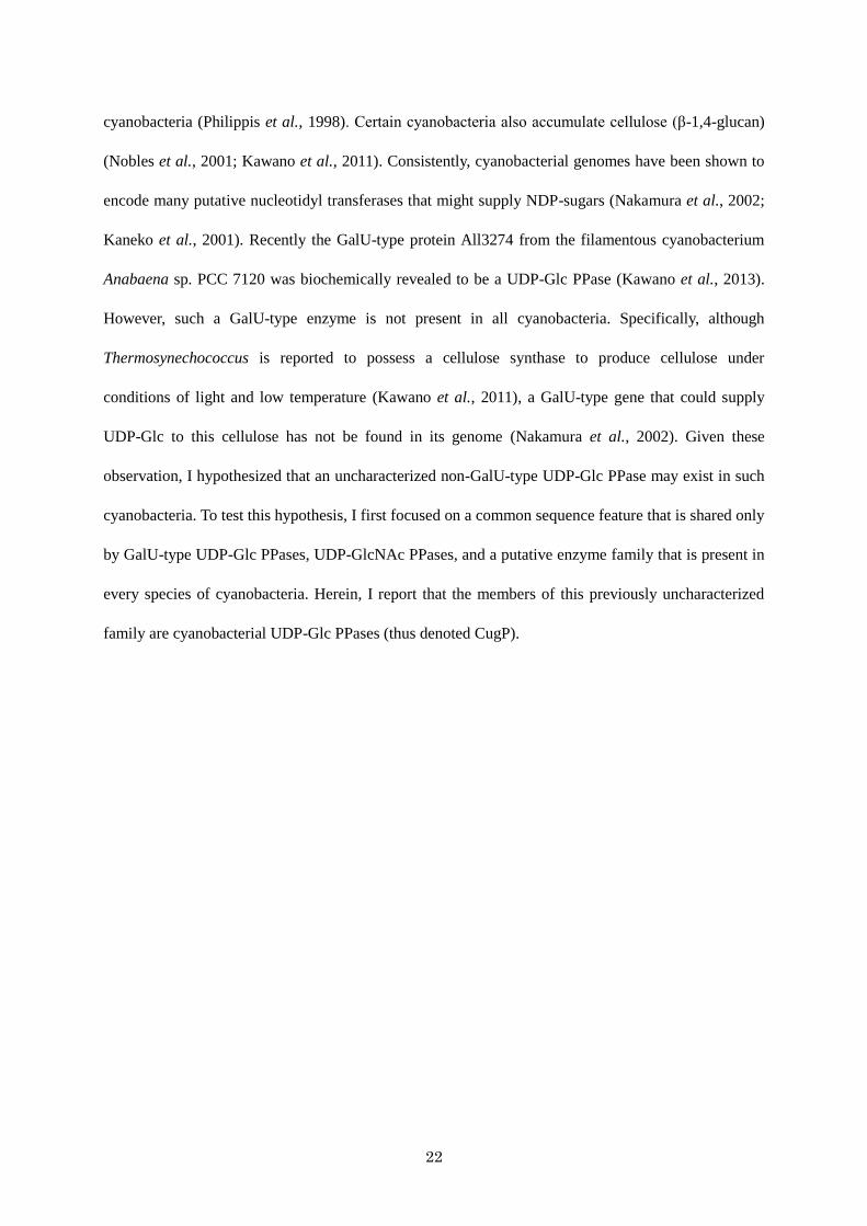
Role of CugP-type UDP-Glc PPase in cyanobacteria
To better understand the role of cyanobacterial CugP-type UDP-Glc PPases, I attempted to
disrupt Sll1558 in the Synechocystis sp. PCC 6803 genome. However, I could not produce such a
mutant, because cyanobacteria typically contain multiple copies of their chromosomes and,
consequently, the mutated Synechocystis sp. PCC 6803 did not cleanly segregate, suggesting that the
CugP-type UDP-Glc is essential for survival of Synechocystis (Fig. I-7). In this context, it must be
mentioned that a galactolipid, monogalactosyldiacylglycerol (MGDG), which is essential in
Synechocystis, is supplied from UDP-Glc via monoglucosyldiacylglycerol (Awai et al., 2006). The
galactolipids MGDG and digalactosyldiacylglycerol (DGDG) are major constituents of the
photosynthetic thylakoid membrane in cyanobacteria and in plant chloroplasts. They bind to the
membrane-spanning photosynthetic complexes and thereby maintain them in a functional state
(Sakurai et al., 2007). Hence, the role of UDP-Glc PPases in providing UDP-Glc must be essential for
growth and survival in cyanobacteria. In this non-segregated mutant, the chlorophyll (Chl) content was
3.47 ± 0.514 µg Chl / OD730, and that of wild type was 4.22 ± 0.0436 µg Chl / OD730 (n = 3). This
difference may reflect the weakened thylakoid membrane in the partially Sll1558-deleted mutant.
Because galactolipids are specific to photosynthetic membranes, the unique CugP-type enzyme, found
mainly in cyanobacteria, may be the result of an evolutionary event in oxygenic photosynthesis.
Certain EPSs secreted by cyanobacteria including Synechocystis often contain Glc as the major
constituent (Philippis et al., 1998). However, it is unknown if they are essential for survival. Cellulose
is selectively deposited in a thermophilic cyanobacterium Thermosynechococcus under conditions of
low temperature and light (Kawano et al., 2011). So, a CugP-type Tlr0611 appears to be the sole
enzyme to provide UDP-Glc for biosynthesis of cellulose and might be induced under such conditions
in this cyanobacterium. Regional distribution of GalU-type PPases in certain cyanobacteria may
account for variation in the requirement of EPSs. Further functional studies of the CugP-type and
GalU-type enzymes are needed to understand the role of UDP-Glc in cyanobacteria.

28
Materials and methods
Sequence alignment and phylogram
The sequences of the cyanobacterial proteins categorized as belonging to the bacterial
nucleotidyl transferase superfamily (Pfam: PF00483, NTP_transferase) (http://pfam.sanger.ac.uk)
were obtained from NCBI (http://www.ncbi.nlm.nih.gov/) and CyanoBase
(http://genome.microbedb.jp/cyanobase) databases. Sequence alignment was performed by
neighbor-joining method using ClustalX2 (Larkin et al., 2007).
Cloning of sll1558 and single amino acid substitution
sll1558 from Synechocystis sp. PCC 6803, which encodes the hypothetical UDP-Glc PPase
Sll1558 was PCR amplified using PrimeSTAR® Max DNA Polymerase (Takara Bio, Otsu, Japan) and
then directly cloned into the expression vector pET-28a(+) (Merck, Darmstadt, Germany) using an
In-Fusion HD Cloning kit reagents (Takara) to produce sll1558-pET28a (The primers were
pET28a-1F; 5′-GGA TCC GAA TTC GAG CTC CGT C-3′, pET28a-2R; 5′-CAT ATG GCT GCC
GCG CGG CAC-3′, sll1558-1F28a; 5′-CGC GGC AGC CAT ATG AAA GCC ATG ATT TTG GCC-3′,
and sll1558-2R28a; 5′-CTC GAA TTC GGA TCC TTA TTC CGG CTG GAG AAG-3′). To generate a
single amino acid substitution within the Sll1558 protein product, the plasmid was amplified by two
sets of primers (sll1558-3FA8G, 5′-GCC GGT GGC AAG GGC ACT CGG GTC AGA CCA ATC-3′,
and sll1558-4RA8G, 5′-GCC CTT GCC ACC GGC CAA AAT CAT GGC TTT CAT-3′) and
self-combined using the aforementioned cloning reagents. These proteins were His-tagged from the
pET28a vector. The resulting DNA constructs were verified by dideoxy sequencing. These proteins
were N-terminally His-tagged, derived from the pET28a vector.
Expression, purification and SDS-PAGE analysis of recombinant proteins
E. coli strain C41 (DE3) carrying sll1558-pET28a or sll1558(A8G)-pET28a were cultured in
Fig. S1

29
1 L of LB medium at 37˚C. When each culture reached an OD600 of 0.4 - 0.8, 1mM isopropyl
β-d-1-thiogalactopyranoside was added (final 100 µM) and the cells were grown at 18˚C for 16 h to
achieve induction. The cells from each culture were then collected by centrifugation at 4,220 g for
15 min and suspended in 20 mM HEPES (pH 7.5) containing 100 mM NaCl and 10 % (w/v) glycerol
and homogenized three times with a French press at 1500 kg cm-2. The soluble proteins were collected
by centrifugation at 50,000 g for 30 min. His-tagged Sll1558 and A8G-Sll1558 were purified by
Ni-affinity column chromatography (HisTrap™ HP; GE Healthcare, Little Chalfont, UK) with the
eluent being a gradient of 30-430 mM imidazole in the aforementioned buffer system. Proteins in each
fraction were subjected to SDS-PAGE followed by Coomassie Brilliant Blue R-250 staining. Low
Molecular Mass Calibration kit standards (GE Healthcare) served as the molecular mass markers. The
fraction enriched in each targeted protein was dialyzed against the aforementioned buffer to remove
the imidazole. Protein concentration was assayed using the Bradford method (Bio-Rad, Hercules, CA)
with bovine serum albumin as the standard.
Nucleotidyl transferase activity assay
Nucleotidyl transferase activities were assayed at 37˚C in the direction of NDP–sugar
formation from NTP and sugar-1P. The reaction rate measurements were determined as the change in
the PPi concentration with time and measured using the EnzChek Pyrophosphate Assay kit (Molecular
Probes, Life Technologies, Carlsbad, CA). The basic reaction medium contained 50 mM Tris-HCl
(pH7.5), 8 mM MgCl2, 200 µM 2-amino-6-mercapto-7-methylpurine ribonucleoside, 1 U purine
nucleoside phosphorylase, 30 mU inorganic pyrophosphatase, and substrates. To reduce contaminating
inorganic phosphate found in the substrate preparations, each reaction solution was preincubated for
20 min at 22C and for 10 min at 37C prior to the addition of the protein. For measurements of
substrate specificity, all substrates were included in the reactions at a 200 µM concentration and the
purified enzymes at 0.66 µM.

30
Structure prediction based on previous studies
The available Protein Data Bank (PDB) data of GalU-type UDP-Glc PPase (PDB ID, 2PA4)
(Thoden et al., 2007a), CDP-Glc PPase (PDB ID, 1TZF) (Koropatkin et al., 2004) and dTDP-Glc
PPase (PDB ID, 1H5T) (Zuccotti et al., 2001) were used to compare the structure and
substrate-binding region of Sll1558.
Gene disruption and mutant analysis
The sll1558 gene was replaced with the chloramphenicol resistance cassette by the
transformation plasmid made by In-Fusion method. Mutants were generated by transformation of
Synechocystis cells with this DNA and selected on BG11 plates containing 20 µg m1-1
chloramphenicol. The segregation was confirmed by PCR with the two sets of primers (sll1558-5F,
5′-GTG TGA TTG AGT TTG AGG -3′, and sll1558-6R, 5′-TTT CCC CCA GTT CTC TTC-3′).
Chlorophyll content was calculated after extraction with 100% methanol as described (Midorikawa et
al., 2009).

31
Table I-1. Substrate specificity of Sll1558 and A8G-Sll1558.
Sll1558 / A8G-Sll1558
sugar-1P
Glc-1P GlcNAc-1P Gal-1P Man-1P
nucleotide
UTP + / + 0 / 0 0 / 0 0 / 0
dTTP 0 / 0 0 / 0 0 / 0 0 / 0
CTP 0 / 0 0 / 0 0 / 0 0 / 0
ATP 0 / 0 0 / 0 0 / 0 0 / 0
GTP 0 / 0 0 / 0 0 / 0 0 / 0
Plus sign indicates the activity. 0 indicates the value is lower than the detection limit (0.01 U / mg
protein).

32
Table I-2. Kinetic parameters for Sll1558 and A8G-Sll1558.
Protein Substrate Vmax
(U mg–1)
Km
(mM)
kcat
(min–1)
kcat /Km
(min–1 mM–1)
Sll1558 UTP 0.63 0.18 28 160
Glc-1P 0.61 0.085 27 320
A8G-Sll1558 UTP 0.64 0.35 29 83
Glc-1P 0.64 0.088 29 330

33
Fig. I-1. Phylogram of cyanobacterial nucleotidyl transferases.
The phylogram of the nucleotidyl transferases from Synechocystis sp. PCC 6803, Anabaena sp. PCC
7120, Arthrospira platensis NIES-39, Synechococcus sp. PCC 7002, Thermosynechococcus elongatus
BP-1, Nostoc punctiforme ATCC 29133 and Gloeobacter violaceus PCC 7421 nucleotidyl transferase
sequences. The families those have Ala just before the G-x-G-t-R motif are shown in gray
backgrounds. The gene of a proteobacteria, Yersinia intermedia, is highlighted by black frame.

34
Fig. I-2. Sequence alignment of the NTP_transferase domain of the nucleotidyl transferases in Fig.
I-1.
The highly conserved residues of the G-x-G-t-R motif are indicated in green. The residues that
correspond to position 8 in Sll1558 are highlighted in red (Ala) and orange (Gly). The residues that are
aligned with Gly-93 in Sll1558 are in blue type. Gly-93 in Sll1558 corresponds to Gly-117 in the
GalU-type UDP-Glc PPase from Corynebacterium glutamicum, as shown in Fig. I-6.

35
Fig. I-3. SDS-PAGE after Sll1558 and A8G-Sll1558expression and purification.
M, marker; S, supernatant of centrifugation; P, purified protein; F, chromatography flow through.

36
Fig. I-4. Lineweaver-Burk plots of the enzyme activity of Sll1558 (A, UTP; B, Glc-1P) and
A8G-Sll1558 (C, UTP; D, Glc-1P).
As a fixed substrate, 1 mM Glc-1P was included in A and C, or 2 mM UTP was included in B and D.
Protein concentration: A and B, 4.5 µg ml–1; C, 18.0 µg ml–1; D, 9.0 µg ml–1. The error bars were based
on SD (A, B, and C, n = 3; D, n = 6).

37
Fig. I-5. A predicted model of substrate-binding in Sll1558 (CugP-type UDP-Glc PPase).
This model is based on the crystal structure of GalU-type UDP-Glc PPase from Corynebacterium
glutamicum in complex with magnesium and UDP-Glc (2PA4) (Thoden et al., 2007a). Only a few key
residues and substrates are shown (color indication: carbon, green; nitrogen, blue; oxygen, red; and
phosphorus, orange). The broken lines denote hypothetical hydrogen bonds.

38
Fig. I-6. Key residues in the active sites of two nucleotidyl transferases and alignment of the
active-site sequences of these two enzymes with the corresponding Sll1558 sequences. A, key
substrate-binding residues in the active site of the GalU-type UDP-Glc PPase from Corynebacterium
glutamicum (PDB ID, 2PA4). B, key substrate-binding residues in the active site of the CDP-Glc
PPase from Salmonella enterica (PDB ID, 1TZF) (color code: carbon, green; nitrogen, blue; oxygen,
red; and phosphorus, orange). The broken lines denote possible hydrogen bonds between transferase
residues and the NDP moieties. C, sequence alignment of Sll1558, the GalU-type UDP-Glc PPase
(C.g.) and CDP-Glc PPase (S.e.). Residues corresponding to Ala8 in Sll1558 are colored red. Residues
corresponding to position 93 in Sll1558 are colored blue.
A B
Ala-20
Gly-21 3.0
2.5
Gly-117
2.8
Gly-8
Gly-9
3.5
2.8
3.5
3.1
Ser-104
3.0
UDP-Glc CDP-Glc
39
Fig. I-7. Agarose gel electrophoresis to check segregation of transformant.
M; marker, WT; wild type, P; transformation plasmid, MT; mutant. The band from wild type genome
is 1.2 kbp (lower arrow), and that from transformation plasmid is 1.4 kbp (upper arrow).
M WT
P MT
PWT

40

41
I - b
Identification of various NDP-sugar pyrophosphorylases
in cyanobacteria and bacteria.

42
将来的に学術雑誌に掲載される予定であるため除外

43
Chapter II
Extracellular cellulose biosynthesis in a thermophilic
cyanobacterium Thermosynechococcus vulcanus.

44
将来的に学術雑誌に掲載される予定であるため除外
45
Chapter III
Biosynthesis and regulation of the viscous sulfated EPS
in Synechocystis sp. PCC 6803 substr. PCC-P.

46
将来的に学術雑誌に掲載される予定であるため、及び特許申請
予定であるため除外

47
General Discussion
将来的に学術雑誌に掲載される予定であるため、及び特許申請予定であ
るため除外

48
References
Agostoni, M., Waters, C.M., Montgomery, B.L. (2016) Regulation of biofilm formation and cellular
buoyancy through modulating intracellular cyclic di ‐ GMP levels in engineered
cyanobacteria. Biotechnology and bioengineering 113: 311-319.
Alonso, M.D., Lomako, J., Lomako, W.M., Whelan, W.J. (1995) A new look at the biogenesis of
glycogen. FASEB. J. 9: 1126-1137.
Åman, P., McNeil, M., Franzén, L.-E., Darvill, A.G., Albersheim, P. (1981) Structural elucidation,
using HPLC-MS and GLC-MS, of the acidic polysaccharide secreted by Rhizobium meliloti
strain 1021. Carbohydrate Research 95: 263-282.
Amikam, D., Galperin, M.Y. (2006) PilZ domain is part of the bacterial c-di-GMP binding protein.
Bioinformatics 22: 3-6.
Awai, K., Kakimoto, T., Awai, C., Kaneko, T., Nakamura, Y., Takamiya, K.-i., Wada, H., et al.
(2006) Comparative genomic analysis revealed a gene for monoglucosyldiacylglycerol
synthase, an enzyme for photosynthetic membrane lipid synthesis in cyanobacteria. Plant
Physiol. 141: 1120-1127.
Awai, K., Ohta, H., Sato, N. (2014) Oxygenic photosynthesis without galactolipids. Proceedings of
the National Academy of Sciences 111: 13571-13575.
Beard, S.J., Hayes, P.K., Pfeifer, F., Walsby, A.E. (2002) The sequence of the major gas vesicle
protein, GvpA, influences the width and strength of halobacterial gas vesicles. FEMS
microbiology letters 213: 149-157.
Becker, A., Katzen, F., Pühler, A., Ielpi, L. (1998) Xanthan gum biosynthesis and application: a
biochemical/genetic perspective. Applied microbiology and biotechnology 50: 145-152.
Bielaszewska, M., Aldick, T., Bauwens, A., Karch, H. (2014) Hemolysin of enterohemorrhagic
Escherichia coli: structure, transport, biological activity and putative role in virulence. Int
J Med Microbiol 304: 521-529.
Borges-Walmsley, M.I., Beauchamp, J., Kelly, S.M., Jumel, K., Candlish, D., Harding, S.E., Price,

49
N.C., et al. (2003) Identification of oligomerization and drug-binding domains of the
membrane fusion protein EmrA. Journal of Biological Chemistry 278: 12903-12912.
Bosco, M., Machtey, M., Iglesias, A., Aleanzi, M. (2009) UDPglucose pyrophosphorylase from
Xanthomonas spp. Characterization of the enzyme kinetics, structure and inactivation
related to oligomeric dissociation. Biochimie 91: 204-213.
Chambers, J., Elbein, A.D. (1970) Biosynthesis of glucans in mung bean seedlings. Formation of
beta-(1,4)-glucans from GDP-glucose and beta-(1,3)-glucans from UDP-glucose. Arch
Biochem Biophys 138: 620-631.
Danishefsky, I., Heritier-Watkins, O. (1967) Nucleoside diphosphate glucose pyrophosphorylases
in mast cell tumors. Biochim. Biophys. Acta. 139: 349-357.
Darnell, C.L., Hussa, E.A., Visick, K.L. (2008) The putative hybrid sensor kinase SypF coordinates
biofilm formation in Vibrio fischeri by acting upstream of two response regulators, SypG
and VpsR. J. Bacteriol. 190: 4941-4950.
De Philippis, R., Vincenzini, M. (1998) Exocellular polysaccharides from cyanobacteria and their
possible applications. FEMS Microbiol. Rev. 22: 151-175.
Dervaux, J., Mejean, A., Brunet, P. (2015) Irreversible collective migration of cyanobacteria in
eutrophic conditions. PloS one 10: e0120906.
Diez, M.a.D.A.n., Peiru ́, S., Demonte, A.M., Gramajo, H., Iglesias, A.A. (2012) Characterization of
recombinant UDP-and ADP-glucose pyrophosphorylases and glycogen synthase to
elucidate glucose-1-phosphate partitioning into oligo-and polysaccharides in Streptomyces
coelicolor. J. Bacteriol. 194: 1485-1493.
Diez, M.D.A., Miah, F., Stevenson, C.E., Lawson, D.M., Iglesias, A.A., Bornemann, S. (2017) The
Production and Utilization of GDP-glucose in the Biosynthesis of Trehalose 6-Phosphate
by Streptomyces venezuelae. Journal of Biological Chemistry 292: 945-954.
DuBois, M., Gilles, K.A., Hamilton, J.K., Rebers, P.t., Smith, F. (1956) Colorimetric method for
determination of sugars and related substances. Analytical chemistry 28: 350-356.

50
Ebrecht, A.C., Orlof, A.M., Sasoni, N., Figueroa, C.M., Iglesias, A.A., Ballicora, M.A. (2015) On the
ancestral UDP-Glucose pyrophosphorylase activity of galF from Escherichia coli. Frontiers
in microbiology 6.
Enomoto, G., Narikawa, R., Ikeuchi, M. (2015) Three cyanobacteriochromes work together to form
a light color-sensitive input system for c-di-GMP signaling of cell aggregation. Proceedings
of the National Academy of Sciences 112: 8082-8087.
Enomoto, G., Nomura, R., Shimada, T., Ni Ni, W., Narikawa, R., Ikeuchi, M. (2014)
Cyanobacteriochrome SesA is a diguanylate cyclase that induces cell aggregation in
Thermosynechococcus. J. Biol. Chem. 289: 24801-24809.
Finn, R.D., Coggill, P., Eberhardt, R.Y., Eddy, S.R., Mistry, J., Mitchell, A.L., Potter, S.C., et al.
(2016) The Pfam protein families database: towards a more sustainable future. Nucleic
Acids Res. 44: D279-D285.
Fisher, M.L., Allen, R., Luo, Y., Curtiss III, R. (2013) Export of extracellular polysaccharides
modulates adherence of the cyanobacterium Synechocystis. PloS one 8: e74514.
Flemming, H.C., Wingender, J. (2010) The biofilm matrix. Nat Rev Microbiol 8: 623-633.
Flores-Diaz, M., Alape-Giron, A., Persson, B., Pollesello, P., Moos, M., von Eichel-Streiber, C.,
Thelestam, M., et al. (1997) Cellular UDP-glucose deficiency caused by a single point
mutation in the UDP-glucose pyrophosphorylase gene. J. Biol. Chem. 272: 23784-23791.
Flowers, H.M., Batra, K.K., Kemp, J., Hassid, W.Z. (1969) Biosynthesis of cellulose in vitro from
guanosine diphosphate D-glucose with enzymic preparations from Phaseolus aureus and
Lupinus albus. J. Biol. Chem. 244: 4969-4974.
Foster, J.S., Havemann, S.A., Singh, A.K., Sherman, L.A. (2009) Role of mrgA in peroxide and light
stress in the cyanobacterium Synechocystis sp. PCC 6803. FEMS Microbiol Lett 293:
298-304.
Fujisawa, T., Narikawa, R., Maeda, S.I., Watanabe, S., Kanesaki, Y., Kobayashi, K., Nomata, J., et
al. (2017) CyanoBase: a large-scale update on its 20th anniversary. Nucleic Acids Res 45:

51
D551-D554.
Fujisawa, T., Narikawa, R., Okamoto, S., Ehira, S., Yoshimura, H., Suzuki, I., Masuda, T., et al.
(2010) Genomic structure of an economically important cyanobacterium, Arthrospira
(Spirulina) platensis NIES-39. DNA Res. 17: 85-103.
Fujishiro, T., Ogawa, T., Matsuoka, M., Nagahama, K., Takeshima, Y., Hagiwara, H. (2004)
Establishment of a pure culture of the hitherto uncultured unicellular cyanobacterium
Aphanothece sacrum, and phylogenetic position of the organism. Appl. Environ. Microbiol.
70: 3338-3345.
Fukaya, Y., Hayashi, K., Wada, M., Ohno, H. (2008) Cellulose dissolution with polar ionic liquids
under mild conditions: required factors for anions. Green Chem. 10: 44-46.
Galvan, E.M., Ielmini, M.V., Patel, Y.N., Bianco, M.I., Franceschini, E.A., Schneider, J.C., Ielpi, L.
(2013) Xanthan chain length is modulated by increasing the availability of the
polysaccharide copolymerase protein GumC and the outer membrane polysaccharide
export protein GumB. Glycobiology 23: 259-272.
Glover, H.E., Prézelin, B.B., Campbell, L., Wyman, M., Garside, C. (1988) A nitrate-dependent
Synechococcus bloom in surface Sargasso Sea water. Nature 331: 161.
Hay, I.D., Rehman, Z.U., Moradali, M.F., Wang, Y., Rehm, B.H. (2013) Microbial alginate
production, modification and its applications. Microbial biotechnology 6: 637-650.
Hayashi, T. (2008) Studies on evaluation of natural products for antiviral effects and their
applications. Yakugaku zasshi: Journal of the Pharmaceutical Society of Japan 128: 61-79.
Helm, R.F., Huang, Z., Edwards, D., Leeson, H., Peery, W., Potts, M. (2000) Structural
characterization of the released polysaccharide of desiccation-tolerant Nostoc
communeDRH-1. J. Bacteriol. 182: 974-982.
Henrissat, B. (1991) A classification of glycosyl hydrolases based on amino acid sequence
similarities. Biochemical Journal 280: 309-316.
Hernández‐Prieto, M.A., Li, Y., Postier, B.L., Blankenship, R.E., Chen, M. (2017) Far‐red light

52
promotes biofilm formation in the cyanobacterium Acaryochloris marina. Environmental
Microbiology.
Higo, A., Katoh, H., Ohmori, K., Ikeuchi, M., Ohmori, M. (2006) The role of a gene cluster for
trehalose metabolism in dehydration tolerance of the filamentous cyanobacterium
Anabaena sp. PCC 7120. Microbiology 152: 979-987.
Hill, J.L., Jr., Hammudi, M.B., Tien, M. (2014) The Arabidopsis cellulose synthase complex: a
proposed hexamer of CESA trimers in an equimolar stoichiometry. Plant Cell 26:
4834-4842.
Hirano, A., Kunito, S., Inoue, Y., Ikeuchi, M. (1997) Light and low temperature induced cell
flocculation of thermophilic cyanobacterium Synechococcus vulcanus. Plant Cell Physiol.
38: s37.
Ho, C.-L. (2015) Phylogeny of Algal Sequences Encoding Carbohydrate Sulfotransferases,
Formylglycine-Dependent Sulfatases, and Putative Sulfatase Modifying Factors. Frontiers
in plant science 6.
Ho, T.D., Waldor, M.K. (2007) Enterohemorrhagic Escherichia coli O157: H7 gal mutants are
sensitive to bacteriophage P1 and defective in intestinal colonization. Infection and
immunity 75: 1661-1666.
Holden, H.M., Rayment, I., Thoden, J.B. (2003) Structure and function of enzymes of the Leloir
pathway for galactose metabolism. J. Biol. Chem. 278: 43885-43888.
Holland, N., Holland, D., Helentjaris, T., Dhugga, K.S., Xoconostle-Cazares, B., Delmer, D.P.
(2000) A comparative analysis of the plant cellulose synthase (CesA) gene family. Plant
Physiol. 123: 1313-1324.
Hong, Y., Cunneen, M.M., Reeves, P.R. (2012) The Wzx translocases for Salmonella enterica O‐
antigen processing have unexpected serotype specificity. Molecular microbiology 84:
620-630.
Hossain, S.A., Tanizawa, K., Kazuta, Y., Fukui, T. (1994) Overproduction and characterization of

53
recombinant UDP-glucose pyrophosphorylase from Escherichia coli K-12. J. Biochem. 115:
965-972.
Islam, S.T., Lam, J.S. (2014) Synthesis of bacterial polysaccharides via the Wzx/Wzy-dependent
pathway. Canadian journal of microbiology 60: 697-716.
Iwai, M., Katoh, H., Katayama, M., Ikeuchi, M. (2004) Improved genetic transformation of the
thermophilic cyanobacterium, Thermosynechococcus elongatus BP-1. Plant and Cell
Physiology 45: 171-175.
Jansson, P.-e., Kenne, L., Lindberg, B. (1975) Structure of the extracellular polysaccharide from
Xanthomonas campestris. Carbohydrate Research 45: 275-282.
Jeamton, W., Dulsawat, S., Tanticharoen, M., Vonshak, A., Cheevadhanarak, S. (2017) Overcoming
Intrinsic Restriction Enzyme Barriers Enhances Transformation Efficiency in Arthrospira
platensis C1. Plant Cell Physiol. 58: 822-830.
Jittawuttipoka, T., Planchon, M., Spalla, O., Benzerara, K., Guyot, F., Cassier-Chauvat, C.,
Chauvat, F. (2013) Multidisciplinary evidences that Synechocystis PCC6803
exopolysaccharides operate in cell sedimentation and protection against salt and metal
stresses. PloS one 8: e55564.
Kaneko, T., Nakamura, Y., Wolk, C.P., Kuritz, T., Sasamoto, S., Watanabe, A., Iriguchi, M., et al.
(2001) Complete genomic sequence of the filamentous nitrogen-fixing cyanobacterium
Anabaena sp. strain PCC 7120. DNA Res. 8: 205-213.
Kanonenberg, K., Schwarz, C.K., Schmitt, L. (2013) Type I secretion systems - a story of
appendices. Res. Microbiol. 164: 596-604.
Katzen, F., Ferreiro, D.U., Oddo, C.G., Ielmini, M.V., Becker, A., Pühler, A., Ielpi, L. (1998)
Xanthomonas campestris pv. campestrisgum Mutants: Effects on Xanthan Biosynthesis
and Plant Virulence. J. Bacteriol. 180: 1607-1617.
Kawaguchi, K., Tanida, S., Matsuda, K., Tani, Y., Ogata, K. (1973) Microbial synthesis of
GDP-glucose. Agricultural and Biological Chemistry 37: 75-81.
54
Kawano, Y., Saotome, T., Ochiai, Y., Katayama, M., Narikawa, R., Ikeuchi, M. (2011) Cellulose
accumulation and a cellulose synthase gene are responsible for cell aggregation in the
cyanobacterium Thermosynechococcus vulcanus RKN. Plant Cell Physiol. 52: 957-966.
Kawano, Y., Sekine, M., Ihara, M. (2013) Identification and characterization of UDP-glucose
pyrophosphorylase in cyanobacteria Anabaena sp. PCC 7120. J. Biosci. Bioeng. 117:
531-538.
Kim, H., Choi, J., Kim, T., Lokanath, N.K., Ha, S.C., Suh, S.W., Hwang, H.Y., et al. (2010)
Structural basis for the reaction mechanism of UDP-glucose pyrophosphorylase. Mol. Cells
29: 397-405.
Kim, S.H., Ahn, S.H., Lee, J.H., Lee, E.M., Kim, N.H., Park, K.J., Kong, I.S. (2003) Genetic
analysis of phosphomannomutase/phosphoglucomutase from Vibrio furnissii and
characterization of its role in virulence. Arch Microbiol 180: 240-250.
Kimura, S., Laosinchai, W., Itoh, T., Cui, X., Linder, C.R., Brown, R.M. (1999) Immunogold
labeling of rosette terminal cellulose-synthesizing complexes in the vascular plant Vigna
angularis. The Plant Cell 11: 2075-2085.
Kobayashi, K., Kondo, M., Fukuda, H., Nishimura, M., Ohta, H. (2007) Galactolipid synthesis in
chloroplast inner envelope is essential for proper thylakoid biogenesis, photosynthesis, and
embryogenesis. Proceedings of the National Academy of Sciences 104: 17216-17221.
Kopf, M., Klähn, S., Scholz, I., Matthiessen, J.K., Hess, W.R., Voß, B. (2014) Comparative analysis
of the primary transcriptome of Synechocystis sp. PCC 6803. DNA Res. 21: 527-539.
Koronakis, V., Eswaran, J., Hughes, C. (2004) Structure and function of TolC: the bacterial exit
duct for proteins and drugs. Annu Rev Biochem 73: 467-489.
Koropatkin, N.M., Holden, H.M. (2004) Molecular structure of alpha-D-glucose-1-phosphate
cytidylyltransferase from Salmonella typhi. J. Biol. Chem. 279: 44023-44029.
Kudlicka, K., Brown Jr, R.M. (1997) Cellulose and callose biosynthesis in higher plants (I.
Solubilization and separation of (1-> 3)-and (1-> 4)-[beta]-glucan synthase activities from
55
mung bean). Plant Physiol. 115: 643-656.
Kumar, M., Atanassov, I., Turner, S. (2017) Functional analysis of cellulose synthase (CESA)
protein class specificity. Plant Physiol. 173: 970-983.
Lai, X., Wu, J., Chen, S., Zhang, X., Wang, H. (2008) Expression, purification, and characterization
of a functionally active Mycobacterium tuberculosis UDP-glucose pyrophosphorylase.
Protein Expr. Purif. 61: 50-56.
Larkin, M., Blackshields, G., Brown, N., Chenna, R., McGettigan, P.A., McWilliam, H., Valentin, F.,
et al. (2007) Clustal W and Clustal X version 2.0. Bioinformatics 23: 2947-2948.
Lei, L., Zhang, T., Strasser, R., Lee, C.M., Gonneau, M., Mach, L., Vernhettes, S., et al. (2014) The
jiaoyao1 Mutant Is an Allele of korrigan1 That Abolishes Endoglucanase Activity and
Affects the Organization of Both Cellulose Microfibrils and Microtubules in Arabidopsis.
Plant Cell 26: 2601-2616.
Letunic, I., Doerks, T., Bork, P. (2015) SMART: recent updates, new developments and status in
2015. Nucleic Acids Res 43: D257-260.
Li, J.-P., Kusche-Gullberg, M. (2016) Chapter Six-Heparan Sulfate: Biosynthesis, Structure, and
Function. International review of cell and molecular biology 325: 215-273.
Linker, A., Jones, R.S. (1966) A new polysaccharide resembling alginic acid isolated from
pseudomonads. Journal of Biological Chemistry 241: 3845-3851.
Lombard, V., Golaconda Ramulu, H., Drula, E., Coutinho, P.M., Henrissat, B. (2013) The
carbohydrate-active enzymes database (CAZy) in 2013. Nucleic Acids Res. 42: D490-D495.
Lori, C., Ozaki, S., Steiner, S., Böhm, R., Abel, S., Dubey, B.N., Schirmer, T., et al. (2015) Cyclic
di-GMP acts as a cell cycle oscillator to drive chromosome replication. Nature 523:
236-239.
Ma, L., Conover, M., Lu, H., Parsek, M.R., Bayles, K., Wozniak, D.J. (2009) Assembly and
development of the Pseudomonas aeruginosa biofilm matrix. PLoS pathogens 5: e1000354.
Ma, Z., Fan, H.-j., Lu, C.-p. (2011) Molecular cloning and analysis of the UDP-Glucose

56
Pyrophosphorylase in Streptococcus equi subsp. zooepidemicus. Mol. Biol. Rep. 38:
2751-2760.
Matthysse, A.G., White, S., Lightfoot, R. (1995) Genes required for cellulose synthesis in
Agrobacterium tumefaciens. J. Bacteriol. 177: 1069-1075.
McNamara, J.T., Morgan, J.L., Zimmer, J. (2015) A molecular description of cellulose biosynthesis.
Annu Rev Biochem 84: 895-921.
Midorikawa, T., Matsumoto, K., Narikawa, R., Ikeuchi, M. (2009) An Rrf2-type transcriptional
regulator is required for expression of psaAB genes in the cyanobacterium Synechocystis
sp. PCC 6803. Plant Physiol. 151: 882-892.
Mika, F., Hengge, R. (2014) Small RNAs in the control of RpoS, CsgD, and biofilm architecture of
Escherichia coli. RNA biology 11: 494-507.
Mitschke, J., Georg, J., Scholz, I., Sharma, C.M., Dienst, D., Bantscheff, J., Voß, B., et al. (2011) An
experimentally anchored map of transcriptional start sites in the model cyanobacterium
Synechocystis sp. PCC6803. Proceedings of the National Academy of Sciences 108:
2124-2129.
Mizanur, R.M., Pohl, N.L. (2009) Phosphomannose isomerase/GDP-mannose pyrophosphorylase
from Pyrococcus furiosus: a thermostable biocatalyst for the synthesis of
guanidinediphosphate-activated and mannose-containing sugar nucleotides. Org Biomol
Chem 7: 2135-2139.
Mollerach, M., Garcia, E. (2000) The galU gene of Streptococcus pneumoniae that codes for a
UDP-glucose pyrophosphorylase is highly polymorphic and suitable for molecular typing
and phylogenetic studies. Gene 260: 77-86.
Mollerach, M., Lopez, R., Garcia, E. (1998) Characterization of the galU gene of Streptococcus
pneumoniae encoding a uridine diphosphoglucose pyrophosphorylase: a gene essential for
capsular polysaccharide biosynthesis. J. Exp. Med. 188: 2047-2056.
Monday, S.R., Schiller, N.L. (1996) Alginate synthesis in Pseudomonas aeruginosa: the role of AlgL
57
(alginate lyase) and AlgX. J. Bacteriol. 178: 625-632.
Morgan, J.L., McNamara, J.T., Zimmer, J. (2014) Mechanism of activation of bacterial cellulose
synthase by cyclic di-GMP. Nat Struct Mol Biol 21: 489-496.
Morgan, J.L., Strumillo, J., Zimmer, J. (2013) Crystallographic snapshot of cellulose synthesis and
membrane translocation. Nature 493: 181-186.
Mota, R., Guimarães, R., Büttel, Z., Rossi, F., Colica, G., Silva, C.J., Santos, C., et al. (2013)
Production and characterization of extracellular carbohydrate polymer from Cyanothece
sp. CCY 0110. Carbohydrate polymers 92: 1408-1415.
Mueller, S.C., Brown, R.M. (1980) Evidence for an intramembrane component associated with a
cellulose microfibril-synthesizing complex in higher plants. The Journal of cell biology 84:
315-326.
Nakai, T., Sugano, Y., Shoda, M., Sakakibara, H., Oiwa, K., Tuzi, S., Imai, T., et al. (2013)
Formation of highly twisted ribbons in a carboxymethylcellulase gene-disrupted strain of a
cellulose-producing bacterium. J Bacteriol 195: 958-964.
Nakamura, Y., Kaneko, T., Sato, S., Ikeuchi, M., Katoh, H., Sasamoto, S., Watanabe, A., et al.
(2002) Complete genome structure of the thermophilic cyanobacterium
Thermosynechococcus elongatus BP-1. DNA Res. 9: 123-130.
Ngatu, N.R., Okajima, M.K., Yokogawa, M., Hirota, R., Eitoku, M., Muzembo, B.A., Dumavibhat,
N., et al. (2012) Anti-inflammatory effects of sacran, a novel polysaccharide from
Aphanothece sacrum, on 2, 4, 6-trinitrochlorobenzene–induced allergic dermatitis in vivo.
Annals of Allergy, Asthma & Immunology 108: 117-122. e112.
Nicaud, J.-M., Mackman, N., Gray, L., Holland, I. (1985) Regulation of haemolysin synthesis in E.
coli determined by HLY genes of human origin. Molecular and General Genetics MGG 199:
111-116.
Ning, B., Elbein, A.D. (2000) Cloning, expression and characterization of the pig liver GDP‐
mannose pyrophosphorylase. The FEBS Journal 267: 6866-6874.

58
Nobles, D.R., Romanovicz, D.K., Brown, R.M. (2001) Cellulose in cyanobacteria. Origin of vascular
plant cellulose synthase? Plant Physiol. 127: 529-542.
Offner, S., Pfeifer, F. (1995) Complementation studies with the gas vesicle‐encoding p‐vac region
of Halobacterium salinarium PHH1 reveal a regulatory role for the p‐gvpDE genes.
Molecular microbiology 16: 9-19.
Ohki, K., Le, N.Q.T., Yoshikawa, S., Kanesaki, Y., Okajima, M., Kaneko, T., Thi, T.H. (2014)
Exopolysaccharide production by a unicellular freshwater cyanobacterium Cyanothece sp.
isolated from a rice field in Vietnam. Journal of applied phycology 26: 265-272.
Ohmori, K., Hirose, M., Ohmori, M. (1992) Function of cAMP as a mat-forming factor in the
cyanobacterium Spirulina platensis. Plant and cell physiology 33: 21-25.
Omadjela, O., Narahari, A., Strumillo, J., Melida, H., Mazur, O., Bulone, V., Zimmer, J. (2013)
BcsA and BcsB form the catalytically active core of bacterial cellulose synthase sufficient
for in vitro cellulose synthesis. Proc. Natl. Acad. Sci. USA 110: 17856-17861.
Panoff, J.-M., Priem, B., Morvan, H., Joset, F. (1988) Sulphated exopolysaccharides produced by
two unicellular strains of cyanobacteria, Synechocystis PCC 6803 and 6714. Archives of
microbiology 150: 558-563.
Pear, J.R., Kawagoe, Y., Schreckengost, W.E., Delmer, D.P., Stalker, D.M. (1996) Higher plants
contain homologs of the bacterial celA genes encoding the catalytic subunit of cellulose
synthase. Proceedings of the National Academy of Sciences 93: 12637-12642.
Pereira, S., Zille, A., Micheletti, E., Moradas-Ferreira, P., De Philippis, R., Tamagnini, P. (2009)
Complexity of cyanobacterial exopolysaccharides: composition, structures, inducing factors
and putative genes involved in their biosynthesis and assembly. FEMS Microbiol. Rev. 33:
917-941.
Pereira, S.B., Mota, R., Vieira, C.P., Vieira, J., Tamagnini, P. (2015) Phylum-wide analysis of
genes/proteins related to the last steps of assembly and export of extracellular polymeric
substances (EPS) in cyanobacteria. Sci Rep 5: 14835.

59
Perez-Mendoza, D., Rodriguez-Carvajal, M.A., Romero-Jimenez, L., Farias Gde, A., Lloret, J.,
Gallegos, M.T., Sanjuan, J. (2015) Novel mixed-linkage beta-glucan activated by c-di-GMP
in Sinorhizobium meliloti. Proc. Natl. Acad. Sci. USA 112: E757-765.
Philippis, R., Vincenzini, M. (1998) Exocellular polysaccharides from cyanobacteria and their
possible applications. FEMS Microbiol. Rev. 22: 151-175.
Quast, C., Pruesse, E., Yilmaz, P., Gerken, J., Schweer, T., Yarza, P., Peplies, J., et al. (2012) The
SILVA ribosomal RNA gene database project: improved data processing and web-based
tools. Nucleic Acids Res. 41: D590-D596.
Ramsey, D.M., Wozniak, D.J. (2005) Understanding the control of Pseudomonas aeruginosa
alginate synthesis and the prospects for management of chronic infections in cystic fibrosis.
Molecular microbiology 56: 309-322.
Ramus, J. (1972) The production of extracellular polysaccharide by the unicellular red alga
Porphyridium aerugineum. Journal of Phycology 8: 97-111.
Rehm, B., Valla, S. (1997) Bacterial alginates: biosynthesis and applications. Applied microbiology
and biotechnology 48: 281-288.
Rehm, B.H. (2010) Bacterial polymers: biosynthesis, modifications and applications. Nature
Reviews Microbiology 8: 578-592.
Reynolds, C.S., Huszar, V., Kruk, C., Naselli-Flores, L., Melo, S. (2002) Towards a functional
classification of the freshwater phytoplankton. Journal of plankton research 24: 417-428.
Romling, U. (2002) Molecular biology of cellulose production in bacteria. Res. Microbiol. 153:
205-212.
Romling, U., Galperin, M.Y. (2015) Bacterial cellulose biosynthesis: diversity of operons, subunits,
products, and functions. Trends Microbiol 23: 545-557.
Romling, U., Galperin, M.Y., Gomelsky, M. (2013) Cyclic di-GMP: the first 25 years of a universal
bacterial second messenger. Microbiol Mol Biol Rev 77: 1-52.
Ross, P., Weinhouse, H., Aloni, Y., Michaeli, D., Weinberger-Ohana, P., Mayer, R., Braun, S., et al.

60
(1987) Regulation of cellulose synthesis in Acetobacter xylinum by cyclic diguanylic acid.
Nature 325: 279-281.
Roth, J. (1995) Compartmentation of glycoprotein biosynthesis. Glycoproteins (eds. J. Montreuil et
al.): 237-312.
Ryan, R.P. (2013) Cyclic di-GMP signalling and the regulation of bacterial virulence. Microbiology
159: 1286-1297.
Sakurai, I., Mizusawa, N., Wada, H., Sato, N. (2007) Digalactosyldiacylglycerol is required for
stabilization of the oxygen-evolving complex in photosystem II. Plant Physiol. 145:
1361-1370.
Sandhoff, K., van Echten, G., Schroder, M., Schnabel, D., Suzuki, K. (1992) Metabolism of
glycolipids: the role of glycolipid-binding proteins in the function and pathobiochemistry of
lysosomes. Biochem. Soc. Trans. 20: 695-699.
Schmid, J., Sieber, V., Rehm, B. (2015) Bacterial exopolysaccharides: biosynthesis pathways and
engineering strategies. Front Microbiol 6: 496.
Silbert, J.E., Sugumaran, G. (1995) Intracellular membranes in the synthesis, transport, and
metabolism of proteoglycans. Biochim. Biophys. Acta. 1241: 371-384.
Silva, E., Marques, A.R., Fialho, A.M., Granja, A.T., Sá-Correia, I. (2005) Proteins encoded by
Sphingomonas elodea ATCC 31461 rmlA and ugpG genes, involved in gellan gum
biosynthesis, exhibit both dTDP-and UDP-glucose pyrophosphorylase activities. Appl.
Environ. Microbiol. 71: 4703-4712.
Smayda, T.J. (1997) Harmful algal blooms: their ecophysiology and general relevance to
phytoplankton blooms in the sea. Limnology and oceanography 42: 1137-1153.
Somerville, C. (2006) Cellulose synthesis in higher plants. Annu. Rev. Cell Dev. Biol. 22: 53-78.
Stanier, R., Kunisawa, R., Mandel, M., Cohen-Bazire, G. (1971) Purification and properties of
unicellular blue-green algae (order Chroococcales). Bacteriological reviews 35: 171.
Stasinopoulos, S.J., Fisher, P.R., Stone, B.A., Stanisich, V.A. (1999) Detection of two loci involved

61
in (1→ 3)-β -glucan (curdlan) biosynthesis by Agrobacterium sp. ATCC31749, and
comparative sequence analysis of the putative curdlan synthase gene. Glycobiology 9:
31-41.
Stolyar, S., Liu, Z., Thiel, V., Tomsho, L.P., Pinel, N., Nelson, W.C., Lindemann, S.R., et al. (2014)
Genome sequence of the thermophilic cyanobacterium Thermosynechococcus sp. strain
NK55a. Genome announcements 2: e01060-01013.
Tal, R., Wong, H.C., Calhoon, R., Gelfand, D., Fear, A.L., Volman, G., Mayer, R., et al. (1998) Three
cdg operons control cellular turnover of cyclic di-GMP in Acetobacter xylinum: genetic
organization and occurrence of conserved domains in isoenzymes. J. Bacteriol. 180:
4416-4425.
Tamaru, Y., Takani, Y., Yoshida, T., Sakamoto, T. (2005) Crucial role of extracellular
polysaccharides in desiccation and freezing tolerance in the terrestrial cyanobacterium
Nostoc commune. Appl. Environ. Microbiol. 71: 7327-7333.
Tamura, J. (2011) 好熱性シアノバクテリアにおける c-di-GMP が調節する生理的機能. master thesis
東京大学大学院 総合文化研究科 広域科学専攻 生命環境科学系
Tan, S.Y., Dutta, A., Jakubovics, N.S., Ang, M.Y., Siow, C.C., Mutha, N.V., Heydari, H., et al. (2015)
YersiniaBase: a genomic resource and analysis platform for comparative analysis of
Yersinia. BMC bioinformatics 16: 9.
Thoden, J.B., Holden, H.M. (2007a) Active site geometry of glucose-1-phosphate
uridylyltransferase. Protein Sci. 16: 1379-1388.
Thoden, J.B., Holden, H.M. (2007b) The molecular architecture of glucose-1-phosphate
uridylyltransferase. Protein Sci. 16: 432-440.
Thomas, S., Holland, I.B., Schmitt, L. (2013) The Type 1 secretion pathway - the hemolysin system
and beyond. Biochim. Biophys. Acta. 1843: 1629-1641.
Tocilj, A., Munger, C., Proteau, A., Morona, R., Purins, L., Ajamian, E., Wagner, J., et al. (2008)
Bacterial polysaccharide co-polymerases share a common framework for control of polymer

62
length. Nat Struct Mol Biol 15: 130-138.
Tran, T.H., Shi, X., Zaia, J., Ai, X. (2012) Heparan sulfate 6-O-endosulfatases (Sulfs) coordinate the
Wnt signaling pathways to regulate myoblast fusion during skeletal muscle regeneration.
Journal of Biological Chemistry 287: 32651-32664.
Uchiyama, I., Mihara, M., Nishide, H., Chiba, H. (2014) MBGD update 2015: microbial genome
database for flexible ortholog analysis utilizing a diverse set of genomic data. Nucleic Acids
Res. 43: D270-D276.
Umena, Y., Kawakami, K., Shen, J.-R., Kamiya, N. (2011) Crystal structure of oxygen-evolving
photosystem II at a resolution of 1.9 Å. Nature 473: 55-60.
Vain, T., Crowell, E.F., Timpano, H., Biot, E., Desprez, T., Mansoori, N., Trindade, L.M., et al.
(2014) The Cellulase KORRIGAN Is Part of the Cellulose Synthase Complex. Plant Physiol.
165: 1521-1532.
Vandavasi, V.G., Putnam, D.K., Zhang, Q., Petridis, L., Heller, W.T., Nixon, B.T., Haigler, C.H., et
al. (2016) A structural study of CESA1 catalytic domain of Arabidopsis cellulose synthesis
complex: evidence for CESA trimers. Plant Physiol. 170: 123-135.
Verbert, A. (1995) From Glc3MangGlcNAc2-protein to Man5GlcNAc2-protein: Transfer 'en bloc'.
Glycoproteins (eds. J. Montreuil et al.): 145-152.
Walsby, A. (1994) Gas vesicles. Microbiological reviews 58: 94-144.
Ward, D.M., Ferris, M.J., Nold, S.C., Bateson, M.M. (1998) A natural view of microbial biodiversity
within hot spring cyanobacterial mat communities. Microbiology and Molecular Biology
Reviews 62: 1353-1370.
Weissborn, A.C., Liu, Q., Rumley, M.K., Kennedy, E.P. (1994) UTP: alpha-D-glucose-1-phosphate
uridylyltransferase of Escherichia coli: isolation and DNA sequence of the galU gene and
purification of the enzyme. J. Bacteriol. 176: 2611-2618.
Whitfield, C. (1995) Biosynthesis of lipopolysaccharide O antigens. Trends in microbiology 3:
178-185.

63
Wijesekara, I., Pangestuti, R., Kim, S.-K. (2011) Biological activities and potential health benefits
of sulfated polysaccharides derived from marine algae. Carbohydrate Polymers 84: 14-21.
Willis, L.M., Whitfield, C. (2013) Structure, biosynthesis, and function of bacterial capsular
polysaccharides synthesized by ABC transporter-dependent pathways. Carbohydrate
research 378: 35-44.
Wolfgang, M.C., Lee, V.T., Gilmore, M.E., Lory, S. (2003) Coordinate regulation of bacterial
virulence genes by a novel adenylate cyclase-dependent signaling pathway. Developmental
cell 4: 253-263.
Wong, H.C., Fear, A.L., Calhoon, R.D., Eichinger, G.H., Mayer, R., Amikam, D., Benziman, M., et
al. (1990) Genetic organization of the cellulose synthase operon in Acetobacter xylinum.
Proc. Natl. Acad. Sci. USA 87: 8130-8134.
Wu, B., Zhang, Y., Zheng, R., Guo, C., Wang, P.G. (2002) Bifunctional phosphomannose
isomerase/GDP-D-mannose pyrophosphorylase is the point of control for GDP-D-mannose
biosynthesis in Helicobacter pylori. FEBS Lett 519: 87-92.
Yildiz, F.H., Visick, K.L. (2009) Vibrio biofilms: so much the same yet so different. Trends in
microbiology 17: 109-118.
Zhang, H., Song, G., Shao, J., Xiang, X., Li, Q., Chen, Y., Yang, P., et al. (2016) Dynamics and
polyphasic characterization of odor-producing cyanobacterium Tychonema bourrellyi from
Lake Erhai, China. Environmental Science and Pollution Research 23: 5420-5430.
Zhao, C., Li, Z., Li, T., Zhang, Y., Bryant, D.A., Zhao, J. (2015) High-yield production of
extracellular type-I cellulose by the cyanobacterium Synechococcus sp. PCC 7002. Cell
Discov 1: 15004.
Zhao, K., Tseng, B.S., Beckerman, B., Jin, F., Gibiansky, M.L., Harrison, J.J., Luijten, E., et al.
(2013) Psl trails guide exploration and microcolony formation in Pseudomonas aeruginosa
biofilms. Nature 497: 388-391.
Zogaj, X., Nimtz, M., Rohde, M., Bokranz, W., Römling, U. (2001) The multicellular morphotypes of

64
Salmonella typhimurium and Escherichia coli produce cellulose as the second component
of the extracellular matrix. Molecular microbiology 39: 1452-1463.
Zuccotti, S., Zanardi, D., Rosano, C., Sturla, L., Tonetti, M., Bolognesi, M. (2001) Kinetic and
crystallographic analyses support a sequential-ordered bi bi catalytic mechanism for
Escherichia coli glucose-1-phosphate thymidylyltransferase. J. Mol. Biol. 313: 831-843.

65
Acknowledgements
I thank Prof. Masahiko Ikeuchi for teaching me everything about scientific research. I thank
Dr. Rei Narikawa, Dr. Takafumi Midorikawa and Dr. Gen Enomoto for their sophisticated advices and
discussion. I thank Dr. Yu Hirose for genome sequencing of Thermosynechococcus vulcanus and
discussion about genome shuffling. I thank Ms. Yukiko Okuda for her cooperation to construct various
transformation DNAs. I thank the members of Ikeuchi laboratory for their kindness. I thank my family
for their support.
This study is supported by Grant-in-Aid for JSPS Fellows 15J07605.