Embed Size (px)
Citation preview

See discussions, stats, and author profiles for this publication at: https://www.researchgate.net/publication/335013574
JIAP April 2019 - Evaluation of Adjunctive Use of Low-Level Diode Laser
Biostimulation with Combined Orthodontic Regenerative Therapy
Article in Journal of the International Academy of Periodontology · August 2019
CITATIONS
0READS
160
1 author:
Some of the authors of this publication are also working on these related projects:
Hypothesis: Rheumatoid arthritis and periodontitis: A new possible link via prolactin hormone View project
Gingival Recession: Etiology & Treatment Options View project
Hala Hazzaa
Al-Azhar University
40 PUBLICATIONS 58 CITATIONS
SEE PROFILE
All content following this page was uploaded by Hala Hazzaa on 07 August 2019.
The user has requested enhancement of the downloaded file.

© International Academy of Periodontology
Journal of the International Academy of Periodontology 2019 21/2: 63–73
Correspondence to: Mai ShafikAttia, 125 OmrAbnElkhatab Street, Al-Zahraa Buildings, Nasr City, Egypt. Mobile Number: +02 01093361936; E-mail address:[email protected]
Introduction
Periodontitis is a polymicrobial infection that results in a destructive host response to the supporting apparatus of the dentition (Nishihara and Koseki, 2004). Periodontal complications maylead to various occlusal problems such as posterior-occlusion collapse, spacing, and overeruption of anterior teeth. These local risk factors in tooth positioning-maycomplicate plaque control, traumatize the periodontium,
Evaluation of Adjunctive Use of Low-Level Diode Laser Biostimulation with Combined Orthodontic Regenerative TherapyMai S. Attia1, HalaH. Hazzaa1, Fatma A. Al-Aziz2, Gasser M. Elewa3
Abstract
Background: The combined orthodontic regenerative therapy approach can greatly enhance periodontal conditions and dentofacial aesthetics in many situations. The purpose of this study was to evaluate the effectiveness of 940nm low-level diode laser biostimulation in enhance-ment of intrabony wound healing with combined orthodontic/regenerative therapy in chronic periodontitis subjects suffering from malocclusion.
Methods: Fifteen chronic periodontitis adult patients with at least two intrabony defects and requiring orthodontic treatment for abnormalities in occlusion were included. A total of 30 defects were divided into two groups and treated in a split mouth design. The defects were treated with combined orthodontic regenerative therapy with laser irradiation (Group I: test group) or with combined orthodontic regenerative therapy alone (Group II: control group). The following hard and soft tissue measurements were recorded at baseline (prior to surgery) and after six and nine months postoperatively: probing depth (PD), clinical attachment level (CAL) by periodontal calibrated probe and bone density (BD) using the DBS-Win software.
Results: Probing depth reduction was 64.57%±9.37 and 64.95%±10.07 with no statistically significant difference between Group I and Group II. Percent change in clinical attachment level gain were 59.77%±12.107and 38.83%±7.56 in Group I and II respectively,with a statistically significant difference (P-value =0.005considered significant). Moreover, defects treated with combined orthodontic regenerative therapy with laser irradiation showed significant preserva-tion of bone density with a percent decrease of 4.14%±3.17 at the end of the study period.
Conclusion: Improvements in clinical and radiographic parameters were observed following the adjunctive use of low-level diode laser therapyand orthodontic regenerative therapy for the management of intrabony defects in chronic periodontitis patients.
Keywords: Orthodontic tooth movement, periodontal regenerative therapy, bone grafts, resorbable membranes, diode laser, low laser therapy, intrabony defects
1Department of Oral Medicine, Periodontology, Diagnosis and Radiology; 2Department of Orthodontics,Faculty of Dental Medicine, Al-Azhar University (Girls Branch), Cairo, Egypt; 3Banha Teaching Hospital, Qalyobia, Egypt
and lead to unsatisfactory esthetics and function (Johal and Ide,1999;Harrel et al., 2006; Harrel and Nunn, 2009).
Research supporting occlusal intervention as an ad-junctive treatment for periodontitis in adults is scarce and leads to theconclusion that no evidence is present for or against use of occlusal intervention in clinical practice (Weston et al., 2008). Orthodontic forces have been used even in patients with reduced alveolar support, howeverit is generally agreed that lower forces should be employed in these patients to avoid adverse effects, including root resorption, and further damage to the periodontal liga-ment, which can lead to excessive tooth mobility (Braun et al., 1993; Ong et al.,1998; Fukunaga et al., 2006;Carda-ropoli and Gaveglio, 2007; Mavreas, 2008).

64 Journal of the International Academy of Periodontology (2019) 22/2
Orthodontic tooth movement generates complex mechanical loading patterns with corresponding com-plex biological responses in the periodontal tissues. This will occur only if the hard tissue around the tooth can undergo proper breakdown and build-up (Skoglund et al., 1997). The primary trigger factor responsible for orthodontic tooth movement is the strain experienced by the periodontal ligament (PDL) cells and the extra-cellular matrix. This strain results in alteration in the gene expression within the cells, with production of various cytokines and chemokines, capillary vasodila-tation within periodontal ligament andmigration of inflammatory cells with more cytokine production and subsequent alveolar bone remodeling in response to mechanical loading (Masella and Meister, 2006).
On application of orthodontic force, the com-pression region within the PDL shows increased osteoclastic activity, whereas in the tension region, there is proliferation of osteoblasts and mineraliza-tion of the extracellular matrix (Zhu and Scott, 2004). In general,molecules that have been linked to tensile strains and act by stimulating osteoblast progenitor cell proliferation in the periodontal ligament, with subsequent boneformation and inhibition of bone resorption similar to orthodontic tooth movement, include transforming growth factor-beta (TGF-b), various bone morphogenic proteins (BMPs), and epi-dermal growth factor (EGF) (Brady et al., 1998; Gao et al., 2002; Mitsui et al., 2006). On the other hand, Interleukin-1 beta (IL-1β), interleukin-6 (IL-6), CC chemokines ligand 2 (CCL2) and other inflammatory cytokines regulate osteoclastic activity through the ac-tivation of the nuclear factor kappa B (RANK) and of the nuclear factor kappa B ligand (RANKL) (Andrade et al., 2009; Taddei et al., 2012).
Regarding various periodontal regenerative modali-ties, bone augmentation materials are used for clinical applications based on the hypothesis that they are osteogenic, osteoinductive, osteoconductive or pos-sess a combination of these properties (Sheikh et al., 2013). In the present study, bioactive glass was used as the grafting material asseveral in vivo and in vitro studies have highlighted the potential for bioactive glass as an effective synthetic regenerative scaffold (Sculean et al., 2002; 2005; Keleset al.,2006).
For guided tissue regeneration (GTR) procedures, membranes are used to provide a barrier to the in-growth of epithelial cells, and to promote the prolifera-tion and differentiation of cells from the periodontal ligament. Individual special cases have been reported in which the use of membranes has succeeded in prevent-ing epithelial attachment from developing with ortho-dontic tooth movement in these regenerated areas by various investigators (Aguirre-Zorzano, 1999; Araujo et al., 2001; Ogihara and Marks, 2006; Cao et al., 2015).
Amongdental lasers, diode lasershave no interac-tions with dental hard tissues making them convenient for soft tissue procedures(Aoki et al., 2004). Low-level laser therapy (LLLT) is used in a variety of clinical applications in periodontics including promotion of wound healing and reduction of pain following non-surgical and surgical procedures (Suresh et al., 2015). Laser enhanced biostimulation has been reported to induce intracellular metabolic changes, resulting in faster cell division, proliferation rate, migration of fibroblasts and rapid matrix production (Kreisler et al., 2003; Pourzarandian et al., 2005). 780 nm diode laser has been shown to inhibit gene expression of the pro-inflammatory interleukin (IL)-1β, modulate MMP activity (Gavish et al., 2006) and reduce mono-cyte chemotacticprotein-1, IL-1α, IL-10, IL-1β, and IL-6 in lipopolysaccharide-stimulated macrophages (Gavish et al.,2008).
Since diode laser with its low laser biostimulation effect could enhance the outcomes of wound healing in periodontitis, the aim of this study was to evaluate the effectiveness of low-level laser biostimulation in enhancement of periodontal healing with combined orthodontic regenerative therapy in chronic periodon-titis subjects.
Material and methods
The present study included fifteen patients (11 females and 4 males), with ages ranging from 25 to 37 years. All patients were selected from the outpatient clinic of the Orthodontic and Oral Medicine Department, Faculty of Dental Medicine, Girls Branch, Al-Azhar University. The individuals were informed about the treatment process, and all of them signed consent forms voluntarily.
The study was approved by the ethics committee of the Faculty. All participants signed a written consent. The selected patients fulfilled the following inclu-sion criteria: (1) patients were free from any systemic disease as evidenced by health questionnaire using cornel medical index (Abramson, 1966); (2) patient had at least two contralateral periodontal intrabony defects; (3) each intrabony defect fulfilled the fol-lowing criteria; a) clinical attachment loss > 5mm, b) probing depth > 5mm, c) radiographic evidence of interproximal alveolar bone loss, (4) each patient had an orthodontic problem such as crowding, elongation of teeth or migration of anterior teeth. Whereas the exclusion criteria were: (1) patient had received previ-ous orthodontic treatment; (2) smokers and patients receiving any medication that could affect healing of soft and hard tissues; (3) pregnant females as well as breast feeding mothers, and (4) history of periodontal surgery or antimicrobial therapy in the six months prior to initiation of our study.

Attia et al.: Low-level laser biostimulation with combined orthodontic regenerative therapy 65
Each patient was asked to pick an envelope from several opaque sealed envelopes after fulfillment of the inclusion criteria and signing the informed consent to be enrolled in the study. The envelope contained the group to which the selected periodontal surgical site was allocated. Consequently, the second surgical site was allocated to the other group.
Presurgical therapy; all participants following initial clinical examination and periapical radiographs were given detailed instructions in self performed plaque control measures, including tooth brushing and inter-dental cleaning with interdental brush and dental floss according to the clinical condition.
The proposed nature of the study was explained. All patients received full mouth scaling and root plan-ning using ultrasonic scaler and hand instruments under local anesthesia to minimize pain. At this time, study casts and a complete radiographic evaluation (intraoral, panoramic and cephalometric evaluation) were made.
Patients were recalled after four weeks following the initial therapy and the following measurements were recorded at baseline (time of surgery), then at six and nine months postoperatively. Clinical measure-ments were performed using a graduated periodontal probe (William’s probe) and recorded to the nearest millimeter: (1) probing depth (PD; Polson et al., 1980); (2) clinical attachment level (CAL; Ramfjord, 1967). Hard tissue measurements were recorded as follows: bone density (BD) was assessed using the DBS-Win software, which is a part of the recently introduced vista scan system. The mean gray value in each region of interest was calculated (256 gray levels of color resolution) by assigning the gray value (0) to black, and the value 256 to white (Yokota et al., 1994). To measure bone density, three successive parallel lines were plotted to cover the surface area of the defect. Then the gray levels at certain points on the lines were recorded. The mean values of those measurements represent the defect (Figure 1).
A total of thirty periodontal intrabony two-three wall defects in the premolar/molar region in a split mouth design were selected and randomly divided into two groups. Each intrabony defect was filled with a bioac-tive glass (Surface activated resorbable bioactive glass, Excellence Pharma, Inc) combined with an overlying collagen membrane (Biocollagen, Bioteck S.R.L. Fermi, Arcugraro VI, Italy) with immediate application of or-thodontic tooth movement. The groups were divided according to the use of diode laser into:
Group I: fifteen periodontal intrabony defects were treated with combined orthodontic regenerative therapy with diode laser irradiation.
Group II: fifteen periodontal intrabony defects were treated with combined orthodontic regenerative therapy with no diode laser irradiation.
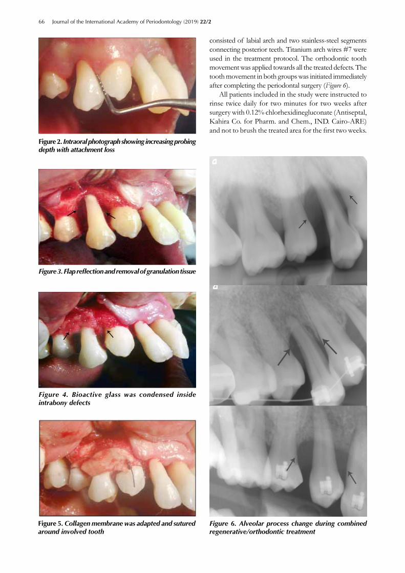
Surgical proceduresLocal anesthesia (2% lidocaine containing 1:100,000 epinephrine) was utilized followed by intrasulcular inci-sion. Full thickness flaps were elevated from both the buccal and the lingual aspects. All granulation tissues were removed from the defects. The 3- or 2-wall defects included in the study were determined after flap reflec-tion. The root surfaces were thoroughly debrided with hand and ultrasonic instruments (Figure 2 and 3). Root conditioning was applied using tetracycline solution for 3 minutes. The wound was rinsed several times with sterile saline solution. In all groups, the defects were filled with a bioactive glass (Figure 4). The collagen membrane was trimmed and adjusted to cover the defect and at least 2-3 mm of the surrounding bone. The coronal portion of the barrier was tightened and sutured on the root with a resorbable sling suture (Figure 5). The flap was replaced and sutured using vertical mattress sutures. In group I; diode laser therapy was performed at the date of surgery then twice weekly for the next four weeks. Laser treatment was performed by using an 870-nm gallium–aluminum–arsenide diode laseri rradiation (Soft Laser SL–202, 870 nm Petrolaser, Russia) in a continu-ous wave mode (CW) in contact to the surface of the gingiva. The surface area of the probe was enough to cover the whole target area of both buccal and lingual aspects of the defect for 60sec. per aspect, reaching a total of 120sec. for each defect. The delivered power was 80 mW through a spot size of 0.55 cm2. The resultant energy density was 8.73 j/cm2 per point of application with a total energy density of 17.46 j/cm2 per defect (Saafan et al., 2013). Both patients and the operator wore protective glasses during laser application.
The patients were treated using the segmented arch technique to change the inclination of extruded, mis-aligned and migrated teeth. The forces used were light and continuous, about 10 to 15g per tooth, depending on the amount of the periodontal support. The anchorage
Figure 1. Densometric analysis using DBS-win software
66 Journal of the International Academy of Periodontology (2019) 22/2
consisted of labial arch and two stainless-steel segments connecting posterior teeth. Titanium arch wires #7 were used in the treatment protocol. The orthodontic tooth movement was applied towards all the treated defects. The tooth movement in both groups was initiated immediately after completing the periodontal surgery (Figure 6).
All patients included in the study were instructed to rinse twice daily for two minutes for two weeks after surgery with 0.12% chlorhexidinegluconate (Antiseptal, Kahira Co. for Pharm. and Chem., IND. Cairo-ARE) and not to brush the treated area for the first two weeks.
29
Fig 2
Figure 2. Intraoral photograph showing increasing probing depth with attachment loss
29
Fig 2
Figure 3. Flap reflection and removal of granulation tissue
30
Fig 3
Fig 4
Figure 4. Bioactive glass was condensed inside intrabony defects
31
Fig 5
The image part with relationship ID rId10 was not found in the file.
31
Fig 5
The image part with relationship ID rId10 was not found in the file.
31
Fig 5
The image part with relationship ID rId10 was not found in the file.
Figure 6. Alveolar process change during combined regenerative/orthodontic treatment
Figure 5. Collagen membrane was adapted and sutured around involved tooth

Attia et al.: Low-level laser biostimulation with combined orthodontic regenerative therapy 67
Systemic antibiotic therapy of doxycycline hyclate (Doxymycin, Nile Co. for Pharm. and Chem., IND. Cairo-ARE), 100 mg every 12 hours for 10 days was prescribed. Recall appointments were carried out every week for the first month and then monthly for profes-sional prophylaxis and oral hygiene reinforcement.
Statistical Analysis Sample size calculationswere achieved using http://biomath.info/power based on a previous study by Saglam et al., (2014). A total sample size of 14 patients (7 in each group) was sufficient to detect the difference based on this calculation. However, the present study was carried on fifteen patients with thirty defects (fifteen in each group).
The collected data were tabulated and statistically analyzed using statistical program SPSS version 16.0 (Statistical Package for Social Sciences, SPSS, Inc., Chicago, IL, USA). Student’s t-test was used to test the effect of group on different measurements within each interval. Paired t-testwas run to test the effect of intervals on different measurements within each group.
Results
All 15 patients completed treatment and had no adverse reactions to therapy. Healing was uneventful in the 30 sites involved in this study.
Clinical parametersProbing depthThe mean PD reduction in the two groups is shown in Figure 7. Mean PD recordings within Group I (the
combined orthodontic regenerative therapy with diode laser application)showed a highly significant reduction during the study period(Pvalue = 0.000), at baseline it was 6.83±0.58 mm, 6 months 2.92±0.67 mm and at 9 months 2.42±0.67 mm with maximum reduction in probing depth noticed after 9 months (2.42±0.67).Throughout the study period there was a highly significant reduction in PD for group II (combined orthodontic regenerative therapy with no diode laser application) between the baseline (6.42±1.16 mm) and 6 months (3.50±0.67 mm). Moreover, a highly statistically significant reduction in probing depth between baseline and 9 month readings was recorded. Nine month read-ings were 2.25±0.96 mm with total reduction in PD 2.17±0.20 mm.As shown in Table 1, in Group I, there was a reduction in PD by 57.25% and 64.57% at 6 and 9 months, respectively. Group II showed a decrease in PD by 45.48% and 64.95% at 6 and 9 months respec-tively, compared to baseline measurements. Statistical analysis regarding mean percent change in PD showed a no statistical significant differences in PD reduction at 6 and 9 months respectively (P = 0.021, P = 0.902) among the two groups.
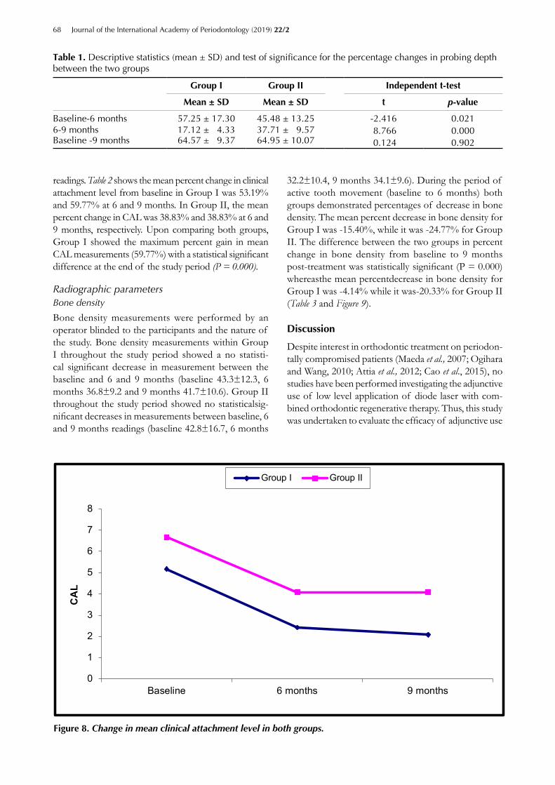
Clinical attachment level The mean clinical attachment level (CAL) results within Group I are shown in Figure 8. Throughout the study pe-riod there was a highly significant gain (baseline 5.17±1.26 mm, 6 months 2.42±1.37 mm, 9 months 2.08±0.79 mm). Maximum gain of clinical attachment was noticed at 9 months which reached 3.09±0.47 mm. There was a non-significant difference between the 6 and 9 months
32
Fig 6
Fig 7
Fig 8
0
1
2
3
4
5
6
7
8
Baseline 6 months 9 months
Prob
ing
dept
h
Group I Group II
0
1
2
3
4
5
6
7
8
Baseline 6 months 9 months
CA
L
Group I Group II
Figure 7. Change in mean probing depth in both groups.
68 Journal of the International Academy of Periodontology (2019) 22/2
readings. Table 2 shows the mean percent change in clinical attachment level from baseline in Group I was 53.19% and 59.77% at 6 and 9 months. In Group II, the mean percent change in CAL was 38.83% and 38.83% at 6 and 9 months, respectively. Upon comparing both groups, Group I showed the maximum percent gain in mean CAL measurements (59.77%) with a statistical significant difference at the end of the study period (P = 0.000).
Radiographic parametersBone density
Bone density measurements were performed by an operator blinded to the participants and the nature of the study. Bone density measurements within Group I throughout the study period showed a no statisti-cal significant decrease in measurement between the baseline and 6 and 9 months (baseline 43.3±12.3, 6 months 36.8±9.2 and 9 months 41.7±10.6). Group II throughout the study period showed no statisticalsig-nificant decreases in measurements between baseline, 6 and 9 months readings (baseline 42.8±16.7, 6 months
32.2±10.4, 9 months 34.1±9.6). During the period of active tooth movement (baseline to 6 months) both groups demonstrated percentages of decrease in bone density. The mean percent decrease in bone density for Group I was -15.40%, while it was -24.77% for Group II. The difference between the two groups in percent change in bone density from baseline to 9 months post-treatment was statistically significant (P = 0.000) whereasthe mean percentdecrease in bone density for Group I was -4.14% while it was-20.33% for Group II (Table 3 and Figure 9).
Discussion
Despite interest in orthodontic treatment on periodon-tally compromised patients (Maeda et al., 2007; Ogihara and Wang, 2010; Attia et al., 2012; Cao et al., 2015), no studies have been performed investigating the adjunctive use of low level application of diode laser with com-bined orthodontic regenerative therapy. Thus, this study was undertaken to evaluate the efficacy of adjunctive use
Group I Group II Independent t-test
Mean ± SD Mean ± SD t p-value
Baseline-6 months 57.25 ± 17.30 45.48 ± 13.25 -2.416 0.0216-9 months 17.12 ± 4.33 37.71 ± 9.57 8.766 0.000Baseline -9 months 64.57 ± 9.37 64.95 ± 10.07 0.124 0.902
Table 1. Descriptive statistics (mean ± SD) and test of significance for the percentage changes in probing depth between the two groups
32
Fig 6
Fig 7
Fig 8
0
1
2
3
4
5
6
7
8
Baseline 6 months 9 months
Prob
ing
dept
h
Group I Group II
0
1
2
3
4
5
6
7
8
Baseline 6 months 9 months
CA
L
Group I Group II
Figure 8. Change in mean clinical attachment level in both groups.

Attia et al.: Low-level laser biostimulation with combined orthodontic regenerative therapy 69
Group I Group II Independent t-test
Mean ± SD Mean ± SD t p-value
Baseline-6 months 53.19 ± 7.89 38.83 ± 10.7 - 4.831 0.0006-9 months 14.04 ± 3.25 1.25 ± 3.3 -12.349 0.000Baseline -9 months 59.77 ± 12.1 38.83 ± 7.56 - 6.564 0.000
Table 2. Descriptive statistics (mean ± SD) and test of significance for the percentage changes in clinical attachment level between the two groups
Group I Group II Independent t-test
Mean ± SD Mean ± SD t p-value
Baseline-6 months 15.40 ± 7.65 24.77 ± 10.39 3.248 0.0026-9 months 13.32 ± 5.87 5.90 ± 3.25 -4.946 0.000Baseline -9 months 4.14 ± 3.17 20.33 ± 5.37 11.611 0.000
Table 3. Descriptive statistics (mean ± SD) and test of significance for the percentage changes in bone density between the two groups
33
Fig 9
0
5
1015
20
25
30
35
40
4550
Baseline 6 months 9 months
Bon
e de
nsity
Group I Group II
Figure 9. Change in mean bone density in both groups.
ofdiode laser biostimulationwith combined orthodontic regenerative therapy clinically and radiographically.
To test effects of orthodontic tooth movement on a reduced periodontium and regenerative periodontal therapy in a reduced periodontium, several studies have been conducted which concluded that orthodontic tooth movement could enhance different regenera-tive procedures (Maeda et al., 2007; Attia et al., 2012; Cao et al., 2015). This contribution to bone formation by mechanical loading forcescould play a significant role in new tissue formation. The integrin-mediated
signal transduction cascade is the main mechanism of mechanotransduction in cells and is associated with osteogenesis. Cell multiplication is the first reaction. In-deed, fibroblast numbers were doubled in the three days after the commencement of tooth movement (Meikle, 2006). Orthodontic tooth movement can stimulate pre-osteoblasts and mesenchymal cells to differentiate into osteoblasts (Faber et al., 2005). Moreover, intense production of TGF-β1 mRNA and the translated protein contributes to angiogenesis and coincides with osteoblast migration, differentiation and the formation

70 Journal of the International Academy of Periodontology (2019) 22/2
of extracellular matrix (Mehrara et al., 1999). Concur-rently, expression of BMP2, 4 and 7 in the connective tissue is also increased (Mehrara et al., 1999).
Both guided tissue regeneration and grafting proce-dures are means of achieving periodontal regeneration (Scantlebury and Ambruster, 2012). Tiefengraber et al. (2002) demonstrated that the transverse dimension of the alveolar bone can be preserved by using a barrier membrane. They also convincingly showed that gingival invagination can be prevented using this technique. Gingival invaginations create niches in patients with poor oral hygiene that may lead to increased marginal bone loss, a reduction in the interdental bone height, and slower orthodontic space closure (Wehrbein et al., 1993).The use of bone grafts might also reduce the risk of gingival invaginations and enhance new bone formation (Reichert et al., 2009).
In this study both experimental groups showed greater reduction in probing depth and gain in clinical attachment level throughout the study interval. Group I which was treated by combined orthodontic regenerative therapy with laser irradiation showed the highest percent gain in clinical attachment at 9 months (59.77±12.1%). This could be due to the effect of regenerative ma-terials and the laser biostimulation to fibroblast and keratinocyte motility, collagen synthesis, angiogenesis and growth factors release, thus facilitating the healing process (Amorim et al., 2006).
The decrease in bone density associated with or-thodontic tooth movement might be attributed to ap-plication of orthodontic force that results in physical distortion of the periodontal ligament and alveolar bone cells. This can also trigger a multilevel cascade of signal transduction pathways, including the prostaglandin E2 (PGE2) pathway, which in turn initiates structural and functional changes (Davidovitch et al., 1988).The production of PGE2, a key factor involved in the destruc-tion of periodontal tissues, is primarily catabolized by cyclooxygenase-2 (COX-2) (Correa et al., 2007; Pejcic et al., 2010). The increased expression of COX-2 results in increased PGE2 and subsequent IL-1β production stimulating osteoclastic activity and further destruction of the supportive periodontal tissues (Trelles, 1987; Sakurai et al., 2000).
Moreover, the cytokine expression pattern found at both the pressure and tension sides shows higher expres-sion of all cytokines when compared to the periodontal ligament of teethnoit subjected to orthodontic forces. The compression side exhibits higher expression of TNF-α, matrix metalloproteinase I (MMP-1) and RANKL, whereas the tension side has a higher expression of type I collagen, IL-10, tissue inhibitor of matrix metalloprotein-ase I (TIMP-1), osteoprotegerin (OPG) and osteocalcin (OCN). The expression of TGF-β is similar in both pressure and tension sides(Garlet et al., 2007).
The reduction in bone density associated with or-thodontic tooth movement was 4.14±3.17 for Group I where laser application was used, while Group II with no laser application showed 20.33±5.37 reduction. There was a significant difference in bone density among both groups at 9 months. This could be due to the incorporation of antimicrobial, anti-inflammatory and bio-stimulating effects of diode laser on the periodontal tissues with combined regenerative therapy in Group I.
On the other hand, the use of low-level laser treat-ment provides anti-inflammatory effects similar to non-steroidal anti-inflammatory drugs (NSAIDs), which are mainly achieved through inhibition of the expression of COX-2, PGE2, and histamine (Chomczynski et al., 1987; Noguchi et al., 2001, Pseveski et al., 2017). Prostaglandin inhibition has been demonstrated following laser appli-cation (Pejcic et al., 2010; Sobouti et al., 2015). Mizutani et al.(2004) showed that low-level laser therapy inhibits the cascade of the arachidonic acid in damaged tissues with subsequent decrease in production of inflammatory cy-tokines. Recently, Saglam et al. (2014) showed adjunctive use of a diode laser provided significant improvements and reduction in IL-1β, IL-6, MMP-1and MMP-8 in the first month following non-surgical periodontal therapy. Di-ode lasers can also prevent plasminogen increased activity, and prostaglandin synthesis. They can also reduce IFN-γ, while having a stimulating effect in the production of platelet derived growth factor (PDGF) and transforming growth factor-ß (TGF-ß). Thus, the anti-inflammatory effect of lasers arise through a number different mecha-nisms (Sobouti et al., 2015).
Conclusion
In conclusion, the combined orthodontic and regenera-tive therapy resulted in favorable clinical and radiographic outcomes. Defectsin which combined orthodontic/regenerative therapy with low laser application (Group I) demonstrated superior results than defects in which combined orthodontic/regenerative therapy was used alone (Group II). Further controlled and prospective studies are necessary to study the effects of diode lasers on orthodontic regenerative therapy.
Conflict of interestThe authors declare that they have no conflict of interest.
References
Abramson J. The corneal medical index as an epide-miological tool. American Journal of Public Health 1966; 56:287-298.
Amorim JC, de Sousa GR, de Barros Silveira L, Prates RA, Pinotti M and Ribeiro MS. Clinical study of the gingiva healing after gingivectomy and low-level laser therapy. Journal of Photomedicine and Laser Surgery 2006; 24:588-594.

Attia et al.: Low-level laser biostimulation with combined orthodontic regenerative therapy 71
Andrade IJ, Taddei SR, Garlet GP, Garlet TP, Teixeira AL, Silva TA, et al. CCR5 down-regulates osteoclast function in orthodontic tooth movement. Journal of Dental Research 2009; 88:1037-1041.
Aguirre-Zorzano AL, Bayona MJ, Remolina A, et al. Pos-torthodontic stability of the new attachment achieved by guided tissue regeneration following orthodontic movement: report of 2 cases. Quintessence Interna-tional 1999; 30:769-774.
Aoki A, Sasaki KM, Watanabe H and Ishikawa I. Lasers in nonsurgical periodontal therapy. Periodontology 2000 2004; 36:59-97.
Araujo MG, Carmagnola D, Berglundh T, Thilander B and Lindhe J. Orthodontic movement in bone defects augmented with Bio-Oss. An experimental study in dogs. Journal of Clinical Periodontology 2001; 28:73-80.
Attia MS, Shoreibah EA, Ibrahim SA and Nassar HA. Regenerative therapy of osseous defects combined with orthodontic tooth movement. Journal International Academy of Periodontology 2012; 14:17-25.
Brady TA, Piesco NP, Buckley MJ, Langkamp HH, Bowen LL, Agarwal S. Autoregulation of periodontal liga-ment cell phenotype and functions by transforming growth factor-l. Journal of Dental Research 1998; 77(10): 1779-1790.
Braun S, Winzler J and Johnson BE. An analysis of orthodontic force systems applied to the dentition with diminished alveolar support. European Journal of Orthodontics 1993; 15:73-77.
Cao T, Xu L, Shi J and Zhou Y. Combined orthodontic-periodontal treatment in periodontal patients with anteriorly displaced incisors. American Journal of Or-thodontics and Dentofacial Orthopedics 2015; 148:805-813.
Cardaropoli D and Gaveglio L. The influence of orth-odontic movement on periodontal tissues level.Semi-nars in Orthodontics 2007; 13:234-245.
Chomczynski P and Sacchi N. Single step method of RNA isolation by acid guanidiniumthiocyanate-phenol-chloroform extraction. Analytical Biochemistry 1987; 162:156-159.
Correa F, Martins RABL, Correa JC, Iversen VV, Joenson J and Bjordal JM. Low-level laser therapy [GaAs =904 nm] reduces inflammatory cell migration in mice with lipopolysaccharide-induced peritonitis. Journal of Pho-tomedicine and Laser Surgery 2007; 25:245-249.
Davidovitch Z, Nicolay OF, Nigan PW and Shanfield JL. Neurotransmitters, cytokines, and the control of alveolar bone remodeling in orthodontics. Dental Clinics of North America 1988; 32:411-435.
Faber J, Azevedo RB, Bao SN. Distraction osteogenesis may promote periodontal bone regeneration. Journal of Dental Research 2005; 84:757-761.
Fukunaga T, Kuroda S, Kurosaka H and Takano-Yamamoto T. Skeletal anchorage for orthodontic co rrection of maxillary protrusion with adult periodontitis. Angle of Orthodontics 2006; 76:148-155.
Gao Q, Zhang S, Jian X, Zeng Q and Ren L. Expres-sion of epidermal growth factor and epidermal growth factor receptor in rat periodontal tissues during orthodontic tooth movement. Zhonghua Kou Qiang Yi XueZaZhi. 2002; 37:294-296.
Garlet TP, Coelho U, Silva JS and Garlet GP.Cytokine expression pattern in compression and tension sides of the periodontal ligament during orthodontic tooth movement in humans. European Journal of Oral Sciences 2007; 115:355-362.
Gavish L, Perez L and Gertz SD. Low-level laser ir-radiation modulates matrix metalloproteinase activ-ity and gene expression in porcine aortic smooth muscle cells. Lasers in Surgery and Medicine 2006; 38:779-786.
Gavish L, Perez LS, Reissman P and Gertz SD. Ir-radiation with 780 nm diode laser attenuates in-flammatory cytokines but upregulates nitric oxide in lipopolysaccharide-stimulated macrophages: implications for the prevention of aneurysm progression. Lasers in Surgery and Medicine 2008; 40:371-378
Harrel SK, Nunn ME, Hallmon WW. Is there an as-sociation between occlusion and periodontal de-struction?: Yes - Occlusal forces can contribute to periodontal destruction. Journal American of Dental Association 2006; 137:1380, 1382, 1384 passim.
Harrel SK, Nunn ME. The association of occlusal contacts with the presence of increased periodontal probing depth. Journal of Clinical Periodontology 2009; 36:1035-1042.
Johal A, Ide M. Orthodontics in the adult patient, with special reference to the periodontally compromised patient. Dental Update 1999; 26:101-108.
Keles GC, Cetinkaya BO, Albayrak D, Koprulu H and Acikgoz G. Comparison of platelet pellet and bioactive glass in periodontal regenerative therapy. Acta Odontologica Scandinavica 2006; 64:327-333.
Kreisler M, Christoffers AB, Willershausen B and d′Hoedt B. Effect of low-level GaAlAs laser irradia-tion on the proliferation rate of human periodontal ligament fibroblasts: An in vitro study. Journal of Clinical Periodontology 2003; 30:353-358.
Maeda S, Maeda Y, Ono Y, Nakamura K and Matsui T. Interdisciplinary approach and orthodontic options for treatment of advanced periodontal disease and malocclusion: A case report. Quintessence International 2007; 38:653-662.
Masella RS and Meister M. Current concepts in the biology of orthodontic tooth movement.American Journal of Orthodontics and Dentofacial Orthopedics 2006; 129:458-468.
Mavreas D. Self-ligation and the periodontally com-promised patient: A different perspective. Seminars in Orthodontics 2008; 14:36-45.
72 Journal of the International Academy of Periodontology (2019) 22/2
Mehrara BJ, Rowe NM, Steinbrech DS, Duziak ME, Saadeh PB, McCarthy JG et al. Rat mandibular distraction osteogenesis: II. Molecular analysis of transforming growth factor beta-1 and osteocalcin gene expression. Plast Reconstructive Surgery 1999; 103:536-547.
Meikle MC. The tissue, cellular and molecular regula-tion of orthodontic tooth movement: 100 years after Carl Sandstedt. European Journal of Orthodontics 2006; 28:221-240.
Mitsui N, Suzuki N, Maeno M, Yanagisawa M, Koy-ama Y, Otsuka K, et al. Optimal compressive force induces bone formation via increasing bone mor-phogenetic proteins production and decreasing their antagonists production by Saos-2 cells. Life Sciences; 2006; 78:2697-2706.
Mizutani K, Musya Y, Wakae K, Kobayashi T, Tobe M, Taira K, et al. A clinical study on serum pros-taglandin E2 with low level laser therapy. Journal of Photomedicine and Laser Surgery 2004; 22:537-539.
Nishihara T, Koseki T. Microbial etiology of periodon-titis. Periodontology 2000 2004; 36:14-26.
Noguchi K, Shitashige M, Endo H, Kondo H, Yot-sumoto Y, Izumi Y, Nitta H and Ishikawa I. In-volvement of cyclooxygenase-2 in serum-induced prostaglandin production by human oral gingival epithelial cells. Journal of Periodontal Research 2001; 6:124-130.
Ogihara S and Marks M. Enhancing the regenerative potential of guided tissue regeneration to treat an intrabony defect and adjacent ridge to deformity by orthodontic extrusive force. Journal of Periodontology 2006; 77:2093-2100.
Ogihara S and Wang HL. Periodontal regeneration with or without limited orthodontics for the treat-ment of 2- or 3-wall infrabony defects. Journal of Periodontology 2010; 81:1734-1742.
Ong MA, Wang HL and Smith FN. Interrelationship between periodontics and adult orthodontics.Journal of Clinical Periodontology 1998; 25:271-277.
Pejcic A, Kojovic D, Kesic L and Obradovic R. The effects of low level laser irradiation on gingival in-flammation. Journal of Photomedicine and Laser Surgery 2010; 28:69-74.
Pesevska S, Gjorgoski I, Ivanovski K, Soldatos NK and Angelov N. The effect of low-level diode laser on COX-2 gene expression in chronic periodontitis patients. Journal of Lasers in Medical Sciences 2017; 32:1463-1468.
Polson AM, Caton JG, Yeaple RN and Zander HA. Histological determination of probe tip penetration into gingival sulcus of humans using an electronic pressure-sensitive probe. Journal of Clinical Periodon-tology 1980; 7:479-488.
Pourzarandian A, Watanabe H, Ruwanpura SM, Aoki A and Ishikawa I. Effect of low-level Er:YAG laser irradiation on cultured human gingival fibroblasts. Journal of Periodontology 2005; 76:187-193.
Ramfjord SP. The periodontal disease index (PDI).Journal of Periodontology 1967; 38:602-610.
Reichert C, Deschner J, Kasaj A, Andreas Jager A. Guided Tissue Regeneration and Orthodontics.A Review of the Literature. Journal of Orofacial Ortho-pedics 2009; 70:6-19.
Saafan A, El-Nahass H, Nasr AS, Radwan R. Effect of low power diode laser 810 nm on TGFβ1 level in GCF in aggressive periodontitis. Journal of Dental Lasers 2013; 7(2): 59-65.
Saglam M, Kantarci A and Dundar N. Clinical and biochemical effects of diode laser as an adjunct to nonsurgical treatment of chronic periodontitis: A randomized, controlled clinical trial. Journal of Lasers in Medical Sciences 2014; 29:37-46.
Sakurai Y, Yamaguchi M and Abiko Y. Inhibitory effect of low level laser irradiation on LPS-stimulated prostaglandin E2 production and cyclooxygenase-2 in human gingival fibroblasts. European Journal of Oral Sciences 2000; 108:29-34.
Scantlebury T, Ambruster J. The development of guided regeneration: making the impossible pos-sible and the unpredictable predictable. J Evid Based Dent Pract2012; 12(3 Suppl):101-17.
Sculean A, Barbe G, Chiantella GC, Arweiler NB, Be-rakdar M and Brecx M. Clinical evaluation of an enamel matrix protein derivative combined with a bioactive glass for the treatment of intrabony peri-odontal defects in humans. Journal of Periodontology 2002; 73:401-408.
Sheikh ZA, Javaid MA and Abdallah MN. Bone re-placement graft materials in dentistry. In: Dental biomaterials (principle and its application), 2nd Ed.; Khurshid Z, Zafar SZ (editors). Paramount Publishing Enterprise: Karachi, Pakistan, 2013.
Skoglund A, Hising P and Young C. A clinical and histological examination in humans of the osse-ous response to implanted natural bone mineral. International Journal of Oral Maxillofacial Implants 1997; 12:194.
Sobouti F, Khatami M, Heydari M and Barati M. The role of low-level laser in periodontal surgeries.Journal of Lasers in Medical Sciences 2015; 6:45-50.
Suresh S, Merugu S, Mithradas N, Sivasankari. Low-level laser therapy: A biostimulation therapy in periodon-tics. SRM Journal Research Dental Science 2015; 6:53-56.
Taddei SR, Andrade I, Queiroz-Junior CM, Garlet TP, Garlet GP, Cunha-Fde Q, et al. Role of CCR2 in orthodontic tooth movement. American Journal of Orthodontics and Dentofacial Orthopedics 2012; 141:153-160.
Attia et al.: Low-level laser biostimulation with combined orthodontic regenerative therapy 73
Tiefengraber J, Diedrich P, Fritz U, et al. Orthodontic space closure in combination with membrane sup-ported healing of extraction sockets (MHE) a pilot study. J OrofacOrthop 2002; 63:422-428.
Trelles M. Bone fracture consolidates faster with low power laser. Lasers in Surgery and Medicine 1987; 7:36-45.
Wehrbein H, Fuhrmann R, Andreas A, et al. Die Bedeu-tung von Gingivaduplikaturen beimorthodontischenLückenschluss:Eineklinisch-radiologischeStudie. FortschrKieferorthop 1993; 54:231-236.
Weston P, Yaziz YA, Moles DR, Needleman I. Oc-clusal interventions for periodontitis in adults. Cochrane Database Syst. Rev. 2008; CD004968. DOI: 10.1002/14651858.CD004968.pub2.
Yokota ET, Miles DA, Newton CW, et al. Interpretation of periapical lesions using radiovisiography. Journal of Endodontics 1994; 20:490-494.
Zhu AJ and Scott MP. Incredible journey: How do developmental signals travel through tissue? Genes and Development 2004; 18:2985-2997.
Yokota ET, Miles DA, Newton CW, et al.Interpretation of periapical lesions using radiovisiography. Journal of Endodontics 1994; 20:490-494.
Zhu AJ and Scott MP. Incredible journey: How do developmental signals travel through tissue? Genes and Development 2004; 18:2985-2997.
View publication statsView publication stats


![REVIEW [REVISIÓN] BIOSTIMULATION IN CATTLE: STIMULATION](https://img.dokumen.tips/doc/110x75/62daefe853e65f03c93991b0/review-revisin-biostimulation-in-cattle-stimulation-.jpg)