Embed Size (px)
Citation preview
Bioorganic & Medicinal Chemistry Letters 24 (2014) 1638–1649
Contents lists available at ScienceDirect
Bioorganic & Medicinal Chemistry Letters
journal homepage: www.elsevier .com/ locate/bmcl
BMCL Digest
Graphene for multi-functional synthetic biology: The last ‘zeitgeist’in nanomedicine
http://dx.doi.org/10.1016/j.bmcl.2014.01.0510960-894X/� 2014 The Authors. Published by Elsevier Ltd.This is an open access article under the CC BY-NC-ND license (http://creativecommons.org/licenses/by-nc-nd/3.0/).
⇑ Corresponding authors. Tel.: +44 1612751800 (K.K.).E-mail addresses: [email protected] (A. Bianco), [email protected]
(M. Prato), [email protected] (K. Kostarelos).
A. Servant a, A. Bianco b,⇑, M. Prato c,⇑, K. Kostarelos a,⇑a Nanomedicine Laboratory, Faculty of Medical and Human Sciences & National Graphene Institute, University of Manchester, AV Hill Building, Manchester M13 9PT, United Kingdomb CNRS, Institut de Biologie Moléculaire et Cellulaire, Laboratoire d’Immunopathologie et Chimie Thérapeutique, 67000 Strasbourg, Francec Dipartimento di Scienze Chimiche e Farmaceutiche, Università di Trieste, 34127 Trieste, Italy
a r t i c l e i n f o
Article history:Received 19 October 2013Revised 13 January 2014Accepted 18 January 2014Available online 10 February 2014
Keywords:NanotechnologyNanomaterialsDrug deliveryCarbon
a b s t r a c t
The high versatility of graphene has attracted significant attention in many areas of scientific researchfrom electronics to physics and mechanics. One of the most intriguing utilisation of graphene remainshowever in nanomedicine and synthetic biology. In particular, the last decade has witnessed an exponen-tial growth in the generation of novel candidate therapeutics of multiple biological activities based ongraphene constructs with small molecules, such as anti-cancer drugs. In this Digest, we summarise thedifferent synthetic strategies and routes available to fabricate these promising graphene conjugatesand the opportunities for the design of multi-functional tools for synthetic biology that they offer.
� 2014 The Authors. Published by Elsevier Ltd. This is an open access article under the CC BY-NC-NDlicense (http://creativecommons.org/licenses/by-nc-nd/3.0/).
Introduction: The landscape for discovering and developingdrugs is evolving at an increasing pace as the direction for newdiagnostic and therapeutic strategies heads towards personalisedmedicine. Personalised medicine aims at improving healthcare byintegrating early detection of disease, preventive medicine, ra-tional drug discovery and development, and monitoring of ther-apy.1 Since the early 1970s, the use of nanotechnologies ingeneral seems to have refashioned the pharmaceutical and drugdelivery field and in particular the administration of medicines.Prior the use of ‘nanomedicines’ that is the development of nanom-aterials containing medicines, it was inconceivable to administer adispersion of solid particles by the route of intravenous injectionconsidering the high risk of embolism. Nowadays, nanoparticlesand nanomaterials have allowed the improvement of solubility,therapeutic activity and reduction of toxicity by increasing uptaketo target sites and changing pharmacokinetic profiles of manytraditional drugs. The design and production of these sophisticatednanoscale vectors has generated a fundamental technologicaland medical breakthrough, allowing the progression over manymilestones in pharmaceutical development.
Despite the astonishing progress made in the development ofdisease-targeted nanoparticles, much effort is still needed in thedesign and engineering of more efficient and safer materials forenhanced targeting and delivery of drugs for which there are no
effective delivery systems. In addition, the creation of multifunc-tional nanomaterials combining for example an imaging and atherapeutic agent (theranostic agent) or different therapeuticagents with complementary biological targets would certainly bethe next challenging step in nanomedicine development towardspersonalised patient medicine.2
To date, many types of nanomaterials have been engineered fordeveloping multifunctional platforms that can fulfil many tasks incancer therapy and diagnosis and in synthetic biology.3 The discov-ery of graphene in 2004 has generated significant interest in manyareas of scientific research and has opened up a plethora of poten-tial applications of its exciting properties encompassing variousareas such as solar cells, transistors, super-capacitors, and memorydevices.4–7 Graphene is a single atom thick and a two dimensionalarrangement of conjugated sp2 carbons in a honeycomb structure.8
Thanks to its unique properties such as an enhanced electrical andthermal conductivity, high charge carrier concentration andmobility, optical transparency, and mechanical strength, grapheneis expected to reform the world of digital technology in a similarway as silicon has transformed the field of electronics. The highsurface to volume ratio and the great chemical versatility ofgraphene make this exclusive material an ideal candidate for thedevelopment of a new generation of nanomedicines with applica-tions encompassing the detection of biomarkers to imaging andcancer therapy.
To date, the exploitation of graphene properties for biomedicalapplications remains at its infancy. Could graphene bring solutionsto the challenges faced by the current nanomedicines and offer an
A. Servant et al. / Bioorg. Med. Chem. Lett. 24 (2014) 1638–1649 1639
efficient platform for theranostics and synthetic biology? Withoutmajor breakthrough, the answer to this question is no. Graphene,in its unmodified state, suffers from one major drawback for its po-tential use in biomedical applications. The physical handling ofgraphene sheets is very delicate as it is not dispersible in aqueousconditions and most organic solvents.9 Graphene tends to formirreversible aggregates or to restack by van der Waals interactionsand p–p stacking. As a result, the conjugation of graphene withtherapeutically active molecules or active biological componentsis extremely challenging.
Chemical functionalisation of graphene sheets may be one wayto address these concerns. The hydrophobic character of graphenecan be altered by the nature of the molecules and the extent ofchemical functionalisation on the graphene sheets, improving thebiocompatibility of this material. Graphene oxide (GO), the ‘hydro-philic derivative’ of graphene has attracted significant attention forits enhanced aqueous dispersibility and colloidal stability overother carbon-based nanomaterials. GO preparation from theHummers method is now common knowledge and is currentlywidely explored for the development of nanomedicines.
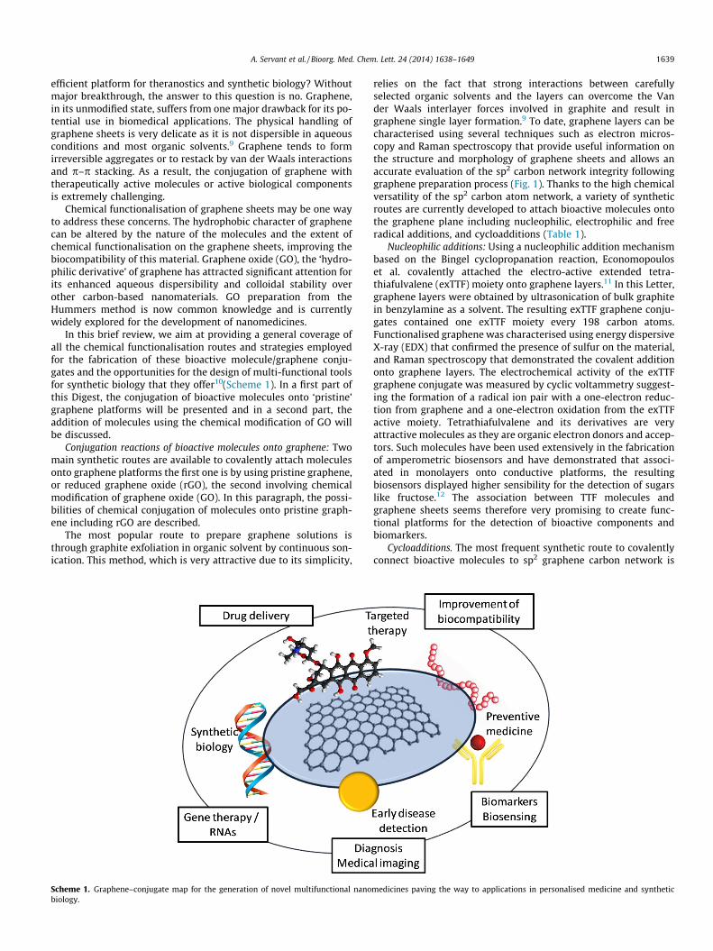
In this brief review, we aim at providing a general coverage ofall the chemical functionalisation routes and strategies employedfor the fabrication of these bioactive molecule/graphene conju-gates and the opportunities for the design of multi-functional toolsfor synthetic biology that they offer10(Scheme 1). In a first part ofthis Digest, the conjugation of bioactive molecules onto ‘pristine’graphene platforms will be presented and in a second part, theaddition of molecules using the chemical modification of GO willbe discussed.
Conjugation reactions of bioactive molecules onto graphene: Twomain synthetic routes are available to covalently attach moleculesonto graphene platforms the first one is by using pristine graphene,or reduced graphene oxide (rGO), the second involving chemicalmodification of graphene oxide (GO). In this paragraph, the possi-bilities of chemical conjugation of molecules onto pristine graph-ene including rGO are described.
The most popular route to prepare graphene solutions isthrough graphite exfoliation in organic solvent by continuous son-ication. This method, which is very attractive due to its simplicity,
Scheme 1. Graphene–conjugate map for the generation of novel multifunctional nanobiology.
relies on the fact that strong interactions between carefullyselected organic solvents and the layers can overcome the Vander Waals interlayer forces involved in graphite and result ingraphene single layer formation.9 To date, graphene layers can becharacterised using several techniques such as electron micros-copy and Raman spectroscopy that provide useful information onthe structure and morphology of graphene sheets and allows anaccurate evaluation of the sp2 carbon network integrity followinggraphene preparation process (Fig. 1). Thanks to the high chemicalversatility of the sp2 carbon atom network, a variety of syntheticroutes are currently developed to attach bioactive molecules ontothe graphene plane including nucleophilic, electrophilic and freeradical additions, and cycloadditions (Table 1).
Nucleophilic additions: Using a nucleophilic addition mechanismbased on the Bingel cyclopropanation reaction, Economopouloset al. covalently attached the electro-active extended tetra-thiafulvalene (exTTF) moiety onto graphene layers.11 In this Letter,graphene layers were obtained by ultrasonication of bulk graphitein benzylamine as a solvent. The resulting exTTF graphene conju-gates contained one exTTF moiety every 198 carbon atoms.Functionalised graphene was characterised using energy dispersiveX-ray (EDX) that confirmed the presence of sulfur on the material,and Raman spectroscopy that demonstrated the covalent additiononto graphene layers. The electrochemical activity of the exTTFgraphene conjugate was measured by cyclic voltammetry suggest-ing the formation of a radical ion pair with a one-electron reduc-tion from graphene and a one-electron oxidation from the exTTFactive moiety. Tetrathiafulvalene and its derivatives are veryattractive molecules as they are organic electron donors and accep-tors. Such molecules have been used extensively in the fabricationof amperometric biosensors and have demonstrated that associ-ated in monolayers onto conductive platforms, the resultingbiosensors displayed higher sensibility for the detection of sugarslike fructose.12 The association between TTF molecules andgraphene sheets seems therefore very promising to create func-tional platforms for the detection of bioactive components andbiomarkers.
Cycloadditions. The most frequent synthetic route to covalentlyconnect bioactive molecules to sp2 graphene carbon network is
medicines paving the way to applications in personalised medicine and synthetic

Figure 1. Graphene structure and physical characterisation; (A) molecular structure of a graphene plan,42 (B) transmission Electron Microscopy image, and (C) high resolutionTEM image; (D) Raman spectroscopy spectrum of few layer graphene; the de-convolution of the 2D band at 2700 cm�1 confirms that it is a few layer graphene from chemicalexfoliation.43,44 (E) Atomic Force Microscopy image of a single layer graphene. The cross-section analysis confirms the thickness of bilayer graphene around 3 nm.
1640 A. Servant et al. / Bioorg. Med. Chem. Lett. 24 (2014) 1638–1649
through cycloaddition. Cycloaddition reactions differ from nucleo-philic and electrophilic additions due to the fact that there is nointermediate formation such as anions or cations as bondrearrangement and bond formation processes occur simulta-neously. Cycloadditions have been widely used for the functionali-sation of fullerenes and carbon nanotubes, and their use ongraphene to modulate graphene solubility in solvents seemspromising.13,14 To date various types of cycloadditions are reportedto functionalise graphene layers; however the two main reactionsemployed for attaching bioactive molecules onto the sp2 carbonatom network remain the cycloaddition through the formation ofaziridine adducts and the 1,3-dipolar cycloaddition.
The insertion of an aziridine moiety onto graphene occurs viathe formation of a nitrene intermediate, which is structurallyrelated to a carbene molecule. This nitrene intermediate is gener-ally obtained through thermal or photo-decomposition of an azidegroup and is subsequently conjugated to the sp2 carbon atoms ofgraphene via a cycloaddition mechanism (Fig. 2). Using thisreaction, Strom et al. described the conjugation of graphene layerswith a modified phenylalanine.15 In this work, Phe was modifiedwith an azido group in para position of the phenyl ring priorcoupling with the graphene through nitrene addition. The final
construct was characterised using standard techniques such asthermo-gravmetric analysis (TGA) and an elemental analysis(XPS), demonstrating high functionalisation with one Phe moietyper 13 carbons. Adding sequences of amino acids onto graphenelayers can be advantageous for improving water solubility and bio-compatibility of graphene. Moreover, amino acid/graphene conju-gates can serve as efficient platforms for the fabrication of noveltherapeutic agents as peptide sequences can be cleaved specificallyby enzymes accessible in cancer cells, facilitating drug targetingand delivery. This strategy was explored previously with multi-walled carbon nanotubes to deliver methotrexate to breast cancercells MCF-7.16
The 1,3-dipolar cycloaddition of azomethine ylides has beenwidely applied to functionalise fullerenes and this chemical func-tionalisation of graphene towards a five-atom ring occurs througha six-electron cycloaddition between a 1,3-dipole and a dipolaro-phile (graphene) (Fig. 3 A).17 This reaction was used to incorporateporphyrin molecules (TTP) onto graphene layers.18 Graphene-TTPconstructs were prepared and the presence of porphyrin moietieswere confirmed by UV–vis spectroscopy and XPS with a relativelyhigh loading estimated at one TTP group per 235 carbon atoms.Porphyrins are very attractive aromatic molecules as they display
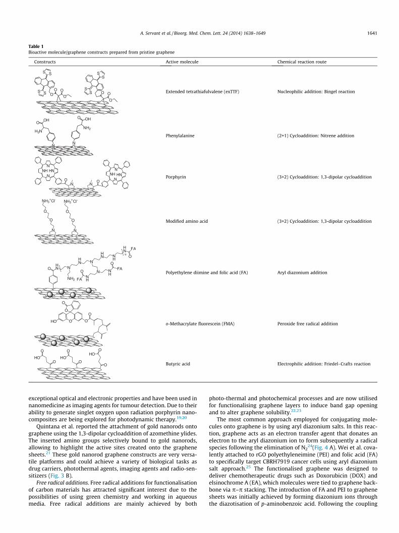
Table 1Bioactive molecule/graphene constructs prepared from pristine graphene
Constructs Active molecule Chemical reaction route
O
OO
SS
SS
OO
OOS S
SS
Extended tetrathiafulvalene (exTTF) Nucleophilic addition: Bingel reaction
N N
O OH
H2N
OHO
NH2
Phenylalanine (2+1) Cycloaddition: Nitrene addition
N N
HNN
NHN
O OHNN
NHN Porphyrin (3+2) Cycloaddition: 1,3-dipolar cycloaddition
N N
O
O
O
O
NH3+ NH3+Cl- Cl-
Modified amino acid (3+2) Cycloaddition: 1,3-dipolar cycloaddition
OHN N
HN N
HN N
H
HN FA
O
NH2
N NH
FAO
n
NHFA
O Polyethylene diimine and folic acid (FA) Aryl diazonium addition
O
O
OOHO
O
o-Methacrylate fluorescein (FMA) Peroxide free radical addition
O OO
OHO
OHO
OHO
Butyric acid Electrophilic addition: Friedel–Crafts reaction
A. Servant et al. / Bioorg. Med. Chem. Lett. 24 (2014) 1638–1649 1641
exceptional optical and electronic properties and have been used innanomedicine as imaging agents for tumour detection. Due to theirability to generate singlet oxygen upon radiation porphyrin nano-composites are being explored for photodynamic therapy.19,20
Quintana et al. reported the attachment of gold nanorods ontographene using the 1,3-dipolar cycloaddition of azomethine ylides.The inserted amino groups selectively bound to gold nanorods,allowing to highlight the active sites created onto the graphenesheets.21 These gold nanorod graphene constructs are very versa-tile platforms and could achieve a variety of biological tasks asdrug carriers, photothermal agents, imaging agents and radio-sen-sitizers (Fig. 3 B).
Free radical additions. Free radical additions for functionalisationof carbon materials has attracted significant interest due to thepossibilities of using green chemistry and working in aqueousmedia. Free radical additions are mainly achieved by both
photo-thermal and photochemical processes and are now utilisedfor functionalising graphene layers to induce band gap openingand to alter graphene solubility.22,23
The most common approach employed for conjugating mole-cules onto graphene is by using aryl diazonium salts. In this reac-tion, graphene acts as an electron transfer agent that donates anelectron to the aryl diazonium ion to form subsequently a radicalspecies following the elimination of N2
24(Fig. 4 A). Wei et al. cova-lently attached to rGO polyethyleneimine (PEI) and folic acid (FA)to specifically target CBRH7919 cancer cells using aryl diazoniumsalt approach.25 The functionalised graphene was designed todeliver chemotherapeutic drugs such as Doxorubicin (DOX) andelsinochrome A (EA), which molecules were tied to graphene back-bone via p–p stacking. The introduction of FA and PEI to graphenesheets was initially achieved by forming diazonium ions throughthe diazotisation of p-aminobenzoic acid. Following the coupling

i ii
iii iv
v vi
A
B
R
NNBF4-
1e-
R
NNBF4-
RN2BF4-
N2
R
BF4-
OHN N
HN N
HN N
H
HN FA
O
NH2
N NH
FAO
n
NHFA
O
Figure 4. Chemical functionalisation of graphene by free radical reaction usingdiazonium salts; (A) mechanism of graphene chemical functionalisation withdiazonium salts; (B) addition of folic acid (FA) onto graphene for cell targetingusing diazonium salts; (i) FA/graphene construct, (ii) TEM images of rGO, (iii) FTIRspectra of rGO and rGO/p aminobenzoic acid, (iv) TEM image of rGO/p aminobenzoic acid constructs; FA/graphene cell internalisation, (v) untreated cells, (vi)cells treated with FA/Cy3 graphene constructs. Reproduced with permission.25
Copyright 2012, Wiley-VCH Verlag 14708 GmbH&Co. KGaA, Weinheim.
B
A
i ii
iii iii
HOHNR1
O
R2 H
O
O
ON R2
R1N
O
R1
R2
O NR2R1
CO2
BocHN O OR1 = R2 = H
N
R1
R2
NR2
R1
N N
O
O
O
O
Figure 3. Chemical functionalisation of graphene via 1,3-dipolar cycloaddition; (A)mechanism of 1,3-dipolar cycloaddition onto graphene platform; (B) characterisa-tion of the gold nanorods onto graphene single layers; (i) schematic of theconstruct, (ii) TEM picture, (iii) thermogravimetric analysis, (iv) Raman spectra ofthe final construct (3), the reaction intermediate (2), graphene (1) and graphite.Reproduced with permission.21 Copyright 2011, American Chemical Society.
A
B
HN OH
O
Boc
N N N N N N
HNBoc OH
O
N
HNBoc OH
O
N2+
NN
HN
OH
O
BocHN
OH
O
Boc
Figure 2. Conjugation of amino acids onto graphene platform by nitrene addition; (A)formation of the intermediate nitrene moiety; (B) mechanism of cycloaddition ofthe nitrene adducts onto graphene.
1642 A. Servant et al. / Bioorg. Med. Chem. Lett. 24 (2014) 1638–1649
with ramified polymer, FA was subsequently added to the aminogroups of PEI to form amide bonds (Fig. 4 B i). The chemical func-tionalisation of rGO sheets with p-aminobenzoic acid was charac-terised with TEM, FTIR and Raman spectroscopy (Fig. 4 B ii, iii,iv). EA and DOX loading onto the graphene constructs were calcu-lated to be around 46% and 29% for EA and DOX, respectively. Cellinternalisation of the graphene constructs was examined by confo-cal laser scanning microscopy (CLSM) and the images revealed agood cell uptake. Moreover, it was demonstrated that these con-structs could effectively enhance the apoptosis of liver cancer cellsand increase the sensitivity of these cells towards drugs and radi-
ation (Fig. 4 B v, vi). However, cell toxicity was induced by thegraphene conjugates in the absence of the drugs above the concen-tration of 12 lg/ml. This could be a serious limitation for translat-ing this delivery system pre-clinically.
The other synthetic route to covalently attach molecules viafree radical addition is using peroxides. Peroxides are a commonsource of radicals, which are formed under photochemical treat-ment. Covalent grafting of polyacrylic acid (PAA) and fluoresceino-methacrylate (FMA) onto rGO was reported by Gollavelli andLing using benzoyl peroxide under microwave irradiation26
(Fig. 5 A, B). These constructs were designed for the generationof promising markers for in vitro and in vivo imaging. In this Letter,GO was converted into ‘magnetic’ graphene (MG) using in situmicrowave assisted reduction and subsequent magnetisation withadsorbing metallic iron nanoparticles prior the covalent addition ofPAA and FMA molecules. The presence of the metallic nanoparti-cles onto the sp2 carbon atom network was confirmed by TEMand AFM (Fig. 5 C, D), while the saturation magnetisation of MGwas measured indicating superparamagnetic properties of MGsheets. The in vitro and in vivo fluorescence of the multifunctionalgraphene (MFG) was demonstrated in HeLa cells and zebrafish,respectively (Fig. 5 E, F). The constructs resulted biocompatibleand resistant to degradation in the lysosomal compatements.These sophisticated constructs due to the presence of the metalliciron nanoparticles are very versatile and could be exploited forMagnetic Resonance Imaging (MRI), magnetically guided drug/gene delivery and photothermal/photodynamic therapy, simulta-neously serving several tasks.

10 nm
A B
C D
E F
O
O
OOHO
O
Figure 5. Chemical functionalisation of graphene with FITC by free radical reactionusing benzoyl peroxide; (A) schematic representation of the FITC/iron oxide/graphene construct; (B) fluorescence spectra (kex 475 nm) of MFG (green line)and fluorescein o-methacrylate alone as a reference (yellow line). Inset representingthe green fluorescence image of MFG aqueous solution upon exposure to UV light;(C) TEM image of FITC/iron oxide/graphene construct at high magnification; (D)AFM image of FITC/iron oxide/graphene construct; (E) CLSM images of treated FITC/iron oxide/graphene construct (incubated with 20 lg/ml of graphene for 24 h) HeLacells; (F) distribution of functionalised graphene inside a fully developed zebrafishusing CLSM, fluorescence image of MFG with a FITC filter, DIC image, and overlayimage. Reproduced with permission.26 Copyright 2011, Elsevier Ltd.
A. Servant et al. / Bioorg. Med. Chem. Lett. 24 (2014) 1638–1649 1643
Electrophilic additions. The electron rich nature of grapheneallows the addition of molecules onto the carbon backbone byelectrophilic substitution. Graphene sheets display high reactivitytowards strong electrophilic molecules. To date, Friedel–Craftsacylation continues to be one of the widely used method to addan aryl ketone onto graphene platforms and has been successfullyperformed to introduce 4-aminobenzoic acid onto graphenelayers.27 The introduction of carboxylic acid moieties onto graph-ene was achieved via Friedel–Crafts by Chen et al.28 to form stabledispersion of exfoliated graphene with grafted butyric acid(Gs-BA). These constructs were designed for developing electro-chemical biosensors and in particular for the detection of glucoseand choline. The authors demonstrated that this type of constructscombined with polyacrylic acid-benzoxazole (PAA-BO), a hydrogenperoxide sensitive probe, displayed higher sensitivity and responsetime than traditional biosensors making graphene hybrid materialsa valuable platform for biosensors with higher capabilities.
Conjugation reactions of bioactive molecules on graphene oxide.GO platforms have been widely exploited as starting materialsfor covalently attaching bioactive molecules, as GO individualisedsheets can be obtained from bulk graphite with relatively highyields. There are different methods to prepare GO, however themost popular synthetic route remains the ‘Hummers’ method
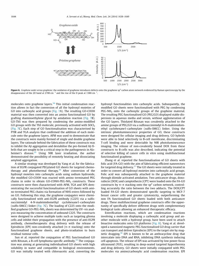
developed by Hummers and Offeman in the fifties.29 GO sheetsare highly functionalised with oxygenated groups such as hydro-xyl, epoxide, ketone and carboxylic acid moieties resulting inremarkably stable dispersions in water and organic solvents. Thestructural differences and notably defects on graphene sp2 carbonnetworks can be easily evidenced using Raman spectroscopy,where the disappearance of the 2D peak at 2700 cm�1 and thehigher D peak at 1380 cm�1 than the G peak at 1576 cm�1 high-lights the large presence of sp3 carbon atoms on GO (Fig. 6).30
The various oxygenated groups attached on GO layers, due to theirdifference in reactivity allow surface modification via numeroussynthetic routes including electrophilic and nucleophilic substitu-tions, free radical addition and condensation reactions; howeverthe most common routes employed for functionalising GO remainthrough nucleophilic and condensation reactions (Table 2).
Condensation reactions. Condensation reactions are reactionsduring which two molecules are combined together, resulting inone single molecule. In case of graphene, this reaction occurs viathe two reactive oxygenated groups present on the GO sheets suchas the hydroxyl and carboxyl functionalities. The amidation reac-tion between an activated carboxyl group from GO and an aminogroup remains nevertheless the most popular reaction for intro-ducing bioactive molecule onto GO platforms. Many groups haveutilised this synthetic route to attach covalently molecules result-ing in graphene conjugates for biomedical applications encompass-ing medical imaging and diagnosis, biosensors, and drug and genedelivery, as described in the next paragraph.
The introduction of biocompatible polymers using condensa-tion reaction has been widely applied in order to develop grapheneconjugates with improved solubility and biocompatibility. Thisstrategy was used by Zhang et al. to engineer functional graphenesheets for investigating the biodistribution of biocompatiblegraphene conjugates.31 In this work, the authors present the conju-gation of GO sheets with dextran (DEX) by condensing the aminogroups of DEX with the carboxylic groups of GO. The size of theGO conjugates was characterised by AFM and TEM and was foundto be ranging from 50 to 100 nm compared to the lateral size of theGO sheets that was comprised between 100 and 500 nm, suggest-ing size reduction resulting from the DEX condensation reactionprocess. The DEX/GO conjugates demonstrated long lasting stabil-ity in aqueous media and mice serum and reduced cell toxicitycompared to unfunctionalised GO sheets. Biodistribution studieswere conducted in order to investigate the in vivo behaviour ofthese constructs by radiolabelling the DEX/GO sheets with radioac-tive 125I. The graphene hybrids accumulated in the reticulo-endo-thelial system including liver and spleen after intravenousinjection and demonstrated relatively long blood circulation times.In addition, the constructs were cleared from the animal bodywithin a week from the injection without significant signs oftoxicity.
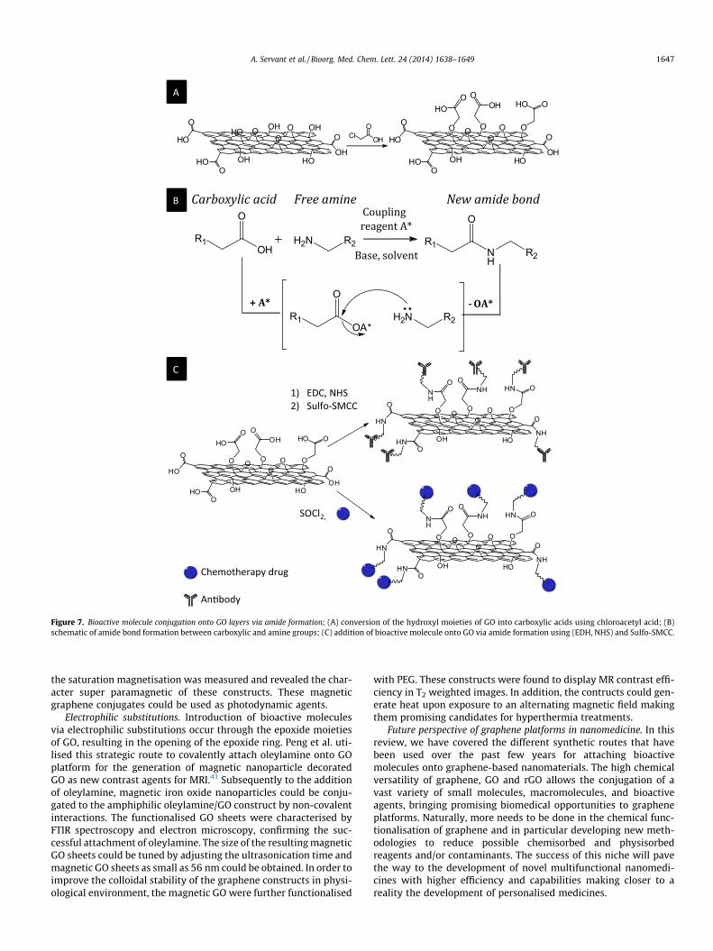
Other functionalised GO constructs, fabricated through the for-mation of an amide group were reported for the development oftherapeutic agents for drug or gene delivery by conjugating ontothe graphene structure thioflavin-S (ThS).32 This molecule isknown to bind specifically to amyloid-b peptides forming the con-jugate GO-ThS-Ab. This construct was designed for the treatmentof Alzheimer’s disease by photothermal therapy. Photothermaltherapy is often presented as good alternative to the traditionalchemotherapy and radiotherapy as it has demonstrated remark-ably reduced side effects and improved selectivity with limitedhealthy tissue exposure to the treatment.33 In this work, GO sheetswere first treated with chloroacetic acid for converting the hydro-xyl functions into carboxylic functions. The condensation betweena hydroxyl group of GO and another electrophilic molecule such aschloroacetic acid has been extensively used for modifying GOsurfaces as a synthetic step for covalently coupling bioactive
0100200300400500600700800
500 1500 2500
Inte
nsit
y (A
.U.)
1/λ (cm-1)
05
101520253035404550
500 1000 1500 2000 2500 3000
Inte
nsit
y (A
.U.)
1/λ (cm-1)
Graphene oxide
Pristine graphene
OH
OOH
HO
OO
OH
HOHO
O
HOO
OH O
Figure 6. Graphene oxide versus graphene; the oxidation of graphene introduces defects onto the graphene sp2 carbon atom network evidenced by Raman spectroscopy by thedisappearance of the 2D band at 2700 cm�1 and the rise of the D peak at 1380 cm�1.
1644 A. Servant et al. / Bioorg. Med. Chem. Lett. 24 (2014) 1638–1649
molecules onto graphene layers.34 This initial condensation reac-tion allows in fact the conversion of all the hydroxyl moieties ofGO into carboxylic acid groups (Fig. 7A). The resulting GO-COOHmaterial was then converted into an amino functionalised GO bygrafting diaminoethylene glycol by amidation reaction (Fig. 7B).GO-ThS was then prepared by condensing the amino-modifiedGO groups with the ThS molecule, previously activated with SOCl2
(Fig. 7C). Each step of GO functionalisation was characterised byFTIR and TGA analysis that confirmed the addition of each mole-cule onto the graphene layers. AFM was used to demonstrate thatthe constructs were mainly formed of single and double graphenelayers. The rationale behind the fabrication of these constructs wasto inhibit the Ab aggregation and destabilise the pre-formed Ab fi-brils that are sought to be a critical step in the pathogenesis in Alz-heimer’s disease.35 Using NIR laser irradiation, the authordemonstrated the possibility of remotely heating and dissociatingamyloid aggregation.
A similar strategy was developed by Yang et al. for the fabrica-tion of EGFR conjugated PEGylated GO sheets for targeted chemo-therapy and photothermal therapy.36 After conversion of thehydroxyl moieties into carboxylic acids using sodium hydroxide,the modified GO-COOH was reacted with amino terminated PEGchains in order to obtain GO-CONH-PEG-NH2 constructs. Theseconstructs were then characterised with AFM, TGA and XPS dem-onstrating the successful functionalisation of GO sheets with ami-no terminated PEG chains via formation of NHS-esters followed byon GO. Following this step, the graphene conjugates were chemi-cally functionalised with anti-EGFR antibody (C225) via a sulfo-succinimidyl 4-N-maleimidomethyl cyclohexane1-carboxylate(sulfo-SMCC) linker (Fig. 7C). The loading of C225 onto the modi-fied graphene GO-PEG-NH2 sheets was determined by UV–vis anal-ysis measuring the concentration of unbound C225. The constructswere designed to achieve multiple tasks such as targeting gliomacells and inhibit their propagation with the presence of anti-EGFRantibody, chemotherapeutic drug delivery with the release ofEpirubicin (EPI) non-covalently attached (p–p stacking) onto thefunctionalised graphene sheets, and photo-irradiation to burnresidual tumour cells.
Sun et al. reported the conjugation of PEG functionalised GOwith Rituxan, a B-cell lymphoma specific antibody.34 The conjuga-tion was aiming at generating individualised GO sheets with highsolubility in water and compatible in biological environments.GO was initially treated with chloroacetic acid, converting the
hydroxyl functionalities into carboxylic acids. Subsequently, themodified GO sheets were functionalised with PEG by condensingPEG-NH2 onto the carboxylic groups of the graphene material.The resulting PEG functionalised GO (PEG/GO) displayed stable dis-persions in aqueous media and serum, without agglomeration ofthe GO layers. Thiolated Rituxan was covalently attached to theamine groups of PEG/GO via a sulfosuccinimidyl 4-N-maleimidom-ethyl cyclohexane1-carboxylate (sulfo-SMCC) linker. Using theintrinsic photoluminescence properties of GO, these constructswere designed for cellular imaging and drug delivery. GO hybridswere able to bind selectively to B-cell membrane, discriminatingT-cell binding and were detectable by NIR photoluminescenceimaging. The release of non-covalently bound DOX from theseconstructs to B-cells was also described, indicating the potentialof selective killing of cancer cells in vitro using multifunctionalfunctionalised graphene.
Zhang et al. reported the functionalisation of GO sheets withfolic acid (FA-GO) with the aim of fabricating efficient nanovectorsfor targeted drug delivery.37 The GO sheets were initially treated inorder to convert all hydroxyl moieties into carboxylic acid groups.Folic acid was subsequently attached to the graphene materialthrough diimide activated amidation. Two anticancer drugs, doxo-rubicin (DOX) and camptothecin (CPT) were loaded onto the FA-GOconstructs by p–p stacking onto the sp2 carbon network, control-ling accurately the ratio between the two adducts. The DOX/CPTloaded FA-GO sheets demonstrated specific targeting to MCF-7breast cancer cells and greater cytotoxicity, outperforming thenon FA functionalised GO sheets loaded with both anticancerdrugs. These multifunctional graphene constructs offer the oppor-tunity of specifically deliver different drugs with complementarybiological tasks allowing an enhanced therapeutic efficacy.
Esterification reactions, which are condensation reactionsinvolving a molecule displaying a carboxylic acid group and an-other molecule with a hydroxyl group, have been used to attachbioactive molecules onto GO platforms (Fig. 8). Yang et al. devel-oped a nanosized magnetic PEG functionalised GO drug carrier thatcan transport and deliver Epirubicin (EPI) to the target site by mag-netic dragging.38 EPI is known to be an anthracycline drug thatintercalate into DNA and RNA, inhibiting DNA synthesis inducingcell apoptosis. The release of EPI was activated by low power focusultrasound (FUS), resulting in deep-seated targeted hyperthermiaand drug delivery. GO sheets were initially conjugated with PEGmolecules via amino/carboxylic acid condensation reaciton. EPI
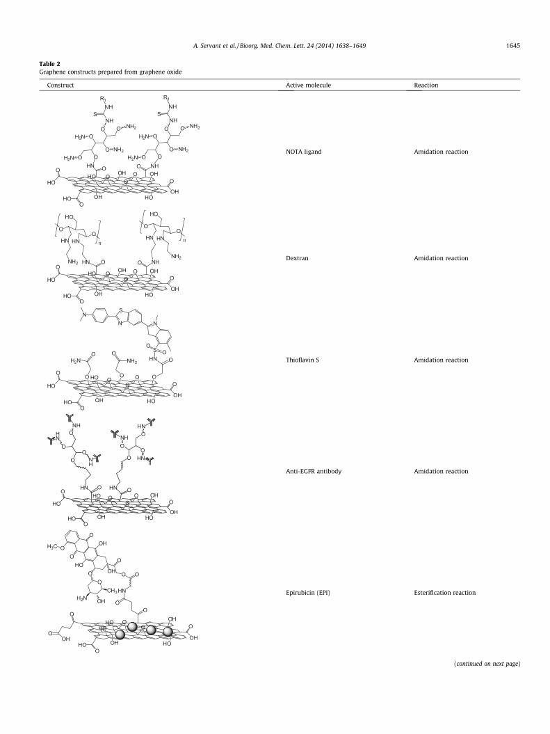
Table 2Graphene constructs prepared from graphene oxide
Construct Active molecule Reaction
OHNOOH2N
O
O
O OH2N
NHNH2
NH2
NHS
R1
NHO
OOH2N
O
O
O OH2N
NHNH2
NH2
NHS
R1
OH
OOH
HO
OO
OH
HOHO
O
HOO
OH O
NOTA ligand Amidation reaction
O
HNO
HN
NH2 HN
HO
n
O
O
HNO
HN
HO
n
NHONH2
OH
OOH
HO
OO
OH
HOHO
O
HOO
OH O
Dextran Amidation reaction
HN
N
S
N
SN
OO
OH
OO
HO
OO
OH
HOHO
O
HOO
O O
H2NO O
NH2 O
O
Thioflavin S Amidation reaction
HN
O
OO
OHN
NH
HN
HN
OO
O
ONH
HN
NH
OH
OOH
HO
OO
OH
HOHO
O
HOO
OOO
Anti-EGFR antibody Amidation reaction
O
OOHOO
OHH2NCH3
OH
HO
O
OOH3C
O
HN
O
O
OH
OOH
HO
OO
OH
HO
OHO
O
HOO
HO
Epirubicin (EPI) Esterification reaction
(continued on next page)
A. Servant et al. / Bioorg. Med. Chem. Lett. 24 (2014) 1638–1649 1645
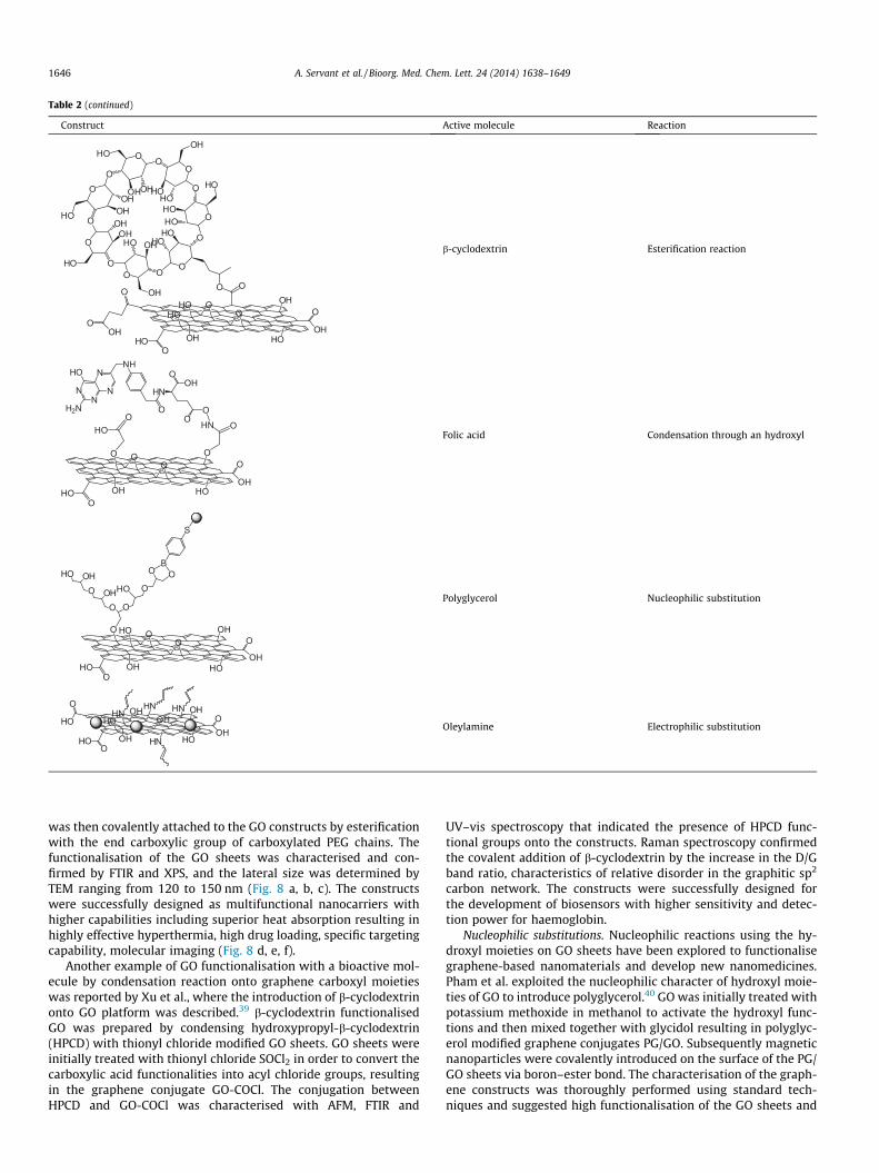
Table 2 (continued)
Construct Active molecule Reaction
OO
O OO
O
O
OO O
O
O
O
OOH HO
HO
HOOH
OH
OHOH
OHHO
HO
HOHO
OH
HO
HO
HOOH
HO
OH O O
OH
OOH
HO
OO
OH
HO
OHO
O
HOO
HO
b-cyclodextrin Esterification reaction
NN
N
NNH
O
HNOH
O
OO
H2N
HO
OH
OO
HO
OO
OH
O
HOO
HOO
OHNFolic acid Condensation through an hydroxyl
OH
OOH
HO
OO
OH
HO
HOO
O
OO
HOOHOOHHO
O
O OB
S
Polyglycerol Nucleophilic substitution
OHO
OH
HO
OHOH
OH
HNHO
O
HOO
HO
HN
HN HN
Oleylamine Electrophilic substitution
1646 A. Servant et al. / Bioorg. Med. Chem. Lett. 24 (2014) 1638–1649
was then covalently attached to the GO constructs by esterificationwith the end carboxylic group of carboxylated PEG chains. Thefunctionalisation of the GO sheets was characterised and con-firmed by FTIR and XPS, and the lateral size was determined byTEM ranging from 120 to 150 nm (Fig. 8 a, b, c). The constructswere successfully designed as multifunctional nanocarriers withhigher capabilities including superior heat absorption resulting inhighly effective hyperthermia, high drug loading, specific targetingcapability, molecular imaging (Fig. 8 d, e, f).
Another example of GO functionalisation with a bioactive mol-ecule by condensation reaction onto graphene carboxyl moietieswas reported by Xu et al., where the introduction of b-cyclodextrinonto GO platform was described.39 b-cyclodextrin functionalisedGO was prepared by condensing hydroxypropyl-b-cyclodextrin(HPCD) with thionyl chloride modified GO sheets. GO sheets wereinitially treated with thionyl chloride SOCl2 in order to convert thecarboxylic acid functionalities into acyl chloride groups, resultingin the graphene conjugate GO-COCl. The conjugation betweenHPCD and GO-COCl was characterised with AFM, FTIR and
UV–vis spectroscopy that indicated the presence of HPCD func-tional groups onto the constructs. Raman spectroscopy confirmedthe covalent addition of b-cyclodextrin by the increase in the D/Gband ratio, characteristics of relative disorder in the graphitic sp2
carbon network. The constructs were successfully designed forthe development of biosensors with higher sensitivity and detec-tion power for haemoglobin.
Nucleophilic substitutions. Nucleophilic reactions using the hy-droxyl moieties on GO sheets have been explored to functionalisegraphene-based nanomaterials and develop new nanomedicines.Pham et al. exploited the nucleophilic character of hydroxyl moie-ties of GO to introduce polyglycerol.40 GO was initially treated withpotassium methoxide in methanol to activate the hydroxyl func-tions and then mixed together with glycidol resulting in polyglyc-erol modified graphene conjugates PG/GO. Subsequently magneticnanoparticles were covalently introduced on the surface of the PG/GO sheets via boron–ester bond. The characterisation of the graph-ene constructs was thoroughly performed using standard tech-niques and suggested high functionalisation of the GO sheets and
A
O
OHCl
OH
OOH
HO
OO
OH
HOHO
O
HOO
OH O
OH
OO
HO
OO
OH
OHO
O
HOO
O O
HOO O
OH OHO
Chemotherapy drug
An�body
C
SOCl2,
1) EDC, NHS2) Sulfo-SMCC
OH
OO
HO
OO
OH
OHO
O
HOO
O O
HOO O
OH OHO
NH
OO
HO
OO
OH
OHN
O
HNO
O O
NH
O ONH OHN
NH
OO
HO
OO
OH
OHN
O
HNO
O O
NH
O ONH OHN
Carboxylic acid Free amine New amide bondCoupling
reagent A*
Base, solvent
+ A* - OA*
R1OH
O
R2H2N
R1NH
O
R2
R1OA*
O
R2H2N
B
Figure 7. Bioactive molecule conjugation onto GO layers via amide formation; (A) conversion of the hydroxyl moieties of GO into carboxylic acids using chloroacetyl acid; (B)schematic of amide bond formation between carboxylic and amine groups; (C) addition of bioactive molecule onto GO via amide formation using (EDH, NHS) and Sulfo-SMCC.
A. Servant et al. / Bioorg. Med. Chem. Lett. 24 (2014) 1638–1649 1647
the saturation magnetisation was measured and revealed the char-acter super paramagnetic of these constructs. These magneticgraphene conjugates could be used as photodynamic agents.
Electrophilic substitutions. Introduction of bioactive moleculesvia electrophilic substitutions occur through the epoxide moietiesof GO, resulting in the opening of the epoxide ring. Peng et al. uti-lised this strategic route to covalently attach oleylamine onto GOplatform for the generation of magnetic nanoparticle decoratedGO as new contrast agents for MRI.41 Subsequently to the additionof oleylamine, magnetic iron oxide nanoparticles could be conju-gated to the amphiphilic oleylamine/GO construct by non-covalentinteractions. The functionalised GO sheets were characterised byFTIR spectroscopy and electron microscopy, confirming the suc-cessful attachment of oleylamine. The size of the resulting magneticGO sheets could be tuned by adjusting the ultrasonication time andmagnetic GO sheets as small as 56 nm could be obtained. In order toimprove the colloidal stability of the graphene constructs in physi-ological environment, the magnetic GO were further functionalised
with PEG. These constructs were found to display MR contrast effi-ciency in T2 weighted images. In addition, the contructs could gen-erate heat upon exposure to an alternating magnetic field makingthem promising candidates for hyperthermia treatments.
Future perspective of graphene platforms in nanomedicine. In thisreview, we have covered the different synthetic routes that havebeen used over the past few years for attaching bioactivemolecules onto graphene-based nanomaterials. The high chemicalversatility of graphene, GO and rGO allows the conjugation of avast variety of small molecules, macromolecules, and bioactiveagents, bringing promising biomedical opportunities to grapheneplatforms. Naturally, more needs to be done in the chemical func-tionalisation of graphene and in particular developing new meth-odologies to reduce possible chemisorbed and physisorbedreagents and/or contaminants. The success of this niche will pavethe way to the development of novel multifunctional nanomedi-cines with higher efficiency and capabilities making closer to areality the development of personalised medicines.
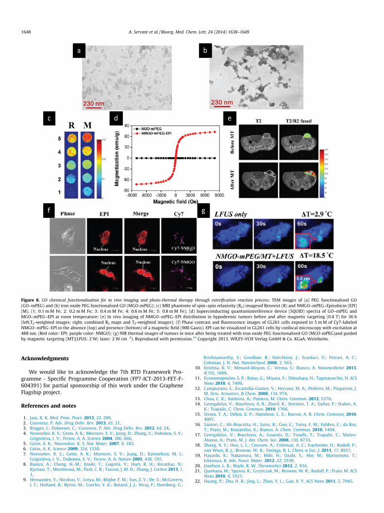
Figure 8. GO chemical functionalisation for in vivo imaging and photo-thermal therapy through esterification reaction process; TEM images of (a) PEG functionalised GO(GO–mPEG) and (b) iron oxide PEG functionalised GO (MGO-mPEG); (c) MRI phantoms of spin–spin relaxivity (R2) imagesof Resovist (R) and NMGO–mPEG–Epirubicin (EPI)(M). (1: 0.1 m M Fe; 2: 0.2 m M Fe; 3: 0.4 m M Fe; 4: 0.6 m M Fe; 5: 0.8 m M Fe); (d) Superconducting quantuminterference device (SQUID) spectra of GO–mPEG andMGO–mPEG–EPI at room temperature; (e) In vivo imaging of NMGO–mPEG–EPI distribution in hypodermic tumors before and after magnetic targeting (0.4 T) for 36 h(left,T2-weighted images; right, combined R2 maps and T2-weighted images); (f) Phase contrast and fluorescence images of GL261 cells exposed to 5 m M of Cy7-labeledNMGO–mPEG–EPI in the absence (top) and presence (bottom) of a magnetic field (900 Gauss). EPI can be visualized in GL261 cells by confocal microscopy with excitation at488 nm. (Red color: EPI; purple color: NMGO); (g) NIR thermal images of tumors in mice after being treated with iron oxide PEG functionalised GO (MGO-mPEG)and guidedby magnetic targeting (MT)(LFUS: 2 W; laser: 2 W cm�2). Reproduced with permission.38 Copyright 2013, WILEY-VCH Verlag GmbH & Co. KGaA, Weinheim.
1648 A. Servant et al. / Bioorg. Med. Chem. Lett. 24 (2014) 1638–1649
Acknowledgments
We would like to acknowledge the 7th RTD Framework Pro-gramme - Specific Programme Cooperation (FP7-ICT-2013-FET-F-604391) for partial sponsorship of this work under the GrapheneFlagship project.
References and notes
1. Jain, K. K. Med. Princ. Pract. 2013, 22, 209.2. Couvreur, P. Adv. Drug Deliv. Rev. 2013, 65, 21.3. Brigger, I.; Dubernet, C.; Couvreur, P. Adv. Drug Deliv. Rev. 2012, 64, 24.4. Novoselov, K. S.; Geim, A. K.; Morozov, S. V.; Jiang, D.; Zhang, Y.; Dubonos, S. V.;
Grigorieva, I. V.; Firsov, A. A. Science 2004, 306, 666.5. Geim, A. K.; Novoselov, K. S. Nat. Mater. 2007, 6, 183.6. Geim, A. K. Science 2009, 324, 1530.7. Novoselov, K. S.; Geim, A. K.; Morozov, S. V.; Jiang, D.; Katsnelson, M. I.;
Grigorieva, I. V.; Dubonos, S. V.; Firsov, A. A. Nature 2005, 438, 197.8. Bianco, A.; Cheng, H.-M.; Enoki, T.; Gogotsi, Y.; Hurt, R. H.; Koratkar, N.;
Kyotani, T.; Monthioux, M.; Park, C. R.; Tascon, J. M. D.; Zhang, J. Carbon 2013, 1,1.
9. Hernandez, Y.; Nicolosi, V.; Lotya, M.; Blighe, F. M.; Sun, Z. Y.; De, S.; McGovern,I. T.; Holland, B.; Byrne, M.; Gun’ko, Y. K.; Boland, J. J.; Niraj, P.; Duesberg, G.;
Krishnamurthy, S.; Goodhue, R.; Hutchison, J.; Scardaci, V.; Ferrari, A. C.;Coleman, J. N. Nat. Nanotechnol. 2008, 3, 563.
10. Krishna, K. V.; Menard-Moyon, C.; Verma, S.; Bianco, A. Nanomedicine 2013,8(10), 1669.
11. Economopoulos, S. P.; Rotas, G.; Miyata, Y.; Shinohara, H.; Tagmatarchis, N. ACSNano 2010, 4, 7499.
12. Campuzano, S.; Escamilla-Gomez, V.; Herranz, M. A.; Pedrero, M.; Pingarron, J.M. Sens. Actuators, B-Chem. 2008, 134, 974.
13. Chua, C. K.; Ambrosi, A.; Pumera, M. Chem. Commun. 2012, 5376.14. Georgakilas, V.; Bourlinos, A. B.; Zboril, R.; Steriotis, T. A.; Dallas, P.; Stubos, A.
K.; Trapalis, C. Chem. Commun. 2010, 1766.15. Strom, T. A.; Dillon, E. P.; Hamilton, C. E.; Barron, A. R. Chem. Commun. 2010,
4097.16. Samori, C.; Ali-Boucetta, H.; Sainz, R.; Guo, C.; Toma, F. M.; Fabbro, C.; da Ros,
T.; Prato, M.; Kostarelos, K.; Bianco, A. Chem. Commun. 2010, 1494.17. Georgakilas, V.; Bourlinos, A.; Gournis, D.; Tsoufis, T.; Trapalis, C.; Mateo-
Alonso, A.; Prato, M. J. Am. Chem. Soc. 2008, 130, 8733.18. Zhang, X. Y.; Hou, L. L.; Cnossen, A.; Coleman, A. C.; Ivashenko, O.; Rudolf, P.;
van Wees, B. J.; Browne, W. R.; Feringa, B. L. Chem.-a Eur. J. 2011, 17, 8957.19. Hayashi, K.; Nakamura, M.; Miki, H.; Ozaki, S.; Abe, M.; Matsumoto, T.;
Ishimura, K. Adv. Funct. Mater. 2012, 22, 3539.20. Josefsen, L. B.; Boyle, R. W. Theranostics 2012, 2, 916.21. Quintana, M.; Spyrou, K.; Grzelczak, M.; Browne, W. R.; Rudolf, P.; Prato, M. ACS
Nano 2010, 4, 3527.22. Huang, P.; Zhu, H. R.; Jing, L.; Zhao, Y. L.; Gao, X. Y. ACS Nano 2011, 5, 7945.

A. Servant et al. / Bioorg. Med. Chem. Lett. 24 (2014) 1638–1649 1649
23. Wang, Q. H.; Jin, Z.; Kim, K. K.; Hilmer, A. J.; Paulus, G. L. C.; Shih, C. J.; Ham, M.H.; Sanchez-Yamagishi, J. D.; Watanabe, K.; Taniguchi, T.; Kong, J.; Jarillo-Herrero, P.; Strano, M. S. Nat. Chem. 2012, 4, 724.
24. Pinson, J.; Podvorica, F. Chem. Soc. Rev. 2005, 34, 429.25. Wei, G. C.; Yan, M. M.; Dong, R. H.; Wang, D.; Zhou, X. Z.; Chen, J. F.; Hao, J. C.
Chem.-A Eur. J. 2012, 18, 14708.26. Gollavelli, G.; Ling, Y. C. Biomaterials 2012, 33, 2532.27. Chua, C. K.; Pumera, M. Chem.-An Asian J. 2012, 7, 1009.28. Chen, H. C.; Chen, Y. H.; Chen, S. L.; Chern, Y. T.; Tsai, R. Y.; Hua, M. Y. Biosens.
Bioelectron. 2013, 46, 84.29. Hummers, W. S.; Offeman, R. E. J. Am. Chem. Soc. 1958, 80, 1339.30. Kuila, T.; Bose, S.; Mishra, A. K.; Khanra, P.; Kim, N. H.; Lee, J. H. Prog. Mater Sci.
2012, 57, 1061.31. Zhang, S. A.; Yang, K.; Feng, L. Z.; Liu, Z. Carbon 2011, 49, 4040.32. Li, M.; Yang, X. J.; Ren, J. S.; Qu, K. G.; Qu, X. G. Adv. Mater. 2012, 24, 1722.33. Huff, T. B.; Tong, L.; Zhao, Y.; Hansen, M. N.; Cheng, J. X.; Wei, A. Nanomedicine
2007, 2, 125.34. Sun, X. M.; Liu, Z.; Welsher, K.; Robinson, J. T.; Goodwin, A.; Zaric, S.; Dai, H. J.
Nano Res. 2008, 1, 203.
35. Yamin, G.; Ono, K.; Inayathullah, M.; Teplow, D. B. Curr. Pharm. Des. 2008, 14,3231.
36. Yang, H. W.; Lu, Y. J.; Lin, K. J.; Hsu, S. C.; Huang, C. Y.; She, S. H.; Liu, H. L.; Lin, C.W.; Xiao, M. C.; Wey, S. P.; Chen, P. Y.; Yen, T. C.; Wei, K. C.; Ma, C. C. M.Biomaterials 2013, 34, 7204.
37. Zhang, L. M.; Xia, J. G.; Zhao, Q. H.; Liu, L. W.; Zhang, Z. J. Small 2010, 6, 537.38. Yang, H.; Hua, M.; Hwang, T.; Lin, K.; Huang, C.; Tsai, R.; Ma, C.; Hsu, P.; Wey, S.;
Hsu, P.; Chen, P.; Huang, Y.; Lu, Y.; Yen, C.; Feng, C.; Lin, C.; Liu, H.; Wei, K. C.Adv. Mater. 2013, 25, 3605.
39. Xu, C. H.; Wang, X. B.; Wang, J. C.; Hu, H. T.; Wan, L. Chem. Phys. Lett. 2010, 498,162.
40. Pham, T. A.; Kumar, N. A.; Jeong, Y. T. Synth. Met. 2010, 160, 2028.41. Peng, E. W.; Choo, E. S. G.; Chandrasekharan, P.; Yang, C. T.; Ding, J.; Chuang, K.
H.; Xue, J. M. Small 2012, 8, 3620.42. Otakar, F. http://www.nanocarbon.cz/.43. Ferrari, A. C.; Basko, D. M. Nat. Nanotechnol. 2013, 8, 235.44. Ferrari, A. C.; Meyer, J. C.; Scardaci, V.; Casiraghi, C.; Lazzeri, M.; Mauri, F.;
Piscanec, S.; Jiang, D.; Novoselov, K. S.; Roth, S.; Geim, A. K. Phys. Rev. Lett. 2006,97, 187401.