Embed Size (px)
Citation preview

J N E R JOURNAL OF NEUROENGINEERINGAND REHABILITATION
Sawacha et al. Journal of NeuroEngineering and Rehabilitation 2012, 9:63http://www.jneuroengrehab.com/content/9/1/63
RESEARCH Open Access
Biomechanical assessment of balance andposture in subjects with ankylosing spondylitisZimi Sawacha1†, Elena Carraro2†, Silvia Del Din1†, Annamaria Guiotto1†, Lara Bonaldo2, Leonardo Punzi3,Claudio Cobelli1* and Stefano Masiero2
Abstract
Background: Ankylosing spondylitis is a major chronic rheumatic disease that predominantly affects axial joints,determining a rigid spine from the occiput to the sacrum. The dorsal hyperkyphosis may induce the patients tostand in a stooped position with consequent restriction in patients’ daily living activities. The aim of this study wasto develop a method for quantitatively and objectively assessing both balance and posture and their mutualrelationship in ankylosing spondylitis subjects.
Methods: The data of 12 healthy and 12 ankylosing spondylitis subjects (treated with anti-TNF-α stabilized), with amean age of 51.42 and 49.42 years; mean BMI of 23.08 and 25.44 kg/m2 were collected. Subjects underwent amorphological examination of the spinal mobility by means of a pocket compass needle goniometer, together with anevaluation of both spinal and hip mobility (Bath Ankylosing Spondylitis Metrology Index), and disease activity (BathAnkylosing Spondylitis Disease Activity Index). Quantitative evaluation of kinematics and balance were performedthrough a six cameras stereophotogrammetric system and a force plate. Kinematic models together with a test forevaluating balance in different eye level conditions were developed. Head protrusion, trunk flexion-extension, pelvic tilt,hip-knee-ankle flexion-extension were evaluated during Romberg Test, together with centre of pressure parameters.
Results: Each subject was able to accomplish the required task. Subjects’ were comparable for demographicparameters. A significant increment was observed in ankylosing spondylitis subjects for knee joint angle with the targetplaced at each eye level on both sides (p< 0.042). When considering the pelvic tilt angle a statistically significantreduction was found with the target placed respectively at 10° (p= 0.034) and at 30° (p= 0.019) less than eye level.Furthermore in ankylosing spondylitis subjects both hip (p= 0.048) and ankle (p= 0.029) joints angles differssignificantly. When considering the posturographic parameters significant differences were observed for ellipse, centerof pressure path and mean velocity (p< 0.04). Goniometric evaluation revealed significant increment of thoracickyphosis reduction of cervical and lumbar range of motion compared to healthy subjects.
Conclusions: Our findings confirm the need to investigate both balance and posture in ankylosing spondylitissubjects. This methodology could help clinicians to plan rehabilitation treatments.
Keywords: Ankylosing Spondylitis, Postural balance, Biomechanics
* Correspondence: [email protected]†Equal contributors1Department of Information Engineering, University of Padova, Padova, ItalyFull list of author information is available at the end of the article
© 2012 Sawacha et al.; licensee BioMed Central Ltd. This is an Open Access article distributed under the terms of the CreativeCommons Attribution License (http://creativecommons.org/licenses/by/2.0), which permits unrestricted use, distribution, andreproduction in any medium, provided the original work is properly cited.

Sawacha et al. Journal of NeuroEngineering and Rehabilitation 2012, 9:63 Page 2 of 11http://www.jneuroengrehab.com/content/9/1/63
BackgroundAnkylosing spondylitis (AS) is a major chronic rheum-atic disease that predominantly affects axial joints, deter-mining a diffuse stiffness and with the advanced stageproducing a rigid spine from the occiput to the sacrum,for a chronic process of inflammation of the fibrous con-nective and bone in the tendon and ligament insertions[1-6]. Prevalence rates of people affected by AS rangesbetween 0.2% and 1.1% (0.9% in northern Europeanwhite population) [7,8]. Young population are primarilyaffected (average age of diagnosis being 24 years), moremale than female [7,8].Sacroiliitis is the distinctive characteristic of AS, with
consequent pain in sacral and gluteus region [4]. Also apossible extension of the pain from the thigh to theproximal calf, with alternated course between the twolower limbs have been reported in association with AS[3]. Hence, the clinical manifestations of AS are pain,stiffness, fatigue, reduced spinal mobility and respiratoryrestriction. Early limitation of spinal mobility has alsobeen identified as one of the most relevant prognosticfactors [8]. Other registered symptoms and objectiveclinical signs of AS are precocious loss of lumbar lordo-sis, increased dorsal kyphosis and inversion of cervicallordosis, abdominal relaxation for diaphragm breath, hipflexion contracture and consequent knee flexion com-pensation [2,9].Some authors reported that the dorsal hyperkyphosis
may induce the patients to stand in a stooped positionunable to see the horizon [3,5,8]. The main drawback ofsuch condition being the consequent restriction inpatients’ daily living activities such as interpersonal com-munication, driving a car, walking down the street ormaintaining personal hygiene [2,4].Spinal kyphosis in AS subjects has also been associated
with a forward and downward shift of the centre of mass(CoM) of the trunk in the sagittal plane, thus inducing aforward and downward shift of the body’s CoM with re-spect to the base of support [3,5,8]. It has been hypothe-sized that in AS subjects extension of the hips, flexion ofthe knees and plantar flexion of the ankles may counter-balance the forward shift of the body CoM relative thebase of support [5,8].However, to date there has been few studies determining
the effect of AS on balance.. Some authors [5] documen-ted balance alterations on a significant proportion of ASpatients both in eyes open and eyes closed conditions,meanwhile Aydog et al. [3] reported AS has no negativeeffect on postural stability. In their study a clinically sig-nificant association was found only between dynamic pos-tural balance and tragus to wall distance. Furthermore theauthors have already demonstrated the presence of bio-mechanics alterations during walking in a group of ASsubjects [10]. Statistically significant alterations were
observed in the sagittal plane at each joint (P < 0.049), to-gether with hip and knee joint extension moments reduc-tion (P< 0.044) [10].Loss of balance in AS patients may be associated with
severe joint deformities and poor posture. Moreover lossof balance may increase the risk of falls. However in theliterature, results concerning assessment of balance inAS subjects are contradictories [3,5,8].Given this scenario it appears the need of performing
an exhaustive evaluation of the consequences of AS alsoon balance and posture in static conditions. This paper,specifically addressed to the static analysis of the ASsubjects’ posture, describes the experimental methoddeveloped herein in order to assess the biomechanicalfeatures in the interaction between balance and posture.As methodological development it was applied on a lim-ited sample of subjects to verify feasibility and validitybefore being transferred to a wide population of subjects.Hence, a quantitative multi-factor analysis was appliedin static conditions, as previously performed during gait[10]. Moreover the methodology proposed herein enablesto assess posture’s modification during balance alterations.This allows identification of distinct postural strategiesused by AS subjects in order to cope with poor balance.This was accomplished by means of clinical and in-
strumental analysis, which allows quantitative evaluationof kinematic and balance through motion capture sys-tems and force plates. In this context a kinematic modelwas developed together with a test for evaluating balancein different eye level conditions like what was previouslyperformed in Benedetti et al. [11] while evaluating theeffects of a physical activity program on flexed postureelderly individuals.
MethodsSubjectsTwelve healthy (control subjects (CS)) and twelve ASsubjects have been analyzed, with a mean age respect-ively of 51.42 (13.91) and 49.42 (10.47) years; a meanBMI respectively of 23.08 (2.37) and 25.44 (3.19) kg/m2
(10 female and 14 male). The mean of AS subjects dis-ease duration was 9.64 ± 6.6 years. Inclusion criteriawere: all patients with AS met the most recent modifiedNew York criteria [9] and were treated with anti-TNF-αwith a stable clinical picture (i.e., with a change in theBath Ankylosing Spondylitis Disease Activity Index(BASDAI: score between 1 and 10) of no more than± 1/10 units in the previous 3 months [1].Exclusion criteria were age older than 70 years, con-
comitant cardiovascular, neurological or psychiatric dis-ease, and severe visual or auditory impairments (reducedvisual acuity was accepted if adequately corrected).Patients with other orthopedic disease or previous sur-gery at the spine and on the lower extremities were also
Sawacha et al. Journal of NeuroEngineering and Rehabilitation 2012, 9:63 Page 3 of 11http://www.jneuroengrehab.com/content/9/1/63
excluded. All AS subjects were consecutively recruitedfrom the patients attending the outpatient clinic of theRehabilitation Department of the University of Padova(Italy). CS consisted of normal subjects enrolled amonghealth personnel of the same Rehabilitation Department;all these subjects had no neurological or orthopedic dis-ease and no severe visual or auditory impairments. Thestudy was approved by the hospital’s ethics committeeand informed consent was obtained from all patientsand controls.
Clinical assessmentPatients were evaluated [10] by means of the BathAnkylosing Spondylitis Metrology Index (BASMI) [12],Bath Ankylosing Spondylitis Disease Activity Index(BASDAI) [13] and Bath Ankylosing Spondylitis Func-tional Index (BASFI) [14].The manual muscle strength test was performed
on the lower limbs using Medical Research CouncilScore [15].Subjects underwent a morphological examination of the
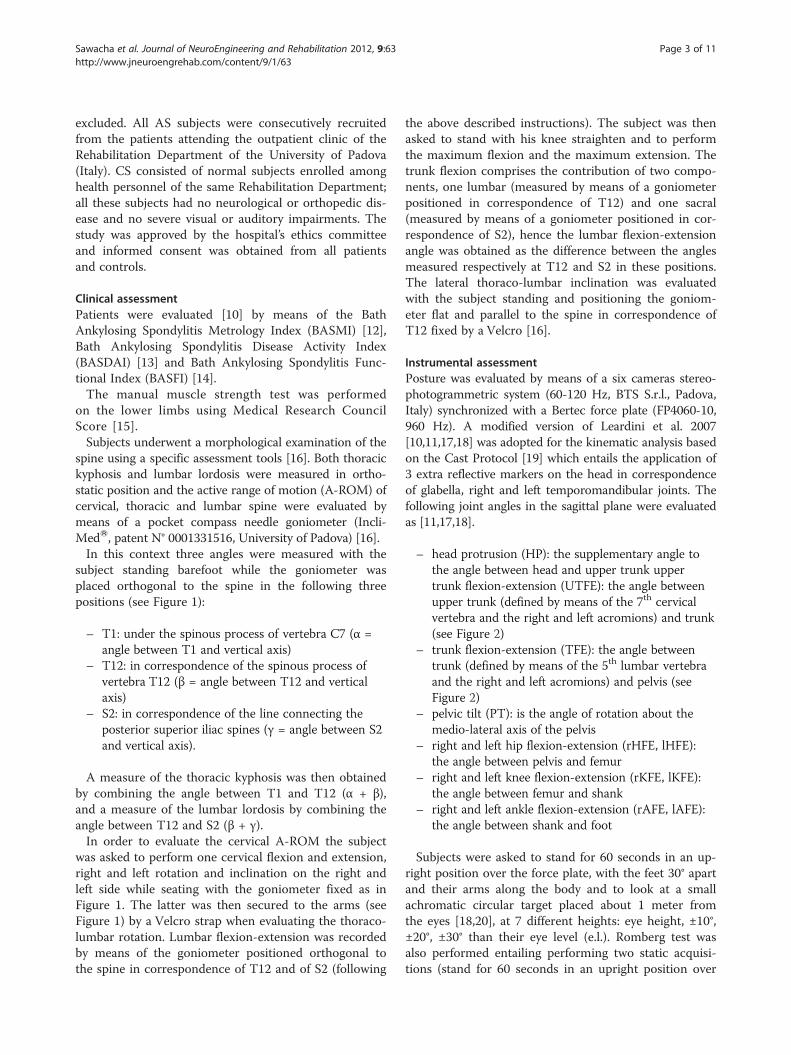
spine using a specific assessment tools [16]. Both thoracickyphosis and lumbar lordosis were measured in ortho-static position and the active range of motion (A-ROM) ofcervical, thoracic and lumbar spine were evaluated bymeans of a pocket compass needle goniometer (Incli-MedW, patent N° 0001331516, University of Padova) [16].In this context three angles were measured with the
subject standing barefoot while the goniometer wasplaced orthogonal to the spine in the following threepositions (see Figure 1):
– T1: under the spinous process of vertebra C7 (α =angle between T1 and vertical axis)
– T12: in correspondence of the spinous process ofvertebra T12 (β = angle between T12 and verticalaxis)
– S2: in correspondence of the line connecting theposterior superior iliac spines (γ = angle between S2and vertical axis).
A measure of the thoracic kyphosis was then obtainedby combining the angle between T1 and T12 (α + β),and a measure of the lumbar lordosis by combining theangle between T12 and S2 (β + γ).In order to evaluate the cervical A-ROM the subject
was asked to perform one cervical flexion and extension,right and left rotation and inclination on the right andleft side while seating with the goniometer fixed as inFigure 1. The latter was then secured to the arms (seeFigure 1) by a Velcro strap when evaluating the thoraco-lumbar rotation. Lumbar flexion-extension was recordedby means of the goniometer positioned orthogonal tothe spine in correspondence of T12 and of S2 (following
the above described instructions). The subject was thenasked to stand with his knee straighten and to performthe maximum flexion and the maximum extension. Thetrunk flexion comprises the contribution of two compo-nents, one lumbar (measured by means of a goniometerpositioned in correspondence of T12) and one sacral(measured by means of a goniometer positioned in cor-respondence of S2), hence the lumbar flexion-extensionangle was obtained as the difference between the anglesmeasured respectively at T12 and S2 in these positions.The lateral thoraco-lumbar inclination was evaluatedwith the subject standing and positioning the goniom-eter flat and parallel to the spine in correspondence ofT12 fixed by a Velcro [16].
Instrumental assessmentPosture was evaluated by means of a six cameras stereo-photogrammetric system (60-120 Hz, BTS S.r.l., Padova,Italy) synchronized with a Bertec force plate (FP4060-10,960 Hz). A modified version of Leardini et al. 2007[10,11,17,18] was adopted for the kinematic analysis basedon the Cast Protocol [19] which entails the application of3 extra reflective markers on the head in correspondenceof glabella, right and left temporomandibular joints. Thefollowing joint angles in the sagittal plane were evaluatedas [11,17,18].
– head protrusion (HP): the supplementary angle tothe angle between head and upper trunk uppertrunk flexion-extension (UTFE): the angle betweenupper trunk (defined by means of the 7th cervicalvertebra and the right and left acromions) and trunk(see Figure 2)
– trunk flexion-extension (TFE): the angle betweentrunk (defined by means of the 5th lumbar vertebraand the right and left acromions) and pelvis (seeFigure 2)
– pelvic tilt (PT): is the angle of rotation about themedio-lateral axis of the pelvis
– right and left hip flexion-extension (rHFE, lHFE):the angle between pelvis and femur
– right and left knee flexion-extension (rKFE, lKFE):the angle between femur and shank
– right and left ankle flexion-extension (rAFE, lAFE):the angle between shank and foot
Subjects were asked to stand for 60 seconds in an up-right position over the force plate, with the feet 30° apartand their arms along the body and to look at a smallachromatic circular target placed about 1 meter fromthe eyes [18,20], at 7 different heights: eye height, ±10°,±20°, ±30° than their eye level (e.l.). Romberg test wasalso performed entailing performing two static acquisi-tions (stand for 60 seconds in an upright position over

Figure 1 Subjects while undergoing a morphological examination of the spine using a specific assessment tool. Both kyphosis andlumbar lordosis were measured and the range of motion of cervical and thoraco-lumbar spine through a pocket compass needle goniometer(IncliMedW). From the top to the bottom and from left to right: first the subject is standing barefoot while the goniometer is placed orthogonalto the spine in correspondence of vertebra T12 performs the maximum extension with his knee straighten, and with two goniometers placed inT12 and S2 performs the maximum flexion of thoraco-lumbar spine; the subject stands and the goniometer is positioned flat and parallel to thespine in correspondence of T12 and performs the lateral thoraco-lumbar inclination; the subject seats with the goniometer fixed in neutralposition; in this position the subject performs one head flexion and extension; the subject seats with the goniometer fixed in neutral position; thesubject performs one head lateral left and right inclination; the subject seats with the goniometer fixed in neutral position; the subject performsleft and right thoraco-lumbar rotation.
Sawacha et al. Journal of NeuroEngineering and Rehabilitation 2012, 9:63 Page 4 of 11http://www.jneuroengrehab.com/content/9/1/63

Figure 2 Description of the joint angles in the sagittal plane.Head protrusion (a), trunk flexion-extension (b), pelvic tilt (c), hipflexion-extension (d), knee flexion-extension (e), ankle flexion-extension (f).
Sawacha et al. Journal of NeuroEngineering and Rehabilitation 2012, 9:63 Page 5 of 11http://www.jneuroengrehab.com/content/9/1/63
the force plate, with the feet 30° apart and their armsalong the body), one in eyes open (e.o.) and one in eyesclosed (e.c.) conditions. Both kinematic and centre ofground reaction forces data were collected.The total body center of pressure (CoP) trajectory over
the support surface was computed from the verticalforce of the force platform, which were recorded for60 seconds at 960 Hz and then filtered by a 3rd order,low-pass Butterworth filter (cut-off frequency 5 Hz). Thefirst 20 seconds of the acquisition were excluded fromthe analysis [18,21,22].A total of 9 CoP measures were computed from the
CoP displacement in the horizontal plane [21-24]. In thetime domain, we obtained 3 measures that characterizedthe CoP trajectory over the support surface, 2 measuresthat estimated the area covered by the CoP, and 3 mea-sures that estimated the velocity of body sway over thesupport surface. The following CoP-based measures werecomputed [21,22]: Sway area (area included in CoP dis-placement per unit of time (mm2/s)), Ellipse 95%, the CoPcoordinate time series, the CoP coordinate time series inantero-posterior (AP) and medio-lateral (ML) directions,mean velocity (total CoP trajectory length/trial duration(mm/s)), mean velocity in AP and ML directions.
Statistical analysisContinuous data were summarized in terms of means andstandard deviation of the mean. The differences betweenAS and CS were investigated by means of the unpaired T-Test followed by a Tukey-Kramer test (Matlab softwarefunction Ttest) when the variances were homogenous(verified with Lilliefors test, Matlab software function lil-lietest) and the Kruskal-Wallis test (Matlab software func-tion kruskalwallis) followed by a Tukey-Kramer test whenthe variances were not homogeneous; p-values under 0.05were considered to be significant. Subjects who averagedmore than 3.5 standard deviations from the mean wereclassified as outliers and removed from the statistical ana-lysis [25].The evaluation of the confidence intervals for the
observed proportions was performed with the staRt Pack-age of R statistical software [26].
ResultsEach subject was able to accomplish the required task.Subjects’ demographic and clinical characteristics werereported in Table 1. The mean BASMI, BASDAI andBASFI scores were respectively 3.38 (1.26), 2.50 (1.59),

Table 1 Subjects demographic and clinical characteristics (mean and standard deviation)
CS AS P
Age [years] 51.4 (13.9) 49.4 (10.5) 0.694
Sex (male/female) 6/6 8/4 0.430
Disease duration [years] 9.6 (6.6)
BMI [kg/m2] 23.1 (2.4) 25.4 (3.2) 0.052
Kyphosis 45.3 (9.4) 54.8 (12.2) 0.044*
Lordosis 36.2 (11.2) 28.8 (14.7) 0.182
Pelvic anteversion 5.7 (2.3) 1.00 (3.6) 0.016*
Flexion-extension of lumbar spine 69 (14.8) 31.3 (13.8) 0.001*
Lateral inclination of thoraco-lumbar spine 58.5 (13.3) 29.1 (18.2) 0.001*
Rotation of thoraco-lumbar spine 90.3 (12.8) 70.2 (23.9) 0.016*
Total range of motion of thoraco-lumbar spine 221.5 (30.1) 130.7 (44.7) 0.001*
Flexion-extension of cervical spine 121.2 (15.0) 90.5 (32.8) 0.007*
Lateral inclination of cervical spine 78.4 (10.5) 52.8 (23.7) 0.002*
Rotation of cervical spine 118.3 (15.5) 102.08 (39.0) 0.193
Total range of motion of cervical spine 318.0 (25.0) 245.4 (84.7) 0.009*
* Statistical significance (P< 0.05).
Sawacha et al. Journal of NeuroEngineering and Rehabilitation 2012, 9:63 Page 6 of 11http://www.jneuroengrehab.com/content/9/1/63
2.22 (1.32). The manual muscle test showed normal ofstrength. Goniometric evaluation revealed in AS subjectsa significant increment of thoracic kyphosis but no lumbarlordosis, and a significant reduction of both cervical andlumbar A-ROM was registered compared to healthy sub-jects. A box plot representation of the results obtainedfrom both joint angles and posturographic parameters hasbeen reported in Figures 3 and 4. In this contest the datarelative to subjects identified as outliers were also reported.Statistically significant differences were observed for kneejoint angle with the target placed at each e.l. on both sides(p< 0.042). When considering the pelvic tilt angle statisti-cally significant differences were found with the targetplaced respectively at 10° (p= 0.034) and at 30° (p=0.019)less than e.l. Furthermore the left side registered significantdifferences in correspondence of hip (at e.l. p = 0.048) andankle (at 10° less than e.l., p = 0.029) joints.When considering the posturographic parameters
results on healthy subjects were found consistent withprevious literature [21]. Significant differences wereobserved, between AS and CS for the following vari-ables: ellipse in e.c. condition (p = 0.03), CoP path in MLdirection in e.o. condition (p = 0.007), CoP mean velocityin e.o. condition (p = 0.04), CoP mean velocity in MLdirection in e.o. condition (p = 0.02), ellipse (p = 0.02)with the target placed at +20° than their eye level.Nevertheless the mean velocity AP registered a p valueof 0.05 when the target was placed at 30° less than e.l.
DiscussionThe key finding of the present paper is that instrumentalassessment of both balance and posture allowed us to
measure either the global postural alignment or spinemobility and its influence on AS balance. Only clinicaland non invasive instrumental evaluation of the spinewere performed on each subject within the presentstudy. This was chosen in agreement with Assessment inSpondyloarthritis International Society (ASAS), whoseguidelines suggests that disease monitoring of patientswith AS should include: patient history (for example,questionnaires), clinical parameters, laboratory tests, andimaging. The frequency of monitoring should be decidedon an individual basis depending on symptoms, severity,and drug treatment [1]. In patients with AS in TNFalpha treatment the imaging assessments should be per-formed at baseline, but more rarely in the follow up.The experimental set up can be considered simple and
the study was carried out with a good compliance by thepatients. Nowadays three dimensional motion analysis ishighly diffuse in clinical settings [23], and allows quanti-tative and objective evaluation of biomechanics para-meters while subjects are performing specific tasks. Inthe present study it was successfully applied to quantita-tively evaluate both AS subjects’ balance and postureduring standing.AS subjects exhibited a poor posture with reduced pel-
vic anteversion and excessive trunk and knee flexion dis-played at each task, even though only the last oneresulted statistically significant. These postural modifica-tions could be associated with a forward and downwardshift of the CoM of the trunk in the sagittal plane, whichhas been previously assessed in relation to an excessivethoracic kyphosis [3]. In particular, excessive kneeflexion has been described by other authors as a typical

Figure 3 (See legend on next page.)
Sawacha et al. Journal of NeuroEngineering and Rehabilitation 2012, 9:63 Page 7 of 11http://www.jneuroengrehab.com/content/9/1/63

(See figure on previous page.)Figure 3 Boxplots of the kinematic parameters: Ankylosing spondylitis (AS) in red, Control Subjects (CS) in blue. Horizontal axis depictsthe 7 eyes eights (eyes level = e.l.); from top to bottom vertical axes represent: the head protrusion (HP), the upper trunk flexion-extension (UTFE),the trunk flexion-extension (TFE), the pelvic tilt (PT) (Figure 3a), the right hip flexion-extension (rHFE), the right knee flexion-extension (rKFE), theright ankle flexion-extension (rAFE), the left hip flexion-extension (lHFE), the left knee flexion-extension (lKFE), and the left ankle flexion-extension(lAFE) (Figure 3b). * Statistical significance (P< 0.05).
Sawacha et al. Journal of NeuroEngineering and Rehabilitation 2012, 9:63 Page 8 of 11http://www.jneuroengrehab.com/content/9/1/63
characteristic of AS posture which, as a major conse-quence, produces earlier fatigue than standing withextended legs due to considerable effort of quadricepsmuscle [27]. The present method was also able to high-light a changing in AS posture strategy when target wasplaced at 10° less than e.l. In this condition AS subjects’ankle dorsiflexion increased while both their pelvic ante-version and hip flexion were reduced. Furthermore a re-duction in pelvic anteversion was observed also at 30°less than e.l. This could determine hip joint extensormuscle contractures. However this extension of the hipsand a posterior rotation of the pelvis could serve as astrategy in order to improve their field of vision, as waspreviously hypothesized by Bot et al. [8]. The same canbe assessed for the reported increment in ankle and kneeflexion that represents a compensatory mechanism tothe displacement of the trunk CoM. Indeed also Botet al. [8] reported similar results for three out of four ASpatients, when asked to adopt a posture, which enablesthem to see the horizon. With regard to relationshipsbetween postural instability and spinal curvature this hasbeen assessed for osteoporosis-related kyphosis subjects[28]. In these patients were also registered excessivethoracic kyphosis and presence of compensatory modifi-cations of the lumbar lordosis [29]. Even though thestudy was carried out on a different population, it hasbeen reported that patients with thoracic hyperkyphosisdisplayed greater use of hip or ankle joint to maintainbalance. This was justified with the significantly lowerboth hip abductor and weight-bearing muscles strengthregistered in osteoporosis-related kyphosis subjects [28-31]. A similar condition can be hypothesized also in ASsubjects where reduced capacity of shock absorption hasbeen also documented, which could be associated withweakness of weight-bearing muscles [10,32]. An overallanalysis of results of the postural analysis revealed thepresence of a strategy in order to maintain their balancewhile performing different tasks in AS subjects. So farthese subjects answer to an increment of their kneeflexion with a dorsiflexed ankle. They answer to an in-crement of the head protrusion with an increment of thetrunk flexion and a reduction of the upper trunk flexionangle. Since AS pathology affects axial joints the abovedescribed strategy was accomplished bilaterally. Theangles chosen in the postural instrumental assessmentcan be considered clinical meaningful; indeed they can
be referred to precise compensatory axial deviations tokyphosis. This has been previously reported also byBenedetti et al. [11] in the assessment of elderly subjectsposture. Based on these results, we can suggest that ina rehabilitative program for AS subjects it could be use-ful introducing exercises for strengthening weight-bearing muscles: strengthening the back extensor couldcounterbalance the attitude to flexed posture, mean-while strengthening and stretching the muscle of thekinetics posterior muscular chain (as hamstrings andtriceps muscles) could either reduce the fatigue or in-crease the endurance. This finds agreement in Masieroet al. 2011 [16].While considering the proposed instrumental balance
evaluation test it included postural assessment duringdifferent conditions. This was chosen in order to evalu-ate the ability of AS subject to cope with a posture,which prevents them from seeing the horizon. Asexpected AS people who are characterized by a stoopposture when asked to watch a target at a higher orlower level than eyes eight adopted compensation strat-egies. In particular they increased their knee flexion witha dorsiflexed ankle. It should be noticed that this specificmethodology allows identifying the presence of balanceimpairment in AS subjects. Indeed an increment of bothEllipse and CoP path was observed on each task with re-spect to the CS group, their mean values representingtwice the one of CS group. Furthermore the values ofthese parameters increased drastically for the task of±20° and ±30° than e.l. These preliminary findings seemto suggest the presence of balance impairment in corres-pondence of an increment of the target angle in bothdirections. This could be associated with their character-istic stooped posture together with excessive kyphosis,which by changing their trunk CoM position could affecttheir balance. Furthermore apparently in contrast withthe literature [8] AS subjects displayed balance impair-ment also during Romberg test both in e.c. and e.o. con-dition. These results suggest that AS subjects balanceproblems should be taken into account when planning arehabilitative treatment. In this context proprioceptiveexercise that increases both the static and dynamic pos-tural stability should be included.When considering the clinical characteristics of the
studied subjects it should be taken into account that ASand CS subjects were comparable for demographic

Figure 4 Boxplots of the posturographic parameters: Ankylosing spondylitis (AS) in red Control Subjects (CS) in blue. Horizontal axisdepicts the 7 eyes eights (eyes level = e.l., eyes closed = e.c.); from top to bottom vertical axes represent: the ellipse 95% (Ellipse95%), the swayarea (Sway Area), the total path (Path), the path in medio-lateral direction (Path ML), the path in anterior-posterior direction (Path AP) (Figure 4a),the total mean velocity (Mean Velocity), the mean velocity in medio-lateral direction (Mean Velocity ML), and the mean velocity in anterior-posterior direction (Mean Velocity AP) (Figure 4b). * Statistical significance (P< 0.05).
Sawacha et al. Journal of NeuroEngineering and Rehabilitation 2012, 9:63 Page 9 of 11http://www.jneuroengrehab.com/content/9/1/63

Sawacha et al. Journal of NeuroEngineering and Rehabilitation 2012, 9:63 Page 10 of 11http://www.jneuroengrehab.com/content/9/1/63
parameters. BASMI, BASDAI and BASFI scores of ourpatients were found to be lower than the patient scoresin other studies (at variance with previous literature ourpatients were treated with anti-TNF-α) [30,31,33-35]. Inorder to allow clinicians to take advantage of this meth-odology the present paper reported also the clinical pos-tural assessment as reported in the literature [8,9,11,33].When comparing the multidimensional goniometricclinical assessment a statistically significant incrementwas noticed in AS kyphosis angle (p = 0.044), AS pelvicretroversion (p = 0.01) together with a reduction in theA-ROM of cervical and lumbar spine on each plane(p < 0.016) with the exclusion of the cervical rotation.These results were found in agreement with the conse-quences of AS reported in the literature which affectingthe axial joints, determines a diffuse stiffness and pro-duces a rigid spine from the occiput to the sacrum [3].Finally it can be hypothesized that these results could berelated to the Pelvis–shoulder coordination alterationsfound by Mangone et al. in AS subjects [36], eventhough their findings were related to walking conditions.
ConclusionsA method to assess both balance and posture in AS sub-jects was developed. The former combines clinical andinstrumental analysis in order to obtain quantitativeevaluation of kinematic and balance through motioncapture systems and force plates. In this context a kine-matic model was developed together with a test forevaluating balance in different eye level conditions. ASsubjects exhibited significant increment in their kyphosisangle, pelvic retroversion and a reduction in the A-ROMof cervical and lumbar spine. A significant incrementwas observed in AS knee joint angle together with areduction of pelvic tilt angle. Finally the CoP pathincreased significantly during Romberg Test in eyesclosed condition (p = 0.04). The methodology allowedidentification of the relationship between kinematic andbalance alterations during static posture. Results couldbe considered very useful as baseline for clinician whoshould plan exercise protocols or rehabilitation treat-ments [16,29,30,34,35,37]. Of course further studies in-cluding a larger sample of subjects should be performedin order to confirm our results.
Competing interestsEach of the authors has read and concurs with the content in the finalmanuscript. The contributing authors guarantee that this manuscript has notbeen submitted, nor published elsewhere. Each of the authors declares thatdon’t have any financial and non-financial competing interests.
Authors' contributionsEach of the authors has read and concurs with the content in the finalmanuscript. ZS, EC, SM and CC participated in conceiving the study. ZS, EC,SM and CC participated in its design and coordination and carried out thedrafting of the manuscript. SD helped to draft the manuscript. ZS, AG, SDcarried out the experimental part of the study relatives to the motion
analysis data collection and carried out and coordinated the data analysis.AG, SD participated to the experimental part of the study relatives to themotion analysis data collection and performed the data analysis. EC, LBcarried out the experimental part of the study relatives to the clinicalevaluation and participated to the motion analysis data collection. LP madethe diagnosis of AS, followed the pharmacological treatment and supervisedthe manuscript. All authors read and approved the final manuscript.
AcknowledgementsWe acknowledge Roberta Ramonda, Alessandro Lo Nigro and RobertaGuglielmin for their support in the subjects’ clinical evaluation.We acknowledge Martina Negretto at the Bioengineering of HumanMovement Laboratory (Department of Information Engineering, University ofPadova) for her assistance in collecting the data.
Author details1Department of Information Engineering, University of Padova, Padova, Italy.2Department of Rehabilitation, University of Padova, Padova, Italy.3Rheumatology Unit, Department of Clinical and Experimental Medicine,University of Padova, Padova, Italy.
Received: 13 February 2012 Accepted: 13 August 2012Published: 29 August 2012
References1. Zochling J, van der Heijde D, Burgos-Vargas R, Collantes E, Davis JC Jr,
Dijkmans B, Dougados M, Géher P, Inman RD, Khan MA, Kvien TK, Leirisalo-Repo M, Olivieri I, Pavelka K, Sieper J, Stucki G, Sturrock RD, van der LindenS, Wendling D, Böhm H, van Royen BJ, Braun J: ASAS/EULARrecommendations for the management of ankylosing spondylitis.Ann Rheum Dis 2006, 65:442–452.
2. Swinkels A, Dolan P: Spinal Position Sense and Disease Progression inAnkylosing Spondylitis, A Longitudinal Study. Spine 2004, 29:1240–1245.
3. Aydog E, Depedibi R, Bal A, Ekşioglu E, Unlu E, Çakci A: Dynamic posturalbalance in ankylosing spondylitis patients. Rheumatology 2006, 45:445–448.
4. Rudwaleit M, Landewé R, van der Heijde D, et al: The development ofassessment of spondyloarthritis international society classification criteriafor axial spondyloarthritis (part I): classification of paper patients by expertopinion including uncertainty appraisal. Ann Rheum Dis 2009, 68:770–776.
5. Murray HC, Elliott C, Barton SE, Murray A: Do patients with ankylosingspondylitis have poorer balance than normal subjects? Rheumatology2000, 39:497–500.
6. Olivieri I, van Tubergen A, Salvarani C, van der Linden S: Seronegativespondyloarthritides. Best Pract Res Clin Rheumatol 2002, 16:723–739.
7. Paladini F, Taccari E, Fiorillo MT, et al: Distribution of HLA-B27 subtypes inSardinia and continental Italy and their association withspondylarthropathies. Arthritis Rheum 2005, 52:3319–3321.
8. Bot SDM, Caspers M, Van Royen BJ, Toussaint HM, Kingma I: Biomechanicalanalysis of posture in patients with spinal kyphosis due to ankylosingspondylitis: a pilot study. Rheumatology 1999, 38:441–443.
9. van der Linden S, Valkenburg HA, Cats A: Arthritis Rheum 1984, 27:361–815.10. Del Din S, Carraro E, Sawacha Z, Guiotto A, Bonaldo L, Masiero S, Cobelli C:
Impaired gait in ankylosing spondylitis. Med Biol Eng Comput 2011,49(7):801–809.
11. Benedetti MG, Berti L, Presti C, Frizziero A, Giannini S: Effects of an adaptedphysical activity program in a group of elderly subjects with flexedposture: clinical and instrumental assessment. Journal of Neuroengineeringand Rehabilitation 2008, 5:32.
12. Jenkinson TR, Mallorie PA, Whitelock HC, Gail Kennedy L, Garrett SL, Calin A:Defining spinal mobility in Ankylosing Spondylitis (AS). The Bath ASMetrology Index. J Rheumatol 1994, 21:1694–1698.
13. Garrett S, Jenkinson T, Kennedy LG, Whitelock H, Gaisford P, Calin A: A newapproach to defining disease status in Ankylosing Spondylitis: The BathAnkylosing Spondylitis Disease Activity Index. J Rheumatol 1994,21(12):2286–2291.
14. Calin A, Garrett S, Whitelock H, Kennedy LG, O'Hea J, Mallorie P, Jenkinson T:A new approach to defining functional ability in Ankylosing Spondylitis:the development of the Bath Ankylosing Spondylitis Functional Index.J Rheumatol 1994, 21:2281–2285.
15. Medical Research Council: Aids to the examination of the peripheral nervoussystem. London: Memorandum no. 45, Her Majesty's Stationery Office; 1981.

Sawacha et al. Journal of NeuroEngineering and Rehabilitation 2012, 9:63 Page 11 of 11http://www.jneuroengrehab.com/content/9/1/63
16. Masiero S, Bonaldo L, Pigatto M, Lo Nigro A, Ramonda R, Punzi L:Rehabilitation treatment in patients with ankylosing spondylitisstabilized with tumor necrosis factor inhibitor therapy. A randomizedcontrolled trial. J Rheumatol 2011, 38(7):1335–1342.
17. Leardini A, Sawacha Z, Paolini G, Nativo R, Ingrosso S, Benedetti MG: A newanatomical based protocol for gait analysis in children. Gait & Posture2007, 26:560–571.
18. Sawacha Z, Guarneri G, Cristoferi G, Guiotto A, Avogaro A, Cobelli C:Diabetic gait and posture abnormalities: A biomechanical investigationthrough three dimensional gait analysis. Clin Biomech 2009, 24:722–728.
19. Cappozzo A, Catani F, Della Croce U, Leardini A: Position and orientation inspace of bones during movement, anatomical frame definition anddetermination. Clin. Biomech 1995, 10:171–178.
20. Kapteyn TS, Njikoktjien CJ, Bles W, Kodde L, Massen CH, Mol JMF:Standardization in platform stabilometry being a part of posturography.Agressologie 1983, 24:321–326.
21. Prieto TE, Myklebust JB, Hoffmann RG, Lovett EG, Myklebust BM: Measuresof postural steadiness: differences between healthy young and elderlyadults. IEEE Trans Biomed Eng 1996, 43:956–966.
22. Chiari L, Cappello A, Lenzi D, Della Croce U: An improved technique forthe extraction of stochastic parameters from stabilograms. Gait & Posture2000, 12:225–234.
23. Baker R: Gait analysis methods in rehabilitation. J Neuroengineering Rehabil2006, 3:4.
24. Rocchi L, Chiari L, Cappello A: Feature selection of stabilometric parametersbased on principal component analysis. Med Biol Eng Comput 2004, 42:71–79.
25. Schilling RJ, Bollt EM, Fulk GD, Skufca JD, Al-Ajlouni AF, Robinson CJ: A quietstanding index for testing the postural sway of healthy and diabetic adultsacross a range of ages. IEEE Trans Biomed Eng 2009, 56(2):292–302.
26. Fabio F: staRt: Inferenza classica con TI-83 Plus. [http://rss.acs.unt.edu/Rdoc/library/staRt/html/00Index.html]
27. De Filippis LG, Balestrieri A, Furfari P, Caliri A, Africa A, Bagnato G: Muscleattivation patterns and gait biomechanics in patients with ankylosingspondylitis. Reumatismo 2006, 58:132–137.
28. Lynn SG, Sinaki M, Westerlind KC: Balance characteristics of persons withosteoporosis. Arch Phys Med Rehabil 1997, 78:273–277.
29. Sinaki M, Brey RH, Hughes CA, Larson DR, Kaufman KR: Balance disorder andincreased risk of falls in osteoporosis and kyphosis: significance of kyphoticposture and muscle strength. Osteoporosis Int 2005, 16:1004–1010.
30. Analay Y, Ozcan E, Karan A, et al: The effectiveness of intensive group exerciseon patients with ankylosing spondylitis. Clin Rehabil 2003, 17:631–636.
31. Karapolat H, Akkoc Y, Sari I, Eyigor S, Akar S, Kirazli Y, Akkoc N: Comparisonof group-based exercise versus home-based exercise in patients withankylosing spondylitis: effects on Bath Ankylosing Spondylitis Indices,quality of life and depression. Clin Rheumatol 2008, 27(6):695–700.
32. Helliwell PS, Smeathers JE, Wright V: Shock absorption by the spinalcolumn in normals and in ankylosing spondylitis. Proc Inst Mech Eng, PartH, (J Eng Med) 1989, 203:187–190.
33. Zebouni L, Helliwell PS, Howe A, Wright V: Gait analysis in ankylosingspondylitis. Annals of Reumatic Diseases 1992, 51:898–899.
34. Viitanen JV, Heikkila S: Functional changes in patients withspondylarthropathy. A controlled trial of the effects of shorttermrehabilitation and 3-year follow-up. Rheumatol Int 2001, 20:211–214.
35. Heikkila S, Viitanen JV, Kautiainen H, et al: Sensitivity to change of mobilitytests; effect of short term intensive physiotherapy and exercise onspinal, hip, and shoulder measurements in spondylarthropathy.J Rheumatol 2000, 27:1251–1256.
36. Mangone M, Scettri P, Paoloni M, Procaccianti R, Spadaro A, Santilli V:Pelvis–shoulder coordination during level walking in patients withankylosing spondylitis. Gait & Posture 2011, 34:1–5.
37. Lubrano E, D'Angelo S, Parsons WJ, Serino F, Tanzillo AT, Olivieri I, PapponeN: Effects of a combination treatment of an intensive rehabilitationprogram and etanercept in patients with ankylosing spondylitis: a pilotstudy. J Rheumatol 2006, 33:2029–2034.
doi:10.1186/1743-0003-9-63Cite this article as: Sawacha et al.: Biomechanical assessment of balanceand posture in subjects with ankylosing spondylitis. Journal ofNeuroEngineering and Rehabilitation 2012 9:63.
Submit your next manuscript to BioMed Centraland take full advantage of:
• Convenient online submission
• Thorough peer review
• No space constraints or color figure charges
• Immediate publication on acceptance
• Inclusion in PubMed, CAS, Scopus and Google Scholar
• Research which is freely available for redistribution
Submit your manuscript at www.biomedcentral.com/submit