Embed Size (px)
Citation preview

Stimulation of Rat Placental Cell DNA Synthesis by Transferri&
MAMATA DE,3 JOAN S. HUNT,4 and MICHAEL J. SOARES2’3
Departments of Physiology3 and Pathology4
Ralph L. Smith Mental Retardation Research Center
University of Kansas Medical Center
Kansas City, Kansas 66103
ABSTRACT
The purpose of the present investigation was to evaluate the in vitro requirements for rat placental cell DNA
synthesis. A cell line established from the labyrinth region of midgestation rat chorioallantoic placentas was
used to examine the actions of various agents. Transferrin was found to stimulate rat placental cell DNA syn-
thesis and cell proliferation. The effects of transferrin on rat placental cell growth paralleled those observed
with fetal bovine serum. Rat placental cells were responsive to both rat and human transferrin. Iron-saturated
(bob-) transferrin was a more potent stimulator of rat placental cell DNA synthesis than was iron-free -(apo-)
transferrin. Addition of insulin, epidermal growth factor, or insulin-like growth factor-lI to serum-free medium
supplemented with rat transferrin did not signIficantly enhance rat placental cell DNA synthesis beyond that
observed with only transferrin. The results demonstrate that a population of cells exists within the rat cborio-
allantoic placenta that are highly responsive to transferrin.
Accepted December 23, 1987.
Received September 24, 1987.‘Supported by grants from the National Institutes of Child Health
and Human Development, HD 20276, the Flossie West Memorial Trust,
and a Mental Retardation Research Center grant from the National
Institutes of Health.
‘Reprint requests.
BIOLOGY OF REPRODUCTION 38, 112 3-1128 (1988)
1123
INTRODUCTION
The regulation of placental morphogenesis is
poorly understood. Morphogenesis in the rat cho-
rioallantoic placenta involves extensive cellular
proliferation, differentiation (including the formation
of glycogen cells, syncytial cells, and giant cells), and
the organization of two structurally and functionally
distinct regions, the labyrinth and junctional zones
(Davies and Glasser, 1968; Soares, 1987; Soares and
Glasser, 1987). A cell line has recently been estab-
lished from the normal rat chorioallantoic placenta
that appears to provide a workable in vitro model
system for identifying factors controlling placental
cell growth and differentiation (Soares et al., 1987).
The placental cell line consists exclusively of epitheloidtype cells as determined by ultrastructural analysis
(Hunt et al., 1988) and cytokeratin expression (Hunt
and Soares, 1988). These cells also express akaline
phosphatase (Soares et al., 1987; Hunt and Soares,
1988) and transferrin receptors (Hunt and Soares,
1988), display low levels of class I (RT1-A) histocom-
patibility antigens (Hunt and Soares, 1988), and have
the potential to differentiate into trophoblast giant
cells (Soares et a!., 1987), all characteristics consistent
with their placental origin.
The purpose of the present study was to use the
placental cell line to examine the in vitro requirements
for rat placental cell DNA synthesis.
Cells
MATERIALS AND METHODS
The cell line used in this study was derived from
chorioallantoic placentas of the Holtzman rat (Soares
et al., 1987) and is designated HRP. The cells were
routinely maintained in RPMI-1640 culture medium
(Hazelton/KC, Lenexa, KS) supplemented with 5%
heat-inactivated fetal bovine serum (FBS, Hazelton/
KC), 5 0�zM j3-mercaptoethanoi (BIORAD, Richmond,
CA), 1 mM sodium pyruvate (Sigma Chemical Com-
pany, St. Louis, MO), 100 units/mi of penicillin, and
100 i.tg/ml of streptomycin (Hazelton/KC) (complete
medium). Experiments determining the effects of
transferrin and other test agents on cell growth were
conducted in the above medium in the absence
of FBS (serum-free medium). The Balb/c mouse
embryo fibroblasts (ATCC CCL 163 Balb/3T3 clone
A3 1) used in some experiments were obtained from
the American Type Culture Collection (Rockville,
MD).

1124 DE ET AL.
Cell Proliferation Assay
HRP cells (1 X 10⁶ ) were plated in 60-mm-diameter
culture dishes in complete medium. The cells were
allowed to attach to the dishes overnight. Complete
medium was replaced with serum-free medium, the
same medium supplemented with 5 pg/mI of rat
transferrin (Pel-Freez, Rogers, AR), or 5% FBS. The
rat transferrin used in these experiments was homo-
geneous by immunoelectrophoresis (Pel-Freez) and
by sodium dodecyl sulfate (SDS)-polyacrylamide gel
electrophoresis (5 pg of transferrin separated in 7.5%
SDS-polyacrylamide gels and stained with Coomassie
Brilliant Blue). Medium was replaced with fresh
medium of the same composition after 2 days of
culture. The experiments were terminated after 4
days. Cells were detached from culture dishes by
exposure to 0.25% trypsin-0.02% ethylenediamine
tetraacetate and were disaggregated by repeated
passage through a 23-gauge needle. The cells were
counted with the aid of a hemacytometer and a light
microscope.
DNA Synthesis Assays
HRP cells (2 X io� ) were plated in 16-mm-diameter
wells in complete medium. The cells were allowed to
attach to the dishes overnight, and the medium was
replaced with serum-free medium the following day.
On the third day of the assay, the medium was
replaced with fresh, serum-free medium containing
various concentrations of rat transferrin or FBS.
After 20 h of incubation, 1 pCi of � H-thymidine was
added to the cultures. After a 4-h incorporation
period, medium was removed. The cells were washed
twice with phosphate-buffered saline (PBS, 10 mM
sodium phosphate, 150 mM sodium chloride pH 7.2),
twice with 10% trichloroacetic acid (TCA), and once
with ethanol:ether (3:1). The cellular residues were
solubilized in 1 ml of 0.2 N sodium hydroxide for 15
mm, then 0.75 ml of the solubilized residue was
transferred to a scintillation vial together with 100 p1
of glacial acetic acid and 5 ml of counting cocktail
(Scinti-Verse, Fisher Scientific, St. Louis, MO). The
radioactivity present in each sample was then deter-
mined with a Packard liquid scintillation counter.
Changes in rat placental cell DNA synthesis follow-
ing the addition of bovine insulin (Sigma), mouse
epidermal growth factor (Sigma), or rat insulin-like
growth factor-I! (Sigma) to serum-free medium
containing rat transferrin (5 pg/mi) was evaluated
with the protocol described above.
The experimental procedure used for the time-
course experiments was identical to that presented
above, except for the duration of transferrin treat-
ment (see Results section for further information).
The protocol used to examine the effects of rat
transferrin on mouse embryo fibroblast DNA synthesis
was also identical to that presented above for the
HRP cells, as were protocols for experiments using
human transferrin (Sigma), iron-free (apo-) human
transferrin and iron-saturated (holo-) human trans-
ferrin (Boehringer-Mannheim Biochemicals Company,
Indianapolis, IN).
HRP cells (1 X 10�) were also plated in Lab-Tek
chambers (Miles Laboratories, Naperville, IL). After
overnight attachment in complete medium, the
medium was replaced with serum-free medium. After
a 24-hour incubation, the medium was replaced with
fresh serum-free medium, or fresh serum-free medium
containing either 5 pg/ml of rat transferrin or 5%
FBS. The cells were incubated for 36 h; the incuba-
tion was followed by the addition of 3H-thymi-
dine at a concentration of lpCi/ml and a subsequent
4-h incubation. The slides were removed from the
Lab-Tek chambers, washed with PBS, and dipped in
photographic emulsion (Kodak, Rochester, NY). The
slides were developed after 3-5 days exposure, and
counterstained with Toluidine blue. Coverslips were
mounted on the slides, which were then analyzed by
light microscopy.
Statistical Analysis
The data were analyzed by analyses of variance.
The source of variation from significant F-ratios was
determined with Dunn’s multiple-comparison test
(Keppel, 1973).
RESULTS
Fetal bovine serum (FBS) was found to be a very
potent stimulator of DNA synthesis by HRP cells
(Fig. 1). As much as tenfold increases in the incor-
poration of � H-thymidine were achieved with con-
centrations of 5-10% of FBS. The minimally effective
concentration of FBS ranged from 0.5 to 1.0%.
The addition of purified rat transferrin to serum-
free culture medium was nearly as effective as FBS in
stimulating DNA synthesis by HRP cells (Fig. 2).
Concentrations of 5-10 pg/ml yielded approximately
eightfold increases in the incorporation of H-thymi-
dine into DNA by HRP cells. The minimally effective

175
,g150
‘0
125
hoc75
�5cCO
25
FIG. 1. Effects of fetal bovine serum (FBS) on the incorporation of
H-thymidine into DNA by Holtzman rat placental cells (HRP cells).
HRP cells were plated in culture medium containing 5% FBS, trans-ferred to serum-free medium after 24 h, and into the respective treat-
ment after an additional 24 h. After 20 h exposure to the treat-
ments, lpCi of H-thymidine was added. The cells were harvested4 h later, and the amount of ‘H-thymidine incorporated into DNA
was determined by liquid scintillation counting. Each point represents
the mean of five to six replicates, and the vertical bars represent the
standard error of the mean. The histogram on the figure represents the
serum-free control treatment (0). Values for FBS concentrations greater
than or equal to 1.0% were significantly different from the serum-free
control, p<0.01.
I,�-1500
125
�
n1OFBS
TRANSFERRIN AND PLACENTAL CELL GROWTH 1125
ci 02 05 1.0 2:0
Psrcsnt Ft1 Bovins S.nan
concentration of rat transferrin was approximately
0.25 pg/mi. Human transferrin was found to stimu-
late DNA synthesis by HRP cells as effectively as rat
transferrin (Table 1). Human holotransferrin was
found to be more potent than human apotrans-
ferrin (Table 2), suggesting a role for iron transport in
the induction of DNA synthesis by transferrin.
Rat transferrin and PBS also stimulated increases
in HRP cell numbers after four days of exposure
(Table 3), and autoradiographic analysis of � H-thymi-
dine incorporation by HRP cells indicated that both
treatments were mitogenic for the rat placental cells
(Pig. 3). HRP cells cultured in serum-free conditions
showed minimal deposition of silver grains (Fig. 3A),
whereas HRP cells exposed to either rat transferrin
(Fig. 3B) or 5% FBS (Pig. 3C) showed abundant
labeling.
The addition of insulin (0.01, 0.1, 1, or 10 pg/ml),
epidermal growth factor (0.1, 1, 10, or 100 ng/ml) or
insulin-like growth factor-Il (0.1, 1, 10, or 100 ng/ml)
did not significantly affect the magnitude of trans-
ferrin’s effect on placental cell DNA synthesis (data
not shown).
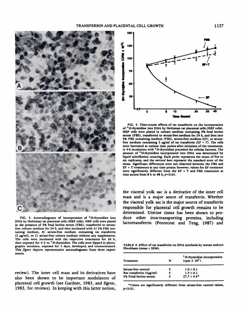
The results from experiments to determine the
kinetics of transferrin effects on HRP cell DNA
synthesis are depicted in Figure 4. An interval of
greater than 24 h in serum-free medium was necessary
to slow DNA synthesis by the HRP cells. There were
no significant differences in cells exposed to Spg/ml
of rat transferrin, 5% PBS, or serum-free medium at 4
h. After 8 h of treatment, a significant difference was
apparent between serum-free-treated cells and cells
exposed to serum or transferrin (p<0.Ol). This
difference was increased through the remainder of the
experiment. Growth curves for transferrin and
serum-treated cells were similar. The magnitude of
___________ the increase in � H-thymidine incorporation by HRP
i.o io �o cells to FBS or transferrin was diminished in cells
preincubated for 48 h in serum-free medium in
comparison to cells preincubated for only 24 h (data
not shown).
Transferrin dependency was not characteristic of
all cell types. Rat transferrin had no significant
effects on the incorporation of 3H-thymidine by
BaIb/c mouse embryo fibroblasts (Table 4). The lack
of stimulation of embryo fibroblasts by transferrin
175
‘1 , . -
0 0.1 0.25 0.5 tO 2.5 50
Rat �..J.rrL. (p’W”��
FIG. 2. Effects of rat transferrin on the incorporation of H-
thymidine into DNA by Holtzman rat placental cells (HRP cells). SeeFigure 1 for further details regarding the experimental protocol. Each
point represents the mean of five to six replicates, and the vertical bars
represent the standard error of the mean. The histograms on the figurerepresent the serum-free control treatment (0) and the 5% fetal bovine
serum control (FBS). Values for transferrin concentrations greater
than or equal to 1.0 pg/mI were significantly different from the serum-
free control, p<0.01.

1126 DE ET AL.
TABLE 1. Effect of human transferrin on DNA synthesis by Holtzman
rat placental cells (mean ± SEM).
Treatment N
3H-thymidine incorporation
(cpm X 1O�)
Serum-free control 6 18.9 ± 1.65% Fetal bovine serum 6 162.2 ± 4.5
Rat transferrin(2.5 pg/mI) 6 126.8 ± 3.2
Human transferrin(2.5 pg/ml 6 127.4 ± 5.3
Values are significantly different from serum-free control values,
p<0.01.
also indicates the absence of contamination of the
transferrin preparation with a number of different
types of growth factors known to stimulate embryo
fibrobiasts (O’Keefe and Pledger, 1983).
DISCUSSION
We have demonstrated that transferrin is a potent
regulator of rat placental cell growth. Exposure of rat
placental cells to transferrin under serum-free condi-
tions resulted jn approximately an eightfold stimula-
tion of DNA synthesis. The stimulatory effects of
transferrin approached those obtained with FBS.
Transferrin also stimulated the proliferation of rat
placental cells in vitro. Transferrin is routinely
TABLE 2. Effects of human apotransferrin and holotransferrin onDNA synthesis by Holtzman rat placental cells(mean ± SEM).�
TreatmentH-thym
(cpm X
idine incorporation
iO�)
Serum-free control 24.6 ± 1.35% Fetal bovine serum 91.0 ± 4.0�Apotransferrin (pg/mI)
0.01 25.9 ± 0.30.1 41.6 ± 3.11.0 72.5 ± 4.6
10.0 75.0 ± 2.8Holotransferrin (pg/mI)
0.01 27.6 ± 11.1
0.1 83.2 ± 95b
1.0 93.5 ± 12.7
10.0 111.9 ± 14.6c
Values represent means of nine measurements (serum-free control
and 5% fetal bovine serum) and means of a representative experimentperformed in triplicate (apotransferrin and holotransferrin).
asignificantly different from serum-free control, p<0.01.
bSignificantly different from apotransferrin, 0.1 pg/mI, p<0.01.
csignificantly different from apotransferrin, 10 pg/mI, p<O.05.
TABLE 3. Rat placental cell proliferation following four days of ex-
posure to rat transferrin (mean ± SEM).
Treatment N Cells (X 106)
Serum-free control 5 2.6 ± 0.3
5% Fetal bovine serum 4 10.3 ± 1.3Rat transferrin (5 pg/mI) 5 7.6 ± 0.6
�Values are significantly different from serum-free control values,
p<0.01.
used as a growth supplement for cell culture but
generally is relatively ineffective in stimulating cell
proliferation without the addition of other growth
factors (Barnes and Sato, 1980). In the present study,
the addition of insulin, epidermal growth factor, or
insulin-like growth factor-I! did not significantly alter
the magnitude of the effect of transferrin on placen-
tal cell DNA synthesis. Although the results obtained
with transferrin on rat placental cells differ somewhat
from those obtained with adult cell types, they are in
agreement with the stimulatory effects of transferrin
on embryonic cell growth (Ekblom et al., 1983).
The rodent placenta has previously been shown to
be a target tissue for transferrin (McArdle et al.,
1984a,b; 1985). Receptors for transferrin have been
preferentially localized to the labyrinth region of the
chorioallantoic placenta of the mouse (Muller et al.,
1983; Adamson, 1986) and rat (Hunt and Soares,
1988). Transferrin has been shown to be synthesized
by a variety of embryonic and extraembryonic cells,
including those forming the visceral yolk sac, a
major source of transferrin during midgestation, and
by cells in adult tissues (Adamson, 1982; Meek and
Adamson, 1985). Although transferrin is not synthe-
sized by the rodent placenta (Adamson, 1986), our
results suggest that placental cells may be dependent
on transferrin for growth.
The growth and differentiation of the chorioallan-
toic placenta are essential for the maintenance of
normal fetal development. Increased placental size
results in increased surface area for nutrient and
waste exchange and increased number of cells capable
of secreting hormones and growth factors that
influence maternal and fetal environments. Some of
the hormones and growth factors produced by the
placenta have also been proposed as regulators of
placental cell growth (see Adamson, 1986, for a
TRANSFERRIN AND PLACENTAL CELL GROWTH
150
125
1127
r
II
8 12
Th� ftm�)
the visceral yolk sac is a derivative of the inner cell
mass and is a major souce of transferrin. Whether
the visceral yolk sac is the major source of transferrin
responsible for placental cell growth remains to be
determined. Uterine tissue has been shown to pro-
duce other iron-transporting proteins, including
lactotransferrin (Pentecost and Teng, 1987) and
TABLE 4. Effect of rat transferrin on DNA synthesis by mouse embryofibroblasts (mean ± SEM).
Values are significantly different from serum-free control values,
p<0.01.
FIG. 3. Autoradiograms of incorporation of 3H-thymidine into
DNA by Holtzman rat placental cells (HRP cells). HRP cells were platedin the presence of 5% fetal bovine serum (FBS), transferred to serum-
free culture medium for 24 h, and then incubated with A) 5% FBS con-
taining medium, B) serum-free medium containing rat transferrin
(5 pg/mI), or C) serum-free culture medium without any supplements.
The cells were incubated with the respective treatments for 36 h,then exposed for 4 h to H-thymidine. The cells were dipped in photo-graphic emulsion, exposed for 3 days, developed, and counterstained.
This figure depicts representative autoradiograms from three experi-
ments.
review). The inner cell mass and its derivatives have
also been shown to be important modulators of
placental cell growth (see Gardner, 1983, and Ilgren,
1983, for reviews). In keeping with this latter notion,
FIG. 4. Time-course effects of rat transferrin on the incorporation
of 3H-thymidine into DNA by Holtzman rat placental cells (HRP cells).
HRP cells were plated in culture medium containing 5% fetal bovine
serum (FBS), transferred to serum-free medium for 24 h, and then into5% FBS containing medium (FBS), serum-free medium (SF), or serum-free medium containing 5 pg/ml of rat transferrin (SF # T). The cellswere harvested at various time points after initiation of the treatments.
A 4-h incubation with H-thymidine preceded the cellular harvests. The
amount of 3H-thymidine incorporated into DNA was determined byliquid scintillation counting. Each point represents the mean of five tosix replicates, and the vertical bars represent the standard error of the
mean. Significant differences were not observed between the FBS andSF + T treatments at any time points; however, values for SF treatmentwere significantly different from the SF + T and FBS treatments attime points from 8 h to 48 h, p<0.0i.
Treatment N
H-thymidine incorporation
(cpm X iOn)
Serum-free control 5 1.0 ± 0.1
Rat transferrin (5pg/mI) 5 1.5 ± 0.15% Fetal bovine serum 5 27.7 ± 4.4

1128 DE ET AL.
uteroferrin (Roberts and Barer, 1985). The role of
these proteins in regulating placental cell growth
is unknown.
The importance of iron delivery in transferrin-
mediated cell growth has been previously demon-
strated (Thesleff et al., 1985). Comparison of the
actions of apotransferrin versus holotransferrin is
consistent with the transfer of iron being an integral
regulatory step in the control of rat placental cell
DNA synthesis.
Our observations on transferrin-stimulated placen-
tal cell growth may not be generalized to all placental
cell types. In this report, we have examined the
effects of transferrin on a population of cells isolated
from the midgestation chorioallantoic placenta
(Soares et al., 1987). This population of placental
cells is relatively autonomous; they produce their
own extracellular matrix (Soares et al., 1987), are
transplantable (Soares et al., 1987), and have minimal
external requirements for growth (present study).
These characteristics are not unlike those expected
for a stem cell population (Adamson, 1986). A more
complex array of growth factors may be required for
placental cells with differentiated phenotypes (Goustin
et al., 1985; Fant et al., 1986; Athanassakis et al.,
1987).
ACKNOWLEDGMENTS
The authors gratefully acknowledge Dr. Paul Terranova for the
availability of the Balb/c mouse embryo fibroblasts and Linda Hicks for
help in the preparation of the manuscript. The authors would also like
to thank Douglas Larsen for his excellent technical assistance.
REFERENCES
Adamson ED, 1982. The location and synthesis of transferrin in mouseembryos and teratocarcinoma cells. Dev Biol 91:227-34
Adamson ED, 1986. Extraembryonic tissues as sources and sinks of
humoral factors in development: teratocarcinoma model systems.In: Serrero G, Hayashi J (eds.), Cellular Endocrinology: Hormonal
Control of Embryonic and Cellular Differentiation. New York:
Alan R. Lisa, Inc., pp. 159-74Athanassakis I, Bleackley RC, Paetkau V. Guilbert L, Barr PJ, Wegmann
TG, 1987. The immunostimulatory effect of T cells and T cell
lymphokines on murine fetally derived placental cells. J Immunol
138:37-44
Barnes DW, Sato G, 1980. Serum-free cell culture: a unifying approach.
Cell 22:649-55Davies J, Glasser DR, 1968. Histological and fine structural observa-
tions on the placenta of the rat. Acta Anat 69:542-608
Ekblom P, Thesleff I, 1985. The role of transferrin and extracellularmatrix components in kidney development. Mod Cell Biol 4:
85-127Ekblom P, Thesleff I, Saxen L, Miettinen A, Timpi R, 1983. Transferrin
as a fetal growth factor: acquisition of responsiveness related to
embryonic induction. Proc Natl Acad Sci USA 80 :2651-55Fant M, Munro H, Moses AC, 1986. An autocrine/paracrine role for
insulin-like growth factors in the regulation of human placental
growth. J Cliii Endocrinol Metab 63:499-505
Gardner RL, 1983. Origin and differentiation of extraembryonic tissue
in the mouse. mt Rev. Exp Pathol 24:63-133
Goustin AS, Betsholtz C, Pfeifer-Ohlsson 5, Persson H, Rydnert J,Bywater M, Holmgren G, Heldun C-H, Westermark B, Ohlsson R,
1985. Coexpression of the sis and myc proto-oncogenes in devel-oping human placenta suggests autocrine control of trophoblast
growth. Cell 41:301-12Hunt JS, Soares MJ, 1988. Expression of histocompatibility antigens,
transferrin receptors, intermediate filaments, and alkaline phos-phatase by in vitro cultured rat placental cells and rat placental
cells in situ. Placenta 9, in press.
Hunt IS, Suzuki Y, Wood GW, Soares MJ, 1988. Ultrastructure of
cultured rat placental cells. Placenta 9, in press.Ilgren EB, 1983. Control of trophoblastic growth. Placenta 4:307-28Keppel G, 1973. Design and Analysis New Jersey: Prentice-Hall:
Englewood Cliffs
McArdle HJ, Douglas AJ, Morgan EH, 1984a. Transferrin binding by
microvillar vesicles isolated from rat placenta. Placenta 5:131-38
McArdle HJ, Douglas AJ, Morgan EH, 1984b. Uptake of transferrin and
iron by cultured rat placental cells. J Cell Physiol 122:405-09McArdle HJ, Morgan EU, 1982. Transferrin and iron movements in the
rat concepturs during gestation. J Reprod Fertil 66:529-36
Meek J, Adainson ED, 1985. Transferrin in fetal and adult mouse
tissues: synthesis, storage, and secretion. J Embryol Exp Morphol
86 :205-18Muller R, Verma I, Adamson ED, 1983. Expression of c-onc genes:
c-fos transcripts accumulate to high levels during development of
mouse placenta, yolk sac and amnion. EMBO J 2:679-84
O’Keefe EJ, Pledger WJ, 1983. A model of cell cycle control: sequentialevents regulated by growth factors. Mol Cell Endocr 31:167-86
Pentecost BT, Teng CT, 1987. Lactotransferrin is the major estrogen
inducible protein of mouse uterine secretions. J Biol Chem 262:
10134-39
Roberts RM, Bazer FW, 1985. Uteroferrin: a protein in search of a
function. Bioassays 1:8-11
Soares MJ, 1987. Developmental changes in the intraplacental distribu-tion of placental lactogen and alkaline phosphatase in the rat. J
Reprod Fertil 79:93-98
Soares MJ, Glasser SR, 1987. Placental lactogen production and func-tional differentiaiton of rat trophoblast cells in vitro. J ReprodFertil 79:335-41
Soares MJ, Schaberg KD, Pinal CS, Dc SK, Bhatia P, Andrews GK,
1987. Establishment of a rat placental cell line expressing charac-teristics of extraembryonic membranes. Dcv Biol 124:134-44
Thesleff I, Partanen A-M, Landschulz W, Trowbidge IS, Ekblom P,
1985. The role of transferrin receptors and iron delivery in mouse
embryonic morphogenesis. Differentiation 30:152-58