Embed Size (px)
Citation preview
ORIGINAL ARTICLE
Biology and distribution of hemichordates (Enteropneusta)with emphasis on Harrimaniidae and descriptionof Protoglossus bocki sp. nov. from Scandinavia
Tomas Cedhagen • Hans G. Hansson
Received: 29 August 2011 / Revised: 29 June 2012 / Accepted: 14 July 2012 / Published online: 14 September 2012
� Springer-Verlag and AWI 2012
Abstract A new species of the genus Protoglossus is
described from the west coast of the Scandinavian Penin-
sula. It lives buried in clay bottoms below the halocline
where the salinity is at least 33–34 psu. Body small, esti-
mated maximal length 1.5 cm. Collar broader than long,
with a forward inclination. The thickest part is the collar
region where it can be up to 1 mm in diameter. Proboscis
colouration light pink to golden yellow; collar white with
transversal yellow bands; branchial, hepatic and intestinal
regions translucent pale yellow to golden yellow; brown
intestine visible through the body wall. Proboscis groove
extends through posterior half of proboscis. Nine to 17
pairs of gill openings, the size of the posteriormost suc-
cessively smaller. It differs from the other European spe-
cies, Protoglossus koehleri, in colouration, smaller size,
fewer gill openings, body shape and proportions. It was
sequenced (18S rRNA gene) and clustered within the
family Harrimaniidae, with Saxipendium as its closest
relative.
Keywords Hemichordata � Enteropneusta �Harrimaniidae � Protoglossus bocki n.sp. � Scandinavian
Peninsula
Introduction
Enteropneusts are poorly known in the Skagerrak–Kattegat
area. Von Willemoes-Suhm (1871) described Harrimania
kupfferi (originally as Balanoglossus Kupfferi) from
Øresund, Denmark. This species was later reported from
the Gullmarfjord area by Theel (1908) and subsequent
authors. A second species, Glossobalanus marginatus
Meek 1922, was obtained from Sweden by Lindroth (1941)
and Silen (1950). The marine fauna of Sweden from the
Norwegian border to Oresund was investigated from 1925
to 1938. Altogether, 440 stations were sampled, but there
are no reports on enteropneusts (Hubendick et al. 1971).
The conventional sieving and sorting methods have prob-
ably destroyed all the fragile enteropneusts.
The new species has been sampled off the west coast of
Sweden since the earlier parts of the twentieth century.
Collectors were Dr. Gunnar Gustafson (1891–1988) at
Kristineberg Marinezoological Station, Fiskebackskil
(Royal Swedish Academy of Sciences), and Prof. Sixten
Bock (1884–1946) at the Swedish Museum of Natural
History, Stockholm. In August 1927, when looking for
other animals, Sixten Bock discovered a small enteropneust
that he found to be very different from Harrimania. He first
believed that it was a specimen of Protoglossus koehleri
(Caullery and Mesnil 1900), which was only found on the
French Atlantic coast at that time, but later understood that
it was an undescribed species of the same genus. The small
size and fragility of the animal explains why this species
has been overlooked. Bock made extensive studies of it in
the field station and histological studies in the laboratory.
He intended to describe it, but this work was interrupted by
his sudden death. Parts of a manuscript draft, including
photographs and drawings, are deposited in the archive
of the Swedish Museum of Natural History. The present
Communicated by H.-D. Franke.
Hans G. Hansson: Deceased.
T. Cedhagen (&)
Department of Biological Sciences, Section of Marine Ecology,
Aarhus University, Building 1135, Ole Worms alle 1,
8000 Aarhus C, Denmark
e-mail: [email protected]
H. G. Hansson
Tjarno Marine Biological Laboratory, University of Gothenburg,
452 96 Stromstad, Sweden
123
Helgol Mar Res (2013) 67:251–265
DOI 10.1007/s10152-012-0320-5
Table 1 Material of Protoglossus bocki n. sp. Swedish Museum of Natural History, Stockholm (SMNH), Natural History Museum, Goteborg,
Sweden (GNM)
Material
number
Collection
number
Locality, date Leg./Det. Notes
1 SMNH-32 Sweden, Bohuslan, Gullmarfjord area,
Humlesacken. 1927-08-16
S. Bock Fixed in Bouin’s fluid. Specimen in
the wet collection and as
microscopic slides
2 SMNH-36 Sweden, Bohuslan, Gullmarfjord area,
near Flatholmen. 1931-05-14
S. Bock Fixed in sublimat. Specimen as
microscopic slides only
3 SMNH-37 Sweden, Bohuslan, Gullmarfjord area,
Humlesacken, 40 m depth. 1931-05-15.
S. Bock Fixed in 5 % alcohol and cold 90 %
sublimat solution. Specimen in the
wet collection and as microscopic
slides
4 SMNH-40 Sweden, Bohuslan, Gullmarfjord area,
near Flatholmen and in Humlesacken.
1931-11-17
S. Bock Specimen in the wet collection.
Holotype
5 SMNH-41 Sweden, Bohuslan, Gullmarfjord area,
near Flatholmen and in Humlesacken.
1931-11-17
S. Bock Fixed in 5 % alcohol and Bouin’s
fluid. Specimen in the wet
collection
6 SMNH-42 Sweden, Bohuslan, Gullmarfjord area,
Tova. 1931-11-18
S. Bock Fixed in 5 % alcohol and Bouin’s
fluid. Specimen as microscopic
slides only
7 SMNH-43 Sweden, Bohuslan, Gullmarfjord area,
Tova. 1931-11-20
S. Bock Anaesthetized in MgCl2 and fixed in
sublimat. Specimen in the wet
collection and as microscopic slides
8 SMNH-45 Sweden, Bohuslan, Gullmarfjord area,
Tova. 1931-11-25
S. Bock Fixed in formalin. Specimen in the
wet collection
9 SMNH-46 Sweden, Bohuslan, Gullmarfjord area,
Tova. 1931-11-25
S. Bock Fixed in 5 % alcohol and hot
sublimat solution. Specimen in the
wet collection
10 SMNH-47 Sweden, Bohuslan, Gullmarfjord area,
Tova. 1931-11-25
S. Bock Fixed in 5 % alcohol and formalin.
Specimen in the wet collection and
as microscopic slides
11 SMNH-48 Sweden, Bohuslan, Gullmarfjord area,
near Flatholmen, 40 m depth. 1932-08-11
S. Bock Fixed in Bouin’s fluid. Specimen as
microscopic slides only
12 SMNH-49 Sweden, Bohuslan, Gullmarfjord area,
Humlesacken, 30 m depth
S. Bock Specimen in the wet collection and as
microscopic slides
13 SMNH-53 Norway, Oslofjorden, Drøbak, near
Degerud depth and Graøy, 150–100 m
depth. 1936-07-1-2
Leg. C. Dons, Det. S. Bock Fixed in Bouin’s solution. Specimen
in the wet collection
14 SMNH-54 Norway, Trondhjem, Vikleiret, 15–20 m
depth. 1936-07-13
Leg. C. Dons, Det. S. Bock Specimen as microscopic slides only.
The slide is labelled 1932 instead of
1936
15 SMNH-57 Norway, Skagerak, Vestfold, 7 km S of
Tvesten, 58�520N, 009�570E, 130-150 m
depth
Sixten Bock’s Skagerak
Expedition 1937 Station
28.7.A. Det. S. Bock
Specimen in the wet collection
16 SMNH-59 Norway, Oslofjorden, Drøbak,
Hakungspollen,. 1938-08-08
S. Bock Specimen in the wet collection
17 SMNH-60 Norway, Oslofjorden, Drøbak,
Hakungspollen, 12 m depth. 1938-08-08
S. Bock Fixed in formalin. Specimen in the
wet collection
18 SMNH-61 Norway, Oslofjorden, Drøbak,
Hakungspollen,. 1938-08-08
S. Bock Fixed in sublimat, picric acid and
formalin. Specimen in the wet
collection
19 Sweden, Skagerak, sledge sample Start:
58�28.01200N; 10�48.9010E. End:
58�28.1410N; 10�49.6750E. Depth:
103-119. 2006-06-05
Leg. R/V Arne Tiselius,
ArtDatabanken, Det. Hans G.
Hansson
Figure 2 in this publication
252 Helgol Mar Res (2013) 67:251–265
123

description is based on Sixten Bock’s results supplemented
with new material.
Silen (1950) used material of this species, collected by
G. Gustafson, for comparative purposes. He referred to it as
Protoglossus sp. Silen did not formally describe the species
because his focus was on the nervous system of another
enteropneust species. He wrote that the nervous system of
the tiny Protoglossus sp. was very difficult to observe, but
supported his general conclusions based on his studies of
the other much larger species, Glossobalanus marginatus,
collected in the same area (Silen 1950). Ake Franzen
(1956) used two individuals of the Protoglossus species for
the studies of sperm morphology. The species is also
included in the discussion by Franzen et al. (1985), but
mainly as a reference to Franzen (1956). Hansson (2009)
and Lundin et al. (2009) used Sixten Bock’s tentative
Fig. 3 Protoglossus bocki n.sp. Fixed individual. Lateral view seen
from right. Scale bar 1 mm. Drawing: Sven Ekblom
Fig. 1 Protoglossus bocki n.sp.
Creeping individual where the
proboscis shape with dorsal
groove, collar colouration,
dorsal ridge and gill openings is
visible. Photograph: Christopher
Reisborg
Fig. 2 Protoglossus bocki n.sp. Living but unhealthy individual with
relaxed proboscis musculature. Photograph: Fredrik Pleijel
Table 1 continued
Material
number
Collection
number
Locality, date Leg./Det. Notes
20 GNM- Sweden, Skagerak,. Sledge sample Start:
58�06,5260N; 11�11,7400E, end: 58�06,3220N; 11�11,1690S. Depth: 68-72 m.
2008-08-20
Leg. R/V Arne Tiselius,
ArtDatabanken, Running no:
228. St. nr: SK68 bsk1. Det.
T. Cedhagen
21 Sweden, Skagerak, Koster area, Aug. 2009 Leg. M. Strand, Det.
T. Cedhagen
Figure 1 in this publication. A
fragment of this individual was
sequenced at SMNH
Helgol Mar Res (2013) 67:251–265 253
123

manuscript name Protoglossus simplex (nomen nudum) for
it. Also, Prof. Bertil Akesson observed the species in
Kristineberg Marine Zoological Station in the 1950s or
1960s (pers. comm.).
Materials and methods
Sixten Bock collected eight individuals between 27 July and
the end of August 1927 in Humlesacken, in the outer area of
Gullmarsfjorden, Sweden. He sampled several times after
the first findings, but in most cases without result. Sediment
was usually collected by the use of an Agassiz trawl at that
time. The conventional sediment-sieving method could not
be used because of the fragility of the animal. Mud was
therefore filled in buckets and covered with just a little water,
left for some time and frequently inspected.
Bock anaesthetized the animals by the addition of ethyl
urethane to the sea water until a strength of 3 %, with a
subsequent treatment with 5 % alcohol in sea water. Sev-
eral specimens were sectioned for microscopy. The internal
structures of some whole mounts of individuals were made
visible after clearing by the use of aniseed oil.
A small number of additional specimens have been
collected in Skagerrak and Kattegat in the beginning of the
twenty-first century, mainly as a result of the activities run
by the Swedish Taxonomy Project. A few of them have
been photographed and preserved in alcohol in order to
allow molecular studies. Sequencing (18S rRNA genes) of
the new species Protoglossus bocki as well as Harrimania
kupfferi was made at the molecular biology laboratory,
Swedish Museum of Natural History. The alignment of the
sequences was done manually using Seaview (Gouy et al.
2010). Phylogenetic analysis was performed using maxi-
mum likelihood (PhyML) programmes as implemented in
Seaview. The support for internal branches was inferred
using 100 bootstrap replicates.
Specimens are deposited in the collections of the
Swedish Museum of Natural History, Stockholm (NRM),
and the Natural History Museum, Gothenburg (GNM),
both in Sweden, and summarized in Table 1. Sixten Bock
identified all materials sampled by himself, and some of
these samples were later verified to belong to Protoglossus
by Cyril Burdon-Jones during his stay at the NRM in 1998.
Taxonomy
The position of the group Hemichordata has been disputed
(Gerhart et al. 2005), but is now regarded as a sister group
Fig. 5 Protoglossus bocki n.sp. Fixed individual showing the median
dorsal ridge. Scale bars 1 mm. Drawing: Sven Ekblom
Fig. 4 Protoglossus bocki n.sp. Fixed individual. Lateral view seen
from left side. Scale bar 1 mm. Drawing: Sven Ekblom
254 Helgol Mar Res (2013) 67:251–265
123
of Echinodermata. Both are together called Ambulacraria,
which is the sister group of the remaining deuterostomes—
the chordates (Hejnol et al. 2009).
The hemichordate group Enteropneusta has recently been
revised by the use of morphological and molecular methods
(Cameron 2005; Cannon et al. 2009). The family Harrima-
niidae, to which the new species belongs, is revised by Deland
et al. (2010). Detailed information on morphology and his-
tology of enteropneusts has been summarized by van der
Horst (1939), Hyman (1959) and Benito and Pardos (1997).
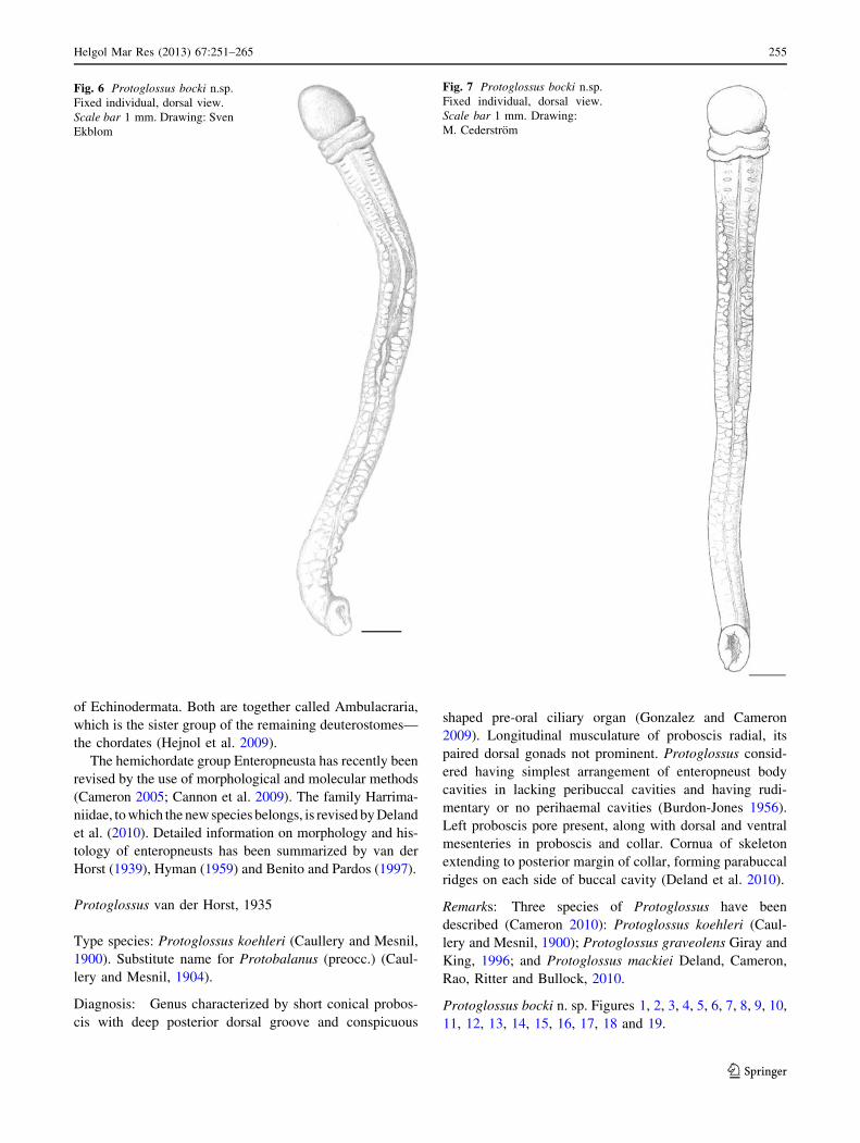
Protoglossus van der Horst, 1935
Type species: Protoglossus koehleri (Caullery and Mesnil,
1900). Substitute name for Protobalanus (preocc.) (Caul-
lery and Mesnil, 1904).
Diagnosis: Genus characterized by short conical probos-
cis with deep posterior dorsal groove and conspicuous
shaped pre-oral ciliary organ (Gonzalez and Cameron
2009). Longitudinal musculature of proboscis radial, its
paired dorsal gonads not prominent. Protoglossus consid-
ered having simplest arrangement of enteropneust body
cavities in lacking peribuccal cavities and having rudi-
mentary or no perihaemal cavities (Burdon-Jones 1956).
Left proboscis pore present, along with dorsal and ventral
mesenteries in proboscis and collar. Cornua of skeleton
extending to posterior margin of collar, forming parabuccal
ridges on each side of buccal cavity (Deland et al. 2010).
Remarks: Three species of Protoglossus have been
described (Cameron 2010): Protoglossus koehleri (Caul-
lery and Mesnil, 1900); Protoglossus graveolens Giray and
King, 1996; and Protoglossus mackiei Deland, Cameron,
Rao, Ritter and Bullock, 2010.
Protoglossus bocki n. sp. Figures 1, 2, 3, 4, 5, 6, 7, 8, 9, 10,
11, 12, 13, 14, 15, 16, 17, 18 and 19.
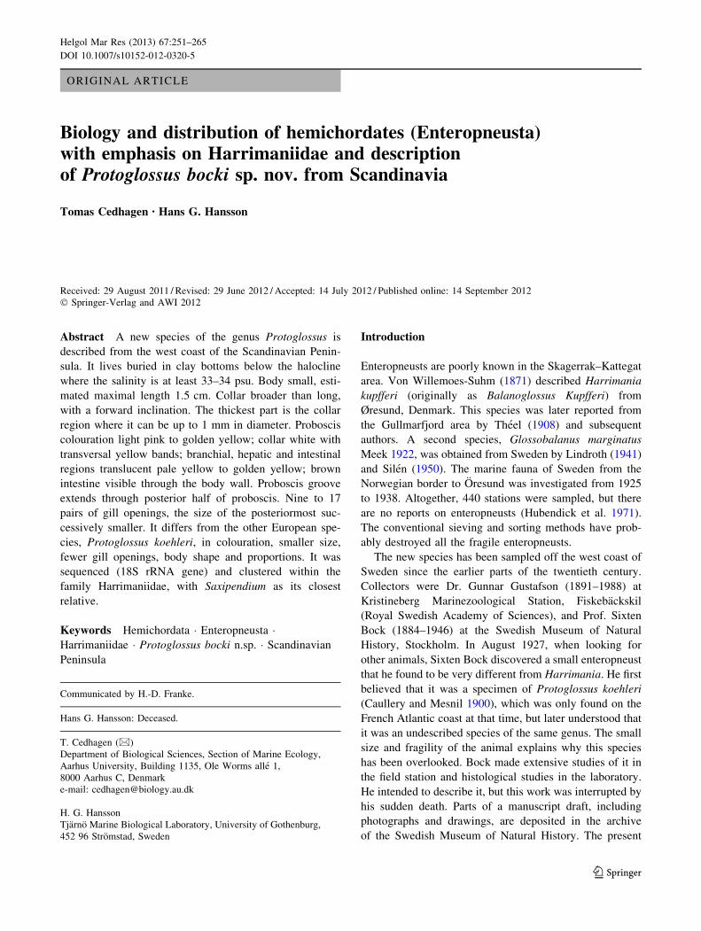
Fig. 7 Protoglossus bocki n.sp.
Fixed individual, dorsal view.
Scale bar 1 mm. Drawing:
M. Cederstrom
Fig. 6 Protoglossus bocki n.sp.
Fixed individual, dorsal view.
Scale bar 1 mm. Drawing: Sven
Ekblom
Helgol Mar Res (2013) 67:251–265 255
123
Synonymy: Protoglossus sp. Silen 1950, pp. 149, 151, 153;
Franzen 1956, pp. 443, 450, 461; Franzen et al. 1985.
p. 303.
Protoglossus simplex. Hansson 2009, p. 313.
Protoglossus ‘‘simplex’’. Lundin et al. 2009, p. 41.
Material: Specimens in the collections of NRM and
GNM (Table 1).
Etymology: We dedicate this species in honour of Pro-
fessor Sixten Bock (1884–1946), who discovered the spe-
cies and made extensive studies of it. Sixten Bock was the
godfather of one of the authors (HGH).
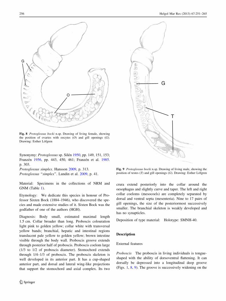
Diagnosis: Body small, estimated maximal length
1.5 cm. Collar broader than long. Proboscis colouration
light pink to golden yellow; collar white with transversal
yellow bands; branchial, hepatic and intestinal regions
translucent pale yellow to golden yellow; brown intestine
visible through the body wall. Proboscis groove extends
through posterior half of proboscis. Proboscis coelom large
(1/3 to 1/2 of proboscis diameter). Stomochord extends
through 1/4–1/3 of proboscis. The proboscis skeleton is
well developed in its anterior part. It has a cup-shaped
anterior part, and dorsal and lateral wing-like projections
that support the stomochord and axial complex. Its two
crura extend posteriorly into the collar around the
oesophagus and slightly curve and taper. The left and right
collar coeloms (mesocoels) are completely separated by
dorsal and ventral septa (mesenteria). Nine to 17 pairs of
gill openings, the size of the posteriormost successively
smaller. The branchial skeleton is weakly developed and
has no synapticles.
Deposition of type material: Holotype: SMNH-40.
Description
External features
Proboscis The proboscis in living individuals is tongue-
shaped with the ability of dorsoventral flattening. It can
dorsally be depressed into a longitudinal deep groove
(Figs. 1, 8, 9). The groove is successively widening on the
G
T
Fig. 9 Protoglossus bocki n.sp. Drawing of living male, showing the
position of testes (T) and gill openings (G). Drawing: Esther Lofgren
G
O
Fig. 8 Protoglossus bocki n.sp. Drawing of living female, showing
the position of ovaries with oocytes (O) and gill openings (G).
Drawing: Esther Lofgren
256 Helgol Mar Res (2013) 67:251–265
123
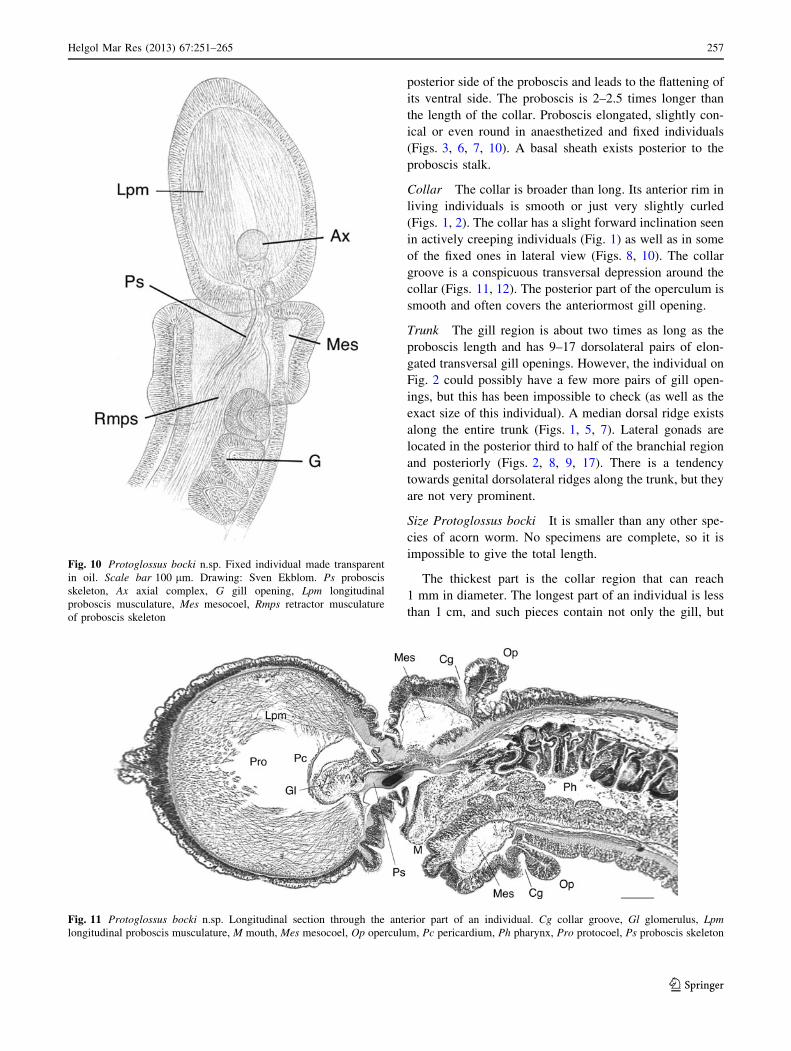
posterior side of the proboscis and leads to the flattening of
its ventral side. The proboscis is 2–2.5 times longer than
the length of the collar. Proboscis elongated, slightly con-
ical or even round in anaesthetized and fixed individuals
(Figs. 3, 6, 7, 10). A basal sheath exists posterior to the
proboscis stalk.
Collar The collar is broader than long. Its anterior rim in
living individuals is smooth or just very slightly curled
(Figs. 1, 2). The collar has a slight forward inclination seen
in actively creeping individuals (Fig. 1) as well as in some
of the fixed ones in lateral view (Figs. 8, 10). The collar
groove is a conspicuous transversal depression around the
collar (Figs. 11, 12). The posterior part of the operculum is
smooth and often covers the anteriormost gill opening.
Trunk The gill region is about two times as long as the
proboscis length and has 9–17 dorsolateral pairs of elon-
gated transversal gill openings. However, the individual on
Fig. 2 could possibly have a few more pairs of gill open-
ings, but this has been impossible to check (as well as the
exact size of this individual). A median dorsal ridge exists
along the entire trunk (Figs. 1, 5, 7). Lateral gonads are
located in the posterior third to half of the branchial region
and posteriorly (Figs. 2, 8, 9, 17). There is a tendency
towards genital dorsolateral ridges along the trunk, but they
are not very prominent.
Size Protoglossus bocki It is smaller than any other spe-
cies of acorn worm. No specimens are complete, so it is
impossible to give the total length.
The thickest part is the collar region that can reach
1 mm in diameter. The longest part of an individual is less
than 1 cm, and such pieces contain not only the gill, but
Fig. 11 Protoglossus bocki n.sp. Longitudinal section through the anterior part of an individual. Cg collar groove, Gl glomerulus, Lpmlongitudinal proboscis musculature, M mouth, Mes mesocoel, Op operculum, Pc pericardium, Ph pharynx, Pro protocoel, Ps proboscis skeleton
Fig. 10 Protoglossus bocki n.sp. Fixed individual made transparent
in oil. Scale bar 100 lm. Drawing: Sven Ekblom. Ps proboscis
skeleton, Ax axial complex, G gill opening, Lpm longitudinal
proboscis musculature, Mes mesocoel, Rmps retractor musculature
of proboscis skeleton
Helgol Mar Res (2013) 67:251–265 257
123

also the genital region. The total length can therefore be
estimated to about one and a half centimetre.
Colour of living individuals The colour of the proboscis
is uniformly light pink to golden yellow (Figs. 1, 2). The
anterior rim of the collar is whitish (Figs. 1, 2, 3, 4, 5, 6, 7,
8, 9). Two slightly darker yellow transversal bands exist
around the collar; the posterior of them coincides with the
narrow circular depression around the collar, the so-called
collar groove. The posterior part of the collar, the oper-
culum, is whitish (Figs. 1, 2, 3, 4, 5, 6, 7, 8, 9). The trunk
colour is uniformly light brownish yellow to golden
yellow. The mucus gland tissue around the gill openings is
slightly lighter yellow than the surrounding body wall. The
intestine looks brown through the body wall (Fig. 1). The
rose ovaries or white testes can be seen through the body
wall (Figs. 2, 8, 9).
Internal features
Proboscis The proboscis is covered by a thick glandular
and ciliated epithelium. Its musculature is well developed
(Figs. 10, 11). The circular muscle fibre layer is thinner
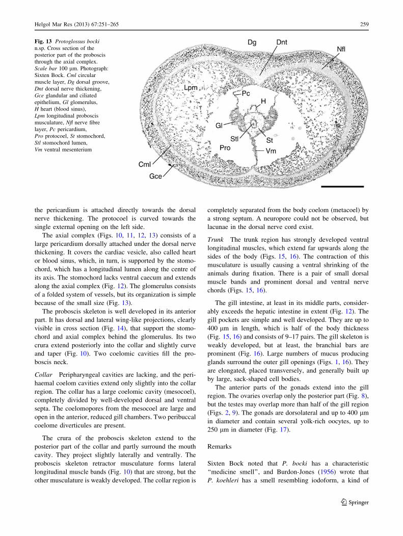
than the nerve fibre layer (Fig. 13). The nerve fibre layer of
the proboscis is thickened middorsally (Fig. 13). There is a
tendency towards a radial arrangement of the fibres in the
longitudinal muscle layer. It is stronger developed laterally
than dorsally, and its internal distribution seen in cross
section coincides with the shape of the dorsal groove in
creeping individuals (Figs. 1, 13).
The new species has a fairly wide protocoel around the
heart and axial complex (Fig. 13). It extends to the anterior
part of the proboscis and has a well-developed ventral
mesenterium (septum). There is no dorsal mesenterium as
GlPc
OpCg
Mes
C
Nch
Ps
St
Mes
M
Lpm
Fig. 12 Protoglossus bocki n.sp. Longitudinal section through the
collar and posterior part of the proboscis. Scale bar 100 lm.
Photograph: Sixten Bock. C collar, Cg collar groove, Gl glomerulus,
Lpm longitudinal proboscis musculature, M mouth, Mes mesocoel,
Nch neurochord, Op operculum, Pc pericardium, Ps proboscis
skeleton, St stomochord
258 Helgol Mar Res (2013) 67:251–265
123
the pericardium is attached directly towards the dorsal
nerve thickening. The protocoel is curved towards the
single external opening on the left side.
The axial complex (Figs. 10, 11, 12, 13) consists of a
large pericardium dorsally attached under the dorsal nerve
thickening. It covers the cardiac vesicle, also called heart
or blood sinus, which, in turn, is supported by the stomo-
chord, which has a longitudinal lumen along the centre of
its axis. The stomochord lacks ventral caecum and extends
along the axial complex (Fig. 12). The glomerulus consists
of a folded system of vessels, but its organization is simple
because of the small size (Fig. 13).
The proboscis skeleton is well developed in its anterior
part. It has dorsal and lateral wing-like projections, clearly
visible in cross section (Fig. 14), that support the stomo-
chord and axial complex behind the glomerulus. Its two
crura extend posteriorly into the collar and slightly curve
and taper (Fig. 10). Two coelomic cavities fill the pro-
boscis neck.
Collar Peripharyngeal cavities are lacking, and the peri-
haemal coelom cavities extend only slightly into the collar
region. The collar has a large coelomic cavity (mesocoel),
completely divided by well-developed dorsal and ventral
septa. The coelomopores from the mesocoel are large and
open in the anterior, reduced gill chambers. Two peribuccal
coelome diverticules are present.
The crura of the proboscis skeleton extend to the
posterior part of the collar and partly surround the mouth
cavity. They project slightly laterally and ventrally. The
proboscis skeleton retractor musculature forms lateral
longitudinal muscle bands (Fig. 10) that are strong, but the
other musculature is weakly developed. The collar region is
completely separated from the body coelom (metacoel) by
a strong septum. A neuropore could not be observed, but
lacunae in the dorsal nerve cord exist.
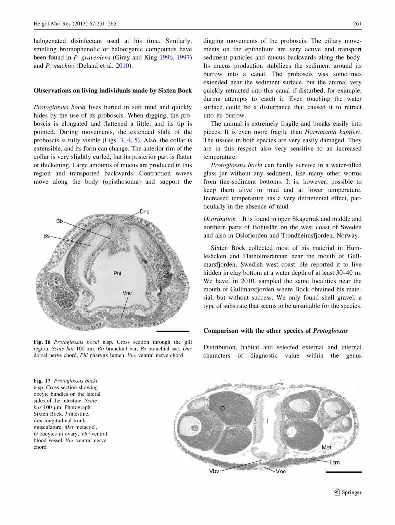
Trunk The trunk region has strongly developed ventral
longitudinal muscles, which extend far upwards along the
sides of the body (Figs. 15, 16). The contraction of this
musculature is usually causing a ventral shrinking of the
animals during fixation. There is a pair of small dorsal
muscle bands and prominent dorsal and ventral nerve
chords (Figs. 15, 16).
The gill intestine, at least in its middle parts, consider-
ably exceeds the hepatic intestine in extent (Fig. 12). The
gill pockets are simple and well developed. They are up to
400 lm in length, which is half of the body thickness
(Fig. 15, 16) and consists of 9–17 pairs. The gill skeleton is
weakly developed, but at least, the branchial bars are
prominent (Fig. 16). Large numbers of mucus producing
glands surround the outer gill openings (Figs. 1, 16). They
are elongated, placed transversely, and generally built up
by large, sack-shaped cell bodies.
The anterior parts of the gonads extend into the gill
region. The ovaries overlap only the posterior part (Fig. 8),
but the testes may overlap more than half of the gill region
(Figs. 2, 9). The gonads are dorsolateral and up to 400 lm
in diameter and contain several yolk-rich oocytes, up to
250 lm in diameter (Fig. 17).
Remarks
Sixten Bock noted that P. bocki has a characteristic
‘‘medicine smell’’, and Burdon-Jones (1956) wrote that
P. koehleri has a smell resembling iodoform, a kind of
Fig. 13 Protoglossus bockin.sp. Cross section of the
posterior part of the proboscis
through the axial complex.
Scale bar 100 lm. Photograph:
Sixten Bock. Cml circular
muscle layer, Dg dorsal groove,
Dnt dorsal nerve thickening,
Gce glandular and ciliated
epithelium, Gl glomerulus,
H heart (blood sinus),
Lpm longitudinal proboscis
musculature, Nfl nerve fibre
layer, Pc pericardium,
Pro protocoel, St stomochord,
Stl stomochord lumen,
Vm ventral mesenterium
Helgol Mar Res (2013) 67:251–265 259
123
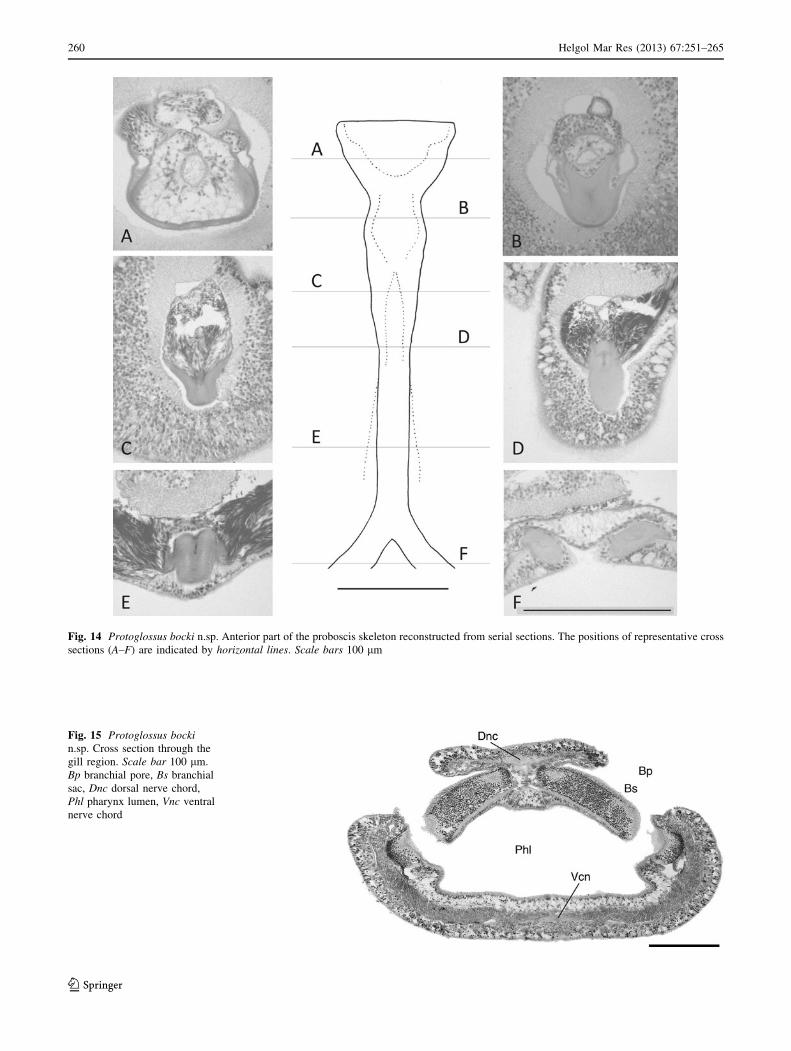
Fig. 14 Protoglossus bocki n.sp. Anterior part of the proboscis skeleton reconstructed from serial sections. The positions of representative cross
sections (A–F) are indicated by horizontal lines. Scale bars 100 lm
Fig. 15 Protoglossus bockin.sp. Cross section through the
gill region. Scale bar 100 lm.
Bp branchial pore, Bs branchial
sac, Dnc dorsal nerve chord,
Phl pharynx lumen, Vnc ventral
nerve chord
260 Helgol Mar Res (2013) 67:251–265
123
halogenated disinfectant used at his time. Similarly,
smelling bromophenolic or haloorganic compounds have
been found in P. graveolens (Giray and King 1996, 1997)
and P. mackiei (Deland et al. 2010).
Observations on living individuals made by Sixten Bock
Protoglossus bocki lives buried in soft mud and quickly
hides by the use of its proboscis. When digging, the pro-
boscis is elongated and flattened a little, and its tip is
pointed. During movements, the extended stalk of the
proboscis is fully visible (Figs. 3, 4, 5). Also, the collar is
extensible, and its form can change. The anterior rim of the
collar is very slightly curled, but its posterior part is flatter
or thickening. Large amounts of mucus are produced in this
region and transported backwards. Contraction waves
move along the body (opisthosoma) and support the
digging movements of the proboscis. The ciliary move-
ments on the epithelium are very active and transport
sediment particles and mucus backwards along the body.
Its mucus production stabilizes the sediment around its
burrow into a canal. The proboscis was sometimes
extended near the sediment surface, but the animal very
quickly retracted into this canal if disturbed, for example,
during attempts to catch it. Even touching the water
surface could be a disturbance that caused it to retract
into its burrow.
The animal is extremely fragile and breaks easily into
pieces. It is even more fragile than Harrimania kupfferi.
The tissues in both species are very easily damaged. They
are in this respect also very sensitive to an increased
temperature.
Protoglossus bocki can hardly survive in a water-filled
glass jar without any sediment, like many other worms
from fine-sediment bottoms. It is, however, possible to
keep them alive in mud and at lower temperature.
Increased temperature has a very detrimental effect, par-
ticularly in the absence of mud.
Distribution It is found in open Skagerrak and middle and
northern parts of Bohuslan on the west coast of Sweden
and also in Oslofjorden and Trondheimsfjorden, Norway.
Sixten Bock collected most of his material in Hum-
lesacken and Flatholmsrannan near the mouth of Gull-
marsfjorden, Swedish west coast. He reported it to live
hidden in clay bottom at a water depth of at least 30–40 m.
We have, in 2010, sampled the same localities near the
mouth of Gullmarsfjorden where Bock obtained his mate-
rial, but without success. We only found shell gravel, a
type of substrate that seems to be unsuitable for the species.
Comparison with the other species of Protoglossus
Distribution, habitat and selected external and internal
characters of diagnostic value within the genus
Fig. 17 Protoglossus bockin.sp. Cross section showing
oocyte bundles on the lateral
sides of the intestine. Scalebar 100 lm. Photograph:
Sixten Bock. I intestine,
Ltm longitudinal trunk
musculature, Met metacoel,
O oocytes in ovary, Vbv ventral
blood vessel, Vnc ventral nerve
chord
Fig. 16 Protoglossus bocki n.sp. Cross section through the gill
region. Scale bar 100 lm. Bb branchial bar, Bs branchial sac, Dncdorsal nerve chord, Phl pharynx lumen, Vnc ventral nerve chord
Helgol Mar Res (2013) 67:251–265 261
123
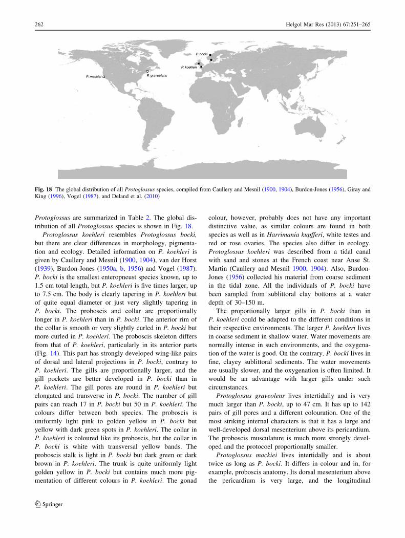
Protoglossus are summarized in Table 2. The global dis-
tribution of all Protoglossus species is shown in Fig. 18.
Protoglossus koehleri resembles Protoglossus bocki,
but there are clear differences in morphology, pigmenta-
tion and ecology. Detailed information on P. koehleri is
given by Caullery and Mesnil (1900, 1904), van der Horst
(1939), Burdon-Jones (1950a, b, 1956) and Vogel (1987).
P. bocki is the smallest enteropneust species known, up to
1.5 cm total length, but P. koehleri is five times larger, up
to 7.5 cm. The body is clearly tapering in P. koehleri but
of quite equal diameter or just very slightly tapering in
P. bocki. The proboscis and collar are proportionally
longer in P. koehleri than in P. bocki. The anterior rim of
the collar is smooth or very slightly curled in P. bocki but
more curled in P. koehleri. The proboscis skeleton differs
from that of P. koehleri, particularly in its anterior parts
(Fig. 14). This part has strongly developed wing-like pairs
of dorsal and lateral projections in P. bocki, contrary to
P. koehleri. The gills are proportionally larger, and the
gill pockets are better developed in P. bocki than in
P. koehleri. The gill pores are round in P. koehleri but
elongated and transverse in P. bocki. The number of gill
pairs can reach 17 in P. bocki but 50 in P. koehleri. The
colours differ between both species. The proboscis is
uniformly light pink to golden yellow in P. bocki but
yellow with dark green spots in P. koehleri. The collar in
P. koehleri is coloured like its proboscis, but the collar in
P. bocki is white with transversal yellow bands. The
proboscis stalk is light in P. bocki but dark green or dark
brown in P. koehleri. The trunk is quite uniformly light
golden yellow in P. bocki but contains much more pig-
mentation of different colours in P. koehleri. The gonad
colour, however, probably does not have any important
distinctive value, as similar colours are found in both
species as well as in Harrimania kupfferi, white testes and
red or rose ovaries. The species also differ in ecology.
Protoglossus koehleri was described from a tidal canal
with sand and stones at the French coast near Anse St.
Martin (Caullery and Mesnil 1900, 1904). Also, Burdon-
Jones (1956) collected his material from coarse sediment
in the tidal zone. All the individuals of P. bocki have
been sampled from sublittoral clay bottoms at a water
depth of 30–150 m.
The proportionally larger gills in P. bocki than in
P. koehleri could be adapted to the different conditions in
their respective environments. The larger P. koehleri lives
in coarse sediment in shallow water. Water movements are
normally intense in such environments, and the oxygena-
tion of the water is good. On the contrary, P. bocki lives in
fine, clayey sublittoral sediments. The water movements
are usually slower, and the oxygenation is often limited. It
would be an advantage with larger gills under such
circumstances.
Protoglossus graveolens lives intertidally and is very
much larger than P. bocki, up to 47 cm. It has up to 142
pairs of gill pores and a different colouration. One of the
most striking internal characters is that it has a large and
well-developed dorsal mesenterium above its pericardium.
The proboscis musculature is much more strongly devel-
oped and the protocoel proportionally smaller.
Protoglossus mackiei lives intertidally and is about
twice as long as P. bocki. It differs in colour and in, for
example, proboscis anatomy. Its dorsal mesenterium above
the pericardium is very large, and the longitudinal
Fig. 18 The global distribution of all Protoglossus species, compiled from Caullery and Mesnil (1900, 1904), Burdon-Jones (1956), Giray and
King (1996), Vogel (1987), and Deland et al. (2010)
262 Helgol Mar Res (2013) 67:251–265
123

proboscis musculature strongly developed, so that only a
very little protocoel is present.
Protoglossus bocki and P. koehleri show the largest
similarities in general morphology, and both live in Euro-
pean waters. The size of the ovaries of the species P. bocki,
P. graveolens and P. koehleri has been described. The
ovary size is very large, which indicates that they have
direct development without a pelagic tornaria larva. Spe-
cies with this type of reproduction often have a limited
ability to disperse and consequently also have a limited
distribution. This also supports that they all are distinct
species.
Phylogeny
A species called Protoglossus sp. collected in South Aus-
tralia is included in an analysis by Cannon et al. (2009).
The corresponding sequences in GenBank are, however,
called Protoglossus koehleri. There are no morphological
data available in order to make it possible to check the
identity of this species. Species of the genus Protoglossus
are described from the east and west coasts of North
America and from Europe but never from Australia or the
Indo-West Pacific region. The presumed limited dispersal
ability of species of Protoglossus as well as Harrimania
Fig. 19 Phylogeny of
Enteropneusta based on
sequences of 18S rRNA genes
in GenBank and two newly
sequences species (Protoglossusbocki n.sp. and Harrimaniakupfferi). Remaining data from
Cannon et al. (2009)
Helgol Mar Res (2013) 67:251–265 263
123

makes it unlikely that the species from Australia is
P. koehleri. The species mentioned as Protoglossus
(EU728432) is located close to Harrimania (AF236799) in
the analysis of Cannon et al. (2009) as well as in our analysis.
We have also included the sequence of Harrimania kupfferi,
previously not sequenced, that clusters as a neighbour of the
former (Fig. 19). We regard the enteropneust from Australia
as an undescribed taxon related to Harrimania. Protoglossus
bocki, the only sequenced species of its genus, clusters close
to the genus Saxipendium despite the morphological simi-
larities with Harrimania (Fig. 19).
Acknowledgments Mrs. Karin Sindemark Kronestedt (NRM,
Stockholm) identified, copied and sent parts of the archive of Sixten
Bock. Mr. Christopher Reisborg (The Swedish Taxonomy Project)
and Prof. Fredrik Pleijel (University of Gothenburg) kindly allowed
us to use their colour photographs. Dr. Malin Strand supplied material
that was sequenced at the Swedish Museum of Natural History,
Stockholm, Sweden, by Prof. Ulf Jondelius, Ms. Karin Nilsson and
Ms. Keyvan Mirbakhsh. Prof. Jan Pawlowski, University of Geneva,
helped with the molecular analysis. Drs. Kristian Moller and Kennet
Lundin as well as Professores emeriti Ake Franzen, Lars Orrhage and
Bertil Akesson are thanked for additional information. The Swedish
Taxonomy Initiative financed expeditions (e.g. BIOSKAG) where
additional material was sampled. Prof. K. Thomas Jensen helped with
translation of parts of the French literature sources. Prof. em. Jørgen
Hylleberg kindly commented the manuscript.
References
Benito J, Pardos F (1997) Hemichordata. In: Harrison FW, Ruppert
EE (eds) Microscopic anatomy of invertebrates, vol 15,
Hemichordata, Chaetognatha, and the invertebrate chordates.
Wiley-Liss, Inc., New York, pp 15–101
Burdon-Jones C (1950a) An enteropneust genus new to the British
Isles. Nature 165(4191):327
Burdon-Jones C (1950b) Records of British Enteropneusta. Nature
165(4199):636–637
Burdon-Jones C (1956) Observations on the enteropneust, Protog-lossus koehleri (Caullery and Mesnil). Proc Zool Soc Lond
127:35–58
Cameron CB (2005) A phylogeny of the hemichordates based on
morphological characters. Can J Zool 83:196–215
Cameron CB (2010) A comprehensive list of extant hemichordate
species with links to images (your guide to ‘global worming’).
http://www.webdepot.umontreal.ca/Usagers/cameroc/MonDepot
Public/Cameron/Species.html. Accessed 12 October 2010
Cannon JT, Rychel AL, Eccleston H, Halanych KM, Swalla BJ
(2009) Molecular phylogeny of hemichordata, with updated
status of deep-sea enteropneusts. Mol Phylogenet Evol 52:17–24
Caullery M, Mesnil F (1900) Sur une normale espece de Balanog-lossus (B. koehleri) habitant les cotes de la Manche. Compt rend
hebd Seanc Mem Soc Biol Paris 52:256–259
Caullery M, Mesnil F (1904) Contribution a l’etude des Enteropn-
eustes. Protobalanus (n. gen.) koehleri Caull. Et Mesn. Zool
Jahrb Anat Ont Tiere 20:227–256 (pls 12–13)
Deland C, Cameron CB, Rao KP, Ritter WE, Bullock TH (2010) A
taxonomic revision of the family Harrimaniidae (Hemichordata,
Table 2 Comparison of distribution, habitat and selected internal and external characters of diagnostic value within the genus Protoglossus(compiled from Burdon-Jones 1956; Giray and King 1996; Deland et al. 2010)
Protoglossus bockin.sp.
Protoglossus koehleri(Caullery and Mesnil, 1900)
Protoglossus graveolensGiray and King, 1996
Protoglossus mackei Deland
et al. 2010
Distribution Boreal (Skagerak,
Norwegian Sea)
Lusitanian (French Atlantic
coast, Irish Sea)
NW Atlantic, Maine, USA Pacific; Moss Beach, San
Mateo, California,
Depth Sublittoral,
30–150 m
Intertidal Intertidal Probably intertidal
Bottom type Clay Sand, gravel Mudflat Probably sand
Max body length (mm) c. 15 75 470 25
# of gill pores 9–17 14–50 [100
Proboscis colouration Uniformly light pink
to golden yellow
Yellow with dark green
spots
Cream-white White
Proboscis stalk
colouration
Light, whitish Dark green or dark brown
Collar colouration White with
transversal yellow
bands
Yellow with dark green
spots
Cream-white to orange-
brown
Yellow
Trunk colouration Uniformly light
golden yellow
Pigmentation of different
colours
Branchial region
translucent
yellow to light yellow-
brown;
hepatic region brown;
intestinal
region pale yellow, fades
posteriorly
White
Dorsal mesenterium
above the pericardium
Absent Well developed Large and well developed Lower half large and well
developed, upper half tapering
264 Helgol Mar Res (2013) 67:251–265
123
Enteropneusta) with descriptions of seven species from the
Eastern Pacific. Zootaxa 2408:1–30
Franzen A (1956) On spermiogenesis, morphology of the spermato-
zoon, and biology of fertilization among invertebrates. Zool Bidr
Upps 31:355–482
Franzen A, Woodwick KH, Sensenbaugh T (1985) Spermiogenesis
and ultrastructure of spermatozoa in Saxipendium coronatum(Hemichordata, Enteropneusta), with consideration of their
relation to reproduction and dispersal. Zoomorph 105:302–307
Gerhart J, Lowe C, Kirschner M (2005) Hemichordates and the origin
of chordates. Curr Opin Genetics Dev 15:461–467
Giray C, King GM (1996) Protoglossus graveolens, a new hemi-
chordare (Hemichordata: Enteropneusta: Harrimanidae) from the
northwest Atlantic. Proc Biol Soc Wash 109(3):430–445
Giray C, King GM (1997) Predator deterrence and 2,4-dibromophenol
conservation by the enteropneusts Saccoglossus bromophenolo-sus and Protoglossus graveolens. Mar Ecol Prog Ser 59:229–238
Gonzalez P, Cameron CB (2009) The gill slits and pre-oral ciliary
organ of Protoglossus (Hemichordata: Enteropneusta) are filter-
feeding structures. Biol J Linn Soc 98:898–906
Gouy M, Guindon S, Gascuel O (2010) SeaView version 4: a
multiplatform graphical user interface for sequence alignment
and phylogenetic tree building. Mol Biol Evol 27:221–224
Hansson HG (2009) Marina sydskandinaviska ‘‘evertebrater’’—ett
naturhistoriskt urval. Preliminar (oillustrerad) utgava. Goteborgs
Universitet, Sven Loven centrum for marina vetenskaper,
Tjarno. http://www.tmbl.gu.se/staff/HansGHanssonP.html. Accessed
November 2009
Hejnol A, Obst M, Stamatakis A, Ott M, Rouse GW, Edgecombe GD,
Martinez P, Baguna J, Bailly X, Jondelius U, Wiens M, Muller
WEG, Seaver E, Wheeler WC, Martindale MQ, Giribet G, Dunn
CW (2009) Assessing the root of bilaterian animals with scalable
phylogenomic methods. Proc R Soc B276:4261–4270
Hubendick B, Hyle G, Sward S (1971) A Survey of the marine
benthonic macro-fauna along the Swedish West Coast
1921–1938 By L.A. Jagerskiold. Acta r Soc Sci Litt Gothobg
Zool 6:1–146
Hyman LH (1959) The Invertebrates: Smaller Coelomate Groups
Chaetognatha, Hemichordata, Pogonophora, Phoronida, Ectopr-
octa, Brachiopoda, Sipunculida. The coelomate Bilateria.
McGraw-Hill Book Co., New York
Lindroth A (1941) Echiurida, Sipunculida und Enteropneusta aus dem
Skagerak 1933. Zool Bidr Upps 20:443–453
Lundin K, Karlsson A, Moller P, Azurduy-Hogstrom C, Andreasson E
(2009) Faunistiskt nytt 2008—marina evertebrater. Goteborgs
Naturhistoriska Musei Arstryck 2009:31–46
Meek A (1922) Glossobalanus marginatus, a new species of
Enteropneusta from the North Sea. Quart J Microsc Sci 66:579–
594
Silen L (1950) On the nervous system of Glossobalanus marginatusMeek (Enteropneusta). Acta Zool 31:149–175
Theel H (1908) Om utvecklingen af Sveriges zoologiska hafsstation
Kristineberg och om djurlifvet i angransande haf och fjordar.
Ark Zool 4(5):1–136 (5 pls, 2 maps)
van der Horst CJ (1939) Hemichordata. Bronns Klassen und
Ordnungen des Tierreichs 4:I–XIII ? 1–737
Vogel P (1987) Protoglossus koehleri (Hemichordes, Enteropneustes)
dans l’Aber de Roscoff. Cah Biol Mar 28:225–229
von Willemoes-Suhm R (1871) Biologische Beobachtungen uber
niedere Meeresthiere. 4. Uber Balanoglossus Kupfferi aus dem
Oeresund. Zeitschr wiss Zool 21:380–396
Helgol Mar Res (2013) 67:251–265 265
123


![Phylum CHORDATA · 370 Phylum CHORDATA Sub-Phylum HEMICHORDATA Class ENTEROPNEUSTA Family Ptychoderidae GLOSSOBALANUS SARNIENSIS Koehler [Burdon-Jones, 1953, p. 342] One incomplete](https://img.dokumen.tips/doc/110x75/5f1036b57e708231d44800aa/phylum-chordata-370-phylum-chordata-sub-phylum-hemichordata-class-enteropneusta.jpg)