Embed Size (px)
Citation preview

UNIVERSIDADE DE LISBOA
FACULDADE DE CIÊNCIAS
DEPARTAMENTO DE BIOLOGIA VEGETAL
BIOLOGICAL RELEVANCE OF MUCOID
VS. NONMUCOID MORPHOTYPE
VARIATION BY BURKHOLDERIA CEPACIA
COMPLEX
Andreia Filipa Campos Tavares
MESTRADO EM BIOLOGIA CELULAR E
BIOTECNOLOGIA
2012

UNIVERSIDADE DE LISBOA
FACULDADE DE CIÊNCIAS
DEPARTAMENTO DE BIOLOGIA VEGETAL
BIOLOGICAL RELEVANCE OF MUCOID VS.
NONMUCOID MORPHOTYPE VARIATION BY
BURKHOLDERIA CEPACIA COMPLEX
Andreia Filipa Campos Tavares
Dissertação Orientada pela Prof. Doutora Leonilde Moreira
Institute for Biotechnology and Bioengineering
Instituto Superior Técnico
e pelo Prof. Doutor Carlos Farinha (FCUL)
MESTRADO EM BIOLOGIA CELULAR E
BIOTECNOLOGIA
2012
iii
Acknowledgments
This work would not be possible without the help and support of several important
people and entities.
Firstly, I would like to address a special thanks to Professor Dr. Leonilde Moreira for
the opportunity to work in her lab, for all the advice and support and for the encouragement to
follow my own ideas.
I want to thank to Inês Silva for guiding me during this year, for sharing her
knowledge with me and for all the patience and willingness to listen and help me even when
time was scarce.
I also thank to every member of the group, especially Mário Santos and Dr. Sofia
Ferreira for all the help and technical and personal support.
I would also like to thank Doctor Jörg Becker from Instituto Gulbenkian de Ciência for all the
help in the microarray data analysis.
I would also like to thank to every people from the Biological Sciences Research
Group (BSRG) that directly or indirectly contributed to this work, to my integration and
welfare in the group.
I also acknowledge to Professor Dr. Carlos Farinha, my internal supervisor in the
scope of the Master in Cellular Biology and Biotechnology, for the help and suggestions
during the writing of my work.
Financial support by FCT (contract: PTDC/QUI-BIQ/118260/2010) is gratefully
acknowledged.
A special thanks to my family, especially my parents and Tiago for the support, trust
and patience, and to my friends that were always there for me.

iv
Resumo
O género Burkholderia é constituído por um grupo de bactérias gram-negativas que
apresentam uma elevada diversidade metabólica e que podem ser encontradas em diversos
nichos ecológicos tais como o solo e a água, em associação com plantas ou como agentes
infeciosos de animais e humanos. Estudos fenotípicos e genotípicos mostraram que algumas
destas bactérias apresentavam uma elevada semelhança entre si e foram portanto agrupadas
num complexo denominado “Burkholderia cepacia complex” (Bcc). Apesar das bactérias
deste complexo serem praticamente inofensivas para indivíduos saudáveis podem infetar
pacientes com doenças pulmonares crónicas como a fibrose quística, bem como indivíduos
com o sistema imunitário debilitado.
A fibrose quística é causada por mutações no gene codificante de uma proteína
transportadora do ião cloreto, conduzindo a uma desregulação da produção de muco nos
pulmões, a uma maior dificuldade na sua remoção e, consequentemente, a uma maior
probabilidade de serem colonizados por microrganismos patogénicos e patogénicos
oportunistas. A presença nos pulmões dos pacientes com fibrose quística de bactérias do
complexo Bcc multi-resistentes a antibióticos contribui para o estabelecimento de infeções
pulmonares crónicas com declínio da função pulmonar que, nos casos mais severos, podem
resultar em septicémias e muitas vezes na morte do paciente.
A patogenicidade das bactérias do complexo Bcc está relacionada com diversos fatores
de virulência, entre os quais o exopolissacárido (EPS) cepaciano. O cepaciano é comumente
produzido dentro do género Burkholderia e confere às bactérias um morfotipo mucóide. As
funções do EPS variam consoante o organismo podendo estar relacionadas com a
patogenicidade da bactéria e com a sua capacidade de adaptação a diferentes ambientes.
Durante as infeções prolongadas com bactérias do complexo Bcc pode acontecer uma
variação de morfotipo que se caracteriza maioritariamente pela conversão de células mucóides
em não-mucóides e que se pensa ser bastante importante para a virulência e adaptação destas
ao ambiente pulmonar. Trabalhos anteriores compararam dois isolados clonais da espécie B.
multivorans isolados sequencialmente de um paciente com fibrose quística em que o primeiro
é mucóide e o segundo é não-mucóide. A caracterização desses dois isolados permitiu
observar que as células mucóides e não-mucóides apresentaram diferenças fenotípicas e de
expressão génica relacionadas com a motilidade, a formação de biofilmes e a resistência aos
antibióticos. O isolado não-mucóide revelou-se também menos virulento do que o isolado

v
mucóide quando se usaram larvas do insecto Galleria mellonella como modelo de infeção.
Foi ainda observado que o isolado mucóide tinha a capacidade de originar variantes não-
mucóides quando era exposto a diversas condições de stress, incluindo a limitação de
nutrientes, a presença de antibióticos, a elevada concentração de sal e de compostos indutores
de stress oxidativo, a temperaturas sub- e supra-ótimas, entre outros. Surgiu então a hipótese
desta possível variação de morfotipo também ocorrer noutras espécies de Burkolderia
pertencentes ou não ao complexo Bcc e provenientes de diferentes nichos ecológicos.
Neste trabalho estudou-se então a variação do morfotipo mucóide a não-mucóide de
diferentes espécies do género Burkholderia quando estas são sujeitas a limitação de nutrientes
e à presença do antibiótico ciprofloxacina. Foram estudadas 16 estirpes de Burkholderia
incluindo 9 isolados clínicos pertencentes ao complexo Bcc (B. multivorans D2095; B.
cepacia IST408; B. cenocepacia J415; B. stabilis LMG18888; B. vietnamiensis PC259; B.
ambifaria CEP0958; B. dolosa CEP0743; e B. anthina FC0974 e FC0967), 5 isolados
ambientais também pertencentes ao complexo Bcc (B. multivorans ATCC 17616; B. cepacia
ATCC 17759; B. stabilis LMG14086; B. ambifaria AMMD; e B. anthina J2552) e dois
isolados ambientais não pertencentes ao complexo Bcc (B. phymatum STM815 e B.
xenovorans LB400). Observou-se que tanto as estirpes clínicas como as ambientais do
complexo Bcc ou não-Bcc tinham a capacidade de originar variantes não-mucóides em
resposta às condições de stress testadas. A infeção de G. mellonella com as estirpes mucóides
e respetivas variantes não-mucóides revelou que algumas destas variantes apresentaram uma
virulência atenuada relativamente aos respetivos isolados mucosos, enquanto outras
mantiveram um grau de virulência semelhante.
Do ponto de vista da caracterização fenotípica compararam-se células mucóides com as
variantes não-mucóides obtidas a partir da incubação com limitação de nutrientes de quatro
estirpes pertencentes a três espécies do complexo Bcc. A comparação englobou a motilidade
“swimming” e “swarming”, a formação de biofilmes e a produção de sideróforos. Avaliou-se
também a suscetibilidade ao detergente SDS e compararam-se os padrões de LPS, para
estudar possíveis alterações membranares. Foi ainda estudada a resistência a agentes
causadores de stress oxidativo (hidroperóxido de cumeno e peróxido de hidrogénio) e a
antimicrobianos das famílias dos beta-lactâmicos (piperacilina/tazobactam, ceftazidima,
imipenemo, aztreonam), aminoglicósidos (amicacina) e quinolonas (ciprofloxacina). No geral
a motilidade “swimming” e a formação de biofilmes apresentou-se sempre alterada entre as
células mucóides e não-mucóides apesar de não ser possível estabelecer nenhuma tendência.
Por outro lado, algumas variantes não-mucóides apresentaram uma maior produção de

vi
sideróforos e maior suscetibilidade aos antibióticos quando comparadas com as células
mucóides.
Com o objetivo de identificar os possíveis mecanismos que conduzem à variação de
morfotipo, analisou-se a expressão génica de células mucóides e não-mucóides da estirpe B.
multivorans ATCC17616 através de uma experiência de microarrays onde foram comparados
os respetivos transcritos. Os resultados mostraram uma diminuição da expressão de genes
codificantes de proteínas do sistema de secreção de tipo VI nas variantes não-mucóides, o que
pode estar relacionado com uma diminuição da virulência. Os genes codificantes de proteínas
constituintes das fimbrias estavam também sub-expressos nas células não-mucóides, enquanto
os genes codificantes de proteínas envolvidas na síntese flagelar e na quimiotaxia tinham a
sua expressão aumentada. Estes resultados estão em concordância com o aumento da
motilidade “swimming” e a diminuição da formação de biofilmes observadas nas células não-
mucóides da estirpe B. multivorans ATCC17616.
No conjunto de dados de expressão génica observaram-se também alterações na
expressão de genes codificantes de vários reguladores de transcrição e de proteínas envolvidas
na transdução de sinal. Entre os possíveis reguladores de transcrição inclui-se o gene
Bmul_2557 que codifica uma proteína da família LysR. Pensa-se que esta família de
reguladores possa estar envolvida em processos celulares como a resistência a antibióticos e a
síntese de fatores de virulência. Iniciou-se então uma estratégia de clonagem para eliminar
esse gene do genoma da estirpe B. multivorans ATCC17616. Esta estratégia consistiu na
amplificação das regiões a montante e a jusante do gene e na sua introdução num vector de
clonagem não replicativo para Burkholderia, juntamente com uma cassete que confere
resistência a trimetoprim, a qual irá substituir o gene-alvo após dupla recombinação do
plasmídeo com o genoma. A obtenção deste mutante é crucial para entender o papel deste
regulador de Burkholderia na conversão de células mucóides em não-mucóides.
Em conclusão, a variação de morfotipo em espécies do complexo Bcc poderá estar
relacionada com alterações fenotípicas e de expressão génica que parecem ser induzidas por
exposição das células mucóides a diferentes stresses. A variação de morfotipo é um fenómeno
relevante no decorrer das infeções crónicas com bactérias do complexo Bcc em doentes com
fibrose quística e a compreensão dos possíveis mecanismos celulares por detrás deste
processo representa uma área-chave para estudos futuros.
Palavras-Chave: Burkholderia, Exopolissacárido, Variação de morfotipo, Condições de
stress, Virulência, Fibrose quística.

vii
Abstract
The genus Burkholderia comprises a group of metabolically diverse gram-negative β-
proteobacteria that can colonize a wide variety of environments including soil, water, plants,
animals and humans. Particularly, bacteria from Burkholderia cepacia complex (Bcc) are
well-known opportunistic pathogens infecting cystic fibrosis (CF) and immunocompromised
patients. Since CF is caused by mutations in a gene encoding a chloride channel, thus
affecting mucus clearance in lungs, infection with antibiotic multi-resistant Bcc bacteria can
lead to chronic lung inflammation and in some cases septicemia. Bcc pathogenicity is related
to several virulence factors, including the exopolysaccharide (EPS) cepacian which is also a
common feature within the genus Burkholderia conferring these bacteria a mucoid phenotype.
Mucoid-to-nonmucoid conversion occurs in Bcc long-term infections of CF patients’ airways
and it is thought to be highly important in bacterial virulence and adaptation.
This study assessed mucoid-to-nonmucoid morphotype variation of different
Burkholderia species upon nutrient starvation and ciprofloxacin treatment. It was shown that
clinical and environmental Bcc and non-Bcc mucoid strains could originate nonmucoid
variants in response to the stress conditions tested. Several of these nonmucoid variants had
an attenuated virulence in Galleria mellonella model of infection, while others remained
similar to the mucoid parental strains. Phenotypic characterization of mucoid isolates and
nonmucoid variants obtained under nutrient starvation was performed. In general, swimming
motility and biofilm formation did always differ between mucoid and nonmucoid cells
although no tendency was established. On the other hand, some nonmucoid variants showed
higher siderophore production and antibiotic susceptibility when compared to the mucoid
isolates.
Also, mucoid and nonmucoid cells evidenced differences in their gene expression
profile, as microarray analysis revealed the altered expression of genes encoding type VI
secretion system proteins, fimbrial proteins and proteins involved in flagellar biosynthesis and
chemotaxis. Understanding morphotype variation signals and mechanisms represents a key
area for future studies.
Keywords: Burkholderia, Exopolysaccharide, Morphotype variation, Stress conditions,
Virulence, Cystic fibrosis.

viii
Abbreviations
AMPs – Antimicrobial peptides An – Amikacin Atm – Aztreonam BATH – Bacterial Adherence to Hydrocarbon Bcc – Burkholderia cepacia complex bp – Base pair CAS – Chrome azurol S Caz – Ceftazidime CF – Cystic Fibrosis CFTR – Cystic fibrosis transmembrane conductance regulator CFU – Colony-forming unit Cip – Ciprofloxacin Cm – Chloramphenicol COGs – Clusters of orthologous groups CSP – Cold-shock protein dNTPs – Deoxyribonucleoside triphosphate EDTA – Ethylenediamine tetraacetic acid EPS – Exopolysaccharide HDTMA – Hexadecyltrimethylammonium bromide Imp – Imipenem Km – Kanamycin LB – Lennox Broth LPS – Lipolysaccharide MIC – Minimum inhibitory concentration MLST – Multilocus sequence typing MM – Mannitol medium P – Piperacillin PBS – phosphate-buffered saline PCR – Polymerase Chain Reaction PFGE – Pulsed-field gel electrophoresis PIA – Pseudomonas isolation agar qRT-PCR – quantitative reverse transcription PCR ROS – Reactive oxygen species rpm – Revolutions per minute SDS – Sodium dodecyl sulphate T – Tazobactam TBE - Tris-borate-EDTA Tp – Trimethoprim YEM - Yest extract mannitol

ix
Table of Contents
Acknowledgments ............................................................................................................... iii
Resumo .......... ...................................................................................................................... iv
Abstract ......... .................................................................................................................... vii
Abbreviations .................................................................................................................... viii
1. Introduction ................................................................................................................... 1
1.1. Burkholderia genus .................................................................................................. 1
1.1.1. Diversity ........................................................................................................ 1
1.1.2. Pathogenesis .................................................................................................. 1
1.2. Virulence factors in Bcc ........................................................................................... 3
1.2.1. Exopolysaccharide (EPS) .............................................................................. 4
1.2.1.1. Roles of EPS in virulence .................................................................... 4
1.3. Mucoid-to-nonmucoid morphotype variation .......................................................... 5
1.4. Objectives ................................................................................................................. 6
2. Materials and Methods ................................................................................................. 7
3. Results .... ..................................................................................................................... 13
3.1. Morphotype variation triggered by nutrient starvation and ciprofloxacin ............. 13
3.2. Survival under nutrient starvation .......................................................................... 15
3.3. Virulence in G. mellonella ..................................................................................... 16
3.4. Phenotypic characterization ................................................................................... 17
3.5. Expression profile of B. multivorans ATCC17616 mucoid isolate and
nonmucoid variant .................................................................................................. 20
3.6. Molecular cloning .................................................................................................. 25
4. Discussion ..................................................................................................................... 27
5. References .................................................................................................................... 32
6. Annex I ... ...................................................................................................................... 37
7. Annex II .. ...................................................................................................................... 39
Introduction
1
1. Introduction
1.1 Burkholderia genus
1.1.1 Diversity
Burkholderia genus is a metabolically and genetically diverse group of gram-negative
β-proteobacteria that can be found in water, soil, in association with plants or as pathogens in
animals and humans (1, 2). The first isolates of this group were primarily described by
William Burkholder in 1950 who classified them as Pseudomonas cepacia (3). Molecular
taxonomic analysis led to the transfer of these bacteria to a new genus distinct from
Pseudomonas, called Burkholderia. Despite the creation of this new genus, heterogeneity was
still very high among these bacteria and their correct identification was very difficult. The
classification of the bacteria from Burkholderia genus has changed during the past two
decades as new members have been identified. A group of phenotypically similar bacteria
were initially clustered in different genomovars that were later identified as new genetic
species and the collective of these species was named “Burkholderia cepacia complex (Bcc)”
(1, 4). Initially, Bcc comprised ten formally named species, classified based on phenotypic
and genotypic analyses: Burkholderia cepacia, Burkholderia multivorans, Burkholderia
cenocepacia, Burkholderia stabilis, Burkholderia vietnamiensis, Burkholderia dolosa,
Burkholderia ambifaria, Burkholderia anthina, Burkholderia pyrrocinia and Burkholderia
ubonensis. A new polyphasic approach based on sequence analysis of the recA gene,
multilocus sequence typing (MLST) studies and phylogenetic comparison of the full-length
16S rRNA gene sequence have allowed the proposal of seven additional species:
Burkholderia latens, Burkholderia diffusa, Burkholderia arboris, Burkholderia seminalis,
Burkholderia metallica, Burkholderia contaminans and Burkholderia sabiae (1, 5). All Bcc
species have high sequence similarity in some particular genes, including 98-100 % in the
16S rRNA gene, 94-95 % in recA gene, and 30-50 % of DNA-DNA hybridization (6).
1.1.2 Pathogenesis
The first identified Burkholderia species were related to some particular plant disease
and consequently they were originally known as plant pathogens. However not all the
relations between Burkholderia species and plants are pathogenic. Some species can develop
symbiotic interactions, for example by fixing atmospheric nitrogen, as B. vietnamiensis,

Introduction
2
Burkholderia kururiensis, Burkholderia xenovorans, Burkholderia silvatlantica and
Burkholderia phymatum, allowing them to colonize the plant roots, stems and leaves (4).
As mentioned above, Burkholderia species are highly versatile and are able to colonize
very different ecological niches. Nowadays a wide number of Burkholderia species is known
as being capable of infecting humans and animals. Two of the first identified species are
Burkholderia mallei, which causes glanders in horses, mules and donkeys, and Burkholderia
pseudomallei, responsible for melioidosis in humans and animals (7, 8). Over the years, other
Burkholderia species, including all the Bcc species, were isolated from both the environment
and human clinical sources (reviewed in (4)).
According to Centers for Disease Control and Prevention in the USA, Bcc species poses
little medical risk to healthy people (9), although one particular situation is the B. cepacia
cutaneous foot infections found in military personnel during swamp training, a lesion called
“swamp foot” (10). In the other hand, these bacteria can cause severe infections in immuno-
compromised patients or in patients with chronic lung diseases like cystic fibrosis (CF).
CF, also known as mucoviscidosis, is an autosomal recessive disorder that affects lungs
and digestive system of about 70000 people worldwide (11). CF is caused by mutations in the
gene encoding the cystic fibrosis transmembrane conductance regulator (CFTR), a chloride
channel (12). CF is characterized by the abnormal function of exocrine glands leading to the
production of unusually thick and sticky mucus. In the lungs this condition is responsible for
the clogging of the airways manifested by wheezing and air trapping. The high mucus
production and decreased mucus clearance may result in chronic cough and frequent
colonization with typical CF pathogens like Staphylococcus aureus, Haemophilus influenzae,
mucoid and nonmucoid Pseudomonas aeruginosa, and Bcc species (13, 14).
Patients infected with Bcc bacteria show a wide variety of symptoms, ranging from
chronic infection that does not alter lung function to serious respiratory infections with rapid
deterioration of lung function, bacteremia and death. This most severe condition is known as
“cepacia syndrome” and occurs mostly in patients with CF (9, 15). Besides severe infection, a
problematic issue of Bcc bacteria is their capability to be transmitted among people by social
contact. This resulted in the application of strict guidelines to avoid any contact between Bcc-
infected people and CF patients not infected with Bcc (5). However, as these rules took some
time to implement, some reports notified the isolation of Bcc strains from CF patients in
multiple countries (16, 17). Different patients infected with the same strain may have a wide
range of symptoms, which makes the diagnosis and treatment difficult. Moreover, if the

Introduction
3
infection is chronic, some Bcc species become highly resistant to antibiotics probably due to
mutation or horizontal gene transfer (18).
Different Bcc species have the capability to cause infections and some of them have
been identified in several CF patients around the world (table 1). B. cenocepacia and B.
multivorans are the most predominant species among these patients representing around 90 %
of all Bcc infections in the world. In Portugal, the most common species are B. cenocepacia
and B. cepacia, having also been identified B. stabilis and B. multivorans. These observations
made B. cenocepacia, B. multivorans and B. cepacia the most studied species among all
Burkholderia species (19, 20).
Table 1 - Distribution of Bcc species in selected populations of CF patients (from (19)).
1.2 Virulence Factors in Bcc
Although Bcc bacteria have only been identified in 3.5 % of CF patients, their presence
contributes to the decline of lung function leading to a more severe health condition (21). For
this reason, intensive research has been made to elucidate the mechanisms of infection, host-
pathogen interaction and virulence factors involved in Bcc colonization. It is now known that
Bcc bacteria share some colonization strategies with other mucosal pathogens, having
virulence factors encoding genes clustered in different regions of their genomes. Some of
these virulence factors include lipopolysaccharide (LPS), exopolysaccharide (EPS), flagella,
cable pili, adhesins, lipases, proteases and siderophores (21–25). These elements can be
involved in different phases of the infection process, like attachment and invasion of the host
cell, bacterial intracellular adaptation and survival, acquisition of iron and virulence
regulation. The study of each of these virulence factors stimulates the development of new
therapeutic strategies that would allow the control and treatment of Bcc infections. Besides,
advances in this field would also help to prevent the spread of Bcc infections, mainly among

Introduction
4
CF patients, through the creation of new vaccines. Currently some studies are testing different
constituents of the LPS of Burkholderia clinical isolates as potential candidates for synthetic
neoglycoconjugate vaccines (26, 27).
1.2.1 Exopolysaccharide (EPS)
EPS is a virulence factor commonly produced by most of the Burkholderia species.
They can produce several types of EPS with different structures and properties, alone or in
mixtures. The most common EPS is called cepacian and some studies evidenced its
production by Bcc and non-Bcc species both from clinical and environmental sources. For this
reason it was considered as “the EPS characteristic of the Bcc” (28, 29).
Cepacian biosynthesis is assigned to two clusters of genes, bce-I and bce-II (29, 30) and
it is structurally characterized by a heptasacharide repeat-unit backbone, three short lateral
chains and 1 to 3 acetyl groups that help bacteria to control EPS properties. The backbone is
composed by units of D-glucose, D-rhamnose, D-mannose, D-galactose and D-glucuronic
acid in the molar ratio of 1:1:1:3:1 and linked by (1→3) glycosidic bonds (28, 31).
The particular type and amount of EPS that is produced by each Burkholderia strain is
probably related to the environment in which that strain usually lives or to the conditions
present during its growth, possibly helping to improve its niche adaptation.
1.2.1.1 Role of EPS in virulence
EPS is an important virulence factor whose functions vary with the producing
microorganism and its ecological niche. Sometimes the capacity and rate of production of
these polymers are related to the pathogenicity of the organism. Their functions include
protection and improvement of bacteria condition in order to better adapt to different
environments (32). Bacteria living in soil are often exposed to adverse conditions as the lack
or excess of water. EPS hydrophilicity possibly contributes to retain water through the
creation of a hydrated layer that acts as a cellular buffer against rapid changes in soil water
content. This function helps to protect against desiccation of the cells, predation by
protozoans and molecular uptake of toxic compounds to the cell (32, 33). Moreover, due to its
negative charge EPS can also aid soil bacteria to sequester positive ions in nutrient poor
environments.
Inside the hosts, polysaccharides are most related to the overcoming of the immune
defenses (34, 35). Inside the lungs, macrophages and neutrophils are two very important types
Introduction
5
of phagocytic cells that constitute one of the first lines of defense against foreign bodies. In
2004, Conway and co-workers showed that EPS leads to an approximately 3-fold decrease in
the interaction of B. cenocepacia cell surface with macrophages and neutrophils. This
suggests that bacterial surface structures are masked by EPS and can no longer be recognized
by these phagocytic cells (36). Moreover, neutrophils do also act by producing reactive
oxygen species (ROS) that constitute a powerful antibacterial agent. It is known that B.
cenocepacia EPS has the ability to scavenge ROS, which can strongly interfere with the
function of immune cells (37). Another ability of the EPS is to interact with antimicrobial
peptides (AMPs) produced by the host immune system. AMPs can change their
tridimensional conformation near the bacterial membrane leading to its permeabilization and
consequent death of the microorganism. The interaction between EPS and AMPs induces a
change in the conformation of AMPs resulting in a decrease of its antimicrobial activity (34).
One common property of Bcc infections is the formation of a biofilm layer, whose
skeleton is mainly composed of EPS, proteins and DNA. These biofilms physically protect
the cells against dangerous chemical agents and contributes to the establishment of a chronic
lung infection. It has been shown that Burkholderia mutants unable to produce cepacian form
less biofilm than the wild-type producing strains (38).
1.3 Mucoid-to-nonmucoid morphotype variation
Morphotype variation is a relatively common process during the adaptation of an
organism to its environment and can be linked to genetic or epigenetic changes. For some
pathogens this switch is related to their virulence, influencing the invasion of mucosal
barriers, adherence to the tissues and immune evasion (39).
Many studies have been focused on P. aeruginosa, one of the most important and well-
known colonizers of CF patients lungs. Usually, the initial colonizers are nonmucoid P.
aeruginosa strains which start to produce alginate (EPS) during the course of infection. The
alginate-containing matrix is associated to a considerable decline in pulmonary function of CF
patients, resulting in a significant increase in morbidity and mortality (40).
Relatively to Bcc, morphotype switch was also identified during the CF patients’ lung
infection period. In opposition to P. aeruginosa, Bcc infections usually initiate with mucoid
strains. Zlosnik and co-workers have studied the prevalence of mucoid and nonmucoid Bcc
strains during 26 years of Bcc infection in 100 CF patients. Thirteen cases have occurred in
which mucoid-to-nonmucoid conversions were observed in infections with B. multivorans, B.
Introduction
6
cenocepacia and B. vietnamiensis. In contrast, only two nonmucoid-to-mucoid conversions
occurred in B. cenocepacia and B. vietnamiensis. Interestingly, B. cenocepacia, the most
virulent specie of the Bcc, has an unusually high frequency of nonmucoid isolates (41),
suggesting that the nonmucoid isolates are more prone to virulence while the mucoid ones to
persistent colonization. It was later shown that the decline of lung function was proportional
to nonmucoid/mucoid Bcc ratio. Patients infected only with nonmucoid strains had a more
rapid decline of lung function than those colonized exclusively with mucoid bacteria (42).
1.4 Objective
Morphotype variation during bacterial colonization of hosts is highly important in
virulence and adaptation. Previous work have evidenced differences at transcriptomic and
phenotypic level between clonal isolates of B. multivorans D2095 (mucoid) and D2214
(nonmucoid), sequentially isolated from a CF patient. Besides EPS production, the main
differences were related to motility, biofilm formation and virulence (43). Further work have
shown that B. multivorans D2095 could originate nonmucoid variants when exposed to
several laboratory stress conditions such as nutrient starvation, osmotic and oxidative stress
agents, antimicrobials and temperatures above and below the optimal for growth, among
others (44). Similarly, mucoid B. cenocepacia C3921 and B. multivorans C5568 isolated from
CF patients showed conversion from mucoid to nonmucoid phenotype after being exposed to
antibiotics such as ciprofloxacin and ceftazidime (42).
The question if other Bcc and non-Bcc species, besides B. multivorans, were also
capable of mucoid-to-nonmucoid conversion when exposed to laboratory stress conditions
was raised. Therefore, the main goal of this work was to test mucoid-to-nonmucoid variation
of different Burkholderia species upon nutrient starvation and in the presence of the
antimicrobial ciprofloxacin and to characterize some of the obtained mucoid/nonmucoid pairs
regarding the phenotypic traits, expression profile and virulence.
A second part of this work was focused on the molecular aspects of EPS production
regulation. Microarray data have shown some transcriptional regulators that were under-
expressed in B. multivorans ATCC17616 nonmucoid variant, in comparison with the
respective mucoid wild-type. One of those genes was Bmul_2557, a LysR family
transcriptional regulator. In this work a deletion mutant for this gene started to be constructed
in order to study its influence on EPS production regulation and morphotype variation.

Materials and Methods
7
2. Materials and Methods
The composition of the culture media, solutions and buffers used in this work is specified in
the Annex I A. and B.
Bacterial strains, plasmids and culture conditions. The bacterial strains and plasmids used
in this study are described in Table 2. E. coli was grown at 37ºC in LB broth, supplemented
with kanamycin (50 µg/ml), chloramphenicol (50 µg/ml) or trimethoprim (80 µg/ml) when
necessary. Burkholderia strains were grown in LB, in Pseudomonas isolation agar (PIA), in
exopolysaccharide producing medium S/SM, in MM or in YEM at 30ºC (for non-Bcc strains)
or 37°C (for Bcc strains).
DNA manipulation techniques. Genomic and plasmid DNA extraction and purification,
DNA restriction analysis and DNA amplification by PCR were performed using standard
protocols (45). Nucleotide sequence determination was obtained as a paid service (Eurofins,
Germany). E. coli transformation was done by electroporation using standard procedures (45).
Bacterial genotyping. Genomic DNA preparation and pulsed-field gel electrophoresis
(PFGE) were performed as previously described (46). Prior to PFGE, immobilized DNA was
digested with 20 U of SpeI restriction endonuclease before being loaded into a 1 % (w/v)
agarose gel in TBE buffer. PFGE was carried out with a Gene Navigator apparatus
(Pharmacia-LKB, Sweden) at 200 V using 5-120 seconds pulse times for 22 h.
Mutant construction. The 1791bp HindIII/XbaI upstream region (L) of Bmul_2557 gene
was amplified by PCR from B. multivorans ATCC17616 genomic DNA using the primers
Bmul2557L-low (5’-GAATCTAGACATGGTCTGAATCTGG-3’; restriction site underlined)
and Bmul2557L-up (5’-CCTAAGCTTGCTTCGAGATATGGC-3’). After digestion with the
appropriate restriction endonucleases, the fragment was cloned into pBCKS- vector giving
rise to pAT312. The 1800 bp XbaI/SacI downstream region (R) of Bmul_2557 was amplified
using the primers Bmul2557R-low (5’-AGCGAGCTCGTTCGAGCATCGGC TT-3’) and
Bmul2557R-up (5’-GACTCTAGACCGGGCCTGCAGTAAA-3’) and cloned into pDrive
vector, resulting in the plasmid pAT712. This XbaI/SacI fragment was then cloned into the
same restriction sites of pAT312 and the resulting plasmid was named pAT812. A fragment

Materials and Methods
8
containing the trimethoprim resistance cassette from pUC-TP was then cloned into the XbaI
site of pAT812 originating pAT812-Tp. To delete the gene Bmul_2557 from B. multivorans
ATCC17616, pAT812-Tp will be introduced into the mucoid isolate by electrotransformation.
The recombinant colonies will be first selected in the presence of trimethoprim. To identify
the ones that will have lost the vector a medium supplemented with chloramphenicol will be
used. Gene deletion will be confirmed by PCR amplification followed by DNA sequence
determination.
Table 2 – Strains and plasmids used in this study. Strain / Plasmid Relevant characteristic(s) Source/Reference
Strains B. multivorans D2095 Cystic fibrosis clinical isolate, Canada (43) B. multivorans ATCC17616 Soil isolate, USA (47) B. cepacia IST408 Cystic fibrosis clinical isolate, Portugal (48) B. cepacia ATCC17759 Soil isolate, Trinidad (49) B. cenocepacia J415 Cystic fibrosis clinical isolate, UK (50) B. stabilis LMG14086 Respirator, UK (51) B. stabilis LMG18888 Human blood, Belgium (52) B. vietnamiensis PC259 Cystic fibrosis clinical isolate, USA (53) B. ambifaria AMMD Root-colonizing bacterium, USA (54) B. ambifaria CEP0958 Cystic fibrosis clinical isolate, Australia (54) B. dolosa CEP0743 Cystic fibrosis clinical isolate, Canada D. P. Speert B. anthina J2552 Soil isolate, UK (55) B. anthina FC0974 Cystic fibrosis clinical isolate, Canada D. P. Speert B. anthina FC0967 Cystic fibrosis clinical isolate, Canada D. P. Speert B. phymatum STM815 Soil isolate, French Guiana; nitrogen fixation (56) B. xenovorans LB400 Soil isolate, USA; degradation of polychlorinated
biphenyl compounds (57)
B. cenocepacia J2315 Cystic fibrosis clinical isolate, UK (58) Escherichia coli DH5α recA1, lacUi69, o80dlacZ ∆M15 Gibco BRL
Plasmids pBCKS- 3.4-kb phagemid derived from pUC19; lac promoter; Cmr Stratagene pDrive 3.85-kb vector; lacZ α-peptide; Ampr Kmr Qiagen pUC-Tp pUC-GM derivative with a 1.1-kb Tpr gene cassette. Tpr,
Ampr (59)
pAT312 pBCKS derivative carrying the upstream region of Bmul_2557 gene
This study
pAT712 pDrive derivative carrying the downstream region of Bmul_2557 gene
This study
pAT812 pAT312 derivative carrying the downstream region of Bmul_2557 gene
This study
pAT812-Tp pAT812 derivative carrying a Tpr cassette between the upstream and downstream region of Bmul_2557 gene
This study
Cm, Chloramphenicol ; Amp, Ampicillin ; Km, Kanamycin ; Tp, Trimethoprim

Materials and Methods
9
Isolation and processing of RNA samples. For RNA analysis mucoid and nonmucoid B.
multivorans ATCC17616 cells were resuspended in RNAprotect bacteria reagent, and total
RNA extraction was carried out using RNeasy MiniKit (Qiagen), following the manufactures’
recommendations. The integrity of RNA was confirmed on an Agilent 2100 Bioanalyser
using an RNA Nano assay. Biological triplicates of RNA from each bacterial culture were
processed and analysed for use on Affymetrix custom dual-species Burkholderia arrays,
according to the manufacturer’s Prokaryotic Target Preparation Assay, as previously
described (43).
Microarray data analysis and validation. Scanned arrays were analysed with Affymetrix
Expression Console software to assure that all quality parameters were in the recommended
range. Subsequent analysis was carried out with DNA-Chip Analyser 2010. The first step was
the application of a digital mask, leaving for analysis only the 9291 probe sets representing B.
multivorans ATCC17616 transcripts. The arrays were then normalized to a baseline array
with median CEL intensity by applying an Invariant Set Normalization Method (60).
Triplicates for each bacterial isolate were weighted gene-wise by using inverse squared
standard error as weights. All genes compared were considered to be differentially expressed
if the 90 % lower confidence bound of the fold change (LCB) between experiment and
baseline was above 1.3. Validation of microarrays expression data was performed by qRT-
PCR as previously described (29). The primer sequences used are shown in Annex II Table
A1. ProC and TrpB were used as housekeeping genes. Relative quantification of gene
expression by qRT-PCR was determined using the ∆∆Ct method (61).
Switching of colony morphology upon stress exposure. Mucoid-to-nonmucoid conversion
was evaluated upon nutrient starvation and in the presence of the antimicrobial ciprofloxacin
(Cip). Triplicates of each Burkholderia strain were grown in test tubes with 3 ml of liquid SM
medium (inoculated to final OD640nm of 0.1). To test nutrient starvation, the cultures were
maintained statically at 37°C (Bcc strains) or 30ºC (non-Bcc strains) for 21 days. Aliquots
were removed at 0, 7, 14, and 21 days, spread plated onto YEM agar, and incubated at 37°C
or 30ºC for 2 days. To test Cip stress, concentrations above the MIC (table 3) were added to
the medium and incubated for 7 days, at 37ºC (Bcc strains) or 30ºC (non-Bcc strains) with no
agitation. After 7 days the cultures were vortexed, spread plated onto YEM agar and
incubated at 37°C or 30ºC for 2 days. In both laboratory conditions plates were examined
with respect to the total number of bacteria and to the proportion of the mucoid and the

Materials and Methods
10
nonmucoid phenotypes. Colonies showing nonmucoid appearance were picked to MM solid
medium and incubated at 37ºC or 30ºC for 2 days. After a second passage, exopolysaccharide
production in MM liquid medium and PFGE genotyping of two representative nonmucoid
colonies were performed.
Long-term nutrient starvation. Bacterial cells grown overnight in LB liquid medium were
harvested, washed twice in NaCl 0,9 % (w/v) and inoculated in duplicate in 50 ml of M63
minimal medium without carbon source to an OD640 of 1.0. Cultures were grown for 28 days
at 37ºC and 250 rpm orbital agitation. Samples were diluted and plated at 0, 15 and 28 days in
MM solid medium, colony forming units (CFUs) were quantified and percentages of survival
were determined.
Phenotypic assays. (i) Exopolysaccharide quantification: The amount of EPS was assessed
based on the dry weight of the ethanol-precipitated polysaccharide recovered from triplicates
of 100 ml culture samples (supplemented with NaCl (0.1 % w/v)) of the different strains
grown in liquid MM medium over 6 days at 30°C with orbital agitation as previously
described (38). (ii) Antimicrobial susceptibility: The resistance of mucoid and nonmucoid Bcc
strains to antimicrobials of different classes was compared using the agar disc diffusion
method (62). Bacterial cultures grown in MM liquid medium for 10.5 h at 37ºC with agitation
were diluted to an OD640 of 0.1 and 100 µl of each culture were inoculated in Mueller-Hinton
(Difco-Laboratories) agar plates. Antibiotic discs of amikacin (30 µg), piperacillin (75 µg)
plus tazobactam (10 µg), aztreonam (30 µg), ciprofloxacin (5 µg), imipenem (10 µg) and
ceftazidime (30 µg) (BD Sensidisc) were applied onto each plate and growth inhibition
diameters were measured after 24 h of incubation at 37ºC. (iii) Zone inhibition assays:
Bacterial cultures grown in MM liquid medium for 10.5 h at 37ºC with agitation were diluted
to an OD640 of 0.1 and 100 µl of each culture were spread onto LB agar plates. Sterile 6mm-
diameter paper discs were placed on the agar surface and were loaded with 10 µl of cumene
hydroperoxide (10 % v/v), H2O2 (0.3 %; 3 % and 30 % v/v) and SDS (10 % w/v). Plates were
incubated at 37ºC and the growth inhibition halos were measured after 24 h. (iv) Siderophores
production: Siderophores production of mucoid and nonmucoid Bcc strains was determined
by modified Chrome azurol S (CAS) agar diffusion assay (63). CAS agar plates were used, in
which CAS solution was mixed with 900 ml MM solid medium. Bacterial cultures grown as
previously described were diluted to an OD640 of 1 and 5 µl spots were inoculated onto CAS-
MM agar plates. Plates were incubated at 37ºC and siderophore production yellow halos were
Materials and Methods
11
measured after 24 h and 48 h. (v) Bacterial hydrophobicity: The cell surface hydrophobicity
was determined by measuring the bacterial adhesion to n-hexadecan, based on the BATH
method proposed by Rosenberg and co-workers (64). Bacterial cultures were inoculated in
MM liquid medium and grown overnight at 37ºC and 250 rpm orbital agitation. Cultures were
harvested by centrifugation, washed twice with phosphate-buffered saline (PBS) and
resuspended in 1.5 mL of PBS to a final OD640 of 0.6. Cell suspensions were mixed with 500
µl of n-hexadecan in test tubes, vigorously vortexed for 20 s and immobilized for 30 min to
allow phases to separate. Aqueous phase DO640 was measured and the percentage of
hydrophobicity was determined using the formula (%) = (1 - OD640 aqueous phase / OD640
initial cell suspension) x 100. (vi) Biofilm formation: Biofilm formation assays were
performed as previously described (38). Overnight bacterial cultures were used to inoculate S
liquid medium with an OD640 of 0.05 and 150 µL of these cell suspensions were inoculated
into wells of a 96-well polystyrene microtiter plate (Greiner Bio-one). Plates were incubated
for 24 h and 48 h at 37ºC without agitation. For biofilm quantification, unattached bacteria
were removed by washing the wells three times with NaCl 0.9 % (w/v). Adherent cells were
stained with 150 µL of 1 % (w/v) crystal violet solution for 15 min at room temperature,
followed by three NaCl 0.9 % (w/v) washes. The stained biofilm cells were resuspended in
150 µL of 96 % ethanol and OD590 was measured using a VERSAmax tunable microplate
reader (Molecular Devices). (vii) Motility assays: Swimming and swarming plates were
prepared as described in Annex I A. For estimation of motility, bacterial cultures grown as
previously described were directly inoculated onto agar surface and plates were incubated at
37ºC. The diameter of the halos was measured after 24 h and 48 h. (viii) LPS analysis: LPS
was obtained by microextraction using Proteinase K digestion, as previously described (65).
Briefly, bacterial cells corresponding to an OD640 of 0.7 in 1.5 mL were harvested by
centrifugation at 14000 g for 5 min and washed with PBS. The pellet was solubilized in 50 µl
of lysis buffer. Bacterial proteins were digested by 1 h incubation at 60ºC with 10 µL of
proteinase K digestion buffer. 10 µl of LPS fractions were analysed by SDS–polyacrylamide
gel electrophoresis on a 15 % (w/v) gel (45). The electrophoresis was run at 12 mA while the
samples were in the stacking gel and then at 15 mA when they reached the resolving gel. The
LPS pattern was revealed by silver nitrate staining. The gel was fixed overnight in 150 ml of
fixing reagent and it was then oxidised for 5 min in oxidising reagent. The gel was washed 8
times for 30 min with distilled water, stained for 10 min with 120 ml of staining solution and
followed by another three 10-min washes with distilled water. The stain was developed with

Materials and Methods
12
the addition of 150 ml of developing solution for approximately 10 min. The gel was quickly
transferred to stopping solution for 3 min and washed twice with distilled water.
Virulence determination in Galleria mellonella. Killing assays were performed as
previously described (66). Larvae were injected with a number of CFUs varying between
1x10⁴ and 7x10⁷ diluted in 10 mM MgSO4 with 1.2 mg/ml ampicillin and the survival rate
evaluated for 7 days post-infection. As a negative control, 10 mM MgSO4 with 1.2 mg/ml
ampicillin was used. Triplicates of ten larvae were used in each experiment.

Results
13
3. Results
3.1 Morphotype variation triggered by nutrient starvation and ciprofloxacin
Morphotype variation upon nutrient starvation and in the presence of ciprofloxacin was
studied for a total of 16 Burkholderia strains (tables 2 and 3). These strains include 9 Bcc
isolates from clinical source (B. multivorans D2095; B. cepacia IST408; B. cenocepacia J415;
B. stabilis LMG18888; B. vietnamiensis PC259; B. ambifaria CEP0958; B. dolosa CEP0743;
and B. anthina FC0974 and FC0967), 5 Bcc environmental isolates (B. multivorans
ATCC17616; B. cepacia ATCC 17759; B. stabilis LMG14086; B. ambifaria AMMD; and B.
anthina J2552) and 2 non-Bcc environmental isolates (B. phymatum STM815 and B.
xenovorans LB400). Each isolate was submitted to nutrient starvation for 21 days or to
inhibitory concentrations of ciprofloxacin (table 3) for 7 days and the percentage of mucoid-
to-nonmucoid conversion was calculated. Eleven of the 16 tested strains have shown
morphotype variation upon one or both stresses (fig. 1).
Table 3 – Ciprofloxacin concentration used to induce morphotype variation in Burkholderia and amount of EPS being produced.
Strain Ciprofloxacin MIC
(µg/mL) Exposure [ciprofloxacin]
(µg/mL) EPS production
(g/L)
B. multivorans D2095 4 12.5 3.7 ± 0.1 B. multivorans ATCC 17616 1 3 0.3 ± 0.1 B. cepacia IST408 1 12.5 1.2 ± 0.5 B. cepacia ATCC 17759 1 12.5 ND B. cenocepacia J415 2 12.5 ND B. stabilis LMG14086 1 2 ND B. stabilis LMG18888 1 12.5 0.2 ± 0.1 B. vietnamiensis PC259 1 12.5 5.7 ± 0.3 B. ambifaria AMMD 1 2 ND B. ambifaria CEP0958 1 6 3.9± 0.3 B. dolosa CEP0743 2 12.5 1.2 ± 0.4 B. anthina J2552 1 12.5 ND B. anthina FC0974 1 2 0.4 ± 0.2 B. anthina FC0967 2 3 1.6 ± 0.6 B. phymatum STM815 8* 3 1.9 ± 0.2 B. xenovorans LB400 8* 3 5.2 ± 0.2
MIC, Minimum inhibitory concentration; ND, not determined; *MIC determined at 30ºC.
Nutrient starvation induced mucoid-to-nonmucoid variation in the two non-Bcc strains,
B. phymatum STM815 and B. xenovorans LB400 and in both the clinical and environmental
Bcc strains B. cepacia IST408, B. multivorans ATCC17616 and D2095, B. vietnamiensis

Results
14
PC259, B. dolosa CEP0743, and B. anthina FC0974 and FC0967. The percentages of
morphotype alteration varied from 1 % to 16 %. In the presence of ciprofloxacin only 4 Bcc
clinical isolates originated nonmucoid colonies (B. multivorans D2095, B. stabilis
LMG18888, B. ambifaria CEP0958 and B. anthina FC0974), with percentages of conversion
between 0.2 % and 4 %.
Figure 1 – Morphotype variation in 16 Burkholderia isolates from clinical (bold) and environmental (not bold) source: B. cepacia ATCC 17759, B. cepacia IST408, B. multivorans ATCC 17616 and D2095, B. cenocepacia J415, B. stabilis LMG14086, B. stabilis LMG18888, B. vietnamiensis PC259, B. dolosa CEP0743, B. ambifaria AMMD and CEP0958, B. anthina J2552, FC0967 and FC0974, B. phymatum STM815 and B. xenovorans LB400. The percentage of mucoid-to-nonmucoid conversion was determined after 21 days of nutrient starvation ( ) or after 7 days in the presence of ciprofloxacin ( ).
All the nonmucoid variants that were obtained and the respective mucoid wild-type
strains were tested for EPS production. EPS quantification was performed after a 6-day
growth in MM liquid medium (table 3). It was observed that the nonmucoid variants produced
none or only trace amounts of EPS in comparison with the wild-type strains.
Figure 2 – PFGE profiles of SpeI-digested genomic DNA of the mucoid parental isolates (P) and the nonmucoid variants (V) obtained under nutrient starvation or in the presence of ciprofloxacin of: B. cepacia IST408 (1); B. multivorans ATCC 17616 (2) and D2095 (3); B. stabilis LMG18888 (4); B. vietnamiensis PC259 (5); B. dolosa CEP0743 (6); B. ambifaria CEP0958 (7); B. anthina FC0974 (8) and FC0967 (9); B. phymatum STM815 (10); and B. xenovorans LB400 (11). Arrowheads indicate differences in banding patterns.
Results
15
To confirm the clonality between the nonmucoid variants and the respective mucoid
wild-types, PFGE was performed. SpeI-restricted profiles were similar in the majority of
mucoid/nonmucoid pairs, with the exception of the variants of B. cepacia IST408, B. stabilis
LMG18888 and B. anthina FC0967 which evidenced some genomic re-arrangements (fig. 2).
Stability tests showed that all the nonmucoid variants obtained under both stresses were
stable. After a 7-days incubation in MM liquid medium the cultures were plated in MM solid
medium and no recurrence of the mucoid morphotype was observed after 48 h.
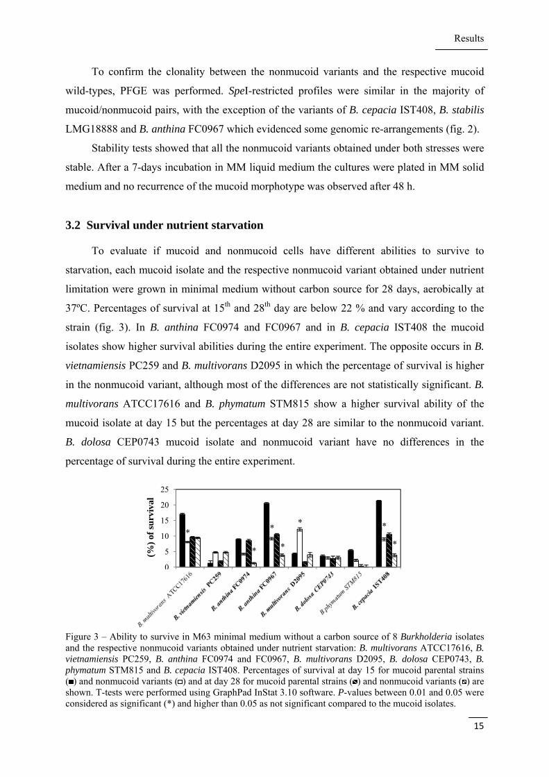
3.2 Survival under nutrient starvation
To evaluate if mucoid and nonmucoid cells have different abilities to survive to
starvation, each mucoid isolate and the respective nonmucoid variant obtained under nutrient
limitation were grown in minimal medium without carbon source for 28 days, aerobically at
37ºC. Percentages of survival at 15th and 28th day are below 22 % and vary according to the
strain (fig. 3). In B. anthina FC0974 and FC0967 and in B. cepacia IST408 the mucoid
isolates show higher survival abilities during the entire experiment. The opposite occurs in B.
vietnamiensis PC259 and B. multivorans D2095 in which the percentage of survival is higher
in the nonmucoid variant, although most of the differences are not statistically significant. B.
multivorans ATCC17616 and B. phymatum STM815 show a higher survival ability of the
mucoid isolate at day 15 but the percentages at day 28 are similar to the nonmucoid variant.
B. dolosa CEP0743 mucoid isolate and nonmucoid variant have no differences in the
percentage of survival during the entire experiment.
Figure 3 – Ability to survive in M63 minimal medium without a carbon source of 8 Burkholderia isolates and the respective nonmucoid variants obtained under nutrient starvation: B. multivorans ATCC17616, B. vietnamiensis PC259, B. anthina FC0974 and FC0967, B. multivorans D2095, B. dolosa CEP0743, B. phymatum STM815 and B. cepacia IST408. Percentages of survival at day 15 for mucoid parental strains ( ) and nonmucoid variants ( ) and at day 28 for mucoid parental strains ( ) and nonmucoid variants ( ) are shown. T-tests were performed using GraphPad InStat 3.10 software. P-values between 0.01 and 0.05 were considered as significant (*) and higher than 0.05 as not significant compared to the mucoid isolates.
0
5
10
15
20
25
(%)
of s
urv
ival
* *
*
*
*
*
*
*
Results
16
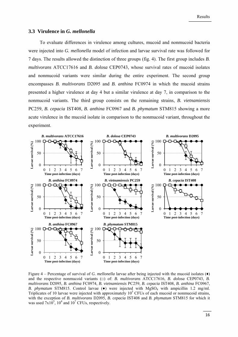
3.3 Virulence in G. mellonella
To evaluate differences in virulence among cultures, mucoid and nonmucoid bacteria
were injected into G. mellonella model of infection and larvae survival rate was followed for
7 days. The results allowed the distinction of three groups (fig. 4). The first group includes B.
multivorans ATCC17616 and B. dolosa CEP0743, whose survival rates of mucoid isolates
and nonmucoid variants were similar during the entire experiment. The second group
encompasses B. multivorans D2095 and B. anthina FC0974 in which the mucoid strains
presented a higher virulence at day 4 but a similar virulence at day 7, in comparison to the
nonmucoid variants. The third group consists on the remaining strains, B. vietnamiensis
PC259, B. cepacia IST408, B. anthina FC0967 and B. phymatum STM815 showing a more
acute virulence in the mucoid isolate in comparison to the nonmucoid variant, throughout the
experiment.
Figure 4 – Percentage of survival of G. mellonella larvae after being injected with the mucoid isolates (♦) and the respective nonmucoid variants (□) of: B. multivorans ATCC17616, B. dolosa CEP0743, B. multivorans D2095, B. anthina FC0974, B. vietnamiensis PC259, B. cepacia IST408, B. anthina FC0967, B. phymatum STM815. Control larvae (●) were injected with MgSO4 with ampicillin 1.2 mg/ml. Triplicates of 10 larvae were injected with approximately 105 CFUs of each mucoid or nonmucoid strains, with the exception of B. multivorans D2095, B. cepacia IST408 and B. phymatum STM815 for which it was used 7x10⁵, 104 and 107 CFUs, respectively.
0
50
100
0 1 2 3 4 5 6 70
50
100
0 1 2 3 4 5 6 70
50
100
0 1 2 3 4 5 6 7
0
50
100
0 1 2 3 4 5 6 70
50
100
0 1 2 3 4 5 6 70
50
100
0 1 2 3 4 5 6 7
0
50
100
0 1 2 3 4 5 6 70
50
100
0 1 2 3 4 5 6 7
B. multivorans ATCC17616 B. dolosa CEP0743 B. multivorans D2095
Lar
vae
surv
ival
(%
)
L
arva
e su
rviv
al (
%)
Lar
vae
surv
ival
(%
)
Time post-infection (days) Time post-infection (days) Time post-infection (days)
B. anthina FC0967 B. phymatum STM815
Time post-infection (days) Time post-infection (days) Time post-infection (days)
Time post-infection (days) Time post-infection (days)
Lar
vae
surv
ival
(%
)
L
arva
e su
rviv
al (
%)
Lar
vae
surv
ival
(%
)
Lar
vae
surv
ival
(%
)
L
arva
e su
rviv
al (
%)
B. anthina FC0974 B. vietnamiensis PC259 B. cepacia IST408
Results
17
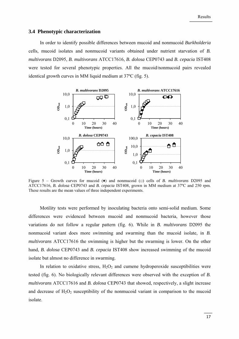
3.4 Phenotypic characterization
In order to identify possible differences between mucoid and nonmucoid Burkholderia
cells, mucoid isolates and nonmucoid variants obtained under nutrient starvation of B.
multivorans D2095, B. multivorans ATCC17616, B. dolosa CEP0743 and B. cepacia IST408
were tested for several phenotypic properties. All the mucoid/nonmucoid pairs revealed
identical growth curves in MM liquid medium at 37ºC (fig. 5).
Figure 5 – Growth curves for mucoid (♦) and nonmucoid (□) cells of B. multivorans D2095 and ATCC17616, B. dolosa CEP0743 and B. cepacia IST408, grown in MM medium at 37ºC and 250 rpm. These results are the mean values of three independent experiments.
Motility tests were performed by inoculating bacteria onto semi-solid medium. Some
differences were evidenced between mucoid and nonmucoid bacteria, however those
variations do not follow a regular pattern (fig. 6). While in B. multivorans D2095 the
nonmucoid variant does more swimming and swarming than the mucoid isolate, in B.
multivorans ATCC17616 the swimming is higher but the swarming is lower. On the other
hand, B. dolosa CEP0743 and B. cepacia IST408 show increased swimming of the mucoid
isolate but almost no difference in swarming.
In relation to oxidative stress, H2O2 and cumene hydroperoxide susceptibilities were
tested (fig. 6). No biologically relevant differences were observed with the exception of B.
multivorans ATCC17616 and B. dolosa CEP0743 that showed, respectively, a slight increase
and decrease of H2O2 susceptibility of the nonmucoid variant in comparison to the mucoid
isolate.
0,1
1,0
10,0
0 10 20 30 400,1
1,0
10,0
0 10 20 30 40
0,1
1,0
10,0
0 10 20 30 400,1
1,0
10,0
100,0
0 10 20 30 40
OD
640
O
D64
0
OD
640
O
D64
0
B. multivorans D2095 B. multivorans ATCC17616
B. dolosa CEP0743 B. cepacia IST408
Time (hours) Time (hours)
Time (hours) Time (hours)

Results
18
Figure 6 – Phenotypic comparison between mucoid isolates ( ) and nonmucoid variants ( ) of B. multivorans D2095 (A) and ATCC17616 (B), B. dolosa CEP0743 (C) and B. cepacia IST408 (D). Swimming and Swarming motilities were assayed in 0.3 % and 0.5 % agar plates, respectively, spotted with 5 µl bacterial cultures and incubated for 24h at 37ºC. H2O2 (30 %), Cumene hydroperoxide (10 %) and SDS (10 %) susceptibility were determined after a 24 h incubation at 37ºC by measuring the diameter of cell growth inhibition. Siderophores production was assessed by spotting CAS-MM plates with 5 µl bacterial cultures and measuring the yellow halo after 48 h of incubation at 37ºC. Biofilm production was determined after a 24 h incubation at 37ºC in S medium. Hydrophobicity of the cells was determined by the BATH method after an overnight incubation in MM liquid medium at 37ºC. These results are the mean values of three independent experiments. T-tests were performed using GraphPad InStat 3.10 software. It was considered P-values under 0.01 as very significant (**), between 0.01 and 0.05 as significant (*) and higher than 0.05 as not significant compared to the mucoid isolates.
To evaluate possible differences in membrane constitution, detergent (SDS)
susceptibility and LPS production were assessed. SDS susceptibility is higher in the
nonmucoid variant of B. multivorans D2095 than in the mucoid isolate. The other strains
showed no significant differences between mucoid and nonmucoid bacteria (fig. 6).
Nevertheless LPS band pattern remains similar in all the four strains tested (Annex II fig. A1).
The four pairs of mucoid/nonmucoid Burkholderia cells were also screened for
siderophores production using the CAS assay. By this method a blue-to-yellow color change
in the solid CAS plate is observed when the siderophores remove the iron from de Fe-CAS-
HDTMA complex in the medium. Siderophore excretion is indicated by the diameter of the
yellow halo around colonies. The results showed that in all the four strains the nonmucoid
variants produced a higher amount of siderophores in comparison to the mucoid wild-types.
0
35
70
0
18
35
0
35
70
0
15
30
45
0
15
30
0
15
30
0
4
8
-200
20406080
Swimming Swarming H2O2 susceptibility
CHP susceptibility SDS susceptibility Siderophores production
Biofilms Hydrophobicity
A B C D A B C D A B C D
A59
0
Gro
wth
inh
ibit
ion
(m
m)
Col
ony
dia
met
er (
mm
)
A B C D A B C D A B C D
(%)
of h
ydro
ph
obic
ity
Gro
wth
inh
ibit
ion
(m
m)
Col
ony
dia
met
er (
mm
)
Dia
met
er o
f th
e
h
alo
(mm
)
Gro
wth
inh
ibit
ion
(m
m)
A B C D A B C D
****
**
**
**
** ** *
** ** ****
****
****
** *
****
**

Results
19
However only B. multivorans D2095 and B. cepacia IST408 seemed to have biologically
significant differences (fig. 6).
The total biofilm formed by each strain was quantified after 24 h and 48 h of incubation
in S medium at 37ºC. The amount of biofilm varied between species and significant
differences were observed between mucoid and nonmucoid cells. In B. multivorans D2095
and B. dolosa CEP0743 the nonmucoid variants formed more biofilm than the mucoid isolates
at 24h (fig. 6). On the other hand B. multivorans ATCC17616 and B. cepacia IST408 seemed
to behave differently as the mucoid WT formed more biofilm than the nonmucoid derivative.
To understand if differences in biofilm formation were related to cell hydrophobicity,
cell surface hydrophobicity was determined using the Bacterial Adhesion to Hydrocarbon
(BATH) method. Mucoid isolates revealed to be more hydrophobic than the nonmucoid
variants in B. multivorans ATCC17616 and B. dolosa CEP0743 with differences of 17 and 47
%, respectively. The opposite occurs with B. cepacia IST408 in which the mucoid isolate
shows no hydrophobicity while the nonmucoid variant is almost 35 % hydrophobic. In B.
multivorans D2095 both mucoid and nonmucoid bacteria are very hydrophobic (around 70
%), with no relevant differences between them (fig. 6).
Figure 7 – Susceptibility to antibiotics of B. multivorans D2095 mucoid isolate ( ) and nonmucoid variant ( ), B. multivorans ATCC17616 mucoid isolate ( ) and nonmucoid variant ( ), B. dolosa CEP0743 mucoid isolate ( ) and nonmucoid variant ( ) and B. cepacia IST408 mucoid isolate ( ) and nonmucoid isolate ( ). Susceptibility to antibiotics was evaluated after a 24 h incubation at 37ºC by measuring the diameter of cell growth inhibition (Pip, piperacillin; Taz, tazobactam; Cip, ciprofloxacin; Cef, ceftazidime; Ami, amikacin). These results are the mean values of three independent experiments. T-tests were performed using GraphPad InStat 3.10 software. It was considered P-values under 0.01 as very significant (**), between 0.01 and 0.05 as significant (*) and higher than 0.05 as not significant compared to the mucoid isolates.
Variation in antimicrobials susceptibility was tested by disc diffusion assay. β-lactams
(ceftazidime, imipenem and aztreonam), an aminoglycoside (amikacin) and a quinolone
(ciprofloxacin) were tested and the results are shown in figure 7. In B. multivorans D2095 and
B. cepacia IST408 the nonmucoid variants were, in general, more susceptible than the mucoid
parental strains to almost all the antibiotics analysed. In fact, in the case of aztreonam the
mucoid isolate of B. multivorans D2095 was totally resistant to the antimicrobial while the
0
15
30
45
AN30 P75/T10 ATM30 CIP5 IMP10 CAZ30
Gro
wth
in
hib
itio
n (
mm
)
**
** ** ** **
** ** ** ** **
* *

Results
20
nonmucoid variant was highly sensitive. In contrast, B. multivorans ATCC17616 and B.
dolosa CEO0743 did not evidence major differences in the susceptibilities to any of the
antibiotics tested. Despite the differences between mucoid and nonmucoid cells of B.
multivorans ATCC17616 are statistically significant for ciprofloxacin resistance, they have
almost no impact in terms of biological meaning.
3.5 Expression profile of B. multivorans ATCC17616 mucoid isolate and
nonmucoid variant
To complement the phenotypic assays, a transcriptional study was performed. B.
multivorans ATCC17616 mucoid isolate and the respective nonmucoid variant obtained under
nutrient starvation were compared. For that study, a dual-species custom Burkholderia
microarray (Bcc1sa520656F) produced by Affymetrix was used. One of the species
represented in the array is B. multivorans ATCC17616, with gene coverage of 99.8 %. Both
mucoid and nonmucoid B. multivorans ATCC17616 were grown in SM medium until early
stationary phase, in which high-molecular-mass EPS is not yet recovered from the mucoid
strain liquid culture. Total RNA was extracted, converted to cDNA, biotin-labelled and
hybridized in triplicate to the array. Hybridization intensities from the nonmucoid variant
were compared to those of the mucoid isolate using dChip (≥1.3-fold-change lower
confidence bound with a resulting false discovery rate of 2.4 %). The results indicated a total
of 174 genes differentially expressed, of which 74 showed a significantly increased
expression and the remaining 100 a significantly decreased expression (See Annex II tables
A2 and A3).
All the genes were divided into clusters of orthologous groups (COGs) (fig. 8). Many of
the genes with increased expression were related to motility, while the genes with decreased
expression include genes related to type VI secretion system, inorganic ion and amino acid
transport and metabolism and cell wall, cell membrane and envelope biogenesis. Genes
related to transcription, signal transduction and intracellular traffic have either over-expressed
as under-expressed genes (table 4).

Results
21
Figure 8 – Functional distribution of COGs of genes with altered expression when comparing B. multivorans ATCC17616 nonmucoid variant with the respective mucoid parental isolate. A total of 174 genes with a statistically significant difference in expression were obtained by using a custom Affymetrix Burkholderia GeneChip.
Table 4 - Selection of a set of differentially expressed genes when comparing the transcriptome of B. multivorans ATCC17616 nonmucoid variant with that of the mucoid isolate, separated by functional groups.
Functional class Gene
identifier LB-FC*
Gene name
Description
Flagellar synthesis, motility, adhesion and chemotaxis
Bmul_0151 2,9 fliC flagellin domain protein
Bmul_0163 1,3 motB OmpA/MotB domain protein
Bmul_3009 1,3 flgL flagellar hook-associated protein 3
Bmul_3017 1,3 flgE flagellar basal body FlaE domain protein
Bmul_3018 1,3 flgD flagellar hook capping protein
Bmul_3019 1,5 flgC flagellar basal body rod protein FlgC
Bmul_3021 1,7 flgA flagellar basal body P-ring biosynthesis protein FlgA
Bmul_0166 1,3 cheW CheW protein
Bmul_3362 1,3 era methyl-accepting chemotaxis sensory transducer
Bmul_0042 1,3 fliN flagellar motor switch protein FliN
Bmul_1610 -1,9 fimC pili assembly chaperone
Bmul_1611 -1,4 fimD fimbrial biogenesis outer membrane usher protein
Bmul_1612 -2,8 fima fimbrial protein
Bmul_2818 1,5 pilB type II secretion system protein E
Virulence and pathogenesis
Bmul_3709 -2,4 adh hemolysin-type calcium-binding region
Bmul_2923 -1,3 bcsE type VI secretion-associated protein, ImpA family
Bmul_2927 -1,3 bcsI type VI secretion system lysozyme-related protein
Bmul_2928 -1,4 bcsJ type VI secretion system effector, Hcp1 family
Bmul_2929 -1,5 bcsK type VI secretion protein, EvpB/VC_A0108 family
Bmul_2930 -1,9 bcsL type VI secretion protein, VC_A0107 family
Bmul_5730 -1,4 - type VI secretion system Vgr family protein
0 5 10 15
Function unknown (S)General function prediction only (R)
Secondary metabolites biosynthesis and transport (Q)Inorganic ion transport and metabolism (P)
Lipid transport and metabolism (I)Coenzyme transport and metabolism (H)Nucleotide transport and metabolism (F)
Amino acid transport and metabolism (E)Carbohydrate transport and metabolism (G)
Energy production and conversion (C)Posttranslational modification, protein turnover (O)
Intracellular traffic (U)Cell motility (N)
Cell wall, membrane, envelope biogenesis (M)Signal transduction (T)
Defense mechanisms (V)Cell division (D)
Replication, recombination, and repair (L)Transcription (K)
Translation, ribossomal structure and biogenesis (J)
Increased expressionDecreased expression

Results
22
Cell Wall / membrane
Bmul_2607 -1,4 - ABC transporter related
Bmul_2608 -1,4 - ABC-2 type transporter
Bmul_0320 1,4 vacJ VacJ family lipoprotein
Bmul_1006 1,3 dgk diacylglycerol kinase
Bmul_1318 -1,4 mscL large conductance mechanosensitive channel protein
Bmul_4401 -1,3 - glycosyl transferase group 1
Bmul_5944 -6,6 - methyltransferase type 12
Bmul_5945 -4,8 - methyltransferase type 12
Transcription regulators
Bmul_1606 -1,7 orbS RNA polymerase, sigma-24 subunit, ECF subfamily
Bmul_1761 1,3 - transcriptional regulator, LysR family
Bmul_2098 1,3 - transcriptional regulator, BadM/Rrf2 family
Bmul_2372 1,5 - transcriptional regulator, GntR family
Bmul_2393 -2,7 cspA cold-shock DNA-binding domain protein
Bmul_2442 1,4 nusB transcription antitermination protein NusB
Bmul_2557 -2,3 - transcriptional regulator, LysR family
Bmul_2962 -1,3 - transcriptional regulator, LysR family
Bmul_4295 -1,3 - LysR family transcriptional regulator
Bmul_4936 -1,3 - transcriptional regulator, AraC family
Signal transdution Bmul_3051 1,5 - putative signal transduction histidine kinase
Bmul_3471 -1,4 - diguanylate phosphodiesterase
Bmul_4254 1,3 - UspA domain protein
Bmul_4389 -1,7 - putative transcriptional regulator, Crp/Fnr family
Bmul_4947 -1,5 - response regulator receiver sensor signal transduction histidine kinase
Bmul_5122 -1,5 - diguanylate cyclase/phosphodiesterase
Bmul_3737 -1,3 - transcriptional regulator, LuxR family
Bmul_5766 -1,3 - two component transcriptional regulator, LuxR family
Bmul_0164 1,4 cheY response regulator receiver protein
Energy production and conversion
Bmul_2649 -1,3 hmpA oxidoreductase FAD/NAD(P)-binding domain protein
Bmul_4546 1,3 coxS (2Fe-2S)-binding domain protein
Bmul_5248 1,4 Ndh FAD-dependent pyridine nucleotide-disulphide oxidoreductase
Bmul_5760 -1,3 - cytochrome c class I
Bmul_5761 -1,3 coxA cytochrome c oxidase subunit I
Bmul_5907 1,3 - cytochrome c, class I
Bmul_5639 -1,4 - alcohol dehydrogenase zinc-binding domain protein
Amino acid transport and metabolism
Bmul_0879 -1,5 glnB nitrogen regulatory protein P-II
Bmul_2990 -1,3 potD extracellular solute-binding protein
Bmul_3036 1,4 dppF dipeptide transporter ATP-binding subunit
Bmul_3701 -1,4 ldcC lysine/arginine decarboxylase
Bmul_3702 -1,3 adiC arginine:agmatin antiporter
Bmul_3719 1,27 pub FAD dependent oxidoreductase
Bmul_4502 -1,4 - ABC transporter related
Bmul_5003 -1,3 - polar amino acid ABC transporter, inner membrane subunit
Inorganic ion transport and metabolism
Bmul_2003 -2,1 katE catalase
Bmul_2016 1,3 phoU phosphate uptake regulator, PhoU
Bmul_3335 -1,3 - transport system permease
Bmul_3336 -1,3 - periplasmic binding protein
Bmul_3337 -1,3 hmuS haemin-degrading family protein
Bmul_3423 1,4 - 2-aminoethylphosphonate ABC transporter/binding protein
Bmul_3736 -1,3 - natural resistance-associated macrophage protein
Bmul_4079 -2,4 mgtA magnesium-translocating P-type ATPase
Bmul_5946 -3,0 - rieske (2Fe-2S) domain protein
Bmul_5949 -1,4 - rieske (2Fe-2S) domain protein
*LB-FC, lower bound of fold-change;

Results
23
In order to validate the data obtained by microarray analysis, the expression of some
genes related to virulence, motility, transcriptional regulation and signal transduction was
analysed by qRT-PCR. Although most of the genes have higher fold-changes when analysed
by qRT-PCR, the results are in agreement with microarray (table 5).
Table 5 - Quantitative real-time RT-PCR analysis performed in B. multivorans ATCC17616 nonmucoid variant in comparison to the mucoid isolate. Data are normalized to proC gene levels.
Gene Microarray lower
bound of fold-change Real-time fold-
change ± sd
Bmul_2929 (bcsK) -1.5 -5.0 ± 1.0
Bmul_3021 (flgA) 1.7 4.2 ± 1.4
Bmul_4936 -1.3 -1.5 ± 0.4
Bmul_4389 -1.7 -10.1 ± 3.8
Bmul_5122 -1.5 -11.6 ± 3.0
The over-expressed motility genes fliC, fliN, flgA/C/D/E/L are mostly involved in
flagellar assembly. This possibly indicates that the nonmucoid variant is more mobile than the
mucoid isolate. Also chemotaxis genes similar to cheW and aer found in other bacteria
showed increased expression in the nonmucoid variant, possibly helping bacteria to find
places with more oxygen and carbon sources. In opposition, some fim genes related to the
synthesis of the pili were under-expressed. These genes are involved in adhesion of the cells
to surfaces which contributes to the idea that the nonmucoid cells are less adherent and,
consequently, more mobile than the mucoid cells. These results agree with the phenotypic
results showed earlier, which evidenced a higher swimming ability and a lower biofilm
formation of B. multivorans ATCC17616 nonmucoid variant.
On the other hand, 7 genes related to type VI secretion system had a decreased
expression in the nonmucoid variant. This secretion system is believed to play a specific role
in the maintenance of Burkholderia cells in an eukaryotic host and to help them defending
against other bacteria in the environment (67).
In relation to energy and intermediary metabolism, it was observed that several genes
involved in inorganic ion and amino acid transport and metabolism were under-expressed in
the nonmucoid variant in comparison with the mucoid isolate. Most of these genes encode for
binding proteins and transporters, however, no particular metabolic pathway was affected.
Genes related to energy production and conversion had differences in expression but did not
evidence a tendency. Genes like Bmul_2649 and Bmul_5639 were under-expressed in the
nonmucoid variant and encode a protein highly homologue to nitric oxide dioxygenase

Results
24
(hmpA) and an alcohol dehydrogenase zinc-binding domain protein, respectively. Genes like
Bmul_4546 and Bmul_5248 were over-expressed and encode a putative aerobic-type carbon
monoxide dehydrogenase (coxS) and a FAD-dependent pyridine nucleotide-disulphide
oxidoreductase, respectively (table 3). These proteins enable bacteria to use different
compounds as inorganic sources of energy. Differences in expression can indicate that
bacteria are able to regulate their metabolism according to their needs and to the
environmental conditions. In addition, the gene encoding catalase, responsible for the
decomposition of H2O2 to water and oxygen, was under-expressed in the nonmucoid variant.
H2O2, nitric oxide and carbon monoxide are three harmful compounds to the cell and the
action of enzymes that use or degrade them is essential to the maintenance of bacterial cells.
In fact, the higher susceptibility of the nonmucoid variant of B. multivorans ATCC17616 to
H2O2 (fig. 6) can be due to this decreased expression of catalase.
It was observed a decrease in expression in a number of genes involved in cell wall, cell
membrane and envelope biogenesis in the nonmucoid variant in comparison with the mucoid
isolate. These genes include two ABC-transporters, two methyltransferases, one glycosyl
transferase and one channel protein sensitive to pressure or hypoosmotic shock. Contrariwise,
genes Bmul_0320 and Bmul_1006 encoding a VacJ family lipoprotein and a diacylglycerol
kinase, respectively, were over-expressed.
In the categories of transcriptional regulators and signal transduction it was observed an
increased expression of the universal stress protein family UspA, which helps bacteria to
survive during the exposure to stress agents. Also members of signal transduction pathways
like the histidine kinase Bmul_4947 and elements of Crp/Fnr and LuxR families were under-
expressed in the nonmucoid variant, as well as two diguanylate phosphodiesterases, Bmul_
3471 and Bmul_5122. These two last proteins are probably related to the degradation of the
signal molecule cyclic di-GMP and they both have regions with high similarity to EAL
domains which may be involved in the regulation of bacterial adhesion.
It was observed an over-expression of genes belonging to the families of transcriptional
regulators LysR, BadM/Rrf2 and GntR and of the transcription antitermination protein NusB,
involved in the regulation of rRNA biosynthesis. Under-expressed genes encompass
transcriptional regulators belonging to the families LysR and AraC, the RNA polimerase
sigma-70 factor and a cold-shock DNA-binding domain protein (CSP). Despite CSP has a
basal level of expression under normal conditions, it is increased when an abrupt drop in
growth temperature occurs. Although its function is not totally understood, it is thought to
regulate mRNA degradation, rRNA translation and the rate of transcription termination (68).

Results
25
One gene encoding a transcriptional regulator significantly under-expressed was
Bmul_2557, belonging to LysR-family of transcriptional regulators. This family encompasses
a wide number of highly conserved LysR-type transcriptional regulators (LTTRs) that can be
involved in cellular processes such as amino acid biosynthesis, CO2 fixation, antibiotic
resistance, degradation of aromatic compounds, nodule formation of nitrogen-fixing bacteria,
and synthesis of virulence factors (69).
3.6 Molecular cloning
One of the genes whose expression was decreased in the nonmucoid variant of B.
multivorans ATCC17616 was the gene Bmul_2557. As referred above, Bmul_2557 gene
encodes for a LysR-family transcriptional regulator possibly involved in a wide variety of
metabolic processes. Gene expression of Bmul_2557 was compared between nonmucoid
variants and the respective mucoid isolates of B. multivorans ATC17616 and D2095, B.
cepacia IST408, B. anthina FC0967 and B. vietnamiensis PC259. In all of these strains the
LysR-family transcriptional regulator was under-expressed in the nonmucoid variant (fig. 9).
Figure 9 - Quantitative real-time RT-PCR analysis of transcript levels of the gene Bmul_2557 in the B. multivorans ATC17616 and D2095, B. cepacia IST408, B. anthina FC0967 and B. vietnamiensis PC259 nonmucoid variants in comparison to the mucoid isolates. Data are normalized to trpB gene levels.
To evaluate the importance of this gene in the mucoid-to-nonmucoid conversion and in
bacteria virulence a strategy to knock-out the gene was initiated (fig. 10). The upstream and
downstream regions of the gene were cloned into the pBCKS- cloning vector and a
trimethoprim (Tp) cassette was inserted between them. The resulting plasmid, pAT812-Tp,
will then be introduced into B. multivorans ATCC17616 and a double recombination with its
-30
-25
-20
-15
-10
-5
0
5
Fol
d-i
ncr
ease
in B
mu
l_25
57
expr
essi
on

Results
26
genome will allow the identification of both wild-type and mutant cells in which the gene
Bmul_2557 will be replaced by the Tp cassette.
Figure 10 – Strategy to eliminate Bmul_2557 of B. multivorans ATCC17616 genome by replacing it by a trimethoprim (Tp) cassette. The stages of the cloning strategy which have already been concluded are represented in darker color, while the following steps are lighter coloured. lacZ – gene coding for β-galactosidase; Cm – gene for chloramphenicol resistance. Amp – gene for ampicillin resistance. Km – gene for kanamycin resistance. TP – gene for trimethoprim resistance. Upstream (L) and downstream (R) regions were cloned into pBCKS- and pDrive vectors, respectively, resulting in pAT312 and pAT712. The R region was then cloned into pAT312. A TP cassette was inserted between L and R originating pAT812-Tp. This final vector will be introduced into B. multivorans ATCC17616 and recombined with a genomic region.

Discussion
27
4. Discussion
Mucoid-to-nonmucoid transition is a very common phenomenon during bacterial
infections and has already been described for different Burkholderia species, including B.
cenocepacia, B. multivorans and B. vietnamiensis (41). Two clonal Burkholderia multivorans
strains, sequentially isolated from a chronically infected CF patient have been previously
characterized in our laboratory (43). These isolates represent a morphotype variation since the
first, D2095, is mucoid and the second, D2214, isolated 6 months later, is nonmucoid.
Differences in the expression of virulence factors, virulence in G. mellonella model of
infection and ability to survive under nutrient limitation were observed (43). Further studies
indicated that morphotype variation did never occur when the strains were grown under
standard conditions (rich medium, aerobically at 37ºC for 7 days). However, mucoid-to-
nonmucoid conversion was observed when D2095 was submitted to some antimicrobials,
nutrient starvation, high or low temperatures, high osmolarity and oxidative stress agents (44).
Some of these stress conditions, as well as iron limitation, anaerobic conditions and growth
within human macrophages can also trigger morphotype alteration in B. pseudomallei (70,
71).
In this study we wanted to understand if stress conditions could also trigger mucoid-to-
nonmucoid variation in other Bcc and non-Bcc species both from clinical and environmental
source. A total of sixteen mucoid strains were tested and the results showed that eight (50 %)
of them gave rise to nonmucoid variants after being incubated for 28 days under nutrient
limitation (fig. 1). These strains include clinical and environmental strains and two non-Bcc
strains, which may indicate that the response to this stress is a common feature of the
Burkholderia genus. In the presence of ciprofloxacin five (30 %) strains had originated
nonmucoid variants (fig. 1). Contrary to nutrient starvation, in the presence of ciprofloxacin
all the strains that originated nonmucoid variants were clinical Bcc strains.
These results contribute to the idea that Burkholderia strains are able to respond to
stresses and to adapt to different growth conditions according to their ecological niche. This
process can occur through specific alterations of pathogenicity and phenotypic traits during
the adaptation of clinical isolates to environment and vice-versa (72–74). Also, Vial and co-
workers have shown that phase variation helps the clinical isolate B. ambifaria HSJ1 to
colonize different ecological niches, as lungs or rizosphere, by changing the type and amount
of virulence factors that are produced (75).
Discussion
28
Normal airway mucus is mainly composed of mucin glycoproteins (76). The sputum of
CF patients’ lungs is characterized by a significant increase in the amount of these
macromolecules as well as by the presence of other large nonmucin polymers such as DNA,
filamentous actin, proteoglycans, biofilms and polypeptides released from inflammatory cells
(reviewed in (77)). The nutrient composition of the sputum is known to influence several
bacterial functions such as the production of virulence factors, motility, biofilm production,
and virulence (reviewed in (78)). In fact, when CF sputum is used as the unique carbon source
for the growth of P. aeruginosa the cells evidence major differences in their physiology,
including the use of amino acids as a carbon source (78). To compare the ability of mucoid
isolates and nonmucoid variants to survive nutrient limitation, the strains were incubated in
minimal medium for 28 days (fig. 3). After 15 days of incubation in three of the eight strains
tested the nonmucoid variant suffered a significantly higher decrease in survival, what seem
to indicate that the mucoid isolates were better adapted to starvation conditions. The opposite
occurs in B. multivorans D2095 in which the nonmucoid variant had a higher survival
capacity. However, after 28 days of incubation, the percentages of survival experienced a
decrease to less than 11 % and the observed differences were not biologically relevant. Since
EPS production is an energy- and carbon-consuming process (79, 80) nonmucoid variants
should save that energy and resources to other metabolic processes. Therefore it would be
expected a higher survival level in starvation conditions in comparison with the mucoid
parental strains.
Virulence assays in G. mellonella suggested that the majority of the nonmucoid variants
were less virulent at day-4 of infection. However, at day-7 half of the strains tested presented
a similar larvae mortality for the mucoid and nonmucoid cells (fig. 4). This results are in
agreement with our previous study which have revealed that the clinical mucoid isolate B.
multivorans D2095 was more virulent than its sequential nonmucoid isolate D2214 (43).
Also, studies performed with mucoid isolates and spontaneous nonmucoid variants of B.
ambifaria evidenced that the second was, in general, less virulent than the first using G.
mellonella as the model of infection. On the other hand strains of B. cenocepacia, the most
virulent Bcc specie, are mainly nonmucoid and it was shown that some virulence factors seem
to be over-expressed in the nonmucoid isolates when compared to the mucoid isolates (41,
81). Besides, data modeling showed that, in general, the intensity of mucoidy is inversely
correlated with the rate of decline in pulmonary function (42). Therefore, the relation between
mucoid and nonmucoid cells’ virulence in G. mellonella appears to be strain dependent.

Discussion
29
Burkholderia species express a wide variety of virulence factors that may be directly or
indirectly related to its virulence. To try to understand the virulence results in G. mellonella, a
phenotypic characterization was performed of four Bcc strains from three different species
commonly isolated from CF patients’ lungs. The results are summarized in table 6.
Table 6 – Phenotypic comparison of nonmucoid variants vs. mucoid isolates of Burkholderia species.
(1) comparison of resistance; (2) the first line is for cumene hidroperoxide and the second for H2O2.
Some of the phenotypic traits remained similar between mucoid and nonmucoid cells of
all strains, including growth rate and LPS pattern. Others like survival in starvation conditions
and resistance to detergents and to oxidative agents had differences in only one or two strains
(table 6).
Our main goal was to understand which characteristics could be related to mucoid-to-
nonmucoid variation and, consequently, would always be altered between the two types of
cells. Although the variations did not evidence a regular tendency in all of the four strains, the
results showed that swimming motility and biofilm formation were always altered between
mucoid and nonmucoid cells (table 6). Swimming motility is important in the dissemination
of bacteria through the organism and it is closely related to the production of flagella, a
virulence factor common to all Bcc species (23). Bcc species can express one or two types of
flagellin that are distinguished by size and RFLP patterns of fliC gene (82). Microarrays have
shown that many genes involved in flagellar assembly are over-expressed in the nonmucoid
variant of B. multivorans ATCC17616 which agrees with its higher swimming motility. Some
studies have shown that flagellin mutants had pronounced deficiencies in invasion of human
lung cells, and a highly decreased capacity to severely injure mice (23, 83). Thereby the
nonmucoid variants of both B. multivorans strains and the mucoid isolates of B. dolosa
CEP0743 and B. cepacia IST408 may be more able to invade human lung cells.

Discussion
30
Flagella are also related to the adhesion of bacterial cells to the host and to the
formation of biofilms (84). Biofilms can be produced by many Bcc species colonizing the
lungs of CF patients, suggesting that its production may be related to the protection from host
antimicrobial defenses (85). The role of biofilms in antimicrobials resistance is controversial
as some studies show that biofilms increase its resistance (86, 87) while others, including this
work, do not evidence any correlation (88). The expression profile analysis showed a decrease
in genes related to the synthesis of pili, also involved in the adhesion of bacterial cells to the
surfaces, in the nonmucoid variants of B. multivorans ATCC17616. This fact contributes to
the idea that these nonmucoid variants are more mobile and less adherent to the host cells. It
would be interesting to evaluate differences in adhesion and invasion of human tissues
between all the mucoid isolates and respective nonmucoid variants.
Siderophores production is also different in three of the four strains tested (table 6). Iron
is a vital element for the bacterial growth since it is an essential co-factor of important
enzymes involved in primary and secondary metabolic pathways. Inside the human host iron
is usually tightly bound to proteins like lactoferrin, transferrin, ferritin and hemoproteins
leaving only a small amount of free serum iron (89). For this reason bacterial pathogens have
to develop mechanisms to acquire iron. One method is the production of siderophores which
are small-molecule compounds (generally <1 kDa) with high-affinity for ferric iron. Members
of Bcc bacteria can produce up to four different siderophores including ornibactin, pyochelin,
cepabactin and cepaciachelin which are thought to be essential for the expression of full
pathogenicity in several hosts namely Caenorhabditis elegans and G. mellonella (24, 90). The
results show that that, in general, the nonmucoid variants produce more siderophores than the
mucoid isolates, possibly indicating its higher ability to acquire iron from the medium. This
increase could lead to a higher activity of iron-dependent enzymes with impact on metabolic
pathways, although significant alterations were not observed in microarray analysis. On the
other hand, some of the results of this work fit the expected results for the absence of the fur
gene demonstrated by Yuhara and co-workers (2008) in B. multivorans ATCC17616,
including a higher production of siderophores, a higher sensitivity to reactive oxygen species
like H2O2 and a decreased expression of catalase and of an enzyme homologue to nitric oxide
dehidrogenase (91). However, there were no significant differences in the expression of the
fur gene and no conclusions can be taken. Moreover, this increase in siderophores production
could lead to an excess of iron that would inhibit enzyme activity and become toxic to the cell
(92). Nevertheless as no differences in growth were observed the major metabolic processes
do not seem to be altered and this hypothesis is not probable to happen.
Discussion
31
Relatively to the susceptibility to antimicrobials only two of the four strains have
evidenced differences between mucoid and nonmucoid cells (table 6). However, when those
differences exist, the nonmucoid variants are always more susceptible than the parental
mucoid isolates. These results are in agreement with our previous study in which the
nonmucoid isolate B. multivorans D2214 is more susceptible to antibiotics than its clonal
mucoid isolate D2095 (43). Due to antibiotic therapies administered to CF patients it would
be expected a higher resistance of the nonmucoid variants. One hypothesis is that the EPS
produced by the mucoid bacteria could be capable of protecting the cell against the action of
several antibiotics.
As these results showed that morphotype alteration is possibly related to different
phenotypic properties we hypothesized that some other alteration, possibly in a regulatory
protein, could be responsible for this morphotype variation. To evaluate that possibility, a
molecular strategy to knock-out the gene Bmul_2557 was initiated. Bmul_2557 encodes a
LysR-family transcriptional regulator that is under-expressed in the nonmucoid variants of B.
multivorans ATCC17616, B. multivorans D2095, B. cepacia IST408, B. anthina FC0967 and
B. vietnamiensis PC259 (fig. 9). Future studies include the evaluation of the role of gene
Bmul_2557 in the cell and its importance on the regulation of mucoid-to-nonmucoid
variation.
In conclusion, this study confirms that morphotype variation can occur in a wide range
of Burkholderia species, belonging or not to the Bcc, by exposing the cells to different
laboratory stress conditions. This variation appears to be related to the original ecological
niche of bacteria. Also, it has shown that mucoid-to-nonmucoid alteration is a complex
process that seems to relate to other phenotypic traits as motilility, biofilm formation and, in a
lesser extent, to the production of siderophores. Further studies are needed to identify the
mechanisms underlying this process and to understand the relationship between the multiple
cellular properties.

References
32
6. References
1. Mahenthiralingam E, Baldwin A, and Dowson CG. 2008. Burkholderia cepacia complex
bacteria : opportunistic pathogens with important natural biology. J. Appl. Microbiol. 104(6):1539–51.
2. Loutet SA and Valvano MA. 2010. A decade of Burkholderia cenocepacia virulence determinant research. Infect. Immun. 78(10):4088–100.
3. Burkholder WH. 1950. Sour skin, a bacterial rot of onion bulbs. Phytopathology 40(1):115–7 . 4. Coenye T and Vandamme P. 2007. Burkholderia: Molecular Microbiology and Genomics. 350.
UK: Horizon Scientific Press. 5. Gautam V, Singhal L, and Ray P. 2011. Burkholderia cepacia complex: Beyond pseudomonas
and acinetobacter. Indian J. Med. Microbiol. 29(1):4–12. 6. Coenye T and Vandamme P. 2003. Diversity and significance of Burkholderia species occupying
diverse ecological niches. Environ. Microbiol. 5(9):719–29. 7. Whitlock GC, Estes DM, and Torres AG. 2007. Glanders : off to the races with Burkholderia
mallei. FEMS Microbiol. Lett. 277(2):115–22. 8. Haase A, Janzen J, Barrett S, and Currie B. 1997. Toxin production by Burkholderia
pseudomallei strains and correlation with severity of melioidosis. J. Med. Microbiol. 46(7):557–63.
9. Centers for Disease Control and Prevention USA. http://www.cdc.gov/. 10. Taplin D, Bassett DC, and Mertz PM. 1971. Foot lesions associated with Pseudomonas cepacia.
Lancet 2(7724):568–71. 11. Cystic Fibrosis Foundation - Home. http://www.cff.org/. 12. Kreindler JL. 2010. Cystic fibrosis: Exploiting its genetic basis in the hunt for new therapies.
Pharmacol. Ther. 125(2):219–29. 13. Yankaskas JR, Marshall BC, Sufian B, Simon RH, and Rodman D. 2004. Cystic fibrosis adult
care: consensus conference report. Chest 125(1 Suppl):1S–39S. 14. Callaghan M and McClean S. 2012. Bacterial host interactions in cystic fibrosis. Curr. Opin.
Microbiol. 15(1):71–7. 15. Jones AM, Dodd ME, and Webb AK. 2001. Burkholderia cepacia: current clinical issues,
environmental controversies and ethical dilemmas. Eur. Repir. J. 17(2):295–301. 16. Johnson WM, Tyler SD, and Rozee KR. 1994. Linkage analysis of geographic and clinical
clusters in Pseudomonas cepacia infections by multilocus enzyme electrophoresis and ribotyping. J. Clin. Microbiol. 32(4):924–30.
17. Mahenthiralingam E, Campbell ME, and Henry DA. 1996. Epidemiology of Burkholderia cepacia infection in patients with cystic fibrosis : Analysis by randomly amplified polymorphic DNA fingerprinting. J. Clin. Microbiol. 34(12):2914–20.
18. Nzula S, Vandamme P, and Govan JRW. 2002. Influence of taxonomic status on the in vitro antimicrobial susceptibility of the Burkholderia cepacia complex. J. Antimicrob. Chemother. 50(2):265–9.
19. Drevinek P and Mahenthiralingam E. 2010. Burkholderia cenocepacia in cystic fibrosis: epidemiology and molecular mechanisms of virulence. Clin. Microbiol. Infect. 16(7):821–30.
20. Ferreira AS, Silva IN, and Moreira LM. 2011. Mechanisms controlling the expression of the exopolysaccharide of Burkholderia and role in niche adaptation. In Biotechnology of Biopolymers, ed. M Elnashar, pp. chapter 8 147–64.
21. McClean S and Callaghan M. 2009. Burkholderia cepacia complex: epithelial cell-pathogen confrontations and potential for therapeutic intervention. J. Med. Microbiol. 58(Pt 1):1–12.
22. Chung JW and Speert DP. 2007. Proteomic identification and characterization of bacterial factors associated with Burkholderia cenocepacia survival in a murine host. Microbiology 153(Pt 1):206–14.
23. Urban TA, Griffith A, Torok AM, Mark E, Burns JL, Goldberg JB, and Smolkin ME. 2004. Contribution of Burkholderia cenocepacia flagella to infectivity and inflammation. Infect. Immun. 72(9):5126–34.
References
33
24. Uehlinger S, Schwager S, Bernier SP, Riedel K, Nguyen DT, Sokol P a, and Eberl L. 2009. Identification of specific and universal virulence factors in Burkholderia cenocepacia strains by using multiple infection hosts. Infect. Immun. 77(9):4102–10.
25. Mil-Homens D and Fialho AM. 2011. Trimeric autotransporter adhesins in members of the Burkholderia cepacia complex: a multifunctional family of proteins implicated in virulence. Front. Cell Infect. Microbiol. 1(13):1–10.
26. Fauré R, Shiao TC, Lagnoux D, Giguère D, and Roy R. 2007. En route to a carbohydrate-based vaccine against Burkholderia cepacia. Org. Biomol. Chem. 5(16):2704–8.
27. Bedini E, Cirillo L, and Parrilli M. 2012. Synthesis of the trisaccharide outer core fragment of Burkholderia cepacia pv. vietnamiensis lipooligosaccharide. Carbohydr. Res. 349:24–32. Elsevier Ltd.
28. Herasimenka Y, Cescutti P, Sampaio Noguera CE, Ruggiero JR, Urbani R, Impallomeni G, Zanetti F, Campidelli S, Prato M, and Rizzo R. 2008. Macromolecular properties of cepacian in water and in dimethylsulfoxide. Carbohydr. Res. 343(1):81–9.
29. Ferreira AS, Leitao JH, Silva IN, Pinheiro PF, Sousa SA, Ramos CG, and Moreira LM. 2010. Distribution of cepacian biosynthesis genes among environmental and clinical Burkholderia strains and role of cepacian exopolysaccharide in resistance to stress conditions. Appl Environ Microbiol 76(2):441–450.
30. Moreira LM, Videira P a, Sousa S a, Leitão JH, Cunha MV, and Sá-Correia I. 2003. Identification and physical organization of the gene cluster involved in the biosynthesis of Burkholderia cepacia complex exopolysaccharide. Biochem. Biophys. Res. Commun. 312(2):323–33.
31. Sist P, Cescutti P, Skerlavaj S, Urbani R, Leitão JH, Sá-Correia I, and Rizzo R. 2003. Macromolecular and solution properties of Cepacian: the exopolysaccharide produced by a strain of Burkholderia cepacia isolated from a cystic fibrosis patient. Carbohydr. Res. 338(18):1861–67.
32. Kumar A and Mody K. 2009. Microbial exopolysacharides: variety and potential applications. In Microbial Production of Biopolymers and Polymer Precursors: Applications and Perspectives, ed. BHA Rehm, pp. 229–54. UK: Caister Academis Press.
33. Wilkinson JF. 1958. The extracellular polysaccharides of bacteria. Bacteriol. Rev. 22(1):46–73. 34. Herasimenka Y, Benincasa M, Mattiuzzo M, Cescutti P, Gennaro R, and Rizzo R. 2005.
Interaction of antimicrobial peptides with bacterial polysaccharides from lung pathogens. Peptides 26(7):1127–32.
35. Lebeer S, Claes IJJ, Verhoeven TL a, Vanderleyden J, and De Keersmaecker SCJ. 2011. Exopolysaccharides of Lactobacillus rhamnosus GG form a protective shield against innate immune factors in the intestine. Microb. Biotechnol. 4(3):368–74.
36. Conway B-AD, Chu KK, Bylund J, Altman E, and Speert DP. 2004. Production of exopolysaccharide by Burkholderia cenocepacia results in altered cell-surface interactions and altered bacterial clearance in mice. J. Infect. Dis. 190(5):957–66.
37. Bylund J, Burgess L-A, Cescutti P, Ernst RK, and Speert DP. 2006. Exopolysaccharides from Burkholderia cenocepacia inhibit neutrophil chemotaxis and scavenge reactive oxygen species. J. Biol. Chem. 281(5):2526–32.
38. Ferreira AS, Leitão JH, Sousa S a, Cosme AM, Sá-Correia I, and Moreira LM. 2007. Functional analysis of Burkholderia cepacia genes bceD and bceF, encoding a phosphotyrosine phosphatase and a tyrosine autokinase, respectively: role in exopolysaccharide biosynthesis and biofilm formation. Appl. Environ. Microbiol. 73(2):524–34.
39. Deitsch KW and Moxon ER. 1997. Shared themes of antigenic variation and virulence in bacterial, protozoal, and fungal infections. Microbiol. Mol. Biol. Rev. 61(3):281–93.
40. Ramsey DM and Wozniak DJ. 2005. Understanding the control of Pseudomonas aeruginosa alginate synthesis and the prospects for management of chronic infections in cystic fibrosis. Mol. Microbiol. 56(2):309–22.
41. Zlosnik JE, Hird TJ, Fraenkel MC, Moreira LM, Henry DA, and Speert DP. 2008. Differential mucoid exopolysaccharide production by members of the Burkholderia cepacia complex. J. Clin. Microbiol. 46(4):1470–3.

References
34
42. Zlosnik JE a, Costa PS, Brant R, Mori PYB, Hird TJ, Fraenkel MC, Wilcox PG, Davidson a GF, and Speert DP. 2011. Mucoid and nonmucoid Burkholderia cepacia complex bacteria in cystic fibrosis infections. Am. J. Respir. Crit. Care. Med. 183(1):67–72.
43. Silva IN, Ferreira AS, Becker JD, Zlosnik JE a, Speert DP, He J, Mil-Homens D, and Moreira LM. 2011. Mucoid morphotype variation of Burkholderia multivorans during chronic cystic fibrosis lung infection is correlated with changes in metabolism, motility, biofilm formation and virulence. Microbiology 157(Pt 11):3124–37.
44. Silva IN, Tavares AC, Ferreira AS, and Moreira L. Stress conditions triggering the emergence of mucoid morphotype variation in Burkholderia and role in virulence. Manuscript submitted for publication.:
45. Sambrook J and Russel DW. 2001. Molecular Cloning: a laboratory manual. NY: Cold Spring Harbor Laboratory.
46. Moreira LM, Da MS, and Sá-correia I. 1997. Comparative genomic analysis of isolates belonging to the six species of the genus Thermus using pulsed-field gel electrophoresis and ribotyping. Arch. Microbiol. 168(2):92–101.
47. Vandamme P, Holmes B, Vancanneyt M, Coenye T, Hoste B, Coopman R, Revets H, et al. 1997. Occurrence of multiple genomovars of Burkholderia cepacia in cystic fibrosis patients and proposal of Burkholderia multivorans sp. nov. Int. J. Syst. Bacteriol. 47(4):1188–200.
48. Richau J a, Leitão JH, Correia M, Lito L, Salgado MJ, Barreto C, Cescutti P, and Sá-Correia I. 2000. Molecular typing and exopolysaccharide biosynthesis of Burkholderia cepacia isolates from a Portuguese cystic fibrosis center. J. Clin. Microbiol. 38(4):1651–5.
49. Stanier RY, Palleroni NJ, and Doudoroff M. 1966. The aerobic pseudomonads: a taxonomic study. J. Gen. Microbiol. 43(2):159–271.
50. Glass S and Govan JR. 1986. Pseudomonas cepacia-fatal pulmonary infection in a patient with cystic fibrosis. J. Infect. 13(2):157–8.
51. Coenye T, Schouls LM, Govan JR, Kersters K, and Vandamme P. 1999. Identification of Burkholderia species and genomovars from cystic fibrosis patients by AFLP fingerprinting. Int. J. Syst. Bacteriol. 49(Pt 4):1657–66.
52. van Laer F, Raes D, Vandamme P, Lammens C, Sion JP, Vrints C, Snoeck J, and Goossens H. 1998. An outbreak of Burkholderia cepacia with septicemia on a cardiology ward. Infect. Control Hosp. Epidemiol. 19(2):112–3.
53. Larsen GY, Stull TL, and Burns JL. 1993. Marked phenotypic variability in Pseudomonas cepacia isolated from a patient with cystic fibrosis. J. Clin. Microbiol. 31(4):788–92.
54. Coenye T, Vandamme P, Govan JRW, and Lipuma JJ. 2001. Taxonomy and Identification of the Burkholderia cepacia complex. J. Clin. Microbiol. 39(10):3427–36.
55. Coenye T, Vandamme P, LiPuma J, Govan J, and Mahenthiralingam E. 2003. Updated version of the Burkholderia cepacia complex experimental strain panel. J. Clin. Microbiol. 41(6):2797–98.
56. Vandamme P, Goris J, Chen W, Vos PD, and Willems A. 2002. Burkholderia tuberum sp. nov. and Burkholderia phymatum sp. nov., nodulate the roots of tropical legumes. Syst. Appl. Microbiol. 25(4):507–12.
57. Goris J, De Vos P, Caballero-Mellado J, Park J, Falsen E, Quensen JF, Tiedje JM, and Vandamme P. 2004. Classification of the biphenyl- and polychlorinated biphenyl-degrading strain LB400T and relatives as Burkholderia xenovorans sp. nov. Int. J. Syst. Evol. Microbiol. 54(Pt 5):1677–81.
58. Govan JR, Brown PH, Maddison J, Doherty CJ, Nelson JW, Dodd M, Greening AP, and Webb AK. 1993. Evidence for transmission of Pseudomonas cepacia by social contact in cystic fibrosis. Lancet 342(8862):15–9.
59. Sokol PA, Darling P, Woods DE, Mahenthiralingam E, and Kooi C. 1999. Role of ornibactin biosynthesis in the virulence of Burkholderia cepacia: Characterization of pvdA, the gene encoding l-ornithine N5-Oxygenase. Infect. Immun. 67(9):4443–55.
60. Li C and Wong WH. Model-based analysis of oligonucleotide arrays : model validation, design issues and standard error application. Genome Biol. 2(8):RESEARCH0032.
61. Pfaffl MW. 2001. A new mathematical model for relative quantification in real-time RT-PCR. Nucleic Acids Res. 29(9):e45.

References
35
62. Bauer AW, Kirby WM, Sherris JC, and Turck M. 1966. Antibiotic susceptibility testing by a standardized single disk method. Am. J. Clin. Pathol. 45(4):493–6.
63. Shin HS, Lim Y, Lee SE, Yang NW, and Rhee JH. 2001. CAS agar diffusion assay for the measurement of siderophores in biological fluids. J. Microbiol. Methods 44(1):89–95.
64. Rosenberg M, Gutnick D, and Rosenberg E. 1980. Adherence of bacteria to hydrocarbons: A simple method for measuring cell-surface hydrophobicity. FEMS Microbiol. Lett. 9(1):29–33.
65. Hitchcock PJ and Brown TM. 1983. Morphological heterogeneity among Salmonella lipopolysaccharide chemotypes in silver-stained polyacrylamide gels. J. Bacteriol. 154(1):269–77.
66. Seed KD and Dennis JJ. 2008. Development of Galleria mellonella as an alternative infection model for the Burkholderia cepacia complex. Infect. Immun. 76(3):1267–75.
67. Schwarz S, West TE, Boyer F, Chiang W-C, Carl MA, Hood RD, Rohmer L, Tolker-Nielsen T, Skerrett SJ, and Mougous JD. 2010. Burkholderia type VI secretion systems have distinct roles in eukaryotic and bacterial cell interactions. PLoS pathog. 6(8):e1001068.
68. Ermolenko DN and Makhatadze GI. 2002. Bacterial cold-shock proteins. Cell. Mol .Life Sci. 59(11):1902–13.
69. Maddocks SE and Oyston PCF. 2008. Structure and function of the LysR-type transcriptional regulator (LTTR) family proteins. Microbiology 154(Pt 12):3609–23.
70. Chantratita N, Wuthiekanun V, Boonbumrung K, Tiyawisutsri R, Vesaratchavest M, Limmathurotsakul D, Chierakul W, et al. 2007. Biological relevance of colony morphology and phenotypic switching by Burkholderia pseudomallei. J. Bacteriol. 189(3):807–17.
71. Tandhavanant S, Thanwisai A, Limmathurotsakul D, Korbsrisate S, Day NP, Peacock SJ, and Chantratita N. 2010. Effect of colony morphology variation of Burkholderia pseudomallei on intracellular survival and resistance to antimicrobial environments in human macrophages in vitro. BMC microbiol. 10(1):303. BioMed Central Ltd.
72. Menard A, Monnez C, Estrada de Los Santos P, Segonds C, Caballero-Mellado J, Lipuma JJ, Chabanon G, and Cournoyer B. 2007. Selection of nitrogen-fixing deficient Burkholderia vietnamiensis strains by cystic fibrosis patients: involvement of nif gene deletions and auxotrophic mutations. Environ. Microbiol. 9(5):1176–85.
73. Ellis CN and Cooper VS. 2010. Experimental adaptation of Burkholderia cenocepacia to onion medium reduces host range. Appl. Environ. Microbiol. 76(8):2387–96.
74. Bernier SP, Silo-suh L, Woods DE, Ohman DE, and Sokol PA. 2003. Comparative analysis of plant and animal models for characterization of Burkholderia cepacia virulence. Infect. Immun. 71(9):5306–13.
75. Vial L, Chapalain A, Groleau M-C, and Déziel E. 2011. The various lifestyles of the Burkholderia cepacia complex species: a tribute to adaptation. Environ. Microbiol. 13(1):1–12.
76. Thornton DJ, Rousseau K, and McGuckin M a. 2008. Structure and function of the polymeric mucins in airways mucus. Annu. Rev. Physiol. 70:459–86.
77. Voynow JA and Rubin BK. 2009. Mucins, mucus, and sputum. Chest 135(2):505–12. 78. Palmer KL, Mashburn LM, Singh PK, and Whiteley M. 2005. Cystic fibrosis sputum supports
growth and cues key aspects of Pseudomonas aeruginosa physiology. J. Bacteriol. 187(15):5267–77.
79. Jarman TR and Pace GW. 1984. Energy requirements for microbial exoplysaccharide synthesis. Arch. Microbiol. 137(3):231–5.
80. Weiner R, Langille S, and Quintero E. 1995. Structure, function and immunochemistry of bacterial exopollysaccharides. J. Ind. Microbiol. 15(4):339–46.
81. Zlosnik JEA and Speert DP. 2010. The role of mucoidy in virulence of bacteria from the Burkholderia cepacia complex: a systematic proteomic and transcriptomic analysis. J. Infect. Dis. 202(5):770–81.
82. Hales BA, Morgan JAW, and Hart CA. 1998. Variation in flagellin genes and proteins of Burkholderia cepacia. J. Bacteriol. 180(5):1110–8.
83. Tomich M, Herfst CA, Golden JW, and Mohr CD. 2002. Role of flagella in host cell invasion by Burkholderia cepacia. Infect. Immun. 70(4):1799–806.
84. O’Toole GAO and Kolter R. 1998. Flagellar and twitching motility are necessary for Pseudomonas aeruginosa biofilm development. Mol. Microbiol. 30(2):295–304.

References
36
85. Conway BD, Venu V, and Speert DP. 2002. Biofilm formation and acyl homoserine lactone production in the Burkholderia cepacia complex. J. Bacteriol. 184(20):5678–85.
86. Caraher E, Reynolds G, Murphy P, McClean S, and Callaghan M. 2007. Comparison of antibiotic susceptibility of Burkholderia cepacia complex organisms when grown planktonically or as biofilm in vitro. Eur. J. Clin. Microbiol. Infect. Dis. 26(3):213–6.
87. Van Acker H, Van Snick E, Nelis HJ, and Coenye T. 2010. In vitro activity of temocillin against planktonic and sessile Burkholderia cepacia complex bacteria. J. Cyst. Fibros. 9(6):450–4. European Cystic Fibrosis Society.
88. Peeters E, Nelis HJ, and Coenye T. 2009. In vitro activity of ceftazidime, ciprofloxacin, meropenem, minocycline, tobramycin and trimethoprim/sulfamethoxazole against planktonic and sessile Burkholderia cepacia complex bacteria. J. Antimicrob. Chemother. 64(4):801–9.
89. Miethke M and Marahiel M a. 2007. Siderophore-based iron acquisition and pathogen control. Microbiol. Mol. Biol. Rev. 71(3):413–51.
90. Thomas MS. 2007. Iron acquisition mechanisms of the Burkholderia cepacia complex. Biometals 20(3-4):431–52.
91. Yuhara S, Komatsu H, Goto H, Ohtsubo Y, Nagata Y, and Tsuda M. 2008. Pleiotropic roles of iron-responsive transcriptional regulator Fur in Burkholderia multivorans. Microbiology 154(Pt 6):1763–74.
92. Andrews SC, Robinson AK, and Rodríguez-Quiñones F. 2003. Bacterial iron homeostasis. FEMS Microbiol. Rev. 27(2-3):215–37.
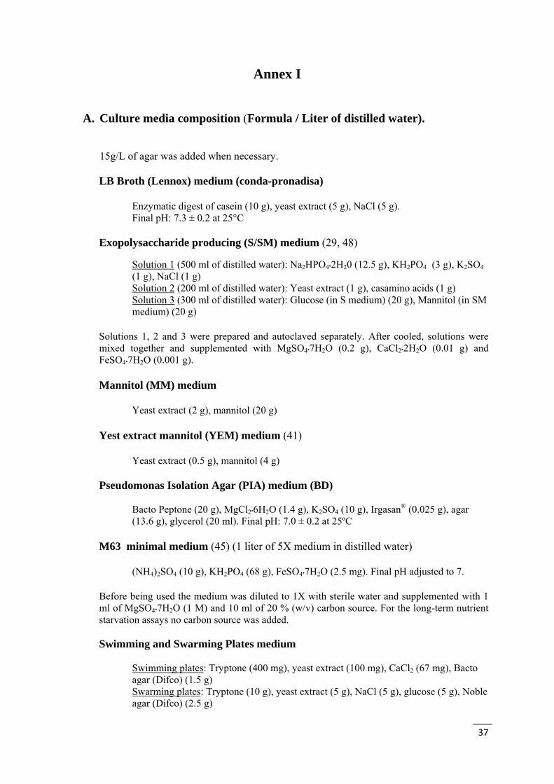
37
Annex I
A. Culture media composition (Formula / Liter of distilled water). 15g/L of agar was added when necessary.
LB Broth (Lennox) medium (conda-pronadisa)
Enzymatic digest of casein (10 g), yeast extract (5 g), NaCl (5 g). Final pH: 7.3 ± 0.2 at 25°C
Exopolysaccharide producing (S/SM) medium (29, 48)
Solution 1 (500 ml of distilled water): Na2HPO4•2H20 (12.5 g), KH2PO4 (3 g), K2SO4 (1 g), NaCl (1 g) Solution 2 (200 ml of distilled water): Yeast extract (1 g), casamino acids (1 g) Solution 3 (300 ml of distilled water): Glucose (in S medium) (20 g), Mannitol (in SM medium) (20 g)
Solutions 1, 2 and 3 were prepared and autoclaved separately. After cooled, solutions were mixed together and supplemented with MgSO4•7H2O (0.2 g), CaCl2•2H2O (0.01 g) and FeSO4•7H2O (0.001 g). Mannitol (MM) medium
Yeast extract (2 g), mannitol (20 g) Yest extract mannitol (YEM) medium (41)
Yeast extract (0.5 g), mannitol (4 g)
Pseudomonas Isolation Agar (PIA) medium (BD)
Bacto Peptone (20 g), MgCl2•6H2O (1.4 g), K2SO4 (10 g), Irgasan® (0.025 g), agar (13.6 g), glycerol (20 ml). Final pH: 7.0 ± 0.2 at 25ºC
M63 minimal medium (45) (1 liter of 5X medium in distilled water)
(NH4)2SO4 (10 g), KH2PO4 (68 g), FeSO4•7H2O (2.5 mg). Final pH adjusted to 7. Before being used the medium was diluted to 1X with sterile water and supplemented with 1 ml of MgSO4•7H2O (1 M) and 10 ml of 20 % (w/v) carbon source. For the long-term nutrient starvation assays no carbon source was added. Swimming and Swarming Plates medium
Swimming plates: Tryptone (400 mg), yeast extract (100 mg), CaCl2 (67 mg), Bacto agar (Difco) (1.5 g) Swarming plates: Tryptone (10 g), yeast extract (5 g), NaCl (5 g), glucose (5 g), Noble agar (Difco) (2.5 g)
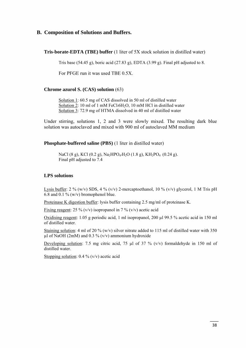
38
B. Composition of Solutions and Buffers.
Tris-borate-EDTA (TBE) buffer (1 liter of 5X stock solution in distilled water)
Tris base (54.45 g), boric acid (27.83 g), EDTA (3.99 g). Final pH adjusted to 8. For PFGE run it was used TBE 0.5X.
Chrome azurol S. (CAS) solution (63)
Solution 1: 60.5 mg of CAS dissolved in 50 ml of distilled water Solution 2: 10 ml of 1 mM FeCl•6H2O, 10 mM HCl in distilled water Solution 3: 72.9 mg of HTMA dissolved in 40 ml of distilled water
Under stirring, solutions 1, 2 and 3 were slowly mixed. The resulting dark blue solution was autoclaved and mixed with 900 ml of autoclaved MM medium Phosphate-buffered saline (PBS) (1 liter in distilled water)
NaCl (8 g), KCl (0.2 g), Na2HPO4•H2O (1.8 g), KH2PO4 (0.24 g). Final pH adjusted to 7.4
LPS solutions
Lysis buffer: 2 % (w/v) SDS, 4 % (v/v) 2-mercaptoethanol, 10 % (v/v) glycerol, 1 M Tris pH 6.8 and 0.1 % (w/v) bromophenol blue.
Proteinase K digestion buffer: lysis buffer containing 2.5 mg/ml of proteinase K.
Fixing reagent: 25 % (v/v) isopropanol in 7 % (v/v) acetic acid
Oxidising reagent: 1.05 g periodic acid, 1 ml isopropanol, 200 µl 99.5 % acetic acid in 150 ml of distilled water.
Staining solution: 4 ml of 20 % (w/v) silver nitrate added to 115 ml of distilled water with 350 µl of NaOH (2mM) and 0.3 % (v/v) ammonium hydroxide
Developing solution: 7.5 mg citric acid, 75 µl of 37 % (v/v) formaldehyde in 150 ml of distilled water.
Stopping solution: 0.4 % (v/v) acetic acid
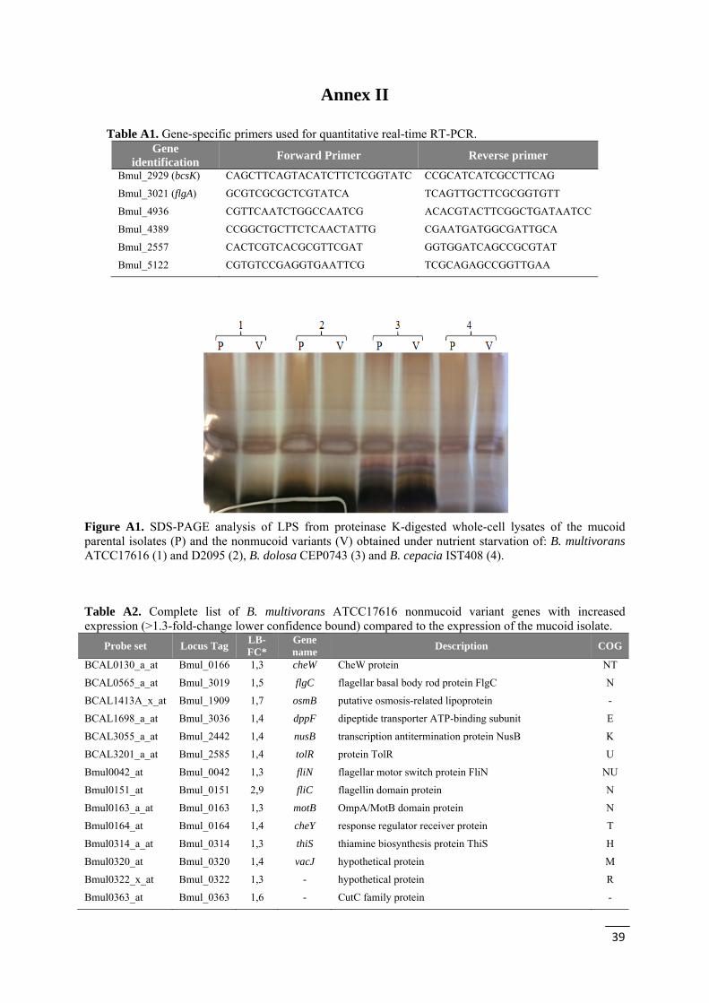
39
Annex II Table A1. Gene-specific primers used for quantitative real-time RT-PCR.
Gene identification
Forward Primer Reverse primer
Bmul_2929 (bcsK) CAGCTTCAGTACATCTTCTCGGTATC CCGCATCATCGCCTTCAG
Bmul_3021 (flgA) GCGTCGCGCTCGTATCA TCAGTTGCTTCGCGGTGTT
Bmul_4936 CGTTCAATCTGGCCAATCG ACACGTACTTCGGCTGATAATCC
Bmul_4389 CCGGCTGCTTCTCAACTATTG CGAATGATGGCGATTGCA
Bmul_2557 CACTCGTCACGCGTTCGAT GGTGGATCAGCCGCGTAT
Bmul_5122 CGTGTCCGAGGTGAATTCG TCGCAGAGCCGGTTGAA
Figure A1. SDS-PAGE analysis of LPS from proteinase K-digested whole-cell lysates of the mucoid parental isolates (P) and the nonmucoid variants (V) obtained under nutrient starvation of: B. multivorans ATCC17616 (1) and D2095 (2), B. dolosa CEP0743 (3) and B. cepacia IST408 (4).
Table A2. Complete list of B. multivorans ATCC17616 nonmucoid variant genes with increased expression (>1.3-fold-change lower confidence bound) compared to the expression of the mucoid isolate.
Probe set Locus Tag LB-FC*
Gene name
Description COG
BCAL0130_a_at Bmul_0166 1,3 cheW CheW protein NT
BCAL0565_a_at Bmul_3019 1,5 flgC flagellar basal body rod protein FlgC N
BCAL1413A_x_at Bmul_1909 1,7 osmB putative osmosis-related lipoprotein -
BCAL1698_a_at Bmul_3036 1,4 dppF dipeptide transporter ATP-binding subunit E
BCAL3055_a_at Bmul_2442 1,4 nusB transcription antitermination protein NusB K
BCAL3201_a_at Bmul_2585 1,4 tolR protein TolR U
Bmul0042_at Bmul_0042 1,3 fliN flagellar motor switch protein FliN NU
Bmul0151_at Bmul_0151 2,9 fliC flagellin domain protein N
Bmul0163_a_at Bmul_0163 1,3 motB OmpA/MotB domain protein N
Bmul0164_at Bmul_0164 1,4 cheY response regulator receiver protein T
Bmul0314_a_at Bmul_0314 1,3 thiS thiamine biosynthesis protein ThiS H
Bmul0320_at Bmul_0320 1,4 vacJ hypothetical protein M
Bmul0322_x_at Bmul_0322 1,3 - hypothetical protein R
Bmul0363_at Bmul_0363 1,6 - CutC family protein -

40
Bmul0434_at Bmul_0434 1,3 - hypothetical protein -
Bmul0528_at Bmul_0528 1,3 kdsC 3-deoxy-D-manno-octulosonate 8-P phosphatase, YrbI family
R
Bmul0627_at Bmul_0627 1,3 dead DEAD/DEAH box helicase domain protein LKJ
Bmul0760_at Bmul_0760 1,3 - hypothetical protein -
Bmul0824_at Bmul_0824 1,4 cysG uroporphyrin-III C-methyltransferase H
Bmul0830_at Bmul_0830 1,4 - hypothetical protein -
Bmul0863_at Bmul_0863 1,3 - hypothetical protein S
Bmul0978_at Bmul_0978 1,4 maf maf protein D
Bmul0987_x_at Bmul_0987 1,3 mgsA methylglyoxal synthase G
Bmul1006_at Bmul_1006 1,3 dgk diacylglycerol kinase M
Bmul1182_at Bmul_1182 1,3 - hypothetical protein S
Bmul1501_at Bmul_1501 1,3 - hypothetical protein -
Bmul1573_at Bmul_1573 1,3 acd acyl-CoA dehydrogenase domain protein I
Bmul1738_at Bmul_1738 1,4 - hypothetical protein -
Bmul1761_at Bmul_1761 1,3 - transcriptional regulator, LysR family K
Bmul2016_a_at Bmul_2016 1,3 phoU phosphate uptake regulator, PhoU P
Bmul2044_at Bmul_2044 1,3 fabD acyl-carrier-protein S-malonyltransferase I
Bmul2098_at Bmul_2098 1,3 - transcriptional regulator, BadM/Rrf2 family K
Bmul2143_at Bmul_2143 1,3 - hypothetical protein S
Bmul2165_a_at Bmul_2165 1,3 nagZ Beta-N-acetylhexosaminidase G
Bmul2205_a_at Bmul_2205 1,5 creA hypothetical protein S
Bmul2210_at Bmul_2210 1,3 - hypothetical protein -
Bmul2266_a_at Bmul_2266 1,3 ubiG ubiquinone biosynthesis O-methyltransferase H
Bmul2372_at Bmul_2372 1,5 - transcriptional regulator, GntR family K
Bmul2373_at Bmul_2373 1,3 gatA amidase J
Bmul2491_at Bmul_2491 1,3 ureE UreE urease accessory domain protein O
Bmul2751_at Bmul_2751 1,4 - protein of unknown function DUF558 S
Bmul2818_at Bmul_2818 1,5 pilB type II secretion system protein E NU
Bmul2960_at Bmul_2960 1,3 - alpha/beta hydrolase fold R
Bmul3009_at Bmul_3009 1,3 flgL flagellar hook-associated protein 3 N
Bmul3017_at Bmul_3017 1,3 flgE flagellar basal body FlaE domain protein N
Bmul3018_at Bmul_3018 1,3 flgD flagellar hook capping protein N
Bmul3021_a_at Bmul_3021 1,7 flgA flagellar basal body P-ring biosynthesis protein FlgA NO
Bmul3022_at Bmul_3022 1,3 flgM anti-sigma-28 factor, FlgM KNU
Bmul3051_at Bmul_3051 1,5 - putative signal transduction histidine kinase T
Bmul3056_at Bmul_3056 1,3 fhlB putative flagellar protein FhlB N
Bmul3151_at Bmul_3151 1,3 - hypothetical protein -
Bmul3239_at Bmul_3239 1,3 benB benzoate 1,2-dioxygenase, small subunit Q
Bmul3342_at Bmul_3342 1,5 - hypothetical protein M
Bmul3362_at Bmul_3362 1,3 era methyl-accepting chemotaxis sensory transducer NT
Bmul3417_at Bmul_3417 1,3 - hypothetical protein -
Bmul3423_at Bmul_3423 1,4 - hypothetical protein P
Bmul3719_at Bmul_3719 1,3 puuB FAD dependent oxidoreductase E
Bmul3760_at Bmul_3760 1,3 - YCII-related protein S
Bmul4080_at Bmul_4080 1,3 - hypothetical protein O
Bmul4128_at Bmul_4128 1,4 marC multiple antibiotic resistance (MarC)-related protein U
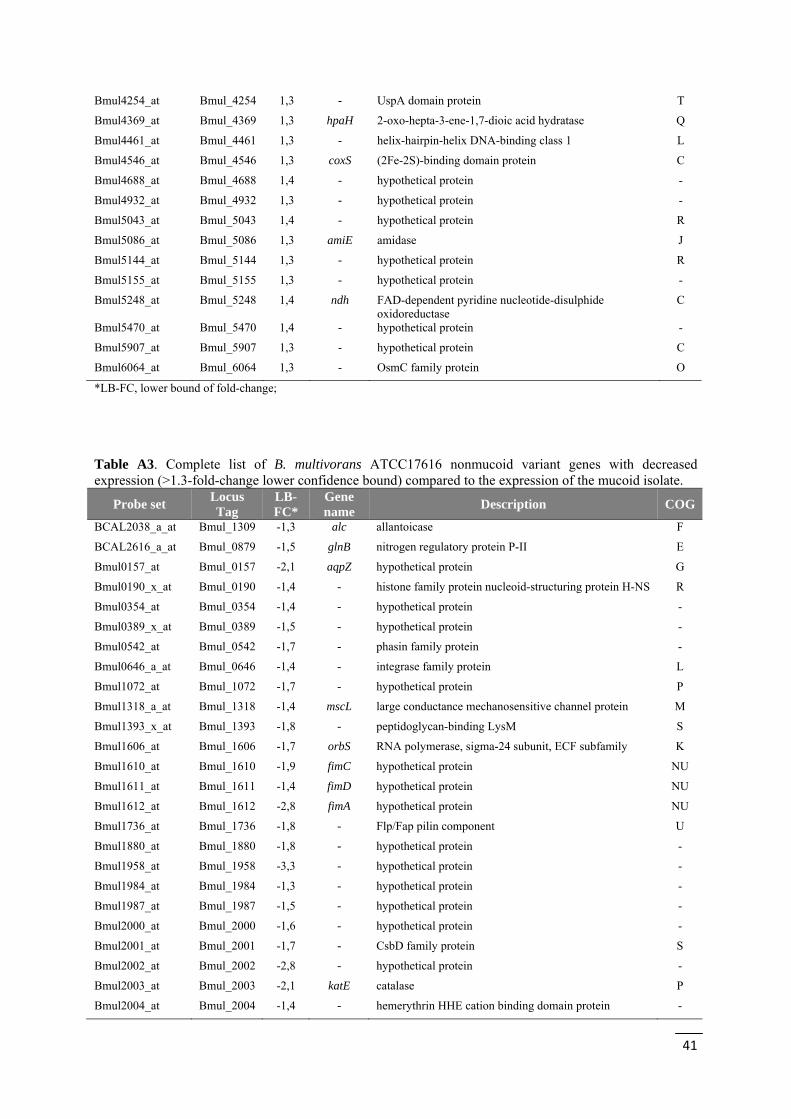
41
Bmul4254_at Bmul_4254 1,3 - UspA domain protein T
Bmul4369_at Bmul_4369 1,3 hpaH 2-oxo-hepta-3-ene-1,7-dioic acid hydratase Q
Bmul4461_at Bmul_4461 1,3 - helix-hairpin-helix DNA-binding class 1 L
Bmul4546_at Bmul_4546 1,3 coxS (2Fe-2S)-binding domain protein C
Bmul4688_at Bmul_4688 1,4 - hypothetical protein -
Bmul4932_at Bmul_4932 1,3 - hypothetical protein -
Bmul5043_at Bmul_5043 1,4 - hypothetical protein R
Bmul5086_at Bmul_5086 1,3 amiE amidase J
Bmul5144_at Bmul_5144 1,3 - hypothetical protein R
Bmul5155_at Bmul_5155 1,3 - hypothetical protein -
Bmul5248_at Bmul_5248 1,4 ndh FAD-dependent pyridine nucleotide-disulphide oxidoreductase
C
Bmul5470_at Bmul_5470 1,4 - hypothetical protein -
Bmul5907_at Bmul_5907 1,3 - hypothetical protein C
Bmul6064_at Bmul_6064 1,3 - OsmC family protein O
*LB-FC, lower bound of fold-change;
Table A3. Complete list of B. multivorans ATCC17616 nonmucoid variant genes with decreased expression (>1.3-fold-change lower confidence bound) compared to the expression of the mucoid isolate.
Probe set Locus Tag
LB-FC*
Gene name
Description COG
BCAL2038_a_at Bmul_1309 -1,3 alc allantoicase F
BCAL2616_a_at Bmul_0879 -1,5 glnB nitrogen regulatory protein P-II E
Bmul0157_at Bmul_0157 -2,1 aqpZ hypothetical protein G
Bmul0190_x_at Bmul_0190 -1,4 - histone family protein nucleoid-structuring protein H-NS R
Bmul0354_at Bmul_0354 -1,4 - hypothetical protein -
Bmul0389_x_at Bmul_0389 -1,5 - hypothetical protein -
Bmul0542_at Bmul_0542 -1,7 - phasin family protein -
Bmul0646_a_at Bmul_0646 -1,4 - integrase family protein L
Bmul1072_at Bmul_1072 -1,7 - hypothetical protein P
Bmul1318_a_at Bmul_1318 -1,4 mscL large conductance mechanosensitive channel protein M
Bmul1393_x_at Bmul_1393 -1,8 - peptidoglycan-binding LysM S
Bmul1606_at Bmul_1606 -1,7 orbS RNA polymerase, sigma-24 subunit, ECF subfamily K
Bmul1610_at Bmul_1610 -1,9 fimC hypothetical protein NU
Bmul1611_at Bmul_1611 -1,4 fimD hypothetical protein NU
Bmul1612_at Bmul_1612 -2,8 fimA hypothetical protein NU
Bmul1736_at Bmul_1736 -1,8 - Flp/Fap pilin component U
Bmul1880_at Bmul_1880 -1,8 - hypothetical protein -
Bmul1958_at Bmul_1958 -3,3 - hypothetical protein -
Bmul1984_at Bmul_1984 -1,3 - hypothetical protein -
Bmul1987_at Bmul_1987 -1,5 - hypothetical protein -
Bmul2000_at Bmul_2000 -1,6 - hypothetical protein -
Bmul2001_at Bmul_2001 -1,7 - CsbD family protein S
Bmul2002_at Bmul_2002 -2,8 - hypothetical protein -
Bmul2003_at Bmul_2003 -2,1 katE catalase P
Bmul2004_at Bmul_2004 -1,4 - hemerythrin HHE cation binding domain protein -

42
Bmul2393_x_at Bmul_2393 -2,7 cspA cold-shock DNA-binding domain protein K
Bmul2557_at Bmul_2557 -2,3 - transcriptional regulator, LysR family K
Bmul2606_at Bmul_2606 -1,4 - protein of unknown function DUF23 -
Bmul2607_at Bmul_2607 -1,4 - ABC transporter related GM
Bmul2608_at Bmul_2608 -1,4 - ABC-2 type transporter GM
Bmul2649_at Bmul_2649 -1,3 hmpA oxidoreductase FAD/NAD(P)-binding domain protein C
Bmul2816_at Bmul_2816 -1,3 - hypothetical protein -
Bmul2885_at Bmul_2885 -1,4 - hypothetical protein -
Bmul2905_at Bmul_2905 -1,8 - hypothetical protein -
Bmul2916_at Bmul_2916 -1,3 - hypothetical protein -
Bmul2917_at Bmul_2917 -1,3 - hypothetical protein -
Bmul2923_at Bmul_2923 -1,3 bcsE type VI secretion-associated protein, ImpA family S
Bmul2927_x_at Bmul_2927 -1,3 bcsI type VI secretion system lysozyme-related protein S
Bmul2928_a_at Bmul_2928 -1,4 bcsJ type VI secretion system effector, Hcp1 family S
Bmul2929_a_at Bmul_2929 -1,5 bcsK type VI secretion protein, EvpB/VC_A0108 family S
Bmul2930_x_at Bmul_2930 -1,9 bcsL type VI secretion protein, VC_A0107 family S
Bmul2931_at Bmul_2931 -1,5 - hypothetical protein S
Bmul2962_at Bmul_2962 -1,3 - transcriptional regulator, LysR family K
Bmul2986_at Bmul_2986 -1,4 - hypothetical protein -
Bmul2990_at Bmul_2990 -1,3 potD hypothetical protein E
Bmul3279_at Bmul_3279 -1,3 - histone family protein nucleoid-structuring protein H-NS R
Bmul3335_at Bmul_3335 -1,3 - hypothetical protein P
Bmul3336_at Bmul_3336 -1,3 - hypothetical protein P
Bmul3337_at Bmul_3337 -1,3 hmuS haemin-degrading family protein P
Bmul3471_at Bmul_3471 -1,4 - diguanylate phosphodiesterase T
Bmul3645_at Bmul_3645 -1,5 - hypothetical protein -
Bmul3695_at Bmul_3695 -1,4 - hypothetical protein S
Bmul3701_at Bmul_3701 -1,4 ldcC lysine decarboxylase E
Bmul3702_at Bmul_3702 -1,3 adiC amino acid permease-associated region E
Bmul3708_at Bmul_3708 -5,1 - hypothetical protein -
Bmul3709_a_at Bmul_3709 -2,4 adh hemolysin-type calcium-binding region -
Bmul3735_at Bmul_3735 -2,0 - hypothetical protein -
Bmul3736_at Bmul_3736 -1,3 - natural resistance-associated macrophage protein P
Bmul3737_at Bmul_3737 -1,3 - transcriptional regulator, LuxR family TK
Bmul3806_at Bmul_3806 -1,3 - histone family protein nucleoid-structuring protein H-NS R
Bmul4078_at Bmul_4078 -1,9 - hypothetical protein -
Bmul4079_at Bmul_4079 -2,4 mgtA magnesium-translocating P-type ATPase P
Bmul4115-2_at Bmul_4115 -1,4 - hypothetical protein S
Bmul4295_at Bmul_4295 -1,3 - hypothetical protein K
Bmul4389_at Bmul_4389 -1,7 - putative transcriptional regulator, Crp/Fnr family T
Bmul4401_at Bmul_4401 -1,3 - glycosyl transferase group 1 M
Bmul4408_at Bmul_4408 -1,5 - lipopolysaccharide biosynthesis protein D
Bmul4502_at Bmul_4502 -1,4 - ABC transporter related E
Bmul4711_at Bmul_4711 -1,3 - hypothetical protein -
Bmul4712_at Bmul_4712 -1,6 - hypothetical protein -
Bmul4822_x_at Bmul_4822 -1,3 xseB exodeoxyribonuclease VII, small subunit L
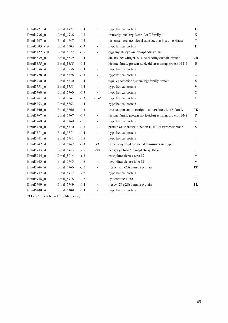
43
Bmul4921_at Bmul_4921 -1,4 - hypothetical protein L
Bmul4936_at Bmul_4936 -1,3 - transcriptional regulator, AraC family K
Bmul4947_at Bmul_4947 -1,5 - response regulator signal transduction histidine kinase T
Bmul5003_a_at Bmul_5003 -1,3 - hypothetical protein E
Bmul5122_a_at Bmul_5122 -1,5 - diguanylate cyclase/phosphodiesterase T
Bmul5639_at Bmul_5639 -1,4 - alcohol dehydrogenase zinc-binding domain protein CR
Bmul5655_at Bmul_5655 -1,4 - histone family protein nucleoid-structuring protein H-NS R
Bmul5656_at Bmul_5656 -1,4 - hypothetical protein -
Bmul5728_at Bmul_5728 -1,3 - hypothetical protein -
Bmul5730_at Bmul_5730 -1,4 - type VI secretion system Vgr family protein S
Bmul5751_at Bmul_5751 -1,4 - hypothetical protein V
Bmul5760_at Bmul_5760 -1,3 - hypothetical protein C
Bmul5761_at Bmul_5761 -1,3 coxA hypothetical protein C
Bmul5763_at Bmul_5763 -1,4 - hypothetical protein -
Bmul5766_at Bmul_5766 -1,3 - two component transcriptional regulator, LuxR family TK
Bmul5767_at Bmul_5767 -1,9 - histone family protein nucleoid-structuring protein H-NS R
Bmul5769_at Bmul_5769 -3,1 - hypothetical protein -
Bmul5770_at Bmul_5770 -1,3 - protein of unknown function DUF125 transmembrane S
Bmul5771_at Bmul_5771 -1,4 - hypothetical protein -
Bmul5941_at Bmul_5941 -1,8 - hypothetical protein -
Bmul5942_at Bmul_5942 -2,3 idi isopentenyl-diphosphate delta-isomerase, type 1 I
Bmul5943_at Bmul_5943 -2,5 dxs deoxyxylulose-5-phosphate synthase HI
Bmul5944_at Bmul_5944 -6,6 - methyltransferase type 12 M
Bmul5945_at Bmul_5945 -4,8 - methyltransferase type 12 M
Bmul5946_at Bmul_5946 -3,0 - rieske (2Fe-2S) domain protein PR
Bmul5947_at Bmul_5947 -2,2 - hypothetical protein -
Bmul5948_at Bmul_5948 -1,7 - cytochrome P450 Q
Bmul5949_at Bmul_5949 -1,4 - rieske (2Fe-2S) domain protein PR
Bmul6209_at Bmul_6209 -1,3 - hypothetical protein -
*LB-FC, lower bound of fold-change;