Embed Size (px)
Citation preview
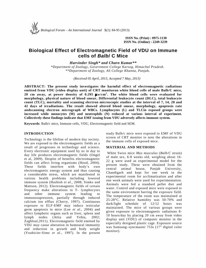
ISSN No. (Print) : 0975-1130ISSN No. (Online) : 2249-3239
Biological Effect of Electromagnetic Field of VDU on Immunecells of Balb/ C Mice
Harvinder Singh* and Charn Kumar***Department of Zoology, Government College Karsog, Himachal Pradesh.
**Department of Zoology, AS College Khanna, Punjab.
(Received 05 April, 2013, Accepted 7 May, 2013)
ABSTRACT: The present study investigates the harmful effect of electromagnetic radiationsemitted from VDU (video display unit) of CRT monitoron white blood cells of male Balb/C mice,20 cm away, at power density of 0.295 w/cm2. The white blood cells were evaluated formorphology, physical nature of blood smear, Differential leukocyte count (DLC), total leukocytecount (TLC), mortality and scanning electron microscopic studies at the interval of 7, 14, 28 and42 days of irradiations. The result showed altered blood smear, morphology, apoptosis rateandscanning electron micrograph of WBCs. Lymphocytes (L) and TLCin exposed groups wereincreased while monocytes (M) and neutrophils (N) reduced at various interval of experiment.Collectively these findings indicate that EMF issuing from VDU adversely affects immune system.
Keywords: Balb/c mice, Immune cells, VDU, Electromagnetic field and SEM.
INTRODUCTION
Technology is the lifeline of modern day society.We are exposed to the electromagnetic fields as aresult of progresses in technology and science.Every electronic equipment used by us in day today life produces electromagnetic fields (Ongelet al, 2009). Despite of benefits electromagneticfields can affect living organisms (Hood, 2004).These fields interfere with body’s ownelectromagnetic energy system and thus causinga considerable stress, which are manifested invarious health problems including loweredimmune system (Hashish et al., 2008; Simko andMattson, 2012). Electromagnetic fields of certainfrequency make alterations in T- lymphocytesand other immune system leading toimmunosupression, partially through inducedcalcium ion efflux (Cherry, 1997). Continuousexposure to ELF-EMF may induce testiculargerm apoptosis in mice (Lee et al., 2004) andaffect lymphetic organs such as liver, spleen andlymph nodes (Attia and Yehia, 2002;Zaghloul,2011). Electromagnetic field emitted byVDU may cause alteration in humoural immunityand reduction in growth and body weight(Youbicier-Simo et al., 1997). In the present
study Balb/c mice were exposed to EMF of VDUscreen of CRT monitor to note the alterations inthe immune cells of exposed mice.
MATERIAL AND METHODS
White Swiss mice Mus musculus (Balb/C strain)of male sex, 6-8 weeks old, weighing about 16-22 g were used as experimental model for thepresent study. These were obtained from thecentral animal house, Panjab University,Chandigarh and kept for one week in theexperimental room for acclimatization and afterone week animals were used for experimentation.Animals were fed a standard pellet diet andwater. Control and exposed mice were exposed tothe same environment barring the exposure field.The temperature of the room was maintained at25-28°C. Relative humidity was 50-70% anddark/light schedule of 12/12 hours wasmaintained. The mice of various groups weregiven exposure to electromagnetic radiations 8-10 hours/day by placing 20 cm away from videodisplay unit (VDU) of computer monitor in theespecially designed plastic cage. Exposure sourcewas Samsung–syncmaster 753s (17” digital colormonitor).
Biological Forum – An International Journal 5(1): 84-95 (2013)

Singh and Kumar 85
Power density (0.295 µw/cm2) was measured with‘RF Field Strength Meter’ at 20 cm in front ofmonitor. The control and exposed group were keptapart so that there is no interference of exposedfield.
Experimental Design: Two groups having fivemice and twenty mice respectively were used forthe present study. The experimental mice weregiven exposure of computer monitor (VDU) for 8-10 hours daily for different time intervals.
GI: This group consisted of five normal controlmice which were not exposed to any source ofradiations.
GII: Twenty mice of this group were exposed toVDU (8-10h/day) for different time intervals i.e.for 7 days, 14 days, 28 days and 42 daysrespectively.
Differential leukocyte count (DLC), totalleukocyte count (TLC) of both groups control (GI)and exposed (GII) was recorded after 7, 14, 28and 42 days. Statistical Analysis of the data waspresented as the mean ± SD for each correlation wascalculated using Microsoft Excel. The variationsobserved in the values of various parametersunder study in VDU exposed group werecompared with control (GI) at different intervalsof experiment. The data were analyzed using SPSSprogram (statistical package for social sciences Inc.Chicago, Illinois).
Mice of GII were sacrificed by jugular veinincision after anesthetizing with diethyl ether onday 7, 14, 28 and 42. Control mice were sacrificedon day 42of experiment. Blood was aspirated incitrate saline (0.85% (w/v) sodium chloride; 3.8%(w/v) sodium citrate). Pooled blood of same groupwas subjected to density gradient centrifugationusing histopaque-1119 (sigma).
Differential Leukocyte Count (DLC):For DLC thin blood smears of were made from tailsnips and stained in Giemsa stain (3% Giemsastock in 0.95 % (w/v) sodium phosphate dibasic,0.91% (w/v) potassium phosphate monobasic) forhalf an hour and observed under microscope(100x) using immersion oil. The percentage ofmononuclear (MN) cells i.e. lymphocytes (L) andmonocytes (M) and polymorphonuclear (PMN)cells i.e. neutrophils (N) was calculated (Fatayer,2006).
Number of M/L/N 100
Total number of WBCs×
Calculation of Total Leukocyte Count (TLC):Using Neuber’s Hemocytometer kit, TLC of allthe mice of both groups (GI and GII) was doneusing Turk’s fluid (10% (w/v)) gentian violet and1.5% glacial acetic acid in distilled water). Thenumber of WBCs will is counted in 4 squares (1, 3, 7& 9) of the slide under microscope at 10x or 40x as perconvenience.
WBC Pipette Neuber’s Slide

Singh and Kumar 86
Using the number of WBCs in 4 squares TLC will becalculated according to following calculations:
Say number of WBCs in square 1,3,7,9 isa + b + c + d = yArea of each square = 1 mm2 , height = 1/10 mmTherefore, volume = 1/10 x 1 = 1/10 mm3
1/10 mm3 contains = y/4 WBCs1 mm3 contain = y/4 x 10 WBCsAs the solution is diluted 20 timesTherefore, 1 mm3 contains =Y 10 20
Z WBC's4
× × =
Separation of WBCs by Density GradientCentrifugation:Blood of mice was aspirated using Pasteur’s pipettein citrate saline by incising jugular vein. Pooledblood of each group of mice was subjected todensity gradient centrifugation using histopaque-1119 to separate WBCs using following procedure(Czuprynski and Brown, 1998).
Separation of white blood cells.
Fluorescent Staining Using Acridine Orange(AO) / Ethidium Bromide (EB):WBCs aspirated by density centrifugation ofblood were subjected to fluorescent staining tostudy apoptosis. WBCs suspension wasincubated with 20µ l of AO/EB solution (1 part100µ g/ml AO in PBS; 1part of 100µ g/ml EB inPBS). The suspension was mixed gently andeach sample was mixed just prior to itsexamination by fluorescent microscope (Leica,Germany) and its quantification, there after(Kasibhatla, 1998).Added 1-2 drops of this suspension on amicroscopic slide and covered with a glasscover slip. Cells were examined underfluorescent microscope using fluorescent filter.500 cells were counted in each group tocalculate the percentage of live and dead cellsby the following formula:
100cellsblood whiteofnumberTotalcellsotic/deadlive/apoptofumberN ×
Live cells stained fluorescent green, whereas,dead cells appeared orange under ultra violetlight.
RESULTS AND DISSCUSSION
Most of the studies pertain to the effect ofelectromagnetic fields from power lines,microwave, color TV screens and self designedinstruments. But little work so far has beenreported on the harms of EMF ofcomputermonitors.Computers are extensivelyused in every sphere of life. During on phagethe screen of CRT monitor can emit EMFranging from X-rays to extremely low frequency(ELF) and very low frequency fields (VLF)(Kavet and Tel,1991; Luketina,1975). In the lastfew decades various studies carried out on mice,rat and human showed that electromagneticfields induced changes in hematologicalparameters in these organisms (High et al.,2000; Ali et al., 2003; Sihem et al., 2006;Hassan and Abdelkawi, 2010).

Singh and Kumar 87
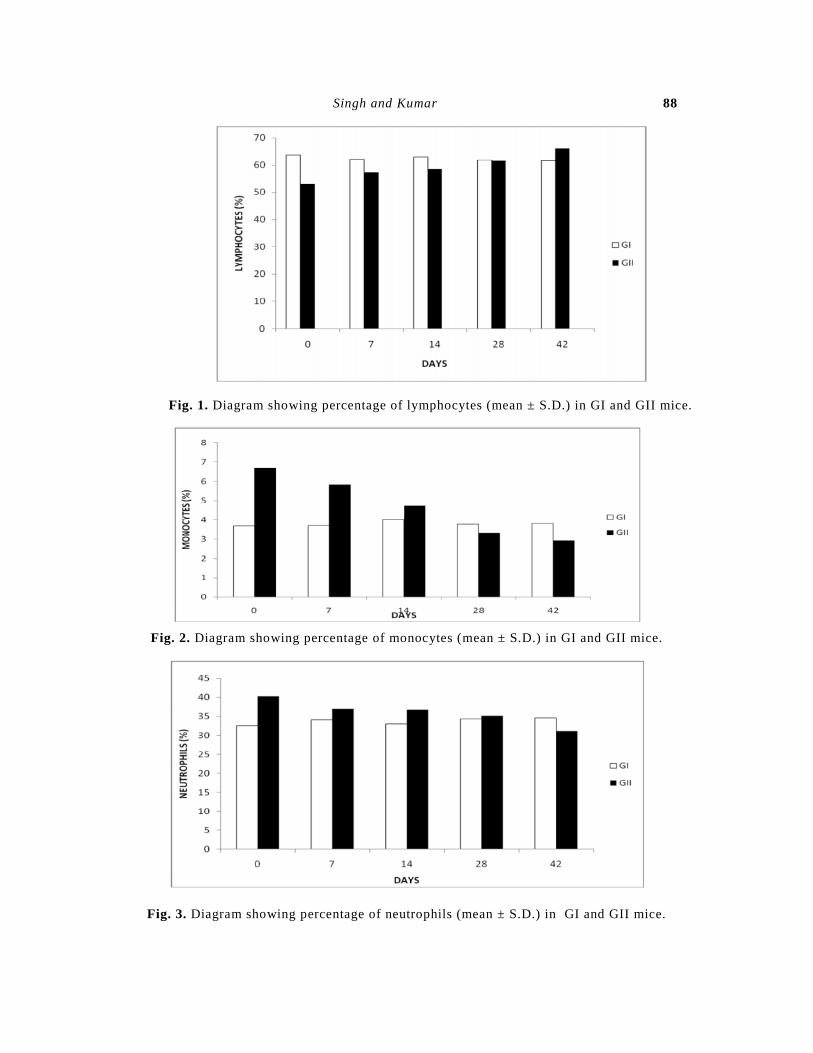
The present study was carried to assess the effectof EMF emitted from VDU of computer monitoron cells of immune of male Balb/c mice. Resultsof present study points to harmful effects of continuouslong term exposure of computer monitors on Balb/Cmice. Percentage of lymphocytes in the exposed groupof mice increased while that of N and M decreased withthe rise in period of exposure (Fig. 1-3). In exposedgroup L showed maximum increase (27.9%) after 42day irradiation while maximum decrease in M (59.9%)and N (29.3%) was reported after 42 days of exposurecompared to control (Table 1). Number of lymphocytesis normally enhanced due to external antigen stimulusor diseased state under normal conditions. The increasein the lymphocytes may be because of reduction in thevalue of monocytes and neutrophils. Declined numberof phagocytic cells (N and M) points to depressed levelof immunity in exposed mice to pathogens. Similarly
Total Leukocyte Count (TLC) in exposed mice groupwas more when compared with the control, whichindicate activation of the immune system as a result ofexposure to electromagnetic radiations (Table 1). Inexposed group maximum increase in the TLC wasobserved after 42 days of exposure (24.7%), whencompared with GI at respective time intervals (Fig. 4).Morphological assessment of apoptosis in WBCswas done by AO/EB staining. Live cells stainedfluorescent green, whereas, dead cells appearedorange under ultra violet light (Fig. 7A, B, C).Maximum percentage of live WBCs was seen innormal mice i.e. 89.16% (Table 1). The percentageof dead white blood cells increased with increasein exposure time in GII. It was 15.8%, 19% and21.4% and 25.54% after days 7, 15, 30 and 45 ofexposure respectively (Fig. 5).
Table 1. Mean± SD of various parameters at different intervals of experiment in GI and GII.
Parameters Day 0 Day 7 Day 14 Day 28 Day 42
L (%) Control63.8±6.67 62.1±6.98 63±9.42 61.8±8.4 61.6±6.9
Exposed53±9.78 57.3±10.76 58.6±11.33 61.47±9.42 66±12.45
M (%) Control3.69±0.6 3.72±0.37 4.02±1 3.78±1.2 3.82±0.68
Exposed6.69±1.2 5.84±1.15 4.73±0.9 3.34±0.62 2.92±0
N (%) Control32.5±8.33 34.2±7.46 33±8 34.4±9.2 34.6±6.48
Exposed40.3±11.4 36.9±10.1 36.7±9.69 35.2±8.53 31.08±11.11
TLC/mm3 Control6420±1220 6850±1403 7200±1469 7766±1463 9080±2651
Exposed6540±1296 6990±1340 7400±1992 7686±1090 7890±1340
Mortality of
WBCs (%)
Live 89.16 84.2 81 78.6 74.46
Dead 10.84 15.8 19 21.4 25.54
Singh and Kumar 88
Fig. 1. Diagram showing percentage of lymphocytes (mean ± S.D.) in GI and GII mice.
Fig. 2. Diagram showing percentage of monocytes (mean ± S.D.) in GI and GII mice.
Fig. 3. Diagram showing percentage of neutrophils (mean ± S.D.) in GI and GII mice.

Singh and Kumar 89
Fig. 4. Diagram showing total leukocyte count (mean ± S.D.) in GI and GII mice.
Fig. 5. Diagram showing the status of live and dead cells obtained by density gradientCentrifugation of pooled peripheral blood of GI and GII mice.
Lymphocytes in exposed mice (GII and GIII) have beenobserved to have less basophilic nucleus, large size andcytoplasm clearly visible (Fig. 6D, G, J, M). Horse shoeshaped nucleus of monocytes has also been observed toswell up in exposed groups (Fig. 6E, H, K, N). Similarly
multi-lobed nucleus of neutrophil has become hypersegmentation. The neutrophil took little stain like that oflymphocytes and monocytes (Fig. 6F, I, L, O). Ourresults confirm the findings of Gagnon et al. (2003) andUsman, A.D. (2012).

Singh and Kumar 90
Fig. 6. Giemsa’s stained blood smear showing different types of leukocytes (1000x) in control (A,B,C) and exposedmice for various intervals i.e. 7 days (D,E,F) , 14 days (G,H,I), 28 days (J,K,L), 42 days (M,N,O).

Singh and Kumar 91
Fig.7. AO/EB staining of WBCs obtained by density gradient centrifugation using histopaque-1119 from normal (A,B) and mice exposed for 7,14,28,42,days(C,D,E,F respectively). Abbreviations: L-Live; D-Dead; A –Apoptotic.
SEM studies of WBCs in exposed mice revealsthat the MN cells were sticking together, and sizewas increased as compared to GI (Fig. 8, 9). PMNcells also showed a variety of shapes. Some ofthe PMN cells were having finger likeprojections and some had small blebs. In exposedgroups PMN cells were decreased in size and the
projections were more prominent All these inexposed mice were reported to become moresevere with increase in exposure period. This willclearly indicate that electromagnetic fields emitted fromthese screens have great influence on the immune cells(Veyret et al., 1991; Novoslova et al. ,1999).

Singh and Kumar 92
Fig. 8. Scanning Electron micrograph of WBC’s of Balb/c mice obtained after separation of blood cells by densitygradient centrifugation. (A and B: MN and PMN cells of normal control mice, C and D: WBC’s of mice exposed for7 days, E and F: WBC’s of mice exposed for 14 days) ABBREVIATIONS: LC- layered surface cells; SC-smoothsurface cells PMN- Polymorphonuclear cells; MN-Morphonuclear Cells.
But our results did not confirm the findings of Hashishet al., 2007.He reported a significant decrease in thecount of monocytes, peripheral lymphocytes as well asspleen total T and B lymphocyte values in micesubjected to static magnetic field and ELF-MFexposure. White blood cells constitute the defense
system of body and any alteration in their value hasbeen associated with various types of infections. But inpresent case only radiations are source of stimulation ofWBC which shows variation in number, morphologyand mortality.

Singh and Kumar 93
Fig. 9. Scanning Electron micrograph of WBC’s of Balb/c mice obtained after separation of blood cells by densitygradient centrifugation. (A and B: WBC’s of mice exposed for 28 days, C and D: WBC’s of mice exposed for 42days) ABBREVIATIONS: LC- layered surface cells; SC-smooth surface cells PMN- Polymorphonuclear cells; MN-Morphonuclear Cells.
Increase in the lymphocytes and TLC points towardsstimulation of immune system of mice after exposure toEMF as observed in infection with pathogens (Shandalaand Vinogradov, 1978; Alghamdi and El- Ehazaly,2012). The increase in lymphocytes may be due to theharmful action of electromagnetic fields exposure thatstimulates the hemopoitic system to release morelymphocytes causing the increase in their number in theblood. Lymphocytes are associated with antibodyproduction and antibody mediated immunity (B-celland T-cell) in the body. Continuous rise inlymphocytes, when there is no any infection in the bodycan leads to many diseases (Bonhomme-Faivre et al.,2004). The blood smear was found altered because ofexposure as observed by Alghamdi and El- Ehazaly2012. The shapes of various leucocytes were distorted,
which may cause lowered cell activity (Gagnon et al.,2003; Usman, A.D. 2012).Majority of WBCs seen in DLC are actuallyapoptotic/ dead cells. It emphasize that the immunesystem is affected because dead WBCs cannot takepart in defense mechanism.The SEM of MN and PMNdid not reveal its normal membrane structure andmorphology may be because of damaging effect ofEMF on cell membrane (Ali et al., 2003).There was decrease in the number of M (phagocytes)
and N in both the exposed groups. This could alsoaffect the immune system of mice. Thus this clearlyindicate that exposure to VDU radiations even for shortperiod of time affect the immune system adversely andmake the body prone more infections.

Singh and Kumar 94
REFERENCES
Alghamdi, M.S. and El- Ehazaly, N.A. 2012. Effects ofexposure to Electromagnetic field on somehematological parameters in mice. OpenJournal of medicinal chemistry. 2: 30-42.
Ali, F.M., Mohamed, W. S. and Mohamed, M.R.2003.Effect of 50 Hz, 0.2 mT magneticfields on RBC properties and heartfunctions of albino rats.Bioelectromagnetics, 24(8): 535-545.
Attia,A.A. and Yehia, M.A. 2002.Histological,Ultrastructural and ImmunohistochemicalStudies of the Low Frequency ElectromagneticField Effect on Thymus, Spleen and Liver ofAlbino Swiss Mice. Pakistan Journal ofBiological Sciences, 5(9): 931-937.
Bonhomme-Faivre, L., Marion, S., Bezie, Y.,Auclair, H. and Fredj, G. 1998. Study ofhuman neurovegetative and hematologiceffects of environmental low frequency(50Hz) electromagnetic fields produced bytransformers. Arch. Environ. Health, 53:92-97.
Bonhomme-Faivre, L., Salma, C., Tanguy, M.L.,Santini, R., Bezie, V., Marion, S., Bottius,L., Pham, N.L. and Orbach-Arbouys, S.2004. Hematologic and cortisol alterationsobserved in young mice placed in front ofa color television screen. ElectromagneticBiology and Medicine, 23(1): 19-27.
Cherry, N. 1997. Potential and Acute Effects of Radiofrequency and microwave Radiations at levelsnear and below 2 w/cm2. Dept. of Nat.Resources Eng., Lincoln, Univ. New Zealand,45.
Clarence, A.S., Maria, C., Rosales – Ronquilluand Silverman, P.H. 1974. Scanningelectron microscope observations of P.bergneiookinetes in primary mosquito cellculture. J. Invert. Pathol., 24: 179-183.
Czuprynski, C.J. and Brown, J.F. 1998. In vitroanalysis isolation and preparation ofLymphocytes from infected animals. In:Immunology of infection. Eds. Stefan,H.E. Kaufmann and Kabelitiz D.,Academic Press, New York. 25: 189-194.
Fatayer, A. R., 2006. Hematology (Theoretical andPractical) Culture Library House forPublication and Distribution. Journal ofEnvironmental Studies, 2: 223- 229.
Gagnon, Z. E., Newkirk, C., Conetta, J. A.,Sama, M. A and Sisselman, S. 2003.
Teratogenic Effect of Broad-BandElectromagnetic Field on Neonatal Mice(Musmusculus). Journal of EnvironmentalScience and Health, 38 (11): 2465–2481.
Hashish, A.H., EI-Missiry, M.A., Abdelkader, H.I.,Abou-Saleh, R.H. 2008. Assessment ofbiological changes of continuous whole bodyexposure to static magnetic field and extremelylow frequency electromagnetic fields inmice.EcotoxicolEnviron. 71(3): 895-902.
Hassan,B.F.2011. Sub chronical effects ofelectromagnatic field exposure of adult femalerats on some hormonal, biochemical andhematological parameters. Diyala AgriculturalSciences Journal, 3(1): 4 7 – 53.
High, W. B., Sikora, J., Ugurbil, K. and Garwod,M. 2000. Subchronicin vivo effects of ahigh static magnetic field (9.4T) in rats. J.Mag. Res. Imag., 12:122-139.
Hood, E. 2001. EMF and DNA effects: Potentialmechanism elucidated. EnvironmentalHealth perspectives, 112(A): 368.
Kasibhatla, S. 1998. Apoptotic assays ed. Jassery, K. in: Cell –a Laboratory manual. Cold SpringHarbour press. United States of America. 1:15.1 -15.7
Kavet, R. and Tel, R. A. 1991. VDTs field levels,epidemiology, and laboratory studies.Health Physics, 63: 31-47.
Lee, J.S., Ahn, S.S.,Jung, K.C.,Kim, Y.W. and Lee,S.K. 2004. Effect of 60 Hzelectromagnetic fields exposure on
testicular germ cell apoptosis. J. CellBiochem, 93(1): 83-92.
Luketina, I.A. 1975. X-ray emissions from colortelevision receivers in New Zealand. N.Z.Med. J., 81(534): 197 – 200.
Novoselova, E.G., Fesenko, E.E., Makar, V.R.andSadovnikov, V.B. 1999,Microwaves and cellular immunity. II.
Immunostimulating effects ofmicrowaves and naturally occurring
antioxidant nutrients. Bioelectrochemistry andBioenergetics, 49(1): 37-41.
Ongel, K., Gumral, N., Ozguner, F.2009. Thepotential effect of electromagnetic field:A review, Cell membrane and free radicalresearch 1(3): 85-89.
Shandala, M.G. and Vinogradov, G.I. 1978.Immunological effects of microwave action.Gigiyena i Sanitariya, no. 10: 34-38, JPRS72956, pp. 16-21, cited by A. Firstenberg(2001).

Singh and Kumar 95
Simko, M. and Mittsson, M.O. 2012. Extremelylow frequency electromagnetic fields aseffector of cellular responces in vitro;possible immune cell activation. Int. J.Cell Biol, 638-897.
Usman, A.D., Wan Ahamad , W.F. AbKabir, M.Z.A.,Mokhtar, M. and Ariffin, R. (2012).Effect of Radiofrequency electromagnetic fieldexposure on hematological parameters ofmice. World applied sciences journal. 16(5):656-664.
Vallejo, D., Picazo, M. L., Sanz, M.P. andBardasano, J.L. 1996. Hematologicalalterations induced after a year’s exposureto extremely low frequency magnetic fieldin mice. Int. J. Dev. Biol., 1: 297S- 298S.
Veyret, B., Bouthet, C., Deschaux, P., De Seze, R.,Geffard, M., Joussot-Dubien, J., LeDiraison, M.
,Moreau, J. M. andCaristan, A. 1991.Antibodyresponses of mice exposed to low-powermicrowaves under combined, pulse andamplitude modulation, Bioelectromagnetics, 12:47-56, cited by Sage 2004.
Youbicier-simo, B.J., Boudard, F., Cabaner, Cand Bastide, M. 1997. Biological effectsof continuous exposure of embryos andyoung chickens to electromagnetic fieldsemitted by video display unitsBioelectromagnetics, 18(7): 514 – 523.
Zaghloul,M.S. 2011 . Effects of Chronic Exposure toStatic Electromagnetic Field on CertainHistological Aspects of the Spleen and SomeHematological Parameters in Albino Rats.Journal of American Science, 7(8): 383-394.