Embed Size (px)
Citation preview

RSC Advances
PAPER
Ope
n A
cces
s A
rtic
le. P
ublis
hed
on 1
6 A
pril
2020
. Dow
nloa
ded
on 3
/15/
2022
6:5
5:00
PM
. T
his
artic
le is
lice
nsed
und
er a
Cre
ativ
e C
omm
ons
Attr
ibut
ion
3.0
Unp
orte
d L
icen
ce.
View Article OnlineView Journal | View Issue
Biological behav
aDepartment of Surgery, Luoyang Central Ho
288 Zhongzhou Road, Luoyang, 471000, Ch
Fax: +86 379 6389 2095; Tel: +86 379 6389bThe Second Affiliated Hospital of Zhengzho
† These two authors should be considere
Cite this: RSC Adv., 2020, 10, 15079
Received 4th December 2019Accepted 2nd March 2020
DOI: 10.1039/c9ra10156j
rsc.li/rsc-advances
This journal is © The Royal Society o
ior exploration of a paclitaxel-eluting poly-L-lactide-coated Mg–Zn–Y–Nd alloyintestinal stent in vivo
Zhanhui Wang, †*a Zongbin Sun,†a Baowei Han,a Qiuxia Zheng,b Shaopeng Liu,a
Bingbing Zhanga and Tinghe Duana
As a new type of intestinal stent, the MAO/PLLA/paclitaxel/Mg–Zn–Y–Nd alloy stent has shown good
degradability, although its biocompatibility in vitro and in vivo has not been investigated in detail. In this
study, its in vivo biocompatibility was evaluated by animal study. New Zealand white rabbits were
implanted with degradable intestinal Mg–Zn–Y–Nd alloy stents that were exposed to different
treatments. Stent degradation behavior was observed both macroscopically and using a scanning
electron microscope (SEM). Energy dispersion spectrum (EDS) and histological observations were
performed to investigate stent biological safety. Macroscopic analysis showed that the MAO/PLLA/
paclitaxel/Mg–Zn–Y–Nd stents could not be located 12 days after implantation. SEM observations
showed that corrosion degree of the MAO/PLLA/paclitaxel/Mg–Zn–Y–Nd stents implanted in rabbits
was significantly lower than that in the PLLA/Mg–Zn–Y–Nd stent group. Both histopathological testing
and serological analysis of in vivo biocompatibility demonstrated that the MAO/PLLA/paclitaxel/Mg–Zn–
Y–Nd alloy stents could significantly inhibit intestinal tissue proliferation compared to the PLLA/Mg–Zn–
Y–Nd alloy stents, thus providing the basis for designing excellent biodegradable drug stents.
1. Introduction
Bowel obstruction due to intestinal stricture formation is a well-known complication of enteral diseases, including malignantand benign strictures. Factors causing benign intestinalobstructions include Crohn's disease and anastomoticstenosis.1 The recommended treatment for strictures involvesself-expanding metal or plastic stents. However, the applicationof these stents is related to several common problems,including new stricture formation, perforation, migration,tissue ingrowths, and repetitive endoscopy.2–6 To avoid thecomplications of permanent metal and plastic stents, biode-gradable polymer alloy stents have recently been introduced.
Biodegradable stents (BDS) made of magnesium alloysdemonstrate superior performance to their polymeric counter-parts due to excellent mechanical properties similar to SS316Lstainless steel, which cannot be achieved by polymers.7 Somestudies have shown that magnesium alloys can be used asorthopedic implants or cardiovascular stents.8–15 However, it iswell known that magnesium alloys will gradually corrode undernatural conditions. And the major limitation of biodegradable
spital Affiliated to Zhengzhou University,
ina. E-mail: [email protected];
2095
u University, Zhengzhou, 450003, China
d equal rst authors.
f Chemistry 2020
magnesium alloy stents is their low corrosion resistance. Tolengthen the degradation time, alloying is used as one of themethods to enhance corrosion resistance and mechanicalstrength. Moreover, scaffold surface polymer coating is alsoimportant for mechanical strength.16 Degradable polymers,such as PLLA, PLGA, have good plasticity, mechanical proper-ties, and biocompatibility, which are most commonly usedbiomaterials for surface modication of alloys to enhance thecorrosion resistance of alloys.17–21 Therefore, PLLA is consideredas a coating material to improve the corrosion resistance ofmagnesium alloy surface in this paper. At present, there aremany ways to improve the corrosion resistance of the alloysurface, besides polymer coating, micro-arc oxidation (MAO)process is also one of the surface modication technologies ofmagnesium alloys. At high breakdown voltage, variousprocesses such as electrochemical, thermodynamic and plasmachemical reactions occur, accompanied by spark discharge toproduce thicker, harder and wear-resistant ceramic coatingsthat can effectively improve the corrosion resistance of the alloysurface.22,23 However, to reduce neointimal growth, biodegrad-able magnesium alloy stents can be coated with polymers con-taining antiproliferative drugs. Even though previous researchreport refered to that the clinical efficacy of paclitaxel wasaffected by drug resistance, in view of its excellent anti-proliferation property in the treaty of breast cancer paclitaxelwas regarded as one of the effective antiproliferative drugs.24 Atthe same time, it has been reported in literatures that both
RSC Adv., 2020, 10, 15079–15090 | 15079
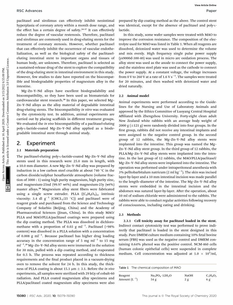
Table 1 The chemical composition of MAO
Reagent Na3PO4$12H2O NaOH C3H8O3
Amount (L�1) 54 g 2 g 6 g
RSC Advances Paper
Ope
n A
cces
s A
rtic
le. P
ublis
hed
on 1
6 A
pril
2020
. Dow
nloa
ded
on 3
/15/
2022
6:5
5:00
PM
. T
his
artic
le is
lice
nsed
und
er a
Cre
ativ
e C
omm
ons
Attr
ibut
ion
3.0
Unp
orte
d L
icen
ce.
View Article Online
paclitaxel and sirolimus can effectively inhibit neointimalhyperplasia of coronary artery within a month dose range, andthe effect has a certain degree of safety.24–27 It can effectivelyreduce the degree of vascular restenosis. Therefore, paclitaxeland sirolimus are commonly used in drug-eluting stents for thetreatment of coronary stenosis. However, whether paclitaxelthat can effectively inhibit the occurrence of vascular endothe-lialization, as well as the biological safety of the paclitaxel-eluting intestinal stent to important organs and tissues ofhuman body, are unknown. Therefore, paclitaxel is selected asa sustained-release drug of the stent to explore the compatibilityof the drug-eluting stent in intestinal environment in this study.However, few studies to date have reported on the biocompat-ible and biodegradable properties of magnesium alloy in theintestine.
Mg–Zn–Y–Nd alloys have excellent biodegradability andbiocompatibility, so they have been used as biomaterials forcardiovascular stent research.28 In this paper, we selected Mg–Zn–Y–Nd alloys as the alloy material of degradable intestinaldrug-eluting stents. The biocompatibility in vitro was evaluatedby the cytotoxicity test. In addition, animal experiments arecarried out by placing scaffolds in different treatment groups.We evaluated the in vivo biocompatibility of a paclitaxel-elutingpoly-L-lactide-coated Mg–Zn–Y–Nd alloy applied as a biode-gradable intestinal stent through animal study.
2. Experiment2.1 Materials preparation
The paclitaxel-eluting poly-L-lactide-coated Mg–Zn–Y–Nd alloystents used in this research were 25.0 mm in length, witha 10.0 mm diameter. A new Mg–Zn–Y–Nd alloy was prepared byinduction in a low carbon steel crucible at about 740 �C in thecarbon dioxide/sulphur hexauoride atmosphere (volume frac-tion of 3000 : 1) using high purity magnesium, high purity zinc,and magnesium-25nd (99.97 wt%) and magnesium-25y (wt%)master alloys.28 Magnesium alloy stent bers were fabricatedusing a single screw extruder. PLLA ((C6H8O4)n, intrinsicviscosity: 3.4 dl g�1 (CHCL3/25 �C)) and paclitaxel were ofreagent grade and purchased from the Science and TechnologyCompany of Solarbio (Beijing, China) and the Academy ofPharmaceutical Sciences (Jinan, China). In this study MAO/PLLA and MAO/PLLA/paclitaxel coatings were prepared usingthe dip coating method. The PLLA was dissolved in dichloro-methane with a proportion of 0.03 g ml�1. Paclitaxel (>98%content) was dissolved in a PLLA solution with a concentrationof 0.008 g ml�1. Because paclitaxel has higher drug loadingaccuracy in the concentration range of 5 mg ml�1 to 15 mgml�1.29 Mg–Zn–Y–Nd alloy stents were immersed in the solutionfor 30 min, pulled with a dip coating method, and evaporatedfor 0.5 h. The process was repeated according to thicknessrequirements and the nal product placed in a vacuum-dryingoven to remove the solvent for 24 h. In this study, the thick-ness of PLLA coating is about 15.1 mm � 3.1. Before the in vivoexperiments, all samples were sterilized with 29 kGy of cobalt-60radiation. And PLLA coated magnesium alloy specimens andPLLA/paclitaxel coated magnesium alloy specimens were also
15080 | RSC Adv., 2020, 10, 15079–15090
prepared by dip coating method as the above. The control stentwas identical, except for the absence of paclitaxel and poly-L-lactide.
In this study, some wafer samples were treated with MAO toimprove the corrosion resistance. The composition of the elec-trolyte used for MAOwas listed in Table 1. When all reagents aredissolved, deionized water was used to determine the volumeand mix evenly. High frequency single pulse power supply(ys9000d-300-40) was used in micro arc oxidation process. Thealloy stent was used as the anode to connect the power supply,and the stainless steel plate was used as the cathode to connectthe power supply. At a constant voltage, the voltage increasesfrom 0 V to 260 V at a rate of 1.6 V s�1. The samples were treatedfor 20 minutes, and then washed with deionized water anddried naturally.
2.2 Animal model
Animal experiments were performed according to the Guide-lines for the Nursing and Use of Laboratory Animals andapproved by the Ethics Committee of Luoyang Central HospitalAffiliated with Zhengzhou University. Forty-eight clean adultNew Zealand white rabbits with an average body weight of2000 g (�121 g) were randomly divided into four groups. In therst group, rabbits did not receive any intestinal implants andwere assigned to the negative control group. In the secondgroup of 12 rabbits, the Mg–Zn–Y–Nd alloy stents wereimplanted into the intestine. This group was named the Mg–Zn–Y–Nd alloy stent group. In the third group of 12 rabbits, thePLLA/Mg–Zn–Y–Nd alloy stents were implanted into the intes-tine. In the last group of 12 rabbits, the MAO/PLLA/paclitaxel/Mg–Zn–Y–Nd alloy stents were implanted into the intestine. Theoperation was performed under intraperitoneal anesthesia with3% pelltobarbitalum natricum (2 ml kg�1). The skin was incisedlayer-by-layer and a 10mm intestinal incision was made parallelto the length–diameter of the intestine. The Mg–Zn–Y–Nd alloystents were embedded in the intestinal incision and theabdomen was sutured layer-by-layer. Aer the operation, about10 ml of sodium chloride were administered to the rabbits. Therabbits were able to conduct regular activities following recoveryof consciousness, including eating and drinking.
2.3 Methods
2.3.1 Cell toxicity assay for paclitaxel loaded in the stent.Indirect contact cytotoxicity test was performed to prove indi-rectly that paclitaxel is loaded in the stent designed in thisstudy. Pure DMEM culturemedium containing 10% fetal bovineserum (FBS) was used as the negative control and DMEM con-taining 0.64% phenol was the positive control. NCM-460 cells(human colonic epithelial cells) were suspended in completemedium. Cell concentration was adjusted at 1.0 � 105/ml,
This journal is © The Royal Society of Chemistry 2020
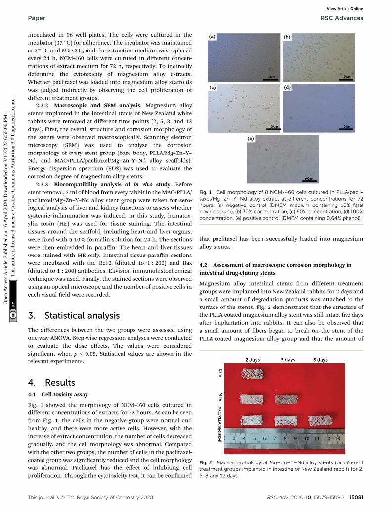
Fig. 1 Cell morphology of 8 NCM-460 cells cultured in PLLA/pacli-taxel/Mg–Zn–Y–Nd alloy extract at different concentrations for 72hours: (a) negative control (DMEM medium containing 10% fetalbovine serum), (b) 30% concentration, (c) 60% concentration, (d) 100%concentration, (e) positive control (DMEM containing 0.64% phenol).
Paper RSC Advances
Ope
n A
cces
s A
rtic
le. P
ublis
hed
on 1
6 A
pril
2020
. Dow
nloa
ded
on 3
/15/
2022
6:5
5:00
PM
. T
his
artic
le is
lice
nsed
und
er a
Cre
ativ
e C
omm
ons
Attr
ibut
ion
3.0
Unp
orte
d L
icen
ce.
View Article Online
inoculated in 96 well plates. The cells were cultured in theincubator (37 �C) for adherence. The incubator was maintainedat 37 �C and 5% CO2, and the extraction medium was replacedevery 24 h. NCM-460 cells were cultured in different concen-trations of extract medium for 72 h, respectively. To indirectlydetermine the cytotoxicity of magnesium alloy extracts.Whether paclitaxel was loaded into magnesium alloy scaffoldswas judged indirectly by observing the cell proliferation ofdifferent treatment groups.
2.3.2 Macroscopic and SEM analysis. Magnesium alloystents implanted in the intestinal tracts of New Zealand whiterabbits were removed at different time points (2, 5, 8, and 12days). First, the overall structure and corrosion morphology ofthe stents were observed macroscopically. Scanning electronmicroscopy (SEM) was used to analyze the corrosionmorphology of every stent group (bare body, PLLA/Mg–Zn–Y–Nd, and MAO/PLLA/paclitaxel/Mg–Zn–Y–Nd alloy scaffolds).Energy dispersion spectrum (EDS) was used to evaluate thecorrosion degree of magnesium alloy stents.
2.3.3 Biocompatibility analysis of in vivo study. Beforestent removal, 3 ml of blood from every rabbit in theMAO/PLLA/paclitaxel/Mg–Zn–Y–Nd alloy stent group were taken for sero-logical analysis of liver and kidney functions to assess whethersystemic inammation was induced. In this study, hematox-ylin–eosin (HE) was used for tissue staining. The intestinaltissues around the scaffold, including heart and liver organs,were xed with a 10% formalin solution for 24 h. The sectionswere then embedded in paraffin. The heart and liver tissueswere stained with HE only. Intestinal tissue paraffin sectionswere incubated with the Bcl-2 (diluted to 1 : 200) and Bax(diluted to 1 : 200) antibodies. Elivision immunohistochemicaltechnique was used. Finally, the stained sections were observedusing an optical microscope and the number of positive cells ineach visual eld were recorded.
3. Statistical analysis
The differences between the two groups were assessed usingone-way ANOVA. Step-wise regression analyses were conductedto evaluate the dose effects. The values were consideredsignicant when p < 0.05. Statistical values are shown in therelevant experiments.
Fig. 2 Macromorphology of Mg–Zn–Y–Nd alloy stents for differenttreatment groups implanted in intestine of New Zealand rabbits for 2,5, 8 and 12 days.
4. Results4.1 Cell toxicity assay
Fig. 1 showed the morphology of NCM-460 cells cultured indifferent concentrations of extracts for 72 hours. As can be seenfrom Fig. 1, the cells in the negative group were normal andhealthy, and there were more active cells. However, with theincrease of extract concentration, the number of cells decreasedgradually, and the cell morphology was abnormal. Comparedwith the other two groups, the number of cells in the paclitaxel-coated group was signicantly reduced and the cell morphologywas abnormal. Paclitaxel has the effect of inhibiting cellproliferation. Through the cytotoxicity test, it can be conrmed
This journal is © The Royal Society of Chemistry 2020
that paclitaxel has been successfully loaded into magnesiumalloy stents.
4.2 Assessment of macroscopic corrosion morphology inintestinal drug-eluting stents
Magnesium alloy intestinal stents from different treatmentgroups were implanted into New Zealand rabbits for 2 days anda small amount of degradation products was attached to thesurface of the stents. Fig. 2 demonstrates that the structure ofthe PLLA-coated magnesium alloy stent was still intact ve daysaer implantation into rabbits. It can also be observed thata small amount of bers began to break on the stent of thePLLA-coated magnesium alloy group and that the amount of
RSC Adv., 2020, 10, 15079–15090 | 15081

Fig. 3 The corrosion morphology observed by SEM of magnesium alloy filaments for different groups of alloy stents after implantation in NewZealand white rabbits for two days: (a and d) bare magnesium alloy stents, (b and e) PLLA coated magnesium alloy stents, (c and f) MAO/PLLA/paclitaxel coated magnesium alloy stents, (a–c) stents located at the unconnected position, (d–f) stents located at the joint.
RSC Advances Paper
Ope
n A
cces
s A
rtic
le. P
ublis
hed
on 1
6 A
pril
2020
. Dow
nloa
ded
on 3
/15/
2022
6:5
5:00
PM
. T
his
artic
le is
lice
nsed
und
er a
Cre
ativ
e C
omm
ons
Attr
ibut
ion
3.0
Unp
orte
d L
icen
ce.
View Article Online
degradation products attached to the surface increased.However, no magnesium alloy stent was found in the rabbitintestinal tract in the bare stent group aer implantation for 5days. Aer stent implantation for 8 days, only the MAO/PLLA/paclitaxel-coated Mg–Zn–Y–Nd stents were found in therabbits. No stents were found in the other two groups. More-over, no magnesium alloy stents were found in the three groupsaer implantation into the rabbit intestinal tract for 12 days.
4.3 SEM corrosion morphology assessment in intestinaldrug-eluting stents
Fig. 3 represents the local corrosion morphology of magnesiumalloy stent bers implanted for 2 days. A few cracks and akingformed on the surface of the silks, while the stent structure foruncoated Mg–Zn–Y–Nd alloy remained intact (Fig. 3a and d).The number of cracks and debris adhesions to the surface ofscaffold threads in the PLLA-coated stent group was muchsmaller than that in the exposed group. However, surface of thescaffold threads in the MAO/PLLA/paclitaxel-coated stent groupwas smoother than that of bare Mg–Zn–Y–Nd alloy stents,showing better resistance to corrosion compared to that of bareMg–Zn–Y–Nd alloy stents (Fig. 3c and f). The surfaces of thethreads at the end of the bracket was rougher than that insidethe stent and there were more small cracks (Fig. 3).
4.4 EDS spectra corrosion morphology assessment inintestinal drug-eluting stents
SEM and EDS results 2 days aer implantation of stents fromdifferent treatment groups are represented in Fig. 4. A lot of
15082 | RSC Adv., 2020, 10, 15079–15090
degradation products were present on the surface of bare scaf-fold laments (Fig. 4a), where the content of P and Ca was10.48% and 15.18%, respectively (Fig. 4b). Gray-white degra-dation products were present on the surface of the PLLA-coatedstent laments (Fig. 4c), with the content of P and Ca of 5.69%and 5.72%, respectively (Fig. 4d). A small amount of degrada-tion products was present on the surface of the MAO/PLLA/paclitaxel coated scaffolds (Fig. 4e), with the content of P andCa of 3.70% and 1.33%, respectively (Fig. 4f). These resultsshowed that degradation products contained some insolubleinorganic substances such as magnesium carbonate andcalcium phosphate. Moreover, the content of P and Ca in theMAO/PLLA/paclitaxel-coated stent group was signicantlysmaller than that in the other two groups (p < 0.05), while thecontents of P and Ca in the PLLA-coated stent group wassignicantly smaller than that in the bare stent group (p < 0.05).EDS analysis showed that the MAO/PLLA/paclitaxel-coated stentgroup had better corrosion resistance than the other twogroups.
4.5 Histopathological assessment of important organs
Pathological assessment images of important rabbit organsaer implantation of the MAO/PLLA/paclitaxel-coated Mg–Zn–Y–Nd alloy stents at 2 and 8 days were shown in Fig. 5.Myocardial ber morphology was presented in Fig. 5a and d,with no obvious abnormality of nuclear structure and no inl-tration of inammatory cells in the interstitial cells found ateach stage. There was no obvious abnormality in themorphology of hepatocytes, as evident from the structure of
This journal is © The Royal Society of Chemistry 2020
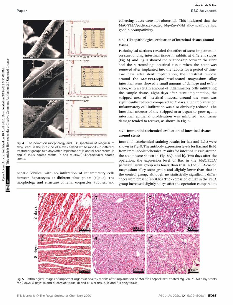
Fig. 4 The corrosion morphology and EDS spectrum of magnesiumalloy stent in the intestine of New Zealand white rabbits in differenttreatment groups two days after implantation: (a and b) bare stents, (cand d) PLLA coated stents, (e and f) MAO/PLLA/paclitaxel coatedstents.
Paper RSC Advances
Ope
n A
cces
s A
rtic
le. P
ublis
hed
on 1
6 A
pril
2020
. Dow
nloa
ded
on 3
/15/
2022
6:5
5:00
PM
. T
his
artic
le is
lice
nsed
und
er a
Cre
ativ
e C
omm
ons
Attr
ibut
ion
3.0
Unp
orte
d L
icen
ce.
View Article Online
hepatic lobules, with no inltration of inammatory cellsbetween hepatocytes at different time points (Fig. 5). Themorphology and structure of renal corpuscles, tubules, and
Fig. 5 Pathological images of important organs in healthy rabbits after imfor 2 days, 8 days: (a and d) cardiac tissue, (b and e) liver tissue, (c and f)
This journal is © The Royal Society of Chemistry 2020
collecting ducts were not abnormal. This indicated that theMAO/PLLA/paclitaxel-coated Mg–Zn–Y–Nd alloy scaffolds hadgood biocompatibility.
4.6 Histopathological evaluation of intestinal tissues aroundstents
Pathological sections revealed the effect of stent implantationon surrounding intestinal tissue in rabbits at different stages(Fig. 6). And Fig. 7 showed the relationship between the stentand the surrounding intestinal tissue when the stent wasremoved aer implanted into the rabbits for a period of time.Two days aer stent implantation, the intestinal mucosaaround the MAO/PLLA/paclitaxel-coated magnesium alloyintestinal stent showed a small amount of damage and exfoli-ation, with a certain amount of inammatory cells inltratingthe sample tissue. Eight days aer stent implantation, thestripped area of intestinal mucosa around the stent wassignicantly reduced compared to 2 days aer implantation.Inammatory cell inltration was also obviously reduced. Theintestinal mucosa of the stripped area began to grow again,intestinal epithelial proliferation was inhibited, and tissuedamage tended to recover, as shown in Fig. 6.
4.7 Immunohistochemical evaluation of intestinal tissuesaround stents
Immunohistochemical staining results for Bax and Bcl-2 wereshown in Fig. 8. The antibody expression levels for Bax and Bcl-2from immunohistochemical results for intestinal tissue aroundthe stents were shown in Fig. 8A(a and b). Two days aer theoperation, the expression level of Bax in the MAO/PLLA/paclitaxel stent group was lower than that in the PLLA-coatedmagnesium alloy stent group and slightly lower than that inthe control group, although no statistically signicant differ-ences were present (p > 0.05). The expression of Bax in the PLLAgroup increased slightly 5 days aer the operation compared to
plantation of MAO/PLLA/paclitaxel coated Mg–Zn–Y–Nd alloy stentskidney tissue.
RSC Adv., 2020, 10, 15079–15090 | 15083
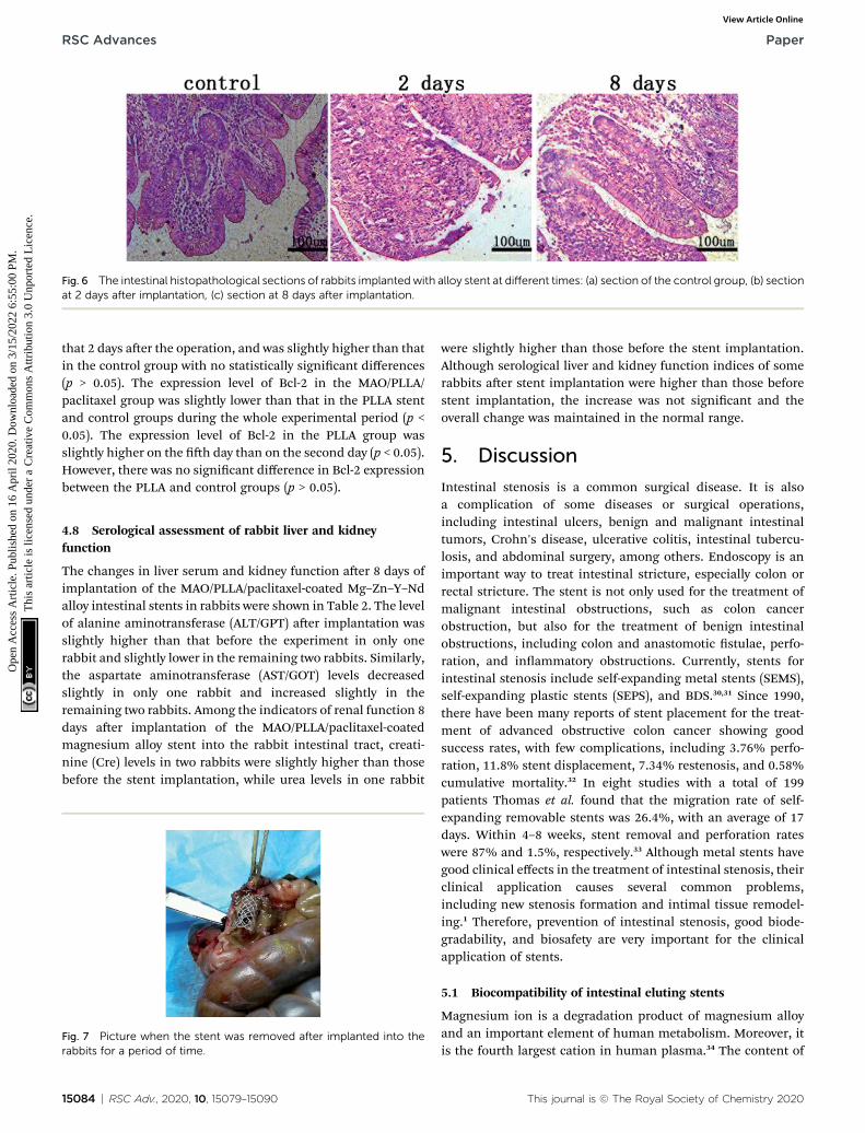
Fig. 6 The intestinal histopathological sections of rabbits implantedwith alloy stent at different times: (a) section of the control group, (b) sectionat 2 days after implantation, (c) section at 8 days after implantation.
RSC Advances Paper
Ope
n A
cces
s A
rtic
le. P
ublis
hed
on 1
6 A
pril
2020
. Dow
nloa
ded
on 3
/15/
2022
6:5
5:00
PM
. T
his
artic
le is
lice
nsed
und
er a
Cre
ativ
e C
omm
ons
Attr
ibut
ion
3.0
Unp
orte
d L
icen
ce.
View Article Online
that 2 days aer the operation, and was slightly higher than thatin the control group with no statistically signicant differences(p > 0.05). The expression level of Bcl-2 in the MAO/PLLA/paclitaxel group was slightly lower than that in the PLLA stentand control groups during the whole experimental period (p <0.05). The expression level of Bcl-2 in the PLLA group wasslightly higher on the h day than on the second day (p < 0.05).However, there was no signicant difference in Bcl-2 expressionbetween the PLLA and control groups (p > 0.05).
4.8 Serological assessment of rabbit liver and kidneyfunction
The changes in liver serum and kidney function aer 8 days ofimplantation of the MAO/PLLA/paclitaxel-coated Mg–Zn–Y–Ndalloy intestinal stents in rabbits were shown in Table 2. The levelof alanine aminotransferase (ALT/GPT) aer implantation wasslightly higher than that before the experiment in only onerabbit and slightly lower in the remaining two rabbits. Similarly,the aspartate aminotransferase (AST/GOT) levels decreasedslightly in only one rabbit and increased slightly in theremaining two rabbits. Among the indicators of renal function 8days aer implantation of the MAO/PLLA/paclitaxel-coatedmagnesium alloy stent into the rabbit intestinal tract, creati-nine (Cre) levels in two rabbits were slightly higher than thosebefore the stent implantation, while urea levels in one rabbit
Fig. 7 Picture when the stent was removed after implanted into therabbits for a period of time.
15084 | RSC Adv., 2020, 10, 15079–15090
were slightly higher than those before the stent implantation.Although serological liver and kidney function indices of somerabbits aer stent implantation were higher than those beforestent implantation, the increase was not signicant and theoverall change was maintained in the normal range.
5. Discussion
Intestinal stenosis is a common surgical disease. It is alsoa complication of some diseases or surgical operations,including intestinal ulcers, benign and malignant intestinaltumors, Crohn's disease, ulcerative colitis, intestinal tubercu-losis, and abdominal surgery, among others. Endoscopy is animportant way to treat intestinal stricture, especially colon orrectal stricture. The stent is not only used for the treatment ofmalignant intestinal obstructions, such as colon cancerobstruction, but also for the treatment of benign intestinalobstructions, including colon and anastomotic stulae, perfo-ration, and inammatory obstructions. Currently, stents forintestinal stenosis include self-expanding metal stents (SEMS),self-expanding plastic stents (SEPS), and BDS.30,31 Since 1990,there have been many reports of stent placement for the treat-ment of advanced obstructive colon cancer showing goodsuccess rates, with few complications, including 3.76% perfo-ration, 11.8% stent displacement, 7.34% restenosis, and 0.58%cumulative mortality.32 In eight studies with a total of 199patients Thomas et al. found that the migration rate of self-expanding removable stents was 26.4%, with an average of 17days. Within 4–8 weeks, stent removal and perforation rateswere 87% and 1.5%, respectively.33 Although metal stents havegood clinical effects in the treatment of intestinal stenosis, theirclinical application causes several common problems,including new stenosis formation and intimal tissue remodel-ing.1 Therefore, prevention of intestinal stenosis, good biode-gradability, and biosafety are very important for the clinicalapplication of stents.
5.1 Biocompatibility of intestinal eluting stents
Magnesium ion is a degradation product of magnesium alloyand an important element of human metabolism. Moreover, itis the fourth largest cation in human plasma.34 The content of
This journal is © The Royal Society of Chemistry 2020

Fig. 8 The histogram represents the expression level of two antibodies and immunohistochemical section: (a and e) section of the controlgroup, (b and f) section at 2 days after implantation, (c and g) section at 5 days after implantation.
Table 2 Serological index content of liver function and renal function
Index of liver function(mmol L�1)
Index of renalfunction (mmol L�1)
ALT/GPT AST/GOT Cre/Cre� UREA
Control M1 107 121 44 5.6M2 112 118 52 4.5M3 114 132 49 5.4
8 days M1 116 106 42 4.4M2 107 120 56 3.6M3 109 138 53 5.7
Paper RSC Advances
Ope
n A
cces
s A
rtic
le. P
ublis
hed
on 1
6 A
pril
2020
. Dow
nloa
ded
on 3
/15/
2022
6:5
5:00
PM
. T
his
artic
le is
lice
nsed
und
er a
Cre
ativ
e C
omm
ons
Attr
ibut
ion
3.0
Unp
orte
d L
icen
ce.
View Article Online
magnesium in bone is about two-thirds of the total amount ofmagnesium in the human body. The other one-third is found intissue and about 1–2% of magnesium is in the extracellularuid.35 In addition, magnesium ions play an important role inregulating the homeostasis of human body. They can inhibit therelease of calcium from sarcoplasmic reticulum in response toa sudden inux of extracellular calcium.36 Early typical symp-toms, such as anorexia, nausea, vomiting, and sleepiness, occurwhen magnesium is decient.37 Research has shown thatdrinking natural mineral water rich in magnesium sulfateseems to be a rst-line solution to functional constipationbefore starting medication.38 Di et al. used degradable magne-sium alloy Mg–1Sr, a new compound, to study its biocompati-bility in vivo in New Zealand white rabbits.39 Histological studiesdid not reveal physiological abnormalities or diseases. It hasbeen reported that appropriate amounts of zinc are veryimportant for regulating the immune response. Zinc supple-mentation has a potential effect on immune function impair-ment caused by a decrease in serum zinc concentration due to
This journal is © The Royal Society of Chemistry 2020
advanced age.40 Without affecting the normal healing process,the main challenge of stenting in the treatment of coronaryheart disease is to prevent restenosis.41 Therefore, stent designwith good compatibility and prevention of intestinal stricture iscritical for the treatment of intestinal stricture.
RSC Adv., 2020, 10, 15079–15090 | 15085

RSC Advances Paper
Ope
n A
cces
s A
rtic
le. P
ublis
hed
on 1
6 A
pril
2020
. Dow
nloa
ded
on 3
/15/
2022
6:5
5:00
PM
. T
his
artic
le is
lice
nsed
und
er a
Cre
ativ
e C
omm
ons
Attr
ibut
ion
3.0
Unp
orte
d L
icen
ce.
View Article Online
In this study, it was proved that paclitaxel had beensuccessfully loaded into magnesium alloy stents through thecytotoxicity test. Aer the MAO/PLLA/paclitaxel/Mg–Zn–Y–Ndalloy stents were implanted into rabbits in this study, nosignicant abnormalities were found in the pathological anal-ysis images of the heart, liver, and kidneys. In addition, sero-logical indicators of liver and kidney function in rabbits showedno signicant abnormalities before and aer stent implanta-tion. This may be related to the fact that the stent does not affectnormal metabolism of magnesium ions in rabbits. Xu et al.prepared organic coatings with PLLA on pure Mg substrate bythe spin-coating method, and the research demonstrated thatPLLA lms could signicantly improve SaOS-2 cell cyto-compatibility of the alloy.17 There are also previous reports thatexcessive magnesium ion concentration or alkaline stress couldproduce negative stimuli for the cell population.42 Moreover,low magnesium diet may lead to higher intra-cellular ratio ofCa : Mg, leading to hypertension and insulin resistance.43 Aprecious study revealed that although low magnesium intake isrelated to constipation, high doses of oral magnesium havea defecation effect.44 This effect of magnesiummay be related tothe following facts: they are absorbed from the intestinal cavityand play a penetrating role to maintain water, therebyincreasing the uidity of the lumen content.45
Paclitaxel is a natural compound, which is isolated from thePacic Redwood. Paclitaxel reacts with b-tubulin inmicrotubuleto induce microtubule polymerization, which leads to cellproliferation stopping at G2/M phase and nally result in tumorcell apoptosis.46 Previous studies have shown that stentimplantation can cause the formation of endothelialization inthe surrounding tissue.47–49 Although the time of stent implan-tation in intestinal tract is short, there may not be obvioustissue proliferation around the stent. However, the number ofepithelial cells and broblasts in intestinal mucosa tissue isincreased in the process of intestinal endothelialization.49
While the antibody used in immunohistochemistry can bedetected by the detection of cell proliferation and expression ofrelated factors were used to indirectly explore the compatibilityof stent on intestinal tissue. In this study, histopathologicalexamination showed that the MAO/PLLA/paclitaxel/Mg–Zn–Y–Nd and PLLA/Mg–Zn–Y–Nd alloy scaffolds did not inducesystemic inammation (p > 0.05), while immunohistochemicalresults showed that degradation products of the MAO/PLLA/paclitaxel/Mg–Zn–Y–Nd alloy scaffolds signicantly inhibitedproliferation of intestinal cells. The PLLA/Mg–Zn–Y–Nd scaffolddegradation products did not induce signicant proliferation ofintestinal tissue cells (especially epithelial cells) (p > 0.05).These results suggest that short-term implantation of the MAO/PLLA/paclitaxel/Mg–Zn–Y–Nd alloy scaffolds can signicantlyinhibit the endothelialization of intestinal epithelial cells. ThePLLA/Mg–Zn–Y–Nd alloy stents were not conducive to theformation of intestinal endothelialization. It has been reportedthat the use of SEPS can cause low epithelial proliferation andmake the stents easy to remove.50 In a study of drug-loadedcardiovascular stents, it was found that proliferation of ratvascular smooth muscle cells (SMCs) was successfully inhibitedwhen paclitaxel was released from the poly(carbonate urethane)
15086 | RSC Adv., 2020, 10, 15079–15090
urea coating.51 Stephen et al. found that a magnesium-baseddrug delivery system had a stronger long-term inhibitoryeffect on the proliferation of SMCs cultured in vitro compared tostainless steel, which may be related to the degradation ofmagnesium alloy matrix greatly accelerating and improving thepharmacokinetics of drug release in vitro.52
In this study, ve rabbits were found to have stents displacedduring their removal. No other organ damage, perforation, orobstruction were found. Geiger et al. analyzed 26 original arti-cles, where 63 patients received long-term treatment with SEMS.Severe complications were found, including bladder perfora-tion, ileostomy, massive hemorrhage, and obstruction.53 Theclinical data for self-expanding metal colon stent implantationin some hospital from January 1996 to May 2012 were retro-spectively analyzed. The surgical technique success rate was92.26% (n¼ 441), with clinical success rate of 78.45% (n¼ 375),and complication rate during follow-up of 18.5%. The incidenceof complications with stainless steel stents was higher than thatwith nickel–titanium alloy stents.54
5.2 Degradation characteristics of intestinal eluting stentsin a rabbit model
It is well known that traditional medical and scaffoldingmaterials such as stainless steel and titanium alloy are non-absorbable. Previous studies have found that long-term reten-tion of these non-absorbable materials in the human body cancause damage to human health.55 Therefore, magnesium alloyscaffolds with biodegradable properties are of great signicancefor the treatment of intestinal diseases. Magnesium-basedsubstrates easily react in aqueous media in the followingmanner:56
Mg(s) + 2H2O(aq) ! Mg(OH)2(s) + H2(g)[ (1)
Mg(OH)2(s) ! Mg2+(aq) + 2OH�(aq) (2)
Hydrogen and insoluble magnesium hydroxide are producedin this reaction, which directly changes the concentration ofmagnesium ion, pH, and other biochemical conditions.However, these changes may have some impact on humanhealth.
In this study, macroscopic corrosion morphology of Mg–Zn–Y–Nd alloy intestinal stents implanted into New Zealand whiterabbits showed that bare stents can be located only aer 2 daysof implantation. PLLA-coated stents could not be found 8 daysaer implantation. The MAO/PLLA/paclitaxel-coated stentswere intact at 2, 5, and 8 days, and no longer present in theintestinal tract at 12 days. This phenomenon may be related tothe stent structure collapse caused by stent degradation aer 8days of implantation in rabbits and its eventual removal fromthe intestine. The bare body scaffold completely degraded 5days aer implantation, most likely because the scaffoldwithout a protective layer comes into direct contact with theintestinal environment and has poor corrosion resistance, thusaccelerating scaffold degradation. In the PLLA/Mg–Zn–Y–Ndalloy scaffold group, a small amount of bers on the scaffoldbegan to break, while the amount of surface degradation
This journal is © The Royal Society of Chemistry 2020
Paper RSC Advances
Ope
n A
cces
s A
rtic
le. P
ublis
hed
on 1
6 A
pril
2020
. Dow
nloa
ded
on 3
/15/
2022
6:5
5:00
PM
. T
his
artic
le is
lice
nsed
und
er a
Cre
ativ
e C
omm
ons
Attr
ibut
ion
3.0
Unp
orte
d L
icen
ce.
View Article Online
products increased. However, the PLLA-coated magnesiumstents were not found in the rabbit intestines aer 8 and 12 daysof stent implantation. This phenomenon indicates that theMAO/PLLA/paclitaxel/Mg–Zn–Y–Nd alloy scaffolds havestronger corrosion resistance than the PLLA/Mg–Zn–Y–Nd alloyscaffolds. SEM analysis indicated that the microwire surface ofthe MAO/PLLA/paclitaxel scaffolds is more complete andcleaner than the other two groups, as shown in Fig. 3. EDSresults in Fig. 4 showed that the content of P and Ca in theMAO/PLLA/paclitaxel stent group is signicantly lower than that inthe other two groups, which is related to better corrosionresistance of the MAO/PLLA/paclitaxel stents in intestinalenvironment and fewer corrosion products. In conclusion,these results suggest that stents treated with the MAO/PLLA/paclitaxel coating can signicantly improve corrosion resis-tance of the magnesium alloy. Previous studies have conrmedthat the PLLA-coated alloy surface can signicantly delay alloydegradation rate, thereby improving corrosion resistance ofalloy materials.52,57 Application of micro-arc oxidation cansignicantly enhance corrosion resistance of biodegradableimplants.58
In clinical application, tissue self-healing usually takes 2weeks to a month, and in a study of a rat model, it was shownthat the internal environment of the wound also returned tonormal when the polymer scaffold was degraded within 14days.59 However, the stent in this study can be completelydegraded within 12 days, and it's a little shorter than expectedin this study. As for the degradation characteristics of the stent,although the degradation time is faster than expected, and thecurrent stent can not meet the clinical needs at present.However, this is only the current degradation characteristics ofthe stent we studied, which will provide new measures for thefuture high corrosion resistance of the stent so as to developa suitable degradation rate to meet the clinical needs of intes-tinal stent to provide reference.
5.3 Current issues associated with magnesium alloyapplications in clinical medicine
This study showed that the MAO/PLLA/paclitaxel/Mg–Zn–Y–Ndalloy scaffolds had better corrosion resistance than bare PLLA/Mg–Zn–Y–Nd alloy scaffolds. One of the necessary conditionsfor good intestinal scaffolds was a stable degradation rate andstructural stability during wound healing. When researchersimplanted intestinal stents in rats, they found that the woundedtissue began to return to normal aer 14 days.60 In this study,the stent completely lost its supporting effect aer 12 days.Rapid degradation characteristics of the stent were not enoughto support the healing of intestinal obstruction, which is relatedto the more active properties of magnesium. Especially ina complex intestinal environment, the contact betweenmagnesium alloy and intestinal organic and inorganicsubstances accelerates its degradation rate. However, appro-priate degradation rate can be achieved by creating a magne-sium alloy stent with a stable structure and sufficient branchesduring tissue healing. Therefore, it is necessary to furtherimprove the degradable magnesium alloy scaffold to delay its
This journal is © The Royal Society of Chemistry 2020
degradation rate. At present, there are many common ways toimprove corrosion resistance by changing the alloy surface,such as micro-arc oxidation to form a ceramic layer, impreg-nation of macromolecule polymers, anodic oxidation treat-ment, steam treatment, alkali heat treatment, uoridetreatment, ion implantation input, and physical vapor deposi-tion.61 However, more research on magnesium alloy degrada-tion rate control is needed. Unremitting efforts have to be madeto improve corrosion resistance of magnesium alloy stents inthe intestinal environment to support intestinal structure, sothat magnesium alloy stents can adapt to the complex andchangeable intestinal environment, thus improving clinicaltreatment of intestinal obstruction diseases.
6. Conclusion
In this study, Mg–Zn–Y–Nd alloy laments were woven into thereticulated scaffolds with inner diameter of 8 mm and length of20 mm using a monolament integral braiding method.Magnesium alloy stents exposed to different treatments wereimplanted into the intestinal tracts of New Zealand whiterabbits and the degradation and supporting properties of theintestinal stents coated with paclitaxel were investigated. Theresults showed that the MAO/PLLA/paclitaxel/Mg–Zn–Y–Ndalloy intestinal stents had better corrosion resistance than thePLLA/Mg–Zn–Y–Nd alloy intestinal stents. When the MAO/PLLA/paclitaxel/Mg–Zn–Y–Nd alloy intestinal stents wereimplanted into rabbits for 8 to 12 days, stents degraded a greatdeal in vivo, which led to stent structure collapse and dischargefrom the body. Pathological observations demonstrated that theMAO/PLLA/paclitaxel double-coated drug-eluting Mg–Zn–Y–Ndalloy stents had no signicant toxicity to important organs andthat the intestinal mucosa around the stent gradually returnedto normal within two weeks. There were no signicant differ-ences in serological parameters at different degradation stages.The scaffolds also exhibited good biocompatibility. Immuno-histochemical evaluation of local intestinal tissue around thestent showed that the PLLA/paclitaxel-coated intestinal stentcould inhibit intestinal tissue proliferation. This mechanismprovided theoretical support for the future treatment design ofintestinal stenosis caused by benign, malignant, and inam-matory hyperplasia.
Data availability statement
The raw/processed data required to reproduce these ndingscannot be shared at this time as the data also forms part of anongoing study.
Conflicts of interest
There are no conicts to declare.
Acknowledgements
This research was funded by the National Natural ScienceFoundation of China (No. U1404825), the Key Scientic and
RSC Adv., 2020, 10, 15079–15090 | 15087
RSC Advances Paper
Ope
n A
cces
s A
rtic
le. P
ublis
hed
on 1
6 A
pril
2020
. Dow
nloa
ded
on 3
/15/
2022
6:5
5:00
PM
. T
his
artic
le is
lice
nsed
und
er a
Cre
ativ
e C
omm
ons
Attr
ibut
ion
3.0
Unp
orte
d L
icen
ce.
View Article Online
Technological Projects of Henan Province of China (No.152102310012), the Natural Science Foundation of HenanProvince (No. 162300410241), and the Science and TechnologyDevelopment Projects of Luoyang City (No. 1503006A-3).
References
1 Z. Wang, L. Nan, L. Rui, Y. Li and L. Ruan, Biodegradableintestinal stents: a review, Prog. Nat. Sci.: Mater. Int., 2014,24(5), 423–432.
2 R. P. Wadhwa, R. A. Kozarek, B. R. E. France, J. J. Brandabur,M. M. Gluck, D. E. Low, W. L. Traverso and M. R. Moonka,Use of self-expandable metallic stents in benign GIdiseases, Gastrointest. Endosc., 2003, 58(2), 207–212.
3 S. Karagul, M. A. Yagci, C. Ara, A. Tardu, I. Ertugrul,S. Kirmizi and F. Sumer, Small bowel perforation due toa migrated esophageal stent: report of a rare case andreview of the literature, International Journal of Surgery CaseReports, 2015, 11, 113–116.
4 J. Huang, S. Hao, F. Yang, Y. Di, L. Yao, J. Li, Y. Jiang,L. Zhong, D. Fu and C. Jin, Endoscopic metal enteral stentplacement for malignant afferent loop syndrome aerpancreaticoduodenectomy, Videosurg. Other MiniinvasiveTech., 2015, 10(2), 257–265.
5 S. Ngamruengphong, R. Z. Sharaiha, A. Sethi, A. A. Siddiqui,C. J. DiMaio, S. Gonzalez, J. Im, J. N. Rogart, S. Jagroop andJ. Widmer, Endoscopic suturing for the prevention of stentmigration in benign upper gastrointestinal conditions:a comparative multicenter study, Endoscopy, 2016, 48(09),802–808.
6 F. Cereatti, F. Fiocca, J.-L. Dumont, V. Ceci, B.-M. Vergeau,T. Tuszynski, B. Meduri and G. Donatelli, Fully coveredself-expandable metal stent in the treatment ofpostsurgical colorectal diseases: outcome in 29 patients,Ther. Adv. Gastroenterol., 2016, 9(2), 180–188.
7 H. Hermawan, D. Dube and D. Mantovani, Developments inmetallic biodegradable stents*, Acta Biomater., 2010, 6(5),1693–1697.
8 L. Xu, F. Pan, G. Yu, L. Yang, E. Zhang and K. Yang, In vitroand in vivo evaluation of the surface bioactivity of a calciumphosphate coated magnesium alloy, Biomaterials, 2009,30(8), 1512–1523.
9 R. Erbel, M. C. Di, J. Bartunek, J. Bonnier, B. B. De,F. R. Eberli, P. Erne, M. Haude, B. Heublein andM. Horrigan, Temporary scaffolding of coronary arterieswith bioabsorbable magnesium stents: a prospective, non-randomised multicentre trial, Lancet, 2007, 369(9576),1869–1875.
10 D. Zhao, F. Witte, F. Lu, J. Wang, J. Li and L. Qin, Currentstatus on clinical applications of magnesium-basedorthopaedic implants: a review from clinical translationalperspective, Biomaterials, 2017, 112, 287–302.
11 R. Radha and D. Sreekanth, Insight of magnesium alloys andcomposites for orthopedic implant applications–a review, J.Magnesium Alloys, 2017, 5(3), 286–312.
12 S. Agarwal, J. Curtin, B. Duffy and S. Jaiswal, Biodegradablemagnesium alloys for orthopaedic applications: a review on
15088 | RSC Adv., 2020, 10, 15079–15090
corrosion, biocompatibility and surface modications,Mater. Sci. Eng., C, 2016, 68, 948–963.
13 H. Mueller, P. Uggowitzer and J. Loeffler, Corrosion resistantstent, US Pat., US2018223406 (A1), 2018.
14 C. Chen, J. Chen, W. Wu, Y. Shi, L. Jin, L. Petrini, L. Shen,G. Yuan, W. Ding and J. Ge, In vivo and in vitro evaluationof a biodegradable magnesium vascular stent designed byshape optimization strategy, Biomaterials, 2019, 221, 119414.
15 D. Jain, E. Mahmood and S. Singhal, Biodegradable Stents, J.Clin. Gastroenterol., 2017, 51(4), 295–299.
16 M. O. Platt, A. J. Roman, A. Wells, D. A. Lauffenburger andL. G. Griffith, Sustained epidermal growth factor receptorlevels and activation by tethered ligand binding enhancesosteogenic differentiation of multi-potent marrow stromalcells, J. Cell. Physiol., 2010, 221(2), 306–317.
17 L. Xu and A. Yamamoto, In vitro degradation ofbiodegradable polymer-coated magnesium under cellculture condition, Appl. Surf. Sci., 2012, 258(17), 6353–6358.
18 T. Ushida, K. Furukawa, K. Toita and T. Tateishi, Three-Dimensional Seeding of Chondrocytes Encapsulated inCollagen Gel into PLLA Scaffolds, Cell Transplant., 2002,11(5), 489–494.
19 M. Diez, M.-H. Kang, S.-M. Kim, H.-E. Kim and J. Song,Hydroxyapatite (HA)/poly-L-lactic acid (PLLA) dual coatingon magnesium alloy under deformation for biomedicalapplications, J. Mater. Sci.: Mater. Med., 2016, 27(2), 34–42.
20 A. Witecka, A. Yamamoto, J. Idaszek, A. Chlanda andW. Swieszkowski, Inuence of biodegradable polymercoatings on corrosion, cytocompatibility and cellfunctionality of Mg-2.0 Zn-0.98 Mn magnesium alloy,Colloids Surf., B, 2016, 144, 284–292.
21 L.-Y. Li, L.-Y. Cui, R.-C. Zeng, S.-Q. Li, X.-B. Chen, Y. Zhengand M. B. Kannan, Advances in functionalized polymercoatings on biodegradable magnesium alloys-A review, ActaBiomater., 2018, 79, 23–36.
22 G. Sundararajan and L. R. Krishna, Mechanisms underlyingthe formation of thick alumina coatings through the MAOcoating technology, Surf. Coat. Technol., 2003, 167(2–3),269–277.
23 J. Cai, F. Cao, L. Chang, J. Zheng, J. Zhang and C. Cao, Thepreparation and corrosion behaviors of MAO coating onAZ91D with rare earth conversion precursor lm, Appl.Surf. Sci., 2011, 257(8), 3804–3811.
24 Y. Shi, L. Sun, G. Chen, D. Zheng, L. Li and W. Wei, Acombination of the telomerase inhibitor, BIBR1532, andpaclitaxel synergistically inhibit cell proliferation in breastcancer cell lines, Targeted Oncology, 2015, 10(4), 565–573.
25 D. I. Axel, W. Kunert, C. Goggelmann, M. Oberhoff,C. Herdeg, A. Kuttner, D. H. Wild, B. R. Brehm, R. Riessenand G. Koveker, Paclitaxel inhibits arterial smooth musclecell proliferation and migration in vitro and in vivo usinglocal drug delivery, Circulation, 1997, 96(2), 636–645.
26 G. Eberhard, S. Sigmund, H. K. Eugen, M. Ralf, B. Lutz,G. Ulrich and E. R. Mary, TAXUS, I: six-and twelve-monthresults from a randomized, double-blind trial on a slow-release paclitaxel-eluting stent for de novo coronarylesions, Circulation, 2003, 107(1), 38–42.
This journal is © The Royal Society of Chemistry 2020
Paper RSC Advances
Ope
n A
cces
s A
rtic
le. P
ublis
hed
on 1
6 A
pril
2020
. Dow
nloa
ded
on 3
/15/
2022
6:5
5:00
PM
. T
his
artic
le is
lice
nsed
und
er a
Cre
ativ
e C
omm
ons
Attr
ibut
ion
3.0
Unp
orte
d L
icen
ce.
View Article Online
27 X. Wang, Q. Zeng, Z. Li, X. Yang, W. Xia and Z. Chen, Adjudinsynergizes with paclitaxel and inhibits cell growth andmetastasis by regulating the sirtuin 3–Forkhead box O3aaxis in human small-cell lung cancer, Thoracic Cancer,2019, 10(4), 642–658.
28 J. Wang, L. Wang, S. Guan, S. Zhu, C. Ren and S. Hou,Microstructure and corrosion properties of as sub-rapidsolidication Mg–Zn–Y–Nd alloy in dynamic simulatedbody uid for vascular stent application, J. Mater. Sci.:Mater. Med., 2010, 21(7), 2001–2008.
29 S. Petersen, S. Kaule, F. Stein, I. Minrath, K. P. Schmitz,U. Kragl and K. Sternberg, Novel paclitaxel-coatedangioplasty balloon catheter based on cetylpyridiniumsalicylate: preparation, characterization and simulated usein an in vitro vessel model, Mater. Sci. Eng., C, 2013, 33(7),4244–4250.
30 N. Srinivasan and R. A. Kozarek, Stents for colonic strictures:materials, designs, and more, Tech. Gastrointest. Endosc.,2014, 16(3), 100–107.
31 M. Jovani, C. Genco, I. Bravata and A. Repici, Stents in themanagement of benign colorectal strictures, Tech.Gastrointest. Endosc., 2014, 16(3), 135–141.
32 S. Shaji, J. Sean, G. Tony, T. William and B. Martin, Pooledanalysis of the efficacy and safety of self-expanding metalstenting in malignant colorectal obstruction, Am. J.Gastroenterol., 2004, 99(10), 2051–2057.
33 T. Thomas, K. R. Abrams, V. Subramanian, J. Mannath andK. Ragunath, Esophageal stents for benign refractorystrictures: a meta-analysis, Endoscopy, 2011, 43(05), 386–393.
34 J. Vormann, Magnesium: nutrition and metabolism, Mol.Aspects Med., 2003, 24(1), 27–37.
35 M. E. Maguire and J. A. Cowan, Magnesium chemistry andbiochemistry, BioMetals, 2002, 203–210.
36 L. T. Iseri and J. H. French, Magnesium: nature's physiologiccalcium blocker, Am. Heart J., 1984, 108(1), 188–193.
37 N. E. L. Saris, E. Mervaala, H. Karppanen, J. A. Khawaja andA. Lewenstam, Magnesium: an update on physiological,clinical and analytical aspects, Clin. Chim. Acta, 2000,294(1), 1–26.
38 D. Christophe, C. Alain and C. Florence, Efficacy and safetyof a magnesium sulfate-rich natural mineral water forpatients with functional constipation, Clin. Gastroenterol.Hepatol., 2014, 12(8), 1280–1287.
39 T. Di, R. Guan, H. Liu, A. Cipriano, Y. Liu, Q. Wang, Y. Huangand N. Hort, An in vivo study on the metabolism andosteogenic activity of bioabsorbable Mg–1Sr alloy, ActaBiomater., 2015, 29, 455–467.
40 J. B. Barnett, M. C. Dao, D. H. Hamer, R. Kandel, G. Brandeis,D. Wu, G. E. Dallal, P. F. Jacques, R. Schreiber and E. Kong,Effect of zinc supplementation on serum zinc concentrationand T cell proliferation in nursing home elderly:a randomized, double-blind, placebo-controlled trial, Am. J.Clin. Nutr., 2016, 103(3), 942–951.
41 P. W. Serruys, B. H. Strauss, H. M. V. Beusekom andW. J. V. D. Giessen, Stenting of coronary arteries: hasa modern Pandora's box been opened?, J. Am. Coll.Cardiol., 1991, 17(6), 143B–154B.
This journal is © The Royal Society of Chemistry 2020
42 Z. Wang, J. Yan, J. Li, Q. Zheng, Z. Wang, X. Zhang andS. Zhang, Effects of biodegradable Mg–6Zn alloy extractson apoptosis of intestinal epithelial cells, Mater. Sci. Eng.,B, 2012, 177(4), 388–393.
43 L. Moore-Schiltz, J. M. Albert, M. E. Singer, J. Swain andN. L. Nock, Dietary intake of calcium and magnesium andthe metabolic syndrome in the National Health andNutrition Examination (NHANES) 2001–2010 data, Br. J.Nutr., 2015, 114(6), 924–935.
44 K. Murakami, S. Sasaki, H. Okubo, Y. Takahashi, Y. Hosoiand M. Itabashi, Association between dietary ber, waterand magnesium intake and functional constipation amongyoung Japanese women, Eur. J. Clin. Nutr., 2007, 61(5),616–622.
45 C. Dupont, A. Campagne and F. Constant, Efficacy and safetyof a magnesium sulfate–rich natural mineral water forpatients with functional constipation, Clin. Gastroenterol.Hepatol., 2014, 12(8), 1280–1287.
46 N. S. El-Sayed, A. N. Shirazi, M. I. Sajid, S. E. Park, K. Parangand R. K. Tiwari, Synthesis and Antiproliferative Activities ofConjugates of Paclitaxel and Camptothecin with a CyclicCell-Penetrating Peptide, Molecules, 2019, 24(7), 1427–1439.
47 M. Yin, Y. Yuan, C. Liu and J. Wang, Development of musseladhesive polypeptide mimics coating for in situ inducing re-endothelialization of intravascular stent devices,Biomaterials, 2009, 30(14), 2764–2773.
48 M. Cejna, S. Thurnher, J. Pidlich, K. Kaserer, M. Schoder andJ. Lammer, Primary implantation of polyester-covered stent-gras for transjugular intrahepatic portosystemic stentshunts (TIPSS): a pilot study, CardioVascular andInterventional Radiology, 1999, 22(4), 305–310.
49 Y. Bai, K. Zhang, R. Xu, H. Liu, F. Guan, H. Luo, Y. Chen andJ. Li, Surface Modication of Esophageal Stent Materials bya Drug-Eluting Layer for Better Anti-Restenosis Function,Coatings, 2018, 8(6), 215–228.
50 C. Ott, N. Ratiu, E. Endlicher, H. C. Rath, C. M. Gelbmann,J. SchoLmerich and F. Kullmann, Self-expanding Polyexplastic stents in esophageal disease: various indications,complications, and outcomes, Surgical Endoscopy, 2007,21(6), 889–896.
51 X. Gu, Z. Mao, S. H. Ye, Y. Koo, Y. Yun, T. R. Tiasha,V. Shanov and W. R. Wagner, Biodegradable, elastomericcoatings with controlled anti-proliferative agent release formagnesium-based cardiovascular stents, Colloids Surf., B,2016, 144, 170–179.
52 S. M. Richardson, J. M. Curran, C. Rui, V. T. Anne, J. A. Hunt,A. J. Freemont and H. Judith Alison, The differentiation ofbone marrow mesenchymal stem cells into chondrocyte-like cells on poly-L-lactic acid (PLLA) scaffolds,Biomaterials, 2006, 27(22), 4069–4078.
53 T. M. Geiger, B. W. Miedema, Z. Tsereteli, E. Sporn andK. Thaler, Stent placement for benign colonic stenosis:case report, review of the literature, and animal pilot data,Int. J. Colorectal Dis., 2008, 23(10), 1007–1012.
54 J. D. S. Wamba, A. F. Martınez, L. G. Pastrana, L. L. Gonzalezand O. B. Arregui, Efficacy and complications in the use of
RSC Adv., 2020, 10, 15079–15090 | 15089

RSC Advances Paper
Ope
n A
cces
s A
rtic
le. P
ublis
hed
on 1
6 A
pril
2020
. Dow
nloa
ded
on 3
/15/
2022
6:5
5:00
PM
. T
his
artic
le is
lice
nsed
und
er a
Cre
ativ
e C
omm
ons
Attr
ibut
ion
3.0
Unp
orte
d L
icen
ce.
View Article Online
self-expanding colonic stents: an analysis of 15 years'experience*, Radiologıa, 2015, 57(5), 402–411.
55 V. Gregor, L. Stefan, B. Holger, L. Peter, M. Matthias, B. Markand O. Udo, Immuno-inammatory tissue reaction tostainless-steel and titanium plates used for internalxation of long bones, Biomaterials, 2003, 24(2), 247–254.
56 Y. Shi, P. Jia, Z. Lei, B. K. Lee, Y. Yun, Z. Jian, Z. Li, G. Song,K. Park and G. Yuan, Understanding the effect ofmagnesium degradation on drug release and anti-proliferation on smooth muscle cells for magnesium-baseddrug eluting stents, Corros. Sci., 2017, 123, 297–309.
57 K. Sternberg, S. Kramer, C. Nischan, N. Grabow, T. Langer,G. Hennighausen and K. P. Schmitz, In vitro study of drug-eluting stent coatings based on poly(L-lactide)incorporating cyclosporine A - drug release, polymerdegradation and mechanical integrity, J. Mater. Sci.: Mater.Med., 2007, 18(7), 1423–1432.
15090 | RSC Adv., 2020, 10, 15079–15090
58 T. S. N. Sankara, N. S. Park and M. H. Lee, Strategies toimprove the corrosion resistance of microarc oxidation(MAO) coated magnesium alloys for degradable implants:prospects and challenges, Prog. Mater. Sci., 2014, 60(3), 1–71.
59 M. Yuan, C. Yeung and B. Chan, Tissue Engineering andRegenerative Medicine International Society Asia PacicMeeting 2011, Proceedings of the TERMIS Asia PacicMeeting, 2011.
60 S. R. Son, R. A. Franco, S. H. Bae, Y. K. Min and B. T. Lee,Electrospun PLGA/gelatin brous tubes for the applicationof biodegradable intestinal stent in rat model, J. Biomed.Mater. Res., Part B, 2013, 101(6), 1095–1105.
61 J. Wang, Y. He, M. F. Maitz, B. Collins, K. Xiong, L. Guo,Y. Yun, G. Wan and N. Huang, A surface-eroding poly(1,3-trimethylene carbonate) coating for fully-biodegradablemagnesium-based stent applications: toward betterbiofunction, biodegradation, and biocompatibility, ActaBiomater., 2013, 9(10), 8678–8689.
This journal is © The Royal Society of Chemistry 2020


