Embed Size (px)
DESCRIPTION
Credits to the original authors and citations
Citation preview

education
J O U R N A L O F WO U N D C A R E VO L 2 3 , N O 1 1 , N OV E M B E R 2 0 1 45 7 0
© 2
01
4 M
A H
eA
lt
Hc
Ar
e l
td
Biofilms in wounds: a review of present knowledge
Following confirmation of the presence of biofilms in chronic wounds, the term biofilm became a buzzword within the wound healing community. For more than a century pathogens have been successfully isolated and identified from wound specimens using techniques that were devised in the nineteenth century by Louis Pasteur and Robert Koch. Although this approach still provides valuable information with which to help diagnose acute infections and to select appropriate antibiotic therapies, it is evident that those organisms isolated from clinical specimens with the conditions normally used in diagnostic laboratories are mainly in a planktonic form that is unrepresentative of the way in which most microbial species exist naturally. Usually microbial species adhere to each other, as well as to living and non-living surfaces, where they form complex communities surrounded by collectively secreted extracellular polymeric substances (EPS). Cells within such aggregations (or biofilms) display varying physiological and metabolic properties that are distinct from those of planktonic cells, and which contribute to their persistence. There are many factors that influence healing in wounds and the discovery of biofilms in chronic wounds has provided new insight into the reasons why. Increased tolerance of biofilms to antimicrobial agents explains the limited efficacy of antimicrobial agents in chronic wounds and illustrates the need to develop new management strategies. This review aims to explain the nature of biofilms, with a view to explaining their impact on wounds.
wound chronicity; extracellular polymeric substances; EPS; immune evasion; biofilm detection; anti-biofilm strategies
The focus on bacterial biofilms has increased in the last 20 years. Until recent-ly, microbiologists have emphasised the planktonic state over the biofilm state. However, the number of conditions where
biofilms are known to be involved are growing each year and it has now been put forward that bacteria predominantly grow as sessile communities, rather than as single cells.1–3
Biofilms have traditionally been studied in sim-ple models in the laboratory. Paul Stoodley and colleagues presented a five-phase model of biofilm formation in vitro under continuous flow condi-tions.4 In the first stage planktonic cells reversibly attach to a surface. Irreversible binding follows this attachment and then multiplication into microcolonies. These microcolonies produce extracellular polymeric substances (EPS), which in turn surround the colonies. After a couple of days the microcolonies attain tower- or mushroom-like structures measuring up to 150μm in the flow-cell.2,4,5 The extracellular matrix contains a mix-ture of polysaccharides, proteins and DNA.6–8 When the biofilm grows to a size not beneficial for bacterial survival and growth, (for example nutri-ent limitations), focal areas of the biofilm are lib-erated. It is hypothesised this enables the other-wise sessile biofilm bacteria to spread and colonise to form a new biofilm. Hence it seems that the bio-
film lifecycle is a dynamic process capable of renewing itself.2,4,5
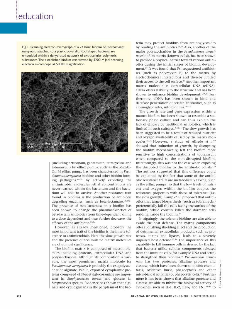
However, it has been shown that biofilms in vitro (Fig 1) have little to do with biofilms found in nature in terms of size and shape.3,9 It seems that biofilms causing harm in the human body are rarely anchored to a solid surface, but rather found in a semi-solid state in the tissue. Furthermore, the size of the infecting biofilms never reaches diameters larger than 100μm, unless the biofilm habitats an undisturbed surface, for example a catheter.3,9
Antimicrobial toleranceThe reason for the augmented interest in bacterial biofilms is their inherent tolerance towards antimi-crobial agents and inflammatory responses of the host. The ability to withstand antimicrobials is divided into two subtypes. Traditionally antibiotic resistance has received most attention, however it is antibiotic tolerance which is the prominent player of biofilm survival. Whereas resistance covers the inherited features that directly impede the efficacy of the antimicrobial, tolerance is the ability to sus-tain with the antibiotic due to the physical state of the bacterium.
Several resistance traits are found in the biofilm mode of growth and there are reports of increased mutation rates in biofilms which enhance resistance development.10–13 The active export of antimicrobials
R.A. Cooper,1 BSc, PhD, PGCE, Professor of Microbiology;T. Bjarnsholt,2,3 DMSc, PhD, Professor;M. Alhede,2, 3 MSc, PhD, Head of Biofilm Test Facility;1. Cardiff School of Health Sciences, Cardiff Metropolitan University, Western Avenue, Cardiff, CF5 2YB, S. Wales, UK2. University of Copenhagen, Faculty of Health Sciences, Department of International Health, Immunology and Microbiology, Blegdamsvej 3B, DK 2200, Copenhagen, Denmark3. Department of Clinical Microbiology 9301, Juliane Mariesvej 22, Copenhagen University Hospital, Copenhagen, Denmark
E-mail: [email protected]
education
J O U R N A L O F WO U N D C A R E VO L 2 3 , N O 1 1 , N OV E M B E R 2 0 1 45 7 2
© 2
01
4 M
A H
eA
lt
Hc
Ar
e l
td
(including aztreonam, gentamicin, tetracycline and tobramycin) by efflux pumps, such as the MexAB-OprM efflux pump, has been characterised in Pseu-domonas aeruginosa biofilms and other biofilm form-ing pathogens.14–19 By actively exporting the antimicrobial molecules lethal concentrations are never reached within the bacterium and the bacte-rium will able to survive. Another resistance trait found in biofilms is the production of antibiotic degrading enzymes, such as beta-lactamase.13,20,21 The presence of beta-lactamase in a biofilm has been shown to change the pharmacokinetics of beta-lactam antibiotics from time-dependent killing to a dose-dependent and thus further decreases the efficacy of the antibiotic.22-24
However, as already mentioned, probably the most important trait of the biofilm is the innate tol-erance to antimicrobials. Here the slow growth rate and the presence of accumulated matrix molecules are of upmost significance.
The biofilm matrix is composed of macromole-cules including proteins, extracellular DNA and polysaccharides. Although its composition is vari-able, the most prominent matrix molecule for Pseudomonas aeruginosa is probably the exopolysac-charide alginate. While, exported cytoplasmic pro-teins composed of N-acetylglucosamine are impor-tant in Staphylococcus aureus and glucans in Streptococcus species. Evidence has shown that algi-nate and cyclic glucans in the periplasm of the bac-
teria may protect biofilms from aminoglycosides by binding the antibiotics.25,26 Also, another of the major polysaccharides in the Pseudomonas aerugi-nosa biofilm matrix (known as Psl), has been shown to provide a physical barrier toward various antibi-otics during the initial stages of biofilm develop-ment.27 It was found that Psl sequestered antibiot-ics (such as polymyxin B) to the matrix by electrochemical interactions and thereby limited their access to the cell surface.27 Another important matrix molecule is extracellular DNA (eDNA). eDNA offers stability to the structure and has been shown to enhance biofilm development.7,28,29 Fur-thermore, eDNA has been shown to bind and decrease penetration of certain antibiotics, such as aminoglycosides, into biofilms.30–32
The growth rate and gene expression within a mature biofilm has been shown to resemble a sta-tionary phase culture and can thus explain the lack of efficacy by traditional antibiotics, which is limited in such cultures.9,33,34 The slow growth has been suggested to be a result of reduced nutrient and oxygen availability caused by the matrix mol-ecules.35,36 However, a study of Alhede et al.9 showed that induction of growth, by disrupting the biofilm mechanically, left the biofilm more sensitive to high concentrations of tobramycin when compared to the non-disrupted biofilm. Interestingly, this was not the case when exposing the disrupted biofilm to the antibiotic colistin.9 The authors suggested that this difference could be explained by the fact that some of the antibi-otic resistance traits are metabolically taxing, such as the efflux pumps, so that the low levels of nutri-ent and oxygen within the biofilm couples the resistance properties with those of tolerance (i.e. the slow growth). Pamp et al. proposed that antibi-otics that target biosynthesis (such as tobramycin) preferentially kill the cells facing the surface of the biofilm, while colistin killed the dormant cells residing inside the biofilm.17
Intriguingly, the tolerant biofilms are also able to evade the host defense. The matrix components offer a fortifying shielding effect and the production of detrimental extracellular products, such as pro-teases, toxins and lipases, leads to a severely impaired host defense.37,38 The importance of this capability to kill immune cells is stressed by the fact that bacteria utilise cellular components released from the immune cells (for example DNA and actin) to strengthen their biofilms.29 Pseudomonas aerugi-nosa has two proteases, alkaline protease and elastase, which have been shown to inhibit chemo-taxis, oxidative burst, phagocytosis and other microbicidal activities of phagocytic cells.39 Further-more it has been shown that alkaline protease and elastase are able to inhibit the biological activity of cytokines, such as IL-1, IL-2, IFN-g and TNF,38–41 to
Fig 1. Scanning electron micrograph of a 24 hour biofilm of Pseudomonas aeruginosa attached to a plastic coverslip. Rod shaped bacteria are embedded within a dehydrated network of extracellular polymeric substances. The established biofilm was viewed by 5200LV Jeol scanning electron microscope at 5000x magnification

educations
J O U R N A L O F WO U N D C A R E VO L 2 3 , N O 1 1 , N OV E M B E R 2 0 1 4 5 7 3
© 2
01
4 M
A H
eA
lt
Hc
Ar
e l
td
cleave human IgA and IgG,42 and to inactivate the complement system.38
An historical review of the discovery of biofilms in woundsThe concept of biofilms in wounds has only recently been coined. However, biofilms have cer-tainly existed historically and wounds containing biofilms have been successfully treated before the concept was born. When the drawings of wound tissue harbouring bacteria are viewed today, it is tempting to speculate that Sir Alexander Ogston may have unwittingly drawn a biofilm in 1880 (Fig 2).43 Another unrecognised clue was found by Bigger in 1944 who observed that soldiers’ infect-ed wounds treated with penicillin during World War II often seemed to respond to treatment, but then relapsed with recurrent infections.44 Today this might arouse suspicion of a tolerant biofilm in a wound.
However, the first recorded observation of a bio-film in a wound is attributed to Gristina and col-leagues following the examination of sutures and staples, removed from healed wounds by scanning electron microscopy and the discovery of several kinds of bacteria in close proximity embedded with-in fibrous material.45 Staphylococcus epidermidis was isolated from all of the wounds examined; yet heal-ing had been accomplished uneventfully without infection or inflammation. The importance of coag-ulase negative staphylococci in wounds was later revised from unimportant skin flora to opportunist pathogens, and their presence in biofilms was asso-
ciated with delayed, recurrent and persistent infec-tions associated with indwelling medical devices.46
Speculation that biofilms might exist in wounds47
was largely founded on animal experiments con-ducted during the 1990s48–51 and from laboratory models where bacteria isolated from wounds were shown to form biofilms relatively quickly under suitable conditions.49,52
Irrefutable evidence of biofilms in wounds came from studies published in 2008. In one study spe-cific bacteria were located in sections of chronic wound tissue using peptide nucleic acid (PNA) probes and fluorescent in situ microscopy (FISH). Pseudomonas aeruginosa was detected in some instances as single cells, but also as aggregates or microcolonies surrounded yet not invaded by host cells.53 In another study epifluorescent microscopy and scanning electron microscopy was utilised to visualise large aggregates of bacteria in wound biop-sies. Gram-positive cocci within an amorphous EPS were most frequently observed, although some bio-films were composed of diverse species and this was confirmed by molecular analysis. Whereas biofilm was only demonstrated in 1 of 16 acute wounds, it was found in 30 of 50 chronic wounds. Hence bio-film was linked to wound chronicity (p>0.001).54
Wounds are a well-suited habitat for bacteria, as the loss of skin integrity provides a moist and often nutrient-rich setting. The microbiota of the deep dermal tissues of chronic wounds is well described and harbours multiple bacterial species.54–57 The use of specific fluorescent probes and confocal laser scanning microscopy (CLSM) has been used to
Fig 2. Copy of a drawing made by Sir Alexander Ogston in 1880. It shows “micrococci in bunches in the wall of an abscess” and was reproduced in The Staphyloccci Proceedings of the Alexander Ogston Centennial Conference, edited by Alexander Macdonald and George Smith and published by Aberdeen University Press in 1981.
References1. Costerton, J.W., Cheng, K.J., Geesey, G.G. et al. Bacterial biofilms in nature and disease. Ann Rev Microbiol 1987; 41: 435–464.2. Davey, M.E., O'Toole, G.A. Microbial biofilms: from ecology to molecular genetics. Microbiol Mol Biol Rev 2000; 64: 4, 847–867.3. Bjarnsholt, T., Alhede, M., Alhede, M. et al. The in vivo biofilm. Trends Microbiol 2013; 21: 9, 466–4744. Stoodley, P., Sauer, K., Davies, D.G., Costerton, J.W. Biofilms as complex differentiated communities. Ann Rev Microbiol 2002; 56: 187–209.5. Costerton, J.W., Lewandowski, Z., Caldwell, D.E. et al. Microbial biofilms. Ann Rev Microbiol 1995; 49: 711–745.6. Wingender, J., Strathmann, M. Rode, A., et al. Isolation and biochemical characterization of extracellular polymeric substances from Pseudomonas aeruginosa. Methods Enzymol 2001; 336: 302–314.7. Whitchurch, C.B., Tolker-Nielsen, T., Ragas, P.C. et al. Extracellular DNA required for bacterial biofilm formation. Science 2002; 295: 5559,1487.8. Costerton, W., Veeh, R., Shirtliff, M., et al. The application of biofilm science to the study and control of chronic bacterial infections. J Clin Invest 2003; 112: 10, 1466–1477.9. Alhede, M., Kragh, K.N., Qvortrup, K. et al. Phenotypes of non-attached Pseudomonas aeruginosa aggregates resemble surface attached biofilm. PLoS One 2011; 6: 11, e27943.10. Driffield, K., Miller, K., Bostock, J.M. et al. Increased mutability of Pseudomonas aeruginosa in biofilms. J Antimicrob Chemother 2008; 61: 5, 1053–1056.11. Tyerman, J.G., Ponciano, J.M., Joyce, P., et al. The evolution of antibiotic susceptibility and resistance during the formation of Escherichia coli biofilms in the absence of antibiotics. BMC Evol Biol 2013; 13: 22.12. Conibear, T.C., Collins, S.L., Webb, J.S. Role of mutation in Pseudomonas aeruginosa biofilm development. PLoS One 2009; 4: 7, e6289.13. Bagge, N., Ciofu, O., Skovgaard, L.T., Hølby, N.

education
J O U R N A L O F WO U N D C A R E VO L 2 3 , N O 1 1 , N OV E M B E R 2 0 1 45 7 4
© 2
01
4 M
A H
eA
lt
Hc
Ar
e l
td
detect biofilms in chronic venous leg ulcers (VLUs),58–60 burns,61,62 malignant wounds associated with breast cancer63 and tissue filler infections.64
The use of molecular techniques to characterise wound flora has revealed the presence of diverse microbial species within chronic wounds. These mixed communities (Table 1) may indicate biofilms, but do not actually provide information on the structural or physiological parameters of the con-stituent member species that would indicate a bio-film phenotype.
Most studies agree on the almost universal pres-ence of Staphylococcus aureus, but another usual sus-pect found in chronic wounds is Pseudomonas aeru-ginosa, which is present in approximately half of the investigated wounds. The organisation and distribu-tion of these two species has been elucidated by employing specific PNA probes for FISH analysis.58,59 These observations revealed that the different bacte-rial species might be present in the same wound, but they do not integrate. Very few aggregates of different bacteria in close proximity to each other were observed and never as part of a truly mixed
population. Based on available evidence it seems that bacteria in chronic infections aggregate mostly as single species.3,58,59,65–67 This is in contrast to when bacteria aggregate in other natural environments such as the floccs in wastewater treatment plants and the soil where several species co-aggregate. This co-aggregation could be explained by the beneficial catabolism and anabolism of compounds among the different bacteria.68 A plausible reason why mul-tispecies biofilms are not common in chronic infec-tions is that the nutrient availability is high and that symbiosis between different species is not a cru-cial requisite for growth. The key challenge for colo-nising bacteria is rather whether they can survive the encounter with the defence system.
Impact of biofilms in woundsBased on the evidence above, the concept of bacte-rial biofilms in chronic wounds is supported, but whether these biofilms play a role in the lack of healing is another question. The biofilm phenotype enables protection of the bacteria from antibiotics, other antimicrobial agents, such as silver and the
Table 1. Microbiota in biofilms of chronic wounds characterised by molecular methods
Wound type Species currently identified Reference
Mixed chronic wounds Pseudomonas spp, Rhodococcus erythropolis, Actinobacterium, Staphylococcus spp, Pseudomonas spp, Haemophilus, Prevotella spp, Clostridium, Streptococcus, Bacteroides, Porphyromonas somerae.
54
Diabetic foot ulcers Corynebacterium, Bacteroides, Peptoniphilus, Finegoldia, Anaerococcus, Streptococcus, Serratia, Staphylococcus, Prevotella, Porphyromonas, Actinomyces, Pseudomonas, Clostridium, Helococcus, Brevibacterium, Varibaculum, Aerococcus, Fusobacterium, Arthrobacter, Bacillus.
139
Staphylococcus, Peptoniphilus, Rhodopseudomonas, Enterococcus, Veillionella, Clostridium, Finegoldia, Haemophilus, Acinetobacter, Morganella, Serratia, Proteus, Dialister, Streptococcus, Stenotrophomonas, Peptococcus niger, Klebsiella, Actinomyces, Gordonia, Delftia, Gemella, Corynebacterium, Salmonella, Fusobacterium, Varibacterium cambriense, Enterobacter, Bacillus, Eikonella, Anaerococcus, Hygenophaga, Alcaligenes faecalis, Escherichia coli, Sphingomonas, Acidovorax, Prevotella, Eubacterium, Bacteroides, Selenomonadaceae, Brevibacterium, Riemerella, Bradyrhizobium, Pantoea, Abiotropica, Citrobacter, Pseudoalteromonas, Granulicatella and unknown bacteria.
56
Pressure ulcers Peptoniphilus, Serratia, Peptococcus niger, Streptococcus, Finegoldia, Dialister, Pectobacterium, Enterobacter, Proteus, Veillionella, Clostridium, Corynebacterium striatum, Delftia, Enterococcus, Staphylococcus, Hydrogenophaga, Eggerthella, Prevotella, Varibaculum, Actinomyces europaeus, Ferrimonas, Bacillus, Fusobacterium, Alcaligenes faecalis, Riemerella, stenotrophomonas, Shewanella, Eubacterium, Anaerococcus, Dialister, Klebsiella, Porphyromonas and unknown bacteria.
56
Venous leg ulcers Enterobacter, Serratia, Stenotrophomonas, Proteus, Salmonella, Clostridium, Alcaligenes faecalis, Pseudomonas, Staphylococcus, Brevundimonas, Streptococcus, Acinetobacter, Enterococcus, Pantoea, Corynebacterium striatum, Peptoniphilus, Escherichia coli, Bacillus, Paenibacillus, Eubacterium, Klebsiella, Xanthomonas, Ferrimonas, Finegoldia, Dendrosporobacter quercicalus, Shewanella algae, Helococcus, Achromobacter xylosoxidans, Shigella and unknown bacteria.
56
Malignant wounds Staphylococcus aureus, Pseudomonas aeruginosa Corynebacterium striatum, Proteus vulgaris. Escherichia coli, Enterococcus faecalis, Klebsiella oxytoca, Fusobaterium necrophorum, Parvimonas micra, Peptoniphilus asaccharolytica, Porphyromonas asaccharolyticus
63
Rapid development in vitro and in vivo of resistance to ceftazidime in biofilm-growing Pseudomonas aeruginosa due to chromosomal beta-lactamase. APMIS 2000; 108: 9, 589–600.14. De Kievit, T.R., Parkins, M.D., Gillis, R.J. et al. Multidrug efflux pumps: expression patterns and contribution to antibiotic resistance in Pseudomonas aeruginosa biofilms. Antimicrob Agents Chemother 2001; 45: 6, 1761–1770.15. Zhang, L., Mah, T.F. Involvement of a novel efflux system in biofilm-specific resistance to antibiotics. J Bacteriol 2008; 190: 13, 4447–4452.16. Soto, S.M. Role of efflux pumps in the antibiotic resistance of bacteria embedded in a biofilm. Virulence 2013; 4: 3, 223–229.17. Pamp, S.J., Gjermansen, M., Johansen,H.K., Tolker-Nielsen, T. Tolerance to the antimicrobial peptide colistin in Pseudomonas aeruginosa biofilms is linked to metabolically active cells, and depends on the pmr and mexAB-oprM genes. Mol Microbiol 2008; 68: 1, 223–240.18. Chiang, W.C., Pamp, S.J., Nilsson, M. et al. The metabolically active subpopulation in Pseudomonas aeruginosa biofilms survives exposure to membrane-targeting antimicrobials via distinct molecular mechanisms. FEMS Immunol Med Microbiol 2012; 65: 2, 245–256.19. Ito, A., Taniuchi, A., May, T. et al. Increased antibiotic resistance of Escherichia coli in mature biofilms. Appl Environ Microbiol 2009; 75: 12, 4093–4100.20. Bagge, N., Ciofu, O. Hentzer, M. et al. Constitutive high expression of chromosomal beta-lactamase in Pseudomonas aeruginosa caused by a new insertion sequence (IS1669) located in ampD. Antimicrob Agents Chemother 2002; 46: 11, 3406–3411.21. Bagge, N., Hentzer, M., Andersen, J. B. et al. Dynamics and spatial distribution of beta-lactamase expression in Pseudomonas aeruginosa biofilms. Antimicrob Agents Chemother 2004; 48: 4,

education
J O U R N A L O F WO U N D C A R E VO L 2 3 , N O 1 1 , N OV E M B E R 2 0 1 45 7 6
© 2
01
4 M
A H
eA
lt
Hc
Ar
e l
td
host defence sytem. This implies that if the bacteria succeed in forming a biofilm in the wound bed, the bacteria will be extremely difficult to eradicate. Data suggest that the presence of certain bacteria, such as Pseudomonas aeruginosa, can induce ulcer enlarge-ment, delay healing66 and lead to the failure of split skin transplantation.69 It has also suggested that bacteria located in the deeper regions of the wounds might play a role in keeping the wounds arrested in a stage dominated by inflammatory processes.70 Evi-dence that biofilm contributes to chronic inflamma-tion in a wound exists, but how that influences wound healing is unclear. We know that biofilms are not the cause of chronic wounds, but they might keep the wound from healing.53
The role of biofilms in wound colonisation and infection was explored with the use of an animal model and Staphylococcus aureus.71 Wounds created on the pig were inoculated with Staphylococcus aureus and treated with either one or two antibiotic preparations within 15 minutes (to simulate an acute infection caused by planktonic bacteria) or after 48 hours (when a biofilm had established). Using electron microscopy and fluorescence micros-copy biofilms were observed in untreated wounds after 48 hours. Wounds treated with antibiotics within 15 minutes of introducing bacteria had no biofilm, showing that planktonic bacteria had been inhibited and that biofilm formation had been pre-vented. Antibiotics applied to wounds 48 hours after inoculation, after a biofilm had been estab-lished, failed to eradicate the biofilm. Decreased sus-ceptibility of biofilms to antimicrobial agents is well documented72 and largely accounts for the persist-ence previously observed by Bigger. In fact the term ‘persister’ was derived by Bigger.44
The difficulties of diagnosing biofilms in wounds When biopsies from chronic wounds of 22 different patients (all allegedly infected by Pseudomonas aeru-ginosa) were investigated, the samples were proc-essed by both standard culturing methods and pep-tide nucleic acid-based fluorescence in situ hybridisation (PNA FISH) for direct visualisation and identification of bacteria.58 The classic culturing methods revealed Staphylococcus aureus to be present in the majority of the wounds, whereas Pseudomonas aeruginosa was cultured less frequently. In contrast, using PNA FISH, Pseudomonas aeruginosa was visual-ised in biofilms in almost half of the wounds. These Pseudomonas aeruginosa biofilms were detected inside the wound bed, whereas Staphylococcus aureus, when present, was detected on the surface of the wounds. Thus, it seems that, although being the gold standard, culturing is not successful for diag-nosing biofilms of Pseudomonas aeruginosa in wounds due to its deep localisation. This is support-
ed by the observations of others showing Staphyloco-ccus aureus in microcolonies on the surface of the wound bed.59,71 It was shown that the distance of the Pseudomonas aeruginosa biofilm to the wound surface was significantly greater than that of the Sta-phylococcus aureus biofilms, suggesting that the dis-tribution of the bacteria in the chronic wounds was non-random.59
As described, the microbiota in chronic wounds has been investigated for several years. In one study Gjødsbol et al. investigated the microbiota by stand-ard culturing.57 Several different bacterial species were found in chronic VLUs, such as Staphylococcus aureus (in 93.5% of the investigated ulcers), Entero-coccus faecalis (71.7%), Pseudomonas aeruginosa (52.2%), coagulase-negative staphylococci (45.7%), Proteus species (41.3%), and anaerobic bacteria (39.1%). Another study also investigated the flora in chronic wounds by culturing and found the most common bacteria to be Staphylococcus (65%), Entero-coccus (62%), Pseudomonas (35%) (Table 1). Molecu-lar techniques have been used to establish the microbiota and in several studies it has been shown that standard culturing of bacteria from wound samples does not reveal on the true bacterial diver-sity in the wounds.56,58 As we mentioned earlier, the localisation, presence and slow growth of biofilms makes culturing difficult. Additionally, a large popu-lation of anaerobic bacteria in wounds has been identified,56 and these bacteria are also difficult to culture. By using molecular techniques, even small populations of a specific bacterium can be detected. The drawback is that these techniques are qualitative , which means that they do not reveal the relative proportions between the different bacteria or how they are organised and distributed in the wounds, as microscopy can do. Another significant drawback is that these techniques cannot be used to identify which bacteria play a key role in the impairment of the wound healing process. Most importantly the bacteria in chronic wounds are very small and heter-ogeneously distributed.55,70 This means that sampling from a chronic wound, especially using biopsies, might show false negative results.
In summary, swabs from chronic wounds are not representative for the microbiota and biopsies might give false negative results. Therefore, it is suggested to combine a thorough swab covering the whole wound surface with several biopsies, which should be investigated by both molecular techniques and culturing (aerobically and anaerobically).73
Biofilm controlWhereas planktonic cells are largely implicated in acute wound infections and control depends on systemic antibiotics, the increased antimicrobial tolerance of microbial cells within established bio-films requires novel control strategies.72 One
1168 –1174.22. Hengzhuang, W., Ciofu, O., Yang, L. et al. High beta-lactamase levels change the pharmacodynamics of beta-lactam antibiotics in Pseudomonas aeruginosa biofilms. Antimicrob Agents Chemother 2013; 57: 1, 196–204.23. Hengzhuang, W., Wu, H., Ciofu, O. et al. In vivo pharmacokinetics/pharmacodynamics of colistin and imipenem in Pseudomonas aeruginosa biofilm infection. Antimicrob Agents Chemother 2012; 56: 5, 2683–2690.24. Hengzhuang, W., Wu,H., Ciofu, O. et al. Pharmacokinetics/pharmacodynamics of colistin and imipenem on mucoid and nonmucoid Pseudomonas aeruginosa biofilms. Antimicrob Agents Chemother 2011; 55: 9, 4469–4474.25. Hentzer, M., Teitzel, G.M., Balzer, G.J. et al. Alginate overproduction affects Pseudomonas aeruginosa biofilm structure and function. J Bacteriol 2001; 183: 18, 5395–5401.26. Mah, T.F., Pitts, B., Pellock, B. et al. A genetic basis for Pseudomonas aeruginosa biofilm antibiotic resistance. Nature 2003; 426: 6964, 306–310.27. Billings, N., Millan, M., Caldara, M. et al. The extracellular matrix Component Psl provides fast-acting antibiotic defense in Pseudomonas aeruginosa biofilms. PLoS Pathog 2013; 9: 8, e1003526.28. Allesen-Holm, M., Barken, K.B., Yang, L. et al. A characterization of DNA release in Pseudomonas aeruginosa cultures and biofilms. Mol. Microbiol 2006; 59: 4, 1114–1128.29. Walker, T.S., Tomlin, K.L., Worthen, G.S. et al. Enhanced Pseudomonas aeruginosa biofilm development mediated by human neutrophils. Infect. Immun. 2005; 73: 6, 3693–3701.30. Chiang, W.C., Nilsson, M., Jensen, P.Ø. et al. Extracellular DNA shields against aminoglycosides in Pseudomonas aeruginosa biofilms. Antimicrob Agents Chemother 2013; 57: 5, 2352–2361.31. Mulcahy, H., Charron-Mazenod, L., Lewenza, S. Extracellular DNA chelates cations and induces

educations
J O U R N A L O F WO U N D C A R E VO L 2 3 , N O 1 1 , N OV E M B E R 2 0 1 4 5 7 7
© 2
01
4 M
A H
eA
lt
Hc
Ar
e l
td
approach is to prevent biofilm formation by inter-fering with either the mechanisms of microbial attachment or the processes involved in biofilm maturation. The other is to remove or disrupt mature biofilm. To date neither strategy has met with unmitigated success; the range of cells with differing physiological and functional variations within a mature biofilm suggests that multiple inhibitory assaults are likely to be more effective than a single antimicrobial intervention.
Interference with attachmentLactoferrin is part of the human innate immune response; it is found in tears, saliva, mucous and milk. It binds to components in the cell walls of Gram-negative bacteria to cause destabilisation, leakiness and ultimately bacterial lysis. It also binds avidly to iron, which is needed for bacterial motility during the initial stages of adherence to surfaces.74 Xylitol is an artificial sweetener that binds to the cell surface of Gram-positive bacteria that blocks adherence.75 Disruption of Pseudomonas aeruginosa biofilm in vitro with lactoferrin or xylitol, both alone or in combination has been reported.76
In the laboratory honey has also been shown to impede attachment of Pseudomonas aeruginosa to the surface of erythrocytes77 and inert surfaces.78
Furthermore, it interferes with binding of Streptococ-cus pyogenes to inert surfaces.79
Interference with quorum sensingOne of the most studied strategies is quorum sens-ing inhibitors (QSIs). Most bacteria regulate a range of behaviours including metabolism, virulence and motility by sensing small secreted molecules in their surroundings (signal molecules). This cooperative behaviour is maintained through inter- and extra-cellular chemical crosstalk comparable to higher organisms.80 This type of bacterial communication was termed quorum sensing (QS).81 QS systems allow bacteria to 'sense' bacterial density in the envi-ronment and respond by changes in gene expres-sion.82 By specifically targeting the QS system the idea is not to kill or detach the biofilm directly but to render the biofilm more susceptible to antibiotics and prevent expression of harmful virulence factors.
The first compounds showing good inhibition of the QS system were the synthetic furanones C-30 and C-56.83,84 In vitro Pseudomonas aeruginosa biofilms were significantly less tolerant to 100 μg /ml tobramy-cin when treated with furanone C-30.83 In addition, in vivo studies in a pulmonary mouse model con-firmed the potential of the furanones by demonstrat-ing that bacteria were cleared faster in furanone-treated versus untreated mice.83,86 Two QSIs from natural sources have recently been isolated: iberin from horseradish and ajoene from garlic.85,86
Using bacterial reporter assays three studies have
demonstrated the ability of different honeys to interfere with quorum sensing in Gram-negative bacteria.87-89 Manuka honey has also been shown to down-regulate three of the four genes essential for functional quorum sensing in Meticillin-resistant Staphylococcus aureus (MRSA), with knock-on effects on virulence and biofilm genes.90
Biofilm disruptionThe use of sharp debridement is one way to reduce biofilm within a wound, but it rarely offers a per-manent solution because, as with dental plaque, any remaining cells are able to regenerate the bio-film. Degradation of biofilm matrix with either cocktails of enzymes (e.g. DNAse) or maggot secre-tions has been reported.91,92 Generation of hydro-gen peroxide by enzymes within an alginogel dis-rupt biofilms in vitro93 and several honeys can also disrupt biofilms.94–97
Ultrasound as antibiofilm treatmentA lot of research has been invested in finding non-invasive applications to overcome the problem of antibiotic resistance and tolerance. Promising studies show that exposing bacteria to ultrasound enhances the antibiotic efficacy. However, the underlying mechanisms of this effect are yet to be elucidated. Additionally, recent studies suggest that any mechanical force (for example ultrasound or shear) can be applied to re-sensitise biofilm bac-teria by tearing the biofilm and stripping off the sessile cells.9 Back in the planktonic state, the bac-teria lose the tolerance provided by the biofilm. Such disruption of the biofilm by ultrasound is denoted destructive ultrasound.
Studies show that exposing Pseudomonas aerugi-nosa simultaneously to low intensity ultrasound and aminoglycosides (such as tobramycin) improves the antibiotic efficacy.98–105 The authors found that ultrasound alone did not affect the cell viability and that the synergistic effect was only observed if the antibiotics were applied during the ultrasonic expo-sure. However, in another study Qian et al. could not detect any structural difference in the biofilm by CLSM during ultrasonic exposure,106 and further documented that the effect was evident on plank-tonic Pseudomonas aeruginosa.100,101,104
An explanation for the antibacterial efficacy of this type of ultrasound was put forward by Liu et al.107 Runyan et al.108 and Nikaido,109 who docu-mented that low intensity ultrasound increased the permeability of Pseudomonas aeruginosa to several tagged molecules. This ultrasonically induced per-meability displayed the same frequency and peak pressure dependence as the above experiments. In addition, studies by Pong et al. showed a similarly increased permeability of phospholipid vesicles.110 Runyan et al. concluded that the effect was due to
antibiotic resistance in Pseudomonas aeruginosa biofilms. PLoS Pathog 2008; 4: 11, e1000213.32. Purdy Drew, K.R., Sanders, L.K., Culumber, Z.W. et al. Cationic amphiphiles increase activity of aminoglycoside antibiotic tobramycin in the presence of airway polyelectrolytes. J Am Chem Soc 2009; 131: 2, 486–493.33. Evans, D.J., Brown, M.R., Allison, D.G. et al. Susceptibility of bacterial biofilms to tobramycin: role of specific growth rate and phase in the division cycle. J Antimicrob Chemother 1990; 25: 4, 585–591.34. Hentzer, M., Eberl, L., Givskov, M. Transcriptome analysis of Pseudomonas aeruginosa biofilm development: anaerobic respiration and iron limitation. Biofilms 2005; 2: 1, 37–61.35. Anderson, G.G., O'Toole, G.A. Innate and induced resistance mechanisms of bacterial biofilms. Curr Top Microbiol Immunol 2008; 322: 85–105.36. Kuhl, M., Rickelt, L.F., Thar, R. Combined imaging of bacteria and oxygen in biofilms. Appl Environ Microbiol 2007; 73: 19, 6289–6295.37. Alhede, M., Bjarnsholt, T. Jensen, P.Ø. et al. Pseudomonas aeruginosa recognizes and responds aggressively to the presence of polymorphonuclear leukocytes. Microbiology 2009; 155: 3500–3508.38. Kharazmi, A. Mechanisms involved in the evasion of the host defence by Pseudomonas aeruginosa. Immunol Lett 1991; 30: 2, 201–205.39. Kharazmi, A., Doring, G., Høiby, N., Valerius, N.H. Interaction of Pseudomonas aeruginosa alkaline protease and elastase with human polymorphonuclear leukocytes in vitro. Infect Immun 1984; 43: 1, 161–165.40. Pedersen, B.K., Kharazmi, A. Inhibition of human natural killer cell activity by Pseudomonas aeruginosa alkaline protease and elastase. Infect Immun 1987; 55: 4, 986–989.41. Theander, T.G., Kharazmi, A., Pedersen, B.K. et al. Inhibition of human lymphocyte proliferation and cleavage of interleukin-2 by Pseudomonas aeruginosa
education
J O U R N A L O F WO U N D C A R E VO L 2 3 , N O 1 1 , N OV E M B E R 2 0 1 45 7 8
© 2
01
4 M
A H
eA
lt
Hc
Ar
e l
td
increased penetration of the antibiotics through the cell membrane of Pseudomonas aeruginosa.108
In addition to the resulting transient permeabili-ty, much attention has been addressed to the destructive ultrasound in order to remove biofilms from implants and wounds.106,111–113 By showing that disruption of biofilms by mechanical force yields an enhanced effect of applied antibiotics, it was proven that biofilm tolerance is reversible.9 This had been hypothesised to be due to disruption of matrix mol-ecules and induction of growth by exposing the cells to nutrients. This inference was supported by the findings of Pitt et al.114
From published studies, it seems that the mode of action of ultrasound has given rise to confusion and that both the terms 'destructive' and 'bioacoustic effect' have been used inconsistently. However, giv-en that the above hypotheses are valid, both destruc-tive ultrasound and the bioacoustic effect enhance the antibiotic efficacy, albeit in entirely different ways: one acting on the biofilm, the other directly on the individual bacterium.
Ultrasound debridement of woundsTreatment of chronic wounds with ultrasound ther-apy has been used with seemingly good results.115, 116 It has been suggested that the positive effect comes from a multitude of factors, such as cellular recruit-ment and stimulation, collagen synthesis, angio-genesis, fibrinolysis.117,118 Recently the knowledge of biofilms in non-healing wounds has led to the hypothesis that the ultrasound, in addition to the above mentioned parameters, aids biofilm disrup-tion and thereby wound healing.119 Measuring wound healing and quantifying the presence of bio-films/bacteria is extremely difficult (if not impossi-ble) and therefore the literature is very limited in this perspective. Escandon and colleagues found a non-significant decline in individual and total bac-terial counts when treating refractory VLUs with non-contact ultrasound therapy.116 It should be not-ed that biofilms able to prevent wound healing are smaller than 100μm in diameter and often situated deep in the wound bed and thereby hard to find by traditional means.3,58,59
More data and possibly also better experimental setups are needed to prove the hypothesis claiming ultrasound to be an efficient antibiofilm strategy. However, the regimen seems safe and the above-mentioned indications are not to be neglected.
Phage therapyOne innovation with the potential to control wound infections is the topical use of lytic bacteri-ophage (or phage). These naturally occurring pred-atory viruses are obligate intracellular parasites that rely on bacteria for their replication. Infection of an appropriate bacterial cell usually leads to rapid
viral replication within that host, followed by lysis and bacterial cell death to release viral progeny without affecting mammalian cells. However, a temperate phage can infect a host bacterial cell, integrate into the host DNA and remain latent for some time; their therapeutic potential is therefore low. Bacteriophages were independently discov-ered in 1915 by Twort in London and by d’Herelle in 1917 in Paris. The antimicrobial potential of lytic phage in treating infections was immediately recognised, particularly by d’Herelle, and several infections were successfully controlled, such as dys-entery, cholera, wound infections and urinary tract infections. However, the antibiotic era saw the demise of bacteriophage therapy, except in eastern European countries such as Georgia, Poland and the former Soviet Union. It is relatively recently that the continued emergence of antibiotic-resist-ant species has prompted a renewed interest in phage and translations of Georgian and Ukrainian studies have lately provided access to this largely forgotten therapeutic approach.
One of the most studied applications of bacteri-ophages has been in the control of Pseudomonas aeruginosa infections in burns, where promising evi-dence of efficacy in animal models of acute infec-tions and against biofilms in vitro has been report-ed.120 MRSA has been eradicated from diabetic foot ulcers using combination therapy of lytic bacteri-ophage and linezolid.121 Most viruses are highly host-specific and treatments with a cocktail of lytic viruses targeted at mixed cultures of bacteria will probably be most effective clinically. Bacterial hosts most likely to be targeted include Pseudomonas aeru-ginosa, Staphylococcus aureus, MRSA, Acinetobacter baumannii and the multi-drug resistant Gram-nega-tive bacteria (or extended-spectrum beta-lactama-ses; ESBLs). Rat and pig models have been used to evaluate the effects of phage cocktails on bacterial counts and wound healing in diabetic cutaneous wounds, with limited success.122
The safety of such an approach has been tested in a phase I trial conducted on VLUs in America. Here 42 patients were treated for 12 weeks with either saline control or a cocktail of phages directed at Pseudomonas aeruginosa, Staphylococcus aureus and Escherichia coli. Neither adverse events nor signifi-cant differences between the two study groups were observed.123 Further clinical data from phase II and III studies is needed, and formulations for delivering suitable phages to the wound bed will have to be developed. Much research is in progress and the licensing of wound dressings incorporating phage is expected within the not too distant future.
Interactions between phage and biofilms are complex and involve not only lysis of bacterial cells, but degradation of EPS by viral enzymes, which is an additional advantage.124 A rabbit-ear
proteases. Infect. Immun. 1988; 56: 7, 1673–1677.42. Doring, G., Goldstein, W., Botzenhart, K. et al. Elastase from polymorphonuclear leucocytes: a regulatory enzyme in immune complex disease. Clin Exp Immunol 1986; 64: 3, 597–605.43. Ogston A. Ueber abscesse. Archiv f Kin Chir 1880; 25: 599. Reproduced in: Macdonald, A., Smith, G. (eds) The staphylococci. Proceedings of the Alexander Ogston Centennial Conference. . Aberdeen University Press, 1981.44. Bigger, J.W. Treatment of staphylococcal infections with penicillin by intermittent sterilization. Lancet 1944; 244: 6320, 497–500.45. Gristina, A.G., Price, J.L., Hobgood, C.D., et al. Bacterial colonisation of percutaneous sutures. Surgery 1985; 98: 1, 12–19.46. Donlan, R.M. Biofilms and device-associated infections. Emerg Infect Dis 2001; 7: 2, 277–281.47. Serralta, V.W., Harrison-Balestra, C., Cazzaniga, A.L., et al. Lifestyles of bacteria in wounds: presence of biofilms? Wounds 2001; 13: 1, 29–34. 48. Akiyama, H., Torigoe, R., Arata, J. Interaction of Staphylococcus aureus cells and silk thread in vitro and in mouse skin. J Dermatol Sci 1993; 6: 3, 247–257. 49. Akiyama, H., Kanzaki, H., Abe, Y., et al. Staphylococcus aureus infection on experimental croton oil-inflammed skin. J Dermatol Sci 1994; 8: 1, 1–1050. Akiyama, H., Kanzaki, H., Tada, J., et al. Staphylococcus aureus infection on cut wounds in the mouse skin: experimental staphylococcal botryomycosis. J Dermatol Sci 1996; 11; 3, 234–238.51. Akiyama, H., Huh, W.K., Yamasaki, O., et al. Confocal laser scanning microscopic observation of glycocalyx by Staphylococcus aureus in mouse skin: does S. aureus generally produce a biofilm on damaged skin? Br J Dermatol 2002;147: 5, 879–885.52. Harrison-Balestra, C., Cazzaniga, A.L., Davis, S., Mertz, P.M. A wound-isolated Pseudomonas aeruginosa grows a biofilm in vitro within 10 hours and is

educations
J O U R N A L O F WO U N D C A R E VO L 2 3 , N O 1 1 , N OV E M B E R 2 0 1 4 5 7 9
© 2
01
4 M
A H
eA
lt
Hc
Ar
e l
td
model was used to investigate the ability of bacte-riophage and sharp debridement to eliminate Sta-phylococcus aureus from a chronic wound. Combi-nation therapy gave better outcomes than bacteriophage or debridement alone.125
Clinical evidence of efficacy of antibiofilm interventions (or lack of it)At present the number of clinical studies in which eradication of biofilms has been investigated is limited and will probably remain so until a routine test to detect biofilm in wound tissue is developed. A concept of biofilm-based wound care (BBWC) has been proposed in which sharp debridement to reduce biofilm is followed by antimicrobial agents to limit biofilm reformation. The rationale for this approach is based on physically removing biofilm and inhibiting the residual bacteria that actively try to reform biofilm before they return to their tolerant status. In a retrospective study of BBWC a 190 patients with critical limb ischaemia were treated by sharp debridement coupled with ultra-sound, followed by lactoferrin and xylitol, silver, cadexomer iodine and antibiotics. Improved heal-ing was observed in these patients compared to a previous study, but the presence of biofilms before and after treatment was not confirmed by electron or confocal scanning laser microscopy.126
A wide range of model systems have been devised to study microbial biofilm biology127 and many bio-film studies conducted in the laboratory have been applied to evaluate wound treatment strate-gies.128–134 Animal models have also been uti-lised.71,135–138 Such studies are important, but the study of wound biofilms is very complicated and it is difficult to make comparisons between different studies, as demonstrated by the conflicting results obtained in evaluating some licensed antimicrobial dressings. Unlike disinfectants there are not yet standardised methods available to determine the efficacy of wound dressings on biofilms. Hence many new compounds and dressings have been evaluated on fast growing reference strains of
bacteria in shaking cultures, rather than on bio-film-growing bacteria commonly present in chron-ic wounds. Even when using a biofilm model, researchers should be aware of the false dogma stating that surface attachment per se makes the biofilm tolerant. This is not true, since young sur-face-attached biofilms still have high growth rates with only a limited matrix shield and therefore are highly susceptible to most antimicrobials. Biofilms across species and models seems to become toler-ant between 20 hours and 48 hours after inocula-tion but continue developing this tolerance with time.3,9 Another important limitation of in vitro models is that they have been developed under artificial conditions that aim to simulate the natu-ral situations in which biofilms are normally estab-lished, and because the validity of these models is questionable, data obtained is not necessarily transferable to clinical practice.
Future prospects Discovering biofilms in wounds has given insight into some of the reasons why wounds fail to heal. It has helped to explain the limited efficacy of antibiotics in chronic wounds and it has stimulat-ed research into innovative anti-biofilm strategies. However, we still face a number of tasks to solve before chronic wounds are history. The range of possible treatment strategies of biofilm infections needs to be expanded and the in vitro models need to be more closely aligned to simulate the wound in vivo. Pseudomonas aeruginosa is the test organism that is commonly used in laboratory biofilm mod-els because it is easy to grow, its genome has been sequenced and knock out mutants are available. Testing a broader range of wound microbiota in both single species and mixed species models might provide a different perspective. Most impor-tantly, in order to prove that biofilm plays the role it is believed to do, we need to improve diagnostic methods to eliminate false negatives. This task is especially important when evaluating treatment strategies in the clinic. n
visualised by light microscopy. Dermatol Surg 2003; 29: 6, 631–635.53. Bjarnsholt, T., Kirketerp-Møller, K., Jensen, P.Ø., et al. Why chronic wounds will not heal: a novel hypothesis. Wound Repair Regen 2008; 16: 1, 2–1054. James, G.A., Swogger, E., Wolcott, R., et al. Biofilms in chronic wounds. Wound Repair Regen 2008; 16: 1, 37–44. 55. Thomsen, T.R., Aasholm, M.S., Rudkjøbing, V.B. et al. The bacteriology of chronic venous leg ulcer examined by culture-independent molecular methods. Wound Repair Regen 2010; 18:1, 38–49.
56. Dowd, S.E., Sun, Y., Secor, P.R. et al. Survey of bacterial diversity in chronic wounds using pyrosequencing, DGGE, and full ribosome shotgun sequencing. BMC Microbiol 2008; 8: 43.57. Gjødsbol, K., Christensen, J.J., Karlsmark, T., et al. Multiple bacterial species reside in chronic wounds: a longitudinal study. Int Wound J 2006; 3: 3, 225–231.58. Kirketerp-Møller, K., Jensen, P.Ø., Fazli, M., et al. Distribution, organization, and ecology of bacteria in chronic wounds. J Clin Microbiol 2008; 46: 8, 2717–2722.59. Fazli, M., Bjarnsholt, T., Kirketerp-Møller, K., et al. Non-random distribution of
Pseudomonas aeruginosa and Staphylococcus aureus in chronic wounds. J Clin Microbiol 2009; 47: 12, 4084–4089. 60. Malic, S., Hill, K.E., Hayes, A., et al. Detection and identification of specific bacteria in wound biofilms using peptide nucleic acid fluorescence in situ hybridization (PNA FISH). Microbiol 2009; 155: 2603–2611.61. Kennedy, P., Brammah, S., Wills, W. Burns, biofilm and a new appraisal of burns sepsis. Burns 2010; 36: 1, 49–56.62. Neut, D., Tijdens-Creusen, E.J.A., Bulstra, S.K., et al. Biofilms in chronic diabetic foot ulcers-a study of 2 cases. Acta Orthopaedica
2011; 82: 3, 383–385.63. Fromantin, I., Seyer, D., Watson, S., et al. Bacterial floras and biofilms of malignant wounds associated with breast cancers. J Clin Microbiol 2013; 51: 10, 3368–3373.64. Alhede, M., Er, Ö., Eickhardt, S. et al. Bacterial biofilm formation and treatment in soft tissue fillers. Pathog Dis 2014; 70: 3, 339–346.65. Rudkjobing, V.B., Thomsen, T.R., Alhede, M., et al. The microorganisms in chronically infected end-stage and non-end-stage cystic fibrosis patients. FEMS Immunol Med Microbiol 2012; 65: 2, 236–44.

education
J O U R N A L O F WO U N D C A R E VO L 2 3 , N O 1 1 , N OV E M B E R 2 0 1 45 8 0
© 2
01
4 M
A H
eA
lt
Hc
Ar
e l
td
66. Burmølle, M., Thomsen, T.R., Fazli, M., et al. Biofilms in chronic infections - a matter of opportunity - monospecies biofilms in multispecies infections. FEMS Immunol Med Microbiol 2010; 59: 3, 324–336.67. Bjarnsholt, T., Jensen, P.Ø., Fiandaca, M.J., et al. Pseudomonas aeruginosa biofilms in the respiratory tract of cystic fibrosis patients. Pediatr Pulmonol 2009; 44: 6, 547–558.68. Bjarnsholt, T. The role of bacterial biofilms in chronic infections. APMIS Suppl 2013; 136: 1–51.69. Høgsberg, T., Bjarnsholt, T., Thomsen, J.S., Kirketerp-Møller, K. Success rate of split-thickness skin grafting of chronic venous leg ulcers depends on the presence of Pseudomonas aeruginosa: A Retrospective Study. PLoS One 2011; 6: 5, e20492.70. Fazli, M., Bjarnsholt, T., Kirketerp-Møller, K., et al. Quantitative analysis of the cellular inflammatory response against biofilm bacteria in chronic wounds. Wound Repair Regen 2011; 19: 3, 387–391. 71. Davis, S.C., Ricotti, C., Cazzaniga, A., et al. Microscopic and physiologic evidence for biofilm-associated wound colonization in vivo. Wound Repair Regen 2008; 16: 23–29.72. Stewart, P.S., Costerton, J.W. Antibiotic resistance of bacteria in biofilms. Lancet 2001; 358: 9276, 135–138. 73. Hall-Stoodley, L., Stoodley, P., Kathju, S., et al. Towards diagnostic guidelines for biofilm-associated infections. FEMS Immunol Med Microbiol 2012; 65: 2,127–145.74. Weinberg, E.D. Suppression of bacterial biofilms by iron limitation. Med Hypotheses 2004; 63: 5, 863–865.75. Tapininen, T., Sormunen, R., Kaijalainen, T., et al. Ultrastructure of Streptococcus pneumoniae after exposure to xylitol. J Antimicrob Chemother 2004; 54: 1, 225–228.76. Ammons, M.C., Ward, L.S., Fisher, S.T., et al. In vitro susceptibility of established biofilms composed of a clinical wound isolate of Pseudomonas aeruginosa treated with lactoferrin and xyltiol. Int J Antimicrob Agents 2009; 33: 3, 230–236.77. Lerrer, B., Zinger-Yosovich, K.D., Avrahami, B., Gilboa-Garber, N. Honey and royal jelly, like human milk, abrogate lectin-dependent infection preceding Pseudomonas aeruginosa adhesion. ISME J 2007; 1: 2, 149–155. 78. Roberts, A.E., Maddocks, S.E., Cooper, R.A. Manuka honey is bactericidal against Pseudomonas aeruginosa and results in differential expression of oprF and algD. Microbiology
2012; 158: 3005–3013.79. Maddocks, S.E., Lopez, M.S., Rowlands, R.S., Cooper, R.A. Manuka honey inhibits the development of Streptococcus pyogenes biofilms and causes reduced expression of two fibronectin binding proteins. Microbiology 2012; 158: 781–790.80. Shapiro, J.A. Thinking about bacterial populations as multicellular organisms. Ann Rev Microbiol 1998; 52: 81–104.81. Fuqua, W.C., Winans, S.C. Greenberg, E.P. Quorum sensing in bacteria: the LuxR-LuxI family of cell density-responsive transcriptional regulators. J Bacteriol 1994; 176: 2, 269–275.82. Waters, C.M., Bassler, B.L. Quorum sensing: cell-to-cell communication in bacteria. Annu Rev Cell Dev Biol 2005; 21: 319–346.83. Hentzer, M., Wu, H., Andersen, J.B. et al. Attenuation of Pseudomonas aeruginosa virulence by quorum sensing inhibitors. EMBO J 2003; 22: 3803–3815.84. Wu, H., Song, Z., Hentzer, M. et al. Synthetic furanones inhibit quorum-sensing and enhance bacterial clearance in Pseudomonas aeruginosa lung infection in mice. J Antimicrob Chemother 2004; 53: 6, 1054–1061.85. Jakobsen, T. H., Bragason, S. K., Phipps, R. K., et al. Food as a source for quorum sensing inhibitors: iberin from horseradish revealed as a quorum sensing inhibitor of Pseudomonas aeruginosa. Appl Environ Microbiol 2012; 78: 7, 2410–2421.86. Jakobsen, T. H., van Gennip, M., Phipps, R. K. et al. Ajoene, a sulfur-rich molecule from garlic, inhibits genes controlled by quorum sensing. Antimicrob Agents Chemother 2012; 56: 5, 2314–2325.87. Truchado, P., Lopez-Galvez, F., Gil, M.I., et al. Quorum sensing inhibitory and antimicrobial activities of honeys and the relationship with individual phenols. Food Chemistry 2009;115: 1337–1344.88. Truchado, P., Gil-Izquierdo, A., Tomas-Barberan, F. et al. Inhibition by chestnut honey of N-Acetyl-L-homoserine lactones and biofilm formation in Erwinia carotovora, Yersinia entercolitica, and Aeromonas hydrophila. J Agric Food Chem 2009; 57: 11186–11193. 89. Wang, R, Starkey, M., Hazan, R., Rahme, L.G. Honey’s ability to counter bacterial infections arises from both bactericidal compounds and QS inhibition. Frontiers Microbiol 2012; 3: 144.90. Jenkins, R.E., Burton, N.F., Cooper, R.A. Proteomic and genomic analysis of methicillin-resistant Staphylococcus aureus (MRSA) exposed to manuka honey
in vitro demonstrated down-regulation of virulence markers. J Antimicrob Chemother 2014; 69: 3, 603–615.91. Nemoto, K., Hirota, K., Murakami, K., et al. Effect of varidase (streptodornase) on biofilm of Pseudomonas aeruginosa. Chemotherapy 2003; 49: 3, 121–125.92. Chambers, L., Woodrow, S., Brown, A.P., et al. Degradation of extracellular matrix components by defined proteinases from the greenbottle larvae Lucilia sericata used for the clinical debridement of non-healing wounds. Br J Dermatol 2003; 148: 1, 14–23.93. Cooper, R.A. Inhibition of biofilms by glucose oxidase, lactoperoxidase and guaiacol: the active antibacterial component in an enzyme alginogel. Int Wound J 2013; 10: 6, 630–637.94. Merckoll, P., Jonassen, T.Ø., Vad, M.E., et al. Bacteria, biofilm and honey: A study of the effects of honey on ‘planktonic’ and biofilm-embedded wound bacteria. Scand J Infect Dis 2009; 41: 5, 341–347.95. Alandejani, T., Marsan, J., Ferris, W., et al. Effectiveness of honey on Staphylococcus aureus and Pseudomonas aeruginosa biofilms. Otolarynology- Head Neck Surg 2009; 141: 1, 114–118.96. Maddocks, S.E., Jenkins, R.E., Rowlands, R.S., et al. Manuka honey inhibits adhesion and invasion of medically important wound bacteria in vitro. Future Microbiol 2013; 8: 12, 1523–1536.97. Cooper, R., Jenkins, L., Hooper, S. Inhibition of biofilms of Pseudomonas aeruginosa by Medihoney in vitro. J Wound Care 2014; 23: 3, 93–104.98. Carmen, J.C., Nelson, J.L., Beckstead, B.L. et al. Ultrasonic-enhanced gentamicin transport through colony biofilms of Pseudomonas aeruginosa and Escherichia coli. J Infect Chemother 2004; 10: 4, 193–199.99. Carmen, J.C., Roeder, B.L., Nelson, J.L. et al. Ultrasonically enhanced vancomycin activity against Staphylococcus epidermidis biofilms in vivo. J Biomater Appl 2004; 18: 4, 237–245.100. Ensing, G.T., Neut, D., van Horn, J.R., et al. The combination of ultrasound with antibiotics released from bone cement decreases the viability of planktonic and biofilm bacteria: an in vitro study with clinical strains. J Antimicrob Chemother 2006; 58: 6, 1287–1290.101. Pitt, W.G., McBride, M.O., Lunceford, J.K., et al. Ultrasonic enhancement of antibiotic action on gram-negative bacteria. Antimicrob Agents Chemother 1994; 38: 11, 2577–2582.102. Rapoport, N., Smirnov, A.I.,
Timoshin, A., et al. Factors affecting the permeability of Pseudomonas aeruginosa cell walls toward lipophilic compounds: effects of ultrasound and cell age. Arch Biochem Biophys 1997; 344: 1, 114–124.103. Rapoport, N., Smirnov, A.I., Pitt, W.G., et al. Bioreduction of Tempone and spin-labeled gentamicin by gram-negative bacteria: kinetics and effect of ultrasound. Arch Biochem Biophys 1999; 362: 2, 233–241.104. Rediske, A.M., Hymas, W.C., Wilkinson,R., Pitt. W.G. Ultrasonic enhancement of antibiotic action on several species of bacteria. J Gen Appl Microbiol 1998; 44: 4, 283–288.105. Rediske, A. M., Roeder, B.L., Brown, M.K., et al. Ultrasonic enhancement of antibiotic action on Escherichia coli biofilms: an in vivo model. Antimicrob Agents Chemother 1999; 43: 5, 1211–1214.106. Qian, Z., Stoodley, P. Pitt, W.G. Effect of low-intensity ultrasound upon biofilm structure from confocal scanning laser microscopy observation. Biomaterials 1996; 17: 20, 1975–1980.107. Liu, J., Lewis, T.N. Prausnitz, M.R. Non-invasive assessment and control of ultrasound-mediated membrane permeabilization. Pharm Res 1998; 15: 6, 918–924.108. Runyan, C. M., Carmen, J.C., Beckstead, B.L., et al. Low-frequency ultrasound increases outer membrane permeability of Pseudomonas aeruginosa. J Gen Appl Microbiol 2006; 52: 5, 295–301.109. Nikaido, H. Molecular basis of bacterial outer membrane permeability revisited. Microbiol Mol Biol Rev 2003; 67: 4, 593–656.110. Pong, M., Umchid, S., Guarino, A. J. et al. In vitro ultrasound-mediated leakage from phospholipid vesicles. Ultrasonics 2006; 45: 133–145.111. Nishikawa, T., Yoshida, A., Khanal, A. et al A study of the efficacy of ultrasonic waves in removing biofilms. Gerodontology 2010; 27: 3, 199–206.112. Karau, M. J., Piper, K.E.,Steckelberg, J.M., et al. In vitro activity of the Quostic Wound Therapy System against planktonic and biofilm bacteria. Adv Skin Wound Care 2010; 23: 7, 316–320.113. Tollefson, D. F., Bandyk, D.F., Kaebnick, H.W., et al. Surface biofilm disruption. Enhanced recovery of microorganisms from vascular prostheses. Arch Surg 1987; 122: 1, 38–43.114. Pitt, W.G., Ross, S.A. Ultrasound increases the rate of bacterial cell growth. Biotechnol Prog 2003; 19: 3, 1038–1044.115. Ramundo, J., Gray, M. Is ultrasonic mist therapy effective for debriding chronic wounds? J

education
J O U R N A L O F WO U N D C A R E VO L 2 3 , N O 1 1 , N OV E M B E R 2 0 1 45 8 2
© 2
01
4 M
A H
eA
lt
Hc
Ar
e l
td
Wound, Ostomy, Continence Nurs 2008; 35: 6, 579–583.116. Escandon, J., Vivas, A.C.,Perez, R., et al. A prospective pilot study of ultrasound therapy effectiveness in refractory venous leg ulcers. Int Wound J 2012; 9: 5, 570–578.117. Vicenti, F.A., Laus, J.L., Costa Neto, J.M. et al. Effects of low-intensity pulsed ultrasound on wound healing in corneas of dogs following keratoplasty. Vet Ophthalmol 2003; 6: 3, 255–263.118. Hess, C.L., Howard, M.A., Attinger, C.E. A review of mechanical adjuncts in wound healing: hydrotherapy, ultrasound, negative pressure therapy, hyperbaric oxygen, and electrostimulation. Ann Plast Surg 2003; 51: 2, 210–218.119. Butcher, G., Pinnuck, L. Wound bed preparation: ultrasonic-assisted debridement. Br J Nurs 2013; 22: 6, S36, S38–43.120. Soothill, J. Use of bacteriophages in the treatment of Pseudomonas aeruginosa infections. Expert Rev Anti Infect Ther 2013; 11: 9, 909–915.121. Chhibber, S., Kaur, T., Kaur, S. Co-therapy using lytic bacteriophage and linezolid: effective treatment in eliminating methicillin-resistant Staphylococcus aureus (MRSA) from diabetic foot infections. PLoS One 2013; 8: 2, e56022.122. Mendes, J.J., Leandro, C.,
Corte-Real, S., et al. Wound healing potential of topical bacteriophage therapy on diabetic cutaneous wounds. Wound Repair Regen 2013; 21: 4, 595–603123. Rhoads, D.D, Wolcott, R.D., Wolcott, M.A., et al. Bacteriophage therapy of venous leg ulcers in humans: results of a phase I safety trial. J Wound Care 2009; 18: 6, 237–243.124. Abedon, S.T. Bacteriophages and biofilms. In: Bailey W.C. (ed) Biofilms: formation, development and properties. Nova Science Publishers Inc, 2011.125. Seth, A.K., Geringer, M.R., Nquyen, K.T., et al. Bacteriophage therapy foot Staphylococcus aureus biofilm-infected wounds: a new approach to chronic wound care. Plast Reconstr Surg 2013; 131: 2, 225–234.126. Wolcott, R.D., Rhoads, D.D. A study of biofilm-based management of subjects with critical limb ischemia. J Wound Care 2008; 17: 4, 145–155.127. Coenye, T., Nelis, H.J. In vitro and in vivo model systems to study microbial biofilm formation. J Microbiol Methods 2014; 83: 2, 89–109. 128. Sun, Y., Dowd, S.E., Smith, E., et al. In vitro multispecies Lubbock chronic wound biofilm model. Wound Repair Regen 2008; 16: 6, 805–813.129. Werthén, M., Henriksson, L.,
Jensen, P.Ø., et al. An in vitro model of bacterial infections in wounds and other soft tissues. APMIS 2010; 118: 2, 156–164.130. Charles, C.A., Ricotti, C.A., Davis, S.C., et al. Use of tissue-engineered skin to study in vitro biofilm development. Dermatol Surg 2009; 35: 9, 1334–1341.131. Thorn, R.M. Nelson, S.M., Greenman, J. Use of a bioluminescent Pseudomonas aeruginosa strain within an in vitro microbiological system, as a model of wound infection, to assess the antimicrobial efficacy of wound dressings by monitoring light production. Antimicrob Agents Chemother 2007; 51: 9, 3217–3224.132. Thorn, R.M., Greenman, J. A novel in vitro flat-bed perfusion biofilm model for determining the potential antimicrobial efficacy of topical wound dressings. J Appl Microbiol 2009; 107: 6, 2070-2079.133. Lipp, C., Kirke, K., Agostinho, A., et al. Testing wound dressings using an in vitro wound model. J Wound Care 2010; 19: 6, 220–226.134. Hill, K.E., Malic, S., McKee, R. et al. An in vitro model of chronic wound biofilms to test wound dressings and assess antimicrobial susceptibilities. J Antimicrob Chemother 2010; 65: 6, 1195–1206. 135. Nakagami, G, Sanada, H., Sugama, J., et al. Detection of Pseudomonas aeruginosa quorum
sensing signals in an infected ischemic wound: an experimental study in rats. Wound Repair Regen 2008; 16: 1, 30–36.
136. Simonetti, O., Cirioni, O., Ghiselli, R., et al. RNAIII-inhibiting peptide enhances healing of wounds infected with methicillin-resistant Staphylococcus aureus. Antimicrob Agents Chemother 2008; 52: 6, 2205–2211.
137. Schierle, C.F., de la Garza, M., Mustoe, T.A., Galiano, R.D. Staphylococcal biofilms impair wound healing by delaying reepithelialisation in a murine cutaneous wound model. Wound Repair Regen 2009; 17: 3, 354–359.
138. Kanno, E., Toriyabe, S., Zhang, L., et al. Biofilm formation on rats skin wounds by Pseudomonas aeruginosa carrying green fluorescent protein gene. Exp Dermatol 2010; 19: 2, 154–156
139. Dowd, S.E., Wolcott, R.D., Sun, Y. et al. Polymicriobial nature of chronic diabetic foot ulcer biofilm infections determined using bacterial tag encoded FLX amplicon pyrosequencing (bTEFAP). PLoS One 2008; 3, 10, e3326.
JWC NOW AVAILABLE ON
Sign up for an account and take advantage of these great bene� ts and features:
• Save JWC articles for quick access
• Save your searches: great if you frequently search for the same criteria
• Get citation alerts to track citations to speci� c articles
Once you’ve registered, you can sign up for table of contents alerts for JWC, delivered to you by email or RSS feed as soon as new content becomes available. Simply visit the journal and click ‘TOC Alerts’ in the menu.
www.magonlinelibrary.com/toc/jwc/current
frequently search for the same criteria

Copyright of Journal of Wound Care is the property of Mark Allen Publishing Ltd and itscontent may not be copied or emailed to multiple sites or posted to a listserv without thecopyright holder's express written permission. However, users may print, download, or emailarticles for individual use.