Embed Size (px)
Citation preview

Biochemistry of
Addison’s Disease

ANATOMICALLY:
• The adrenal gland is situated on the anteriosuperior aspect of the kidney and receives its blood supply from the adrenal arteries.
HISTOLOGICALLY:• The adrenal gland consists
of two distinct tissues of different embryological origin, the outer cortex and inner medulla.

The adrenal cortex comprises three zones based on cell type and function: Zona glomerulosa The outermost zone
aldosterone (the principal mineralocorticoid).
The deeper layers of the cortex:
Zona fasciculata glucocorticoids – mainly
cortisol (95%)Zona reticularis
Sex hormones
Steroid Hormone Synthesis Steroid Hormone Synthesis
Cholesterol
Pregnenolone (C21)3-β-Hydroxysteroid dehydrogenase
Progesterone (C21)
17-α-Hydroxylase
17-α-Hydroxyprogesterone (C21)
Androstenedione (C19)
Testosterone (C19)
Estradiol (C18)
11-Deoxycortisol (C21)11-Deoxycorticosterone (C21)
Cortisol (C21)
21-α-Hydroxylase
11- β -Hydroxylase
Corticosterone
Aldosterone (C21)
Per
iph
eral
tis
sues

Aldosterone Hormone:• The principal physiological function of
aldosterone is to conserve Na+, mainly by facilitating Na+ reabsorption and reciprocal K+ or H+ secretion in the distal renal tubule.
• aldosterone is a major regulator of water and electrolyte balance, as well as blood pressure.

• Aldosterone, by acting on the distal convoluted tubule of kidney, leads to:
• potassium excretion • sodium and water reabsorption
• Renin-Angiotensin system is the most important regulatory mechanism for aldosterone secretion

The renin - angiotensin system:
• It is the most important system controlling aldosterone secretion.
• It is involved in B.P. regulation.
Renin:• a proteolytic enzyme produced by the juxtaglomerular
cells of the afferent renal arteriole.
• Sensitive to B.P. changes through baroreceptors
• released into the circulation in response to :– a fall in circulating blood volume. – a fall in renal perfusion pressure.– loss of Na+.

Renin
Angiotensin I
Angiotensin II
ACE
Vasoconstriction
B.P
• Aldosterone sec.• Renin release
Degraded
Angiotensin III
Angiotensinogen (α2-Globulin made in the liver)

Causes of adrenocortical hypofunction
A. Primary destruction of adrenal gland: AutoimmuneInfection, e.g., tuberculosisInfiltrative lesions, e.g., amylodosis
B. Secondary to pituitary disease:Pituitary tumorsVascular lesions TraumaHypothalmic diseasesIatrogenic (steroid therapy, surgery or radiotherapy)

Signs and symptoms of primary adrenal failure (Addison’s
disease)The symptoms are precipitated by trauma,
infection or surgery:Lethargy, weakness, nausea & weight loss.
Hypotension especially on standing (postural)
Hyperpigmentation (buccal mucosa, skin creases, scars)
Deficiency of both glucocorticoids and mineralocorticoids
Hypoglycemia, Na+, K+ and raised urea
Life threatening and need urgent care.

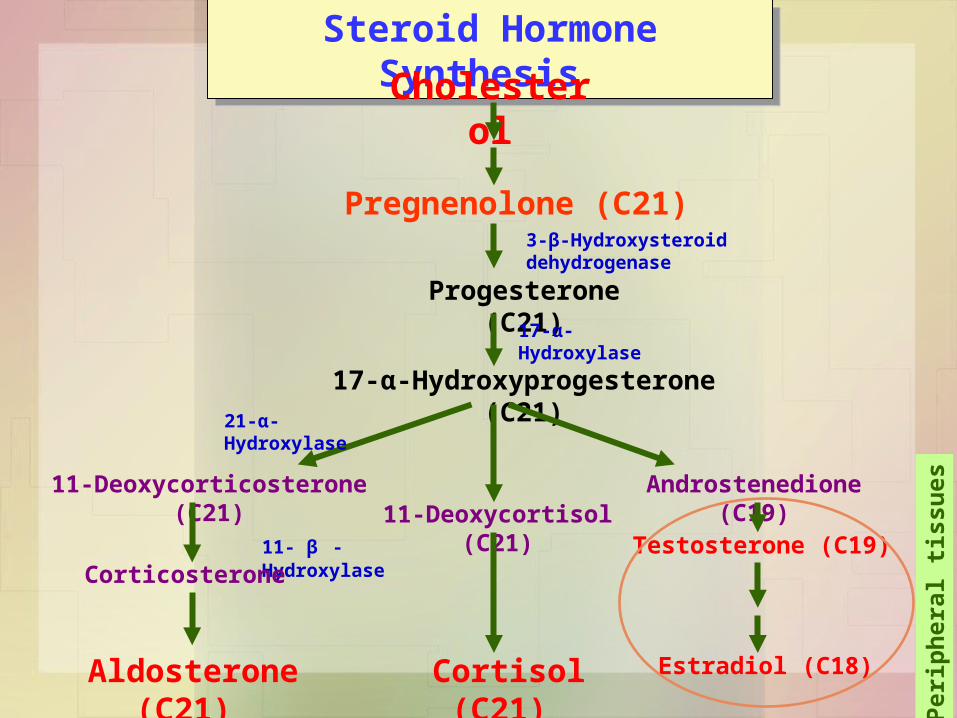
Hyperpigmentation in Addison’s disease
Hyperpigmentation occurs because melanocyte-stimulating hormone (MSH) and (ACTH) share the same precursor molecule, Pro-opiomelanocortin (POMC).
The anterior pituitary POMC is cleaved into ACTH, γ-MSH, and β-lipotropin.
The subunit ACTH undergoes further cleavage to produce α-MSH, the most important MSH for skin pigmentation.
In secondary adrenocortical insufficiency, skin darkening does not occur.

Investigation of Addison’s disease (AD)
• The patient should be hospitalized
• Basal measurement of:Serum urea, Na+, K+ & glucoseSerum cortisol and plasma ACTH
• Definitive diagnosis and confirmatory tests should be done later after crisis.

Investigation of Addison’s disease (AD)
• Normal serum cortisol and UFC does not exclude AD.
• Simultaneous measurement of cortisol and ACTH improves the accuracy of diagnosis of primary adrenal failure:
Low serum cortisol ( <200nmol/L) and High plasma ACTH (>200 ng/L)
Cont’d

Confirmatory Tests:1. Short tetracosactrin (Synacthen) test
(Short ACTH stimulation test)
• Measure basal S. cortisol• Stimulate with I.M. synthetic ACTH (0.25 mg)• Measure S. cortisol 30 min after I/M injection• Normal: of S. cortisol to >500 nmol/L • Failure of S. cortisol to respond to stimulation,
confirm AD.• Abnormal results:
– emotional stress– glucocorticoid therapy– estrogen contraceptives.

Confirmatory Tests:2. Adrenal antibodies
• Detection of adrenal antibodies in serum of patients with autoimmune Addison’s disease
3. Imaging (Ultrasound/CT)
Ultrasound or CT for adrenal glands for identifying the cause of primary adrenal failure

Investigation of Secondary AC Insufficiency
• Low serum cortisol with low plasma ACTH
• No response to short synacthen test: Adrenocortical cells fail to respond to short ACTH stimulation
• Depot Synacthen test (confirmatory test) 1. Measure basal S. cortisol
2. Stimulate with I.M. synthetic ACTH (1.0 mg) on each of three consecutive days
3. Measure S. cortisol at 5 hours after I.M. injection on each of the three days

Investigation of Secondary AC Insufficiency
Depot Synacthen test …. Cont’d
Interpretation of results:- Addison’s disease: No rise of S. cortisol >600 nmol/L
at 5 h after 3rd injection.
- Secondary AC: Stepwise increase in the S. cortisol after successive injections
- Limitations: Hypothyroidism: Thyroid deficiency must be
corrected before testing of adrenocortical functions
Prolonged steroid therapy

Investigation of Secondary AC Insufficiency …. Cont’dOther Investigations
• Insulin-induced hypoglycemia:
Adrenal failure secondary to pituitary causes
• MRI for pituitary gland

Screenin
g
• Basal plasma ACTH and basal serum cortisol, glucose, urea and electrolytes
• High ACTH and Low cortisol
Confirmati
on
• Short ACTH stimulation test: No response
Others
• Adrenal autoantibodies• Ultrasound/CT adrenal glands
Investigation for Addison’s disease

Screenin
g
• Low ACTH and Low cortisol
Confirmati
on
• Long ACTH stimulation test: Stepwiseincrease in S. cortisol
Others
• Insulin-induced hypoglycemia• MRI pituitary gland
Investigation for Secondary AC Insufficiency

Lecture Notes in Clinical Chemistry – 8th/9th edition