Embed Size (px)
Citation preview

Review
Bioastronautics: The Influence of Microgravityon Astronaut Health
Elizabeth Blaber,1,2 Helder Marcal,1,2,3 and Brendan P. Burns1,2
Abstract
For thousands of years different cultures around the world have assigned their own meaning to the Universe.Through research and technology, we have begun to understand the nature and mysteries of the Cosmos. Lastyear marked the 40th anniversary of our first steps on the Moon, and within two decades it is hoped thathumankind will have established a settlement on Mars. Space is a harsh environment, and technological ad-vancements in material science, robotics, power generation, and medical equipment will be required to ensurethat astronauts survive interplanetary journeys and settlements. The innovative field of bioastronautics aims toaddress some of the medical issues astronauts encounter during space travel. Astronauts are faced with severalhealth risks during both short- and long-duration spaceflight due to the hostile environment presented in space.Some of these health problems include bone loss, muscle atrophy, cardiac dysrhythmias, and altered orientation.This review discusses the effects of spaceflight on living organisms, in particular, the specific effects of micro-gravity on the human body and possible countermeasures to these effects. Key Words: Bioastronautics—Microgravity—Human physiology. Astrobiology 10, 463–473.
1. Introduction
The hostile environment of space is known to have animpact on human physiology (Buckey, 1999; Williams,
2002; Clement and Slenzka, 2006; Kalb and Solomon, 2007).The predominant hostile conditions are those of radiation andthe absence of gravity. A new field called bioastronautics,developed by NASA, has emerged in life sciences to ensurethe health and safety of humans during space travel (Wil-liams, 2002).
The National Aeronautics and Space Administration(NASA) defines bioastronautics as the study of the biologicaland medical effects of spaceflight on living organisms(Charles, 2005). Bioastronautics aims to expand extramuralcollaboration and leverage new and unique capabilities ofthe scientific community and federal governments, whileapplying the knowledge gained to Earth-based problems(Williams, 2002, 2003). Specifically, as humans advance inexploratory planetary missions, systemic and interrelatedadaptations of the human body to microgravity are inevita-ble for humans to survive (Vernikos, 1996; Stewart et al.,2007).
Furthermore, operational challenges, such as the man-agement of waste disposal systems, life-support systems,food and nutrition, medical care, and psychosocial healthdue to long-term confinement, will have an impact on
human health during long-term space missions (Klein et al.,1988; Williams, 2002; Horneck, 2008). Therefore, an under-standing of the response of the human body to the envi-ronment of space at the systemic integration system level andat the molecular and cellular level is warranted. This reviewaims to discuss the response of the human body to the en-vironment of space. In particular, it examines the response ofspecific physiological systems of the human body to condi-tions of microgravity and possible countermeasures to these.One of the major gaps in our knowledge in this area is theeffect of microgravity at the cellular level, and new researchdirections are thus discussed at the end of this review.
1.1. The bioastronautics roadmap
The bioastronautics roadmap is a risk reduction strategyfor human space exploration. The risks associated with spaceexploration are identified in NASA’s bioastronautics road-map, which is the framework for identifying, assessing, andreducing the risks of crew exposure to the hazardous envi-ronments of space (Charles, 2005). The roadmap identifies 45risks of space travel and categorizes these risks into 5 mainareas: human health and countermeasures, autonomousmedical care, behavioral health and performance, radia-tion health, and advanced human support technologies(Charles, 2005). The roadmap aims to prioritize research and
1Australian Centre for Astrobiology, 2School of Biotechnology and Biomolecular Sciences, and 3The Graduate School of BiomedicalEngineering, The University of New South Wales, Sydney, Australia.
ASTROBIOLOGYVolume 10, Number 5, 2010ª Mary Ann Liebert, Inc.DOI: 10.1089/ast.2009.0415
463

technology and communicate these technologies by encom-passing operations and policies related to the risks associatedwith human spaceflight. Additionally, the roadmap aspiresto establish tolerance limits to the space environment anddevelop countermeasures to overcome these problems.
Astronauts experience muscle atrophy, bone loss, andcardiovascular changes during short-term space missions(Ilyina-Kakueva and Burkovskaya, 1991; Kaplansky et al.,1991; Blomqvist et al., 1994; Fritsch-Yelle et al., 1996; Caillot-Augusseau et al., 1998; Williams, 2003; Dai et al., 2007; Stewartet al., 2007). In addition, some astronauts experience cardiacdysrhythmias due to reduced aerobic capabilities (Williams,2002; Diedrich et al., 2007). Other significant effects includepost-flight orthostatic intolerance, decreased bone density,ataxia, and poor coordination (Clement et al., 2005; Con-vertino, 2005; Mano, 2005; Norsk, 2005; Souvestre et al., 2008).
Therefore, one of the major challenges in the evolution ofspace medicine is to determine how the physiological ad-aptation to space may alter the pathophysiology of diseaseand the manifestation of illness and injury in space. Toovercome these challenges, ground-based (simulated micro-gravity) and space studies are needed.
2. Microgravity
The absence of gravity in space, also known as zerogravity, can be simulated on Earth as microgravity. The in-fluential effects of microgravity on human physiology are
best studied in space; however, this is not always practical oreconomical. Simulated microgravity is thus used to study theeffects of microgravity on cellular systems on Earth. Themost common small-scale method for simulating micro-gravity is conducted with the use of a device known as arotating-wall vessel (RWV) bioreactor (Fig. 1), which facili-tates cellular models under conditions of simulated micro-gravity. Cellular models may be used for investigating theeffects of microgravity on cell-specific tissues and organswithin the human body. However, little research has beenconducted to date on the specific responses of cells to con-ditions of microgravity, with the majority of researchfocusing on the systemic effects of microgravity.
The RWV bioreactor is a NASA-designed tissue culturevessel that can be used to simulate microgravity while re-ducing the shear and turbulence associated with impeller-driven and stirred bioreactors (Fig. 1) (Schwarz et al., 1992;Goodwin et al., 1993; Freed et al., 1999). The system is basedon two important design principles:
(a) Solid body rotation about a horizontal axis.(b) Active or passive diffusion of oxygen through a sili-
cone rubber membrane (Schwarz et al., 1992; Goodwinet al., 1993; Unsworth and Lelkes, 1998).
Solid body rotation is achieved by a horizontal rotatingvessel that is filled with culture medium and has no gas-liquid interface. The initial rotational speed of the vessel isadjusted to ensure that the culture medium and cell con-
FIG. 1. Image of Rotary Cell Culture System (RCCS) containing Rotating-Wall Vessel (RWV) bioreactor illustrating themajor functional components. Color images available online at www.liebertonline.com/ast.
464 BLABER ET AL.

structs rotate synchronously with the vessel, enabling anefficient and low-shear mass transfer of nutrients and wastes(Fig. 1) (Unsworth and Lelkes, 1998; Navran, 2008). TheRWV bioreactors utilize a centrally located silicone mem-brane for oxygenation, which is in direct contact with theculture medium and, thus, minimizes cellular damage(Schwarz et al., 1992; Goodwin et al., 1993). Incubator air iscirculated through the central shaft of the vessel by an ex-ternal air pump, which thus eliminates the introduction of airbubbles into the culture medium (Schwarz et al., 1992;Goodwin et al., 1993).
When the vessel and its contents reach an appropriatespeed, the mass remains suspended close to a stationarypoint relative to a ground observer (Freed et al., 1999;Nickerson et al., 2003). During these conditions, at any giventime, the gravitational vectors are randomized to simulatemicrogravity; due to the dynamic equilibrium that existsbetween gravitational force (Fg), centrifugal force (Fc), anddrag forces (Fd), the cell mass remains in constant free falland thus is exposed to simulated microgravity (Fig. 2)(Unsworth and Lelkes, 1998; Freed et al., 1999). By incorpo-rating the design principles of solid body rotation andmembrane oxygenation, the RWV is an extremely low-shear,low-turbulence microgravity environment that allows for theculture of high-density three-dimensional mammalian cells(Schwarz et al., 1992; Goodwin et al., 1993; Freed et al., 1999;Nickerson et al., 2003; Navran, 2008).
In addition to cellular models of simulated microgravity,animal models have also been developed to study the effectsof microgravity on whole systems. Such models includehind-limb unloading and head-down bed rest (Colleran et al.,2000; Aubert et al., 2005; Bao et al., 2007; Delp, 2008; Radeket al., 2008). Hind-limb unloading is a method of simulatingthe effects of microgravity on Earth. By suspending an animal(usually rat) from its tail or hind limbs, the effect of gravita-tional loading on the hind limbs is removed, which enablesstudy of the effects of weightlessness on this area of the animal(Morey-Holton and Globus, 1998; Basso et al., 2005). Suchmodels enable the study of the effects of microgravity onEarth; however, as only part of the animal remains suspended(weightless), such studies have limitations that can only beovercome by conducting experiments in the true microgravityenvironment of space. Another model that is used to simulatethe environment of space on Earth is head-down tilt-bed rest(HDTBR), whereby a patient’s head is tilted at an angle that is6 degrees lower than the patient’s feet (Regnard et al., 2001;
Baisch, 2002; Trappe et al., 2006). By conducting similar ex-periments in the microgravity of space and using Earth-basedsimulations of microgravity (HDTBR), Trappe et al. (2006)found that HDTBR was an adequate analogue for spaceflight.However, it has also been noted that in-flight data can varyfrom data collected via ground-based models such as HDTBR(Regnard et al., 2001). These discrepancies could be due to thealtered physical forces that affect tissues; in HDTBR, gravita-tional forces compress body tissues, whereas in microgravity,there is negative pressure around the body (Convertino et al.,1997; Regnard et al., 2001).
These discrepancies need to be taken into account whenusing Earth-based simulations to model microgravity.
3. Primary Areas of Microgravity Research
To date, research has mainly focused on the systemic ef-fects of microgravity on human physiology, which includesthe musculoskeletal system, cardiovascular system, sensory-motor system, and the immune system (Table 1).
3.1. The musculoskeletal system
The musculoskeletal system, an organ system comprisedprimarily of the skeleton and skeletal muscle, has been one ofthe main focuses of microgravity research in the last 10 years.
3.1.1. The skeleton. Research has shown that micro-gravity causes immense changes in bone tissue that result indecreased bone mass and lead to early onset osteoporosis inmany astronauts (Kaplansky et al., 1991; Caillot-Augusseauet al., 1998; Carmeliet and Bouillon, 1999; Colleran et al., 2000;Vico et al., 2000; Bucaro et al., 2007; Dai et al., 2007). Thesechanges produce a condition known as spaceflight osteope-nia, which is characterized by an increase in bone resorptionby osteoclasts and a decrease in bone formation by osteo-blasts (Bucaro et al., 2007).
Bone is a multifunctional tissue that has two main func-tions: to provide mechanical integrity for both movementand protection, and to help regulate mineral homeostasisthrough involvement in metabolic pathways. The architec-ture of bone is related to the mechanical stresses that areexerted on it. In a microgravity environment, however, thesemechanical stresses are nonexistent, which results in rapidand severe uncoupling between bone formation and boneresorption. It is estimated that in a microgravity environmentapproximately 1–2% of the skeleton is mobilized and lost
FIG. 2. Rotating Wall Vessel (RWV) bioreactor displaying tissue constructs (A). When the vessel is rotating at the correctspeed, the constructs (B) remain in continual free fall due to a dynamic equilibrium between the acting forces (gravitationalforce Fg, centrifugal force Fc, and drag forces Fd). Color images available online at www.liebertonline.com/ast.
EFFECTS OF MICROGRAVITY ON HUMAN PHYSIOLOGY 465
each month (Carmeliet and Bouillon, 1999), which causessignificant bone loss during long-duration spaceflight.However, the precise mechanism by which this occurs re-mains unknown.
By collecting biochemical data from astronauts during a180-day spaceflight, Caillot-Augusseau et al. (1998) foundthat all bone formation parameters (bone alkaline phos-photase, osteocalcin, and type I procollagen peptide) weredecreased and bone resorption markers (procollagen C-telopeptide) were increased during flight (Caillot-Augusseauet al., 1998). Previous studies have also shown a decrease inbone formation and an increase in bone resorption; however,these changes were primarily found in weight-bearing bones(Kaplansky et al., 1991; Carmeliet and Bouillon, 1999; Col-leran et al., 2000). Caillot-Augusseau et al. (1998), on the otherhand, observed that the systemic variations in these markerswere so large after a 6-month spaceflight they concluded thatnon-weight-bearing bones are also affected. This was inagreement with Vico et al. (2000), who observed a bone massloss in tibiae and radii in humans after 6 months of space-flight. Bucaro et al. (2007) hypothesized that the decrease inbone formation in microgravity conditions is due to micro-gravity directly inducing osteoblast apoptosis. Apoptosis is aprocess of programmed cell death by which cells undergo anordered sequence of events that lead to death of the cell.However, they found that simulated microgravity did notdirectly increase the induction of apoptosis in osteoblasts,and it was proposed that the decrease in the number of os-teoblasts observed could be due to microgravity causing anincrease in osteoclast-mediated resorption of bone (Bucaroet al., 2007). This increase in bone resorption could then causean increase in matrix apoptogen release, which would resultin increased apoptosis of osteoblasts (Bucaro et al., 2007).
An alternative hypothesis has also been postulated to ac-count for the loss of bone mass by the cephalic fluid shift thatoccurs in microgravity (Colleran et al., 2000). This may affectbone blood flow and could, therefore, potentially alter in-terstitial fluid pressures and flows. If such alterations were tooccur in weight-bearing bones, then the mechanical envi-ronment of osteo-progenitor cells, osteoblasts, and osteo-clasts would be altered, which would thus alter boneperfusion rates. To confirm the mechanism by which mi-crogravity causes bone loss in both astronauts and cosmo-nauts during spaceflight, further human studies must be
conducted with the use of both ground-based models(HDTBR) and space models.
3.1.2. The skeletal muscle. Changes have also beenidentified in the other primary component of the musculo-skeletal system, the skeletal muscle (Table 1) (Fitts et al., 2000,2001; Slentz et al., 2001; Kalb and Solomon, 2007; Trappe et al.,2009). Unimpaired motor function is necessary for effectiveinteraction of an extraplanetary explorer with that environ-ment. Reduction in muscle function is first detectable withindays of existence in microgravity and progresses throughoutthe duration of the mission (Tesch et al., 2008). The effects ofmicrogravity on skeletal muscles are most pronounced inantigravity muscles, that is, muscles that play a postural rolein standard gravity conditions (1 g), such as the calf muscles(soleus and gastrocnemius) and the quadriceps (Fitts et al.,2000; Slentz et al., 2001; Kalb and Solomon, 2007). Moststudies have also shown that extensor muscles are affected toa greater extent by microgravity than are their antagonisticcounterparts, the flexors (Kalb and Solomon, 2007). Follow-ing a 17-day spaceflight, Widrick et al. (1999) observed thatfibers from flexors (e.g., gastrocnemius muscle of the calf ) areless affected by spaceflight than are extensors (soleus muscleof the calf ). However, as the duration of the missionlengthened, all skeletal muscles appeared to be affected bymicrogravity conditions in a similar manner (Fitts et al.,2000).
Although the rate of muscle deterioration is dependent onthe function of the muscle, fibers within the muscle are alsoaffected by microgravity conditions (Tesch et al., 2008). Re-cent studies using rats have shown that type I muscle fibers,which are slow and resistant to fatigue, atrophy preferen-tially in microgravity (Fitts et al., 2001; Kalb and Solomon,2007). Human studies found that type II fibers (type IIa, fastand resistant to fatigue, and type IIb, fast and fatigues rap-idly) were affected as much as type I fibers were (Edgertonet al., 1995; Widrick et al., 1999; Kalb and Solomon, 2007).Edgerton et al. (1995) observed, after an 11-day spaceflight, asignificant decrease in the cross-sectional area of fibers fromthe vastus lateralis muscle with the decline being greatest inthe type II fibers and least in type I fibers.
Another study proposed that there are three sets of factorsthat underlie the observed decrease in muscle function (Kalband Solomon, 2007). The first is the removal of the antigravity
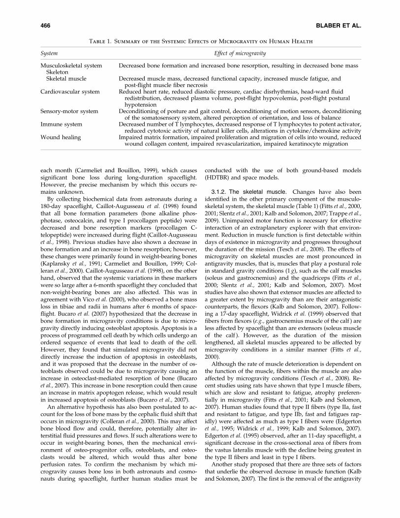
Table 1. Summary of the Systemic Effects of Microgravity on Human Health
System Effect of microgravity
Musculoskeletal system Decreased bone formation and increased bone resorption, resulting in decreased bone massSkeletonSkeletal muscle Decreased muscle mass, decreased functional capacity, increased muscle fatigue, and
post-flight muscle fiber necrosisCardiovascular system Reduced heart rate, reduced diastolic pressure, cardiac disrhythmias, head-ward fluid
redistribution, decreased plasma volume, post-flight hypovolemia, post-flight posturalhypotension
Sensory-motor system Deconditioning of posture and gait control, deconditioning of motion sensors, deconditioningof the somatosensory system, altered perception of orientation, and loss of balance
Immune system Decreased number of T lymphocytes, decreased response of T lymphocytes to potent activator,reduced cytotoxic activity of natural killer cells, alterations in cytokine/chemokine activity
Wound healing Impaired matrix formation, impaired proliferation and migration of cells into wound, reducedwound collagen content, impaired revascularization, impaired keratinocyte migration
466 BLABER ET AL.

load that is most evident in antigravity muscles such as thesoleus. The unloading of antigravity muscles (the removal ofthe gravitational load) may lead to a decrease in muscle genetranscription and protein translation (Fitts et al., 2000; Kalband Solomon, 2007). The second factor is a reduction in neuraldrive to the muscle (Kalb and Solomon, 2007). Antonutto et al.(1998) found that there is a greater loss of leg muscle powerthan can be attributed to reduction in muscle mass in astro-nauts after 21 days of spaceflight, which indicates that mi-crogravity induces changes in motor control and coordination.By using electromyographic techniques on Rhesus monkeysunder microgravity and 1 g conditions, Recktenwald et al.(1999) confirmed the existence of altered neuronal recruitmentpatterns. However, studies in humans need to be performedto further validate these hypotheses. The third set of factorsthat contribute to the loss of muscle mass are systemic factors,including hormone alteration and changes in metabolism thatoccur during spaceflight (Kalb and Solomon, 2007).
Upon returning to Earth, astronauts often experienceweakness and delayed onset muscle soreness associated withtheir reacquaintance with the gravitational field of Earth.Myopathology of muscles, such as fiber necrosis, interstitialoedema, and activated macrophages, are observable severaldays after landing (Kalb and Solomon, 2007). This effect ofreloading proposes serious consequences for interplanetarymissions, as reloading of the muscles will cause substantialmuscle damage and may incapacitate the astronauts. Theestablishment of interplanetary settlements on Mars has beenproposed for as early as 2020; however, missions involvedwith such settlements would expose astronauts to extendedperiods of zero gravity, followed by a period of extremegravity (up to 6 g) during reentry and landing, and thenlowered gravity on Mars (38% of that on Earth) (Buckey,1999; Horneck, 2008). Muscle atrophy has been documentedto increase in severity upon re-exposure to gravitationalfields (Antonutto et al., 1998; Buckey, 1999). It remains to beseen what effects exposure to hypergravity during reentrywill have on damaged muscles and whether exposure tolowered gravity on Mars will enable this damage to be re-versed. Investigations into the effect of hypergravity couldpossibly be conducted with the use of animal models onEarth, after initial exposure of these animals to microgravityconditions for a period of time. Although this does not rep-resent true zero gravity, it is the most effective way to modelzero gravity on Earth. These questions need to be examinedand countermeasures determined before interplanetary set-tlements can be pursued without serious consequences toastronaut health.
3.2. The cardiovascular system
Spaceflight also has significant effects on the cardiovas-cular system. Studies on animals (rats and Rhesus monkeys)have found consistent decreases in heart rate during flight(Fuller, 1985). Similarly, by using 12 astronauts over 6 mis-sions, Fritsch-Yelle et al. (1996) found that the in-flight heartrates of astronauts were reduced. However, it is suggestedthat these reductions are due to the cardiovascular systemoperating with lower sympathetic activity and vascular re-sistance in space. This hypothesis is supported by the find-ings of Eckberg et al. (1988) and Wallin et al. (1992), whofound that plasma catecholamines are reduced in flight and
plasma catecholamine levels correlate with muscle sympa-thetic activity, which is also linked to diastolic pressure andheart rate (Eckberg et al., 1988; Wallin et al., 1992; Fritsch-Yelle et al., 1996; Aubert et al., 2005). Serious cardiac dys-rhythmias with potentially fatal consequences have also beenreported as an effect of spaceflight and exposure to micro-gravity on the human body (Fritsch-Yelle et al., 1996). Al-though this study found an occurrence of dysrhythmias inthe 12 astronauts studied (Fritsch-Yelle et al., 1996), the fre-quency of disrhythmias was small, and consistent data in thefield have yet to be provided.
Many changes have also been reported in blood andplasma volume during spaceflight (Bao et al., 2007; Diedrichet al., 2007) (Fig. 3). Blood, including plasma, circulatesthroughout the body to supply oxygen and energy to organsand tissues. The regulation of this process relies on the car-diovascular system, fluid- and electrolyte-regulating hor-mones, the nervous system, and the renal system to keepblood volume in a physiologically narrow range (Norsk,2005). The loss of gravitational forces that occurs duringspaceflight causes characteristic facial oedema during thefirst few days of spaceflight and post-flight thin legs causedby head-ward fluid redistribution, as seen in Fig. 3 (Norsk,2005). Additionally, post-flight hypovolemia (decreasedblood volume) has been reported in conjunction with or-thostatic intolerance (Blomqvist et al., 1994), and recentstudies have shown plasma volume is decreased by 10–17%(Blomqvist et al., 1994; Diedrich et al., 2007). It is now thoughtthat diminished blood volume in space is the result of re-duced fluid intake with a smaller reduction in urinary out-put, reduced red cell mass, and fluid shifts fromintravascular to interstitial space due to reduced compres-sion of tissues by gravitational forces (Blomqvist et al., 1994;Norsk, 2005; Diedrich et al., 2007).
The most well-documented symptom in astronauts andcosmonauts is post-flight orthostatic intolerance. This is acondition that occurs when a patient’s body is unable, viabasic cardiovascular mechanisms, to compensate for the ra-pid translocation of blood from the upper body to the lowerbody while standing under normal Earth gravity conditions(Fritsch-Yelle et al., 1996). Post-flight orthostatic intolerance isthought to be due to a combination of alterations that occurduring spaceflight, including hypovolemia, cardiovascularde-conditioning, and reduced vasoconstrictor response.Cardiovascular measures during and after landing alsosuggest considerable cardiovascular stress (Fritsch-Yelle et al.,1996). This study recorded heart rates of >160 beats perminute during landing and systolic pressure drops of>25 mm Hg subsequent to landing (Fritsch-Yelle et al., 1996).It was also recorded that heart rate, plasma catecholaminelevels, and peripheral vascular resistance levels were all el-evated post-flight (Fritsch-Yelle et al., 1996).
Furthermore, the effects of interplanetary travel and set-tlement on Mars on the cardiovascular system are unknown.Previous work has shown that spaceflight leaves astronautsunprepared for the return to Earth’s gravitational field (1 g);however, the cardiovascular stressors associated with hy-pergravity (up to 6 g) during entry to Mars and adaptation tothe lowered gravitational field on Mars (38% of Earth’sgravity) are unknown. It would be expected that cardiovas-cular reconditioning and reduced vasoconstrictor responsewould occur, and additionally astronauts would be expected
EFFECTS OF MICROGRAVITY ON HUMAN PHYSIOLOGY 467

to suffer from post-flight orthostatic intolerance as has beenexperienced on Earth following spaceflight. Furthermore, itis not known whether the reduced gravitational field onMars would cause these effects to worsen. Cardiovascularchanges that occur during zero gravity and hypergravityrequire further investigation before interplanetary missionscan be achieved safely.
3.3. The sensory-motor system
The sensory-motor system, which consists of both sensoryand motor neurons and their corresponding targets, is one ofthe most critical systems affected by microgravity. Duringlong-term interplanetary missions, the sensory motor systemreconditions from exposure to microgravity in space, andthis can cause a variety of effects that may be detrimental toboth astronaut health and the success of the mission (seeTable 1 for summary of effects).
Adaptation to microgravity requires the reorganization ofcentral nervous system processing of the three major sourcesof spatial information—visual, vestibular, and somatosen-sory (Souvestre et al., 2008). Recent experimental resultsindicate that, during microgravity conditions, adaptivechanges occur in neural strategies that are used to resolveambiguous linear accelerations detected by the otolith sys-tem (Kalb and Solomon, 2007; Souvestre et al., 2008). In theabsence of gravitational fields, visual references are vital toastronaut orientation during spaceflight (Kalb and Solomon,2007; Souvestre et al., 2008; Dyde et al., 2009). It has also beenfound that, in the absence of gravity, signals from the centralvestibular system, peripheral pressure receptors, and visualcues become misleading to a point where disorientation oc-curs (Mano, 2005; Souvestre et al., 2008). This produces acondition known as space adaptation syndrome (SAS),which is the main cause of space motion sickness (SMS).Commonly recorded symptoms of SAS and SMS includedizziness, vertigo, headaches, cold sweating, fatigue, nausea,and vomiting (Souvestre et al., 2008). Current research sug-gests that SAS is caused by a sensory conflict between
sensory-motor control inputs from visual and tactile senses,and input coming from the vestibular organs in the inner ear(Lackner and Dizio, 2006), which can then lead to SMS(Souvestre et al., 2008).
The central nervous system has the ability to adapt andcompensate under altered stimulus conditions such as thoseexperienced during spaceflight (Kalb and Solomon, 2007;Souvestre et al., 2008). The perception of locomotion andpositions are a result of the brain’s ability to integrate visualand auditory signs with vestibular input and proprioceptiveinformation (motion, pressure, and temperature receptors inthe tendons, muscles, joints, and skin) (Kalb and Solomon,2007; Souvestre et al., 2008). If the input the brain receivesfrom these sensors is modified due to altered gravity con-ditions, then the central nervous system is forced to developnew interpretations of the stimuli and thus develop alter-native adjustment strategies to compensate for the alteredstimuli (Lackner and Dizio, 2006; Souvestre et al., 2008).However, if the new interpretation of stimuli does not matchfundamental, specific patterns in the brain, then SAS and,consequently, SMS are likely to occur.
Although the symptoms of SAS and SMS are well docu-mented (Lackner and Dizio, 2006; Kalb and Solomon, 2007;Souvestre et al., 2008; Dyde et al., 2009), effective therapies orcountermeasures are yet to be produced (Lackner and Dizio,2006; Souvestre et al., 2008). One study, however, has pro-posed that exposure to artificial gravity by way of a centri-fuge may facilitate the ability of astronauts and cosmonautsto respond normally to gravitational fields that are encoun-tered at their destination (Kalb and Solomon, 2007).
3.4. The immune system and wound healing
The immune system consists of immunological cells,lymph nodes, and organs that are also altered under mi-crogravity conditions (Table 1). Alterations in the regulationand efficiency of the immune system could have profoundeffects on the ability of the host to protect itself from invasionby foreign entities. These include infectious agents and
FIG. 3. The major effects of gravity onplasma volume during upright position (A)and during weightlessness (B) (adapted fromDiedrich et al., 2007).
468 BLABER ET AL.

tumors, and effects on the ability of the body to repair itself,particularly subsequent to cutaneous or intrinsic wounds.Wound healing is a complex and multistage process thatconsists of intricate interactions of immunological cells, suchas neutrophils and macrophages, soluble factors, the extra-cellular matrix, and tissue cells such as fibroblasts (Pollack,1984; Davidson et al., 1999).
Several studies, including ground-based models andspaceflight experiments, have investigated the effects of mi-crogravity on the immune system (Woods and Chapes, 1994;Sonnenfeld, 2002; Gridley et al., 2009). The overall goal ofthese studies was to determine the effect microgravity has onthe efficiency and effectiveness of the immune system in aspace environment. Deterioration of this system in such anenvironment can lead to secondary immunodeficiency andraise the risk of contracting diseases such as bacterial andviral infections, allergies, and autoimmunity pathologies(Rykova et al., 2008). Such conditions can then decrease thecapacity of astronauts to complete a mission successfully.Additionally, the establishment of interplanetary settlementsrequires a thorough understanding of the host’s ability toprotect itself from foreign entities to ensure the health ofthose living there. It is essential to establish the effects thatspaceflight has on immunological cells, cytokines, chemo-kines, and other pro-inflammatory and regulatory proteins.
Gridley and associates (2009) investigated the effectspaceflight has on T lymphocytes. T lymphocytes are dividedlargely into two subsets: CD4þ T helper cells and CD8þ Tcytotoxic cells (Gridley et al., 2009). T helper cells secretecytokines that regulate the immune system when facing in-fection with foreign organisms, while T cytotoxic cells di-rectly kill cells that are recognized as foreign (Gridley et al.,2009). By studying mice flown in space for 13 days, Gridleyand colleagues (2009) found that spaceflight significantlyaffects T lymphocytes. This study demonstrated that T cellnumbers were reduced and decreased in response to a potentT cell activator. Additionally, analysis of the thymus of flightmice indicated that spaceflight had a significant impact onthe expression of many cancer-related genes, of which somechanges indicate an increase in carcinogenesis (Gridley et al.,2009). In conjunction with the radiation that is experienced inspace, these results indicate the serious possibility for astro-nauts to develop cancer during long-term missions. Thepotential for astronauts to develop cancer and tumors inspace is further enhanced by alterations in natural killer cellfunction in microgravity conditions. Natural killer cells killboth tumor cells and virally infected cells by releasinggranules contained within their cytoplasm that cause thedeath of the target cell (Sonnenfeld, 2002). Rykova and as-sociates (2008) studied the effect of spaceflight on 30 astro-nauts aboard the International Space Station. This studyfound that the cytotoxic activity of natural killer cells wasreduced, and additionally the number of natural killer cellscirculating in the blood was decreased following long-duration flights (Rykova et al., 2008).
Furthermore, alterations in cytokine and chemokine pro-duction have also been shown to occur during microgravityconditions. Studies have shown a severe decrease in theability of leukocytes to produce interferon a/b followingspaceflight. Interferon a/b is an important cytokine involvedin the antiviral immune response as well as functioning as animmunoregulatory cytokine (Sonnenfeld, 2002). This could
potentially lead to decreased resistance to viral infectionduring spaceflight conditions. Additionally, Woods andChapes (1994) found that the ability of tumor necrosis factor(TNF)-a, which is a potent cytokine involved in immuno-regulation, induction of inflammation, and inhibition oftumorigenesis, to induce apoptosis in diseased cells wasinhibited during spaceflight (Woods and Chapes, 1994). Theimpairment of both immune cells and cytokines/chemokinesthat activate, stimulate, and regulate immunological cells hasconsiderable effects on the ability of the human body to resistinfection during spaceflight conditions. In addition, the im-pairment of the immune system during spaceflight also hasthe potential to alter dramatically the ability of the body torepair itself following cutaneous or intrinsic wounds.
Little is known about the effects of spaceflight on woundhealing; however, several studies have indicated thatspaceflight can negatively affect tissue repair in both boneand skeletal muscle (Ilyina-Kakueva and Burkovskaya, 1991;Kaplansky et al., 1991; Davidson et al., 1999). On Earth, whendisruptive injury occurs to the skin, the wound repair pro-cess is characterized into several stages: the inflammatoryphase, the proliferative phase, epithelialization, and matu-ration and remodelling (Pollack, 1984). Each of these phasesconsists of a complex interplay of inflammatory and tissuecells, biochemical cascades and factors that can potentially bealtered under conditions of microgravity that lead to im-paired wound healing.
During a skin injury, blood enters the wound site andbrings with it cellular elements and plasmic constituents,including platelets and fibrinogen (Mutsaers et al., 1997;Diegelmann and Evans, 2004; Baum and Arpey, 2005).Platelets aggregate at the site to form a fibrin clot, which actsto halt bleeding from the wound site (Diegelmann and Evans,2004). The fibrin clot serves as a temporary lattice for mi-grating fibroblasts, inflammatory cells, and growth factors(Baum and Arpey, 2005). If the dynamic matrix of proteinsand cells that compose the fibrin clot are impaired, appro-priate wound healing does not occur (Pollack, 1984; Baum andArpey, 2005). Davidson et al (1999) studied the effect of mi-crogravity on intrinsic wound healing in rats by subcutane-ously implanting sponges that released one of several growthfactors or a placebo. This investigation into the capacity of ratsto form granulation tissue and collagen during spaceflightfound that matrix formation was significantly inhibited underspaceflight conditions; thus stimulated proliferation andmigration of cells into the wound was absent. (Davidson et al.,1999). From these results, it is possible to hypothesize thatimpaired formation of the initial matrix structure has negativeeffects on the success of the wound healing process; however,further investigation into this area is required.
Following the formation of a fibrin clot, blood vesselssurrounding the site dilate, which results in the initiation ofthe inflammatory response (Pollack, 1984). The inflammatoryresponse involves a vast number of cell types, includingpolymorphonuclear neutrophils, monocytes, T lymphocytes,fibrocytes, and eosinophils (Diegelmann and Evans, 2004;Baum and Arpey, 2005). Additionally, a vast array of serumproteins, growth factors, cytokines, and chemokines are re-quired to attract inflammatory cells to the wound site andstimulate their function. As mentioned previously, manyimmunological cell types are impaired under conditions ofmicrogravity, and this may have a significant effect on the
EFFECTS OF MICROGRAVITY ON HUMAN PHYSIOLOGY 469

success of the wound healing process. Through their space-flight experiments, Davidson et al. (1999) discovered thatdeficits in immune cell infiltration occurred during exposureto microgravity conditions and led to impaired woundhealing. Specifically, this study found that microgravity al-tered the capacity of rats to generate adequate macrophagepopulations that could possibly cause impaired woundhealing during spaceflight (Davidson et al., 1999).
The proliferative phase of wound healing is marked by thereplacement of the extracellular matrix of the wound withgranulation tissue (Pollack, 1984; Diegelmann and Evans,2004). Granulation tissue includes components that becomepermanent residents of the repaired dermis, including fi-broblasts and collagen (Baum and Arpey, 2005). Fibroblastsproduce collagen, which is one of the most important pro-teins in wound healing, as it provides increased woundstrength while facilitating the movement of other cells intothe wound site (Clark, 1985; Mutsaers et al., 1997; Baum andArpey, 2005). Davidson et al. (1999) discovered that ratsflown in the spaceflight environment had considerably re-duced wound collagen concentration compared to groundcontrols, and collagen content remained decreased aftergrowth factor stimulation. Due to the importance of collagenfor wound strength and migration of other cell types, pri-marily endothelial cells, and macrophages, it is likely thatseverely reduced wound collagen content can have a drasticeffect on the ability of a wound to heal successfully underspaceflight conditions.
In addition to collagen synthesis, the proliferative phase ofwound healing also consists of angiogenesis (i.e., the for-mation of new blood vessels) and reepithelialization,whereby intact epidermis is reestablished over granulationtissue (Clark, 1985; Mutsaers et al., 1997; Baum and Arpey,2005). Angiogenesis is stimulated by a variety of changes inthe local tissue environment, such as low oxygen tension,which reflect deficient tissue perfusion due to damagedcapillaries at the wound site (Pollack, 1984; Mutsaers et al.,1997). However, several growth factors and cytokines alsostimulate angiogenesis, including vascular endothelialgrowth factor, fibroblast growth factor, and transforminggrowth factor b (TGF-b) (Baum and Arpey, 2005). Radek et al.(2008) found that vascular endothelial growth factor con-centration in hind-limb unloaded rats was approximately25% less than that of control animals and hypothesized thatthis decrease could be responsible for impaired revasculari-zation in the wound area. This study also investigated ker-atinocyte migration during hind-limb unloading (Radek et al.,2008). Keratinocytes, in healthy wound healing, migratefrom opposite sides of the wound and leave in their path astratified layer of proliferating keratinocytes, until cells fromopposite sides of the wound reestablish contact (Clark, 1985;Diegelmann and Evans, 2004). Radek et al. (2008) found thatkeratinocyte migration was impaired during wound healingin mechanically unloaded rats, which could result in im-paired wound healing under spaceflight conditions.
The final phase of wound healing, maturation, and re-modeling consists of wound contraction and wound matu-ration. Wound contraction is a cell-directed process in whichthe edges of the wound migrate toward each other corre-lating with the phenotypic switch of fibroblasts to myofi-broblasts stimulated by TGF-b (Clark, 1985; Baum andArpey, 2005). The contractile actions of myofibroblasts cause
the edges of the wound to contract toward each other, whichresults in wound closure (Pollack, 1984). This action isstimulated by growth factors, including TGF-b; however,growth factor stimulation has been shown to be diminishedin the spaceflight environment (Davidson et al., 1999). Thismay lead to diminished myofibroblast action and, hence,inadequate wound closure and wound healing; however,further investigation into the effect of diminished myofi-broblast action is needed.
Wound healing is a complex and multistage process thatinvolves multiple cellular components as well as a vast arrayof growth factors and biochemical cascades. Research con-ducted into the effects of microgravity conditions on thiscomplex process has revealed significant impairment of in-dividual components involved in the wound healing process.During long-duration spaceflight and interplanetary mis-sions, the occurrence of cutaneous abrasions is inevitable.Furthermore, interplanetary settlements will most likely in-cur the incidence of surgery, which would thus require ef-ficient intrinsic wound healing. Our understanding of theeffects of spaceflight and microgravity on both cutaneousand intrinsic wound healing is vital to the future of spacetravel and settlement.
3.5. Cellular effects of microgravity
Thus far, the focus of this review has been on the systemiceffects of exposure to microgravity conditions as experiencedduring spaceflight. To gain a comprehensive understandingof these effects, the effects microgravity has on the humanbody at a cellular level must continue to be analyzed.
However, insufficient research has been undertaken at thislevel. Nevertheless, adaptive changes to cells in microgravityare expected, since basic life processes that occur within thecell are dominated by gravitational influences (Clement andSlenzka, 2006). Additionally, fluid dynamics in microgravityconditions differ to those experienced on Earth, and this mayconsequently alter or affect the mechanical, biochemical, andphysiological processes that occur at a cellular level (Clementand Slenzka, 2006). Gravity is also thought to affect the spatialrelationships among cellular organelles and structures. Bio-chemical and biosynthetic pathways related to the cellularstructure may also be altered under microgravity conditions(Clement and Slenzka, 2006). Furthermore, DNA replication,RNA transcription, and protein migration to cellular organ-elles are strongly related to cellular polarity. Many transpor-tation methods such as active transport of ions and moleculesare thought to be dependent, to some extent, on the force ofgravity. The effects of microgravity at a cellular level must beexplored in order to gain further insight into the responses ofcells in the human body to the absence of gravity.
Some studies have been conducted into the effects of mi-crogravity on human mesenchymal stem cells (Meyers et al.,2005; Yuge et al., 2006; Huang et al., 2009), human fibroblasts,(Woods and Chapes, 1994; Liu and Wang, 2008) and mouseembryonic fibroblasts (Kawahara et al., 2009). Huang et al.(2009) investigated the effect of simulated microgravity andhypergravity on rat bone marrow mesenchymal stemcells (rBMSCs) to determine the differentiation fate of thesecells under such conditions. By using flow cytometry,transcriptional analyses, and proteomic analysis, it wasfound that gravity is an important regulator of rBMSC dif-
470 BLABER ET AL.

ferentiation. As mesenchymal stem cells are multipotent cellsthat can differentiate into osteoblasts (bone cells), chon-drocytes (cartilage cells), endotheliums (cells that line bloodvessels), myocytes (muscle cells), and adipocytes (adiposetissue cells) (Deans and Moseley, 2000), the alteration ofmultipotency in these cells may cause significant changes toan organism’s physiology. Huang et al. (2009) found thatsimulated microgravity inhibited the ability of rBMSCs todifferentiate into cardiomyocytes and osteoblasts and in-creased the ability of rBMSCs to differentiate into adipocytes(Huang et al., 2009). Kawahara et al. (2009) similarly foundthat mouse embryonic stem cells remain undifferentiated insimulated microgravity in a serum-free environment. Thiswas also in agreement with Pan et al. (2008), who found thathind-limb unloading of rats decreased the ability of ratmesenchymal stem cells to differentiate into osteblasts. An-other study found that rBMSC proliferation and osteogenesiswas decreased under simulated microgravity and growthfactors had only a slight effect on the cells’ proliferativeability under such conditions (Dai et al., 2007). Additionally,it was found that osteoblast gene markers were decreased incells induced by osteogenic conditions (Pan et al., 2008).
As previously mentioned, microgravity decreases boneformation and increases bone resorption in astronauts and inhind-limb unloaded rats (Kaplansky et al., 1991). If adultmesenchymal stem cells, which have the ability to differen-tiate into osteoblasts, lose their differentiation capabilities inmicrogravity, then this could give insight into the possiblecauses of spaceflight osteopenia. Huang et al. (2009) alsoreported that rBMSC proliferation was decreased when cul-tured under simulated microgravity conditions, which wasproposed to be caused by disruption of integrin signalingpathways of the mitogen-activated protein kinase pathways(Huang et al., 2009). Such pathways are involved in the dif-ferentiation and proliferation of osteoblasts and cardiomyo-cyte differentiation (Liu and Wang, 2008; Huang et al., 2009).However, this hypothesis was in contradiction of researchconducted by Liu and Wang (2008), who used transcriptionalanalysis to determine the effect of microgravity stress onhuman fibroblasts. It was found that mitogen-activatedprotein kinase pathways were upregulated in microgravityconditions, as were stress response signaling pathways suchas DNA repair pathways (Liu and Wang, 2008). Such con-tradictions in current literature display the need for furtherin-depth research into the effects of microgravity on cellularfunction with the use of ground-based models and in-flightexperiments.
Finally, the effect of altered gravity on a lower eukaryote,the slime mould Physarum polycephalum, was studied as analternative model for study of the molecular biology of thecell cycle in varying gravity (He et al., 2008). These authorsfound that the cytoskeleton of Physarum polycephalumis highly sensitive to changes in the gravity environment (Heet al., 2008). Microfilament dynamics of Physarum poly-cephalum were found to be affected by altered gravity, whichresulted in some filaments losing their preferential orienta-tion. Crawford-Young (2006) also found that microgravitycauses a range of cell types to exhibit cytoskeletal changeswhen first exposed to microgravity. Additionally, it has beenfound that some of these cells recover after approximatelythree days; however, these cells often still show changes inthe nucleus and in cell shape (Crawford-Young, 2006). These
changes to the cytoskeleton of cells could have a detrimentaleffect on individual cells, as the cytoskeleton forms the mainstructural component and consists of interactions betweenmicrofilaments, microtubules, intermediate filaments, andassociated proteins (Clement and Slenzka, 2006; Crawford-Young, 2006). Expression of some proteins in microgravityconditions that were not expressed in 1 g conditions has alsobeen reported in Physarum polycephalum, and this indicatesthat these proteins may be involved in the adaptive processesof the cell to conditions of microgravity (He et al., 2008).
4. Conclusions and Future Implications
This review has summarized our current knowledge in thegrowing field of bioastronautics, with an emphasis on theeffects of microgravity on human physiology. Althoughthe previous section has described some work that has beendone, an area that has received insufficient attention is theeffect of microgravity on cellular function. To advance thecurrent knowledge in the field of bioastronautics, it is criticalthat in-depth research is undertaken to determine the preciseeffects that microgravity and the absence of gravity have oncellular processes and how they affect organisms.
At present, our laboratory is examining the effects of mi-crogravity on human stem cells. Due to the potential of thesecells to differentiate into multiple cell lines and their regen-erative capacities, they are ideal subjects by which to deter-mine the effects of microgravity at a cellular level.Preliminary results have suggested significant differences inproteins expressed in microgravity compared to normalgravity, and this information is key in understanding thecellular basis for the various health issues described earlier.These results will thus facilitate an insight into the effectmicrogravity has at the cellular level and can be translated tothe systemic level.
In addition, further investigation into the influential effectsmicrogravity has on the 208 different specific cell lines withinthe human body would afford a comprehensive under-standing of the basic responses of the body to conditions ofmicrogravity. This will not only allow an understanding ofthe effects that microgravity has on the cellular level but willalso allow a more comprehensive understanding of the re-sponses of the human body to microgravity at the level ofintegrated systems. Although it would be ideal to conduct in-flight genomic and proteomic experiments that would pro-vide a wealth of data on the effects of gravity at the cellularlevel on various cells, research time aboard the InternationalSpace Station and shuttles is both rare and costly. Therefore,in addition to these endeavors, the use of bioreactors such asthe NASA-approved RWV to simulate microgravity is aninvaluable tool for space research and will provide an ex-cellent platform to further our understanding of the effects ofmicrogravity and enable effective countermeasures to theseeffects to be produced.
Author Disclosure Statement
No competing financial interests exist.
Abbreviations
HDTBR, head-down tilt-bed rest; rBMSCs, rat bonemarrow mesenchymal stem cells; RWV, rotating-wall vessel;
EFFECTS OF MICROGRAVITY ON HUMAN PHYSIOLOGY 471

SAS, space adaptation syndrome; SMS, space motion sickness;TGF-b, transforming growth factor b.
References
Antonutto, G., Bodem, F., Zamparo, P., and di Prampero, P.E.(1998) Maximal power and EMG of lower limbs after 21 daysspaceflight in one astronaut. J. Gravit. Physiol. 5:63–66.
Aubert, A.E., Beckers, F., and Verheyden, B. (2005) Cardiovas-cular function and basics of physiology in microgravity. Acta.Cardiol. 60:129–151.
Baisch, F.J. (2002) Head down tilt combined with breathing as-sistance by the ‘‘IRON LUNG.’’ A new simulation model forcardiovascular deconditioning, skin, and kidney function inweightlessness? J. Gravit. Physiol. 9:67–68.
Bao, J.X., Zhang, L.F., and Ma, J. (2007) Angiotensinogen andAT1R expression in cerebral and femoral arteries during hin-dlimb unloading in rats. Aviat. Space Environ. Med. 78:852–858.
Basso, N., Bellows, C.G., and Heersche, J.N. (2005) Effect ofsimulated weightlessness on osteoprogenitor cell number andproliferation in young and adult rats. Bone 36:173–183.
Baum, C.L. and Arpey, C.J. (2005) Normal cutaneous woundhealing: clinical correlation with cellular and molecularevents. Dermatol. Surg. 31:674–686.
Blomqvist, C.G., Buckey, J.C., Gaffney, F.A., Lane, L.D., Levine,B.D., and Watenpaugh, D.E. (1994) Mechanisms of post-flightorthostatic intolerance. J. Gravit. Physiol. 1:122–124.
Bucaro, M.A., Zahm, A.M., Risbud, M.V., Ayyaswamy, P.S.,Mukundakrishnan, K., Steinbeck, M.J., Shapiro, I.M., andAdams, C.S. (2007) The effect of simulated microgravity onosteoblasts is independent of the induction of apoptosis. J. CellBiochem. 102:483–495.
Buckey, J.C., Jr. (1999) Preparing for Mars: the physiologic andmedical challenges. Eur. J. Med. Res. 4:353–356.
Caillot-Augusseau, A., Lafage-Proust, M.H., Soler, C., Pernod, J.,Dubois, F., and Alexandre, C. (1998) Bone formation and re-sorption biological markers in cosmonauts during and after a180-day space flight (Euromir 95). Clin. Chem. 44:578–585.
Carmeliet, G. and Bouillon, R. (1999) The effect of microgravityon morphology and gene expression of osteoblasts in vitro.FASEB J. 13:129–134.
Charles, J.B. (2005) Bioastronautics Roadmap, Vol. 2009, edited byJ.B. Charles, National Aeronautics and Space Administration,Washington DC.
Clark, R.A. (1985) Cutaneous tissue repair: basic biologic con-siderations. I. J. Am. Acad. Dermatol. 13:701–725.
Clement, G. and Slenzka, K., editors. (2006) Fundamentals of SpaceBiology: Research on Cells, Animals and Plants in Space, SpringerScienceþBusiness Media, New York.
Clement, G., Reschke, M., and Wood, S. (2005) Neurovestibularand sensorimotor studies in space and Earth benefits. Curr.Pharm. Biotechnol. 6:267–283.
Colleran, P.N., Wilkerson, M.K., Bloomfield, S.A., Suva, L.J.,Turner, R.T., and Delp, M.D. (2000) Alterations in skeletalperfusion with simulated microgravity: a possible mechanismfor bone remodeling. J. Appl. Physiol. 89:1046–1054.
Convertino, V.A. (2005) Consequences of cardiovascular adap-tation to spaceflight: implications for the use of pharmaco-logical countermeasures. Gravit. Space. Biol. Bull. 18:59–69.
Convertino, V.A., Bloomfield, S.A., and Greenleaf, J.E. (1997) Anoverview of the issues: physiological effects of bed rest andrestricted physical activity. Med. Sci. Sports Exerc. 29:187–190.
Crawford-Young, S.J. (2006) Effects of microgravity on cell cy-toskeleton and embryogenesis. Int. J. Dev. Biol. 50:183–191.
Dai, Z.Q., Wang, R., Ling, S.K., Wan, Y.M., and Li, Y.H. (2007)Simulated microgravity inhibits the proliferation and osteo-genesis of rat bone marrow mesenchymal stem cells. CellProlif. 40:671–684.
Davidson, J.M., Aquino, A.M., Woodward, S.C., and Wilfinger,W.W. (1999) Sustained microgravity reduces intrinsic woundhealing and growth factor responses in the rat. FASEB J.13:325–329.
Deans, R.J. and Moseley, A.B. (2000) Mesenchymal stem cells:biology and potential clinical uses. Exp. Hematol. 28:875–884.
Delp, M.D. (2008) Unraveling the complex web of impairedwound healing with mechanical unloading and physical de-conditioning. J. Appl. Physiol. 104:1262–1263.
Diedrich, A., Paranjape, S.Y., and Robertson, D. (2007) Plasmaand blood volume in space. Am. J. Med. Sci. 334:80–85.
Diegelmann, R.F. and Evans, M.C. (2004) Wound healing: anoverview of acute, fibrotic and delayed healing. Front. Biosci.9:283–289.
Dyde, R.T., Jenkin, M.R., Jenkin, H.L., Zacher, J.E., and Harris,L.R. (2009) The effect of altered gravity states on the percep-tion of orientation. Exp. Brain Res. 194:647–660.
Eckberg, D.L., Rea, R.F., Andersson, O.K., Hedner, T., Pernow,J., Lundberg, J.M., and Wallin, B.G. (1988) Baroreflex modu-lation of sympathetic activity and sympathetic neurotrans-mitters in humans. Acta Physiol. Scand. 133:221–231.
Edgerton, V.R., Zhou, M.Y., Ohira, Y., Klitgaard, H., Jiang, B.,Bell, G., Harris, B., Saltin, B., Gollnick, P.D., Roy, R.R., Day,M.K., and Greenisen, M. (1995) Human fiber size and enzy-matic properties after 5 and 11 days of spaceflight. J. Appl.Physiol. 78:1733–1739.
Fitts, R.H., Riley, D.R., and Widrick, J.J. (2000) Physiology of amicrogravity environment invited review: microgravity andskeletal muscle. J. Appl. Physiol. 89:823–839.
Fitts, R.H., Riley, D.R., and Widrick, J.J. (2001) Functional andstructural adaptations of skeletal muscle to microgravity.J. Exp. Biol. 204:3201–3208.
Freed, L.E., Pellis, N., Searby, N., de Luis, J., Preda, C., Bordo-naro, J., and Vunjak-Novakovic, G. (1999) Microgravity cul-tivation of cells and tissues. Gravit. Space Biol. Bull. 12:57–66.
Fritsch-Yelle, J.M., Charles, J.B., Jones, M.M., and Wood, M.L.(1996) Microgravity decreases heart rate and arterial pressurein humans. J. Appl. Physiol. 80:910–914.
Fuller, C.A. (1985) Homeostasis and biological rhythms in the ratduring spaceflight. Physiologist 28:199–200.
Goodwin, T.J., Prewett, T.L., Wolf, D.A., and Spaulding, G.F.(1993) Reduced shear stress: a major component in the abilityof mammalian tissues to form three-dimensional assemblies insimulated microgravity. J. Cell. Biochem. 51:301–311.
Gridley, D.S., Slater, J.M., Luo-Owen, X., Rizvi, A., Chapes, S.K.,Stodieck, L.S., Ferguson, V.L., and Pecaut, M.J. (2009) Space-flight effects on T lymphocyte distribution, function and geneexpression. J. Appl. Physiol. 106:194–202.
He, J., Zhang, X., Gao, Y., Li, S., and Sun, Y. (2008) Effects ofaltered gravity on the cell cycle, actin cytoskeleton and pro-teome in Physarum polycephalum. Acta Astronaut. 63:915–922.
Horneck, G. (2008) Astrobiological aspects of Mars and humanpresence: pros and cons. Hippokratia 1:49–52.
Huang, Y., Dai, Z.Q., Ling, S.K., Zhang, H.Y., Wan, Y.M., andLi, Y.H. (2009) Gravity, a regulation factor in the differenti-ation of rat bone marrow mesenchymal stem cells. J. Biomed.Sci. 16:87.
Ilyina-Kakueva, E.I. and Burkovskaya, T.E. (1991) The micro-gravity effect on a repair process in M. soleus of the rats flownon Cosmos-2044. Physiologist 34:141–142.
472 BLABER ET AL.

Kalb, R. and Solomon, D. (2007) Space exploration, Mars, andthe nervous system. Arch. Neurol. 64:485–490.
Kaplansky, A.S., Durnova, G.N., Burkovskaya, T.E., and Vor-otnikova, E.V. (1991) The effect of microgravity on bone fracturehealing in rats flown on Cosmos-2044. Physiologist 34:196–199.
Kawahara, Y., Manabe, T., Matsumoto, M., Kajiume, T., andYuge, L. (2009) LIF-free embryonic stem cell culture in simu-lated microgravity. PLoS One 4:e6343.
Klein, K.E., Bluth, B.J., and Wegmann, H.M. (1988) Assessmentof space station design and operation through bioastronautics.Acta. Astronaut. 17:207–212.
Lackner, J.R. and Dizio, P. (2006) Space motion sickness. Exp.Brain Res. 175:377–399.
Liu, Y. and Wang, E. (2008) Transcriptional analysis of normalhuman fibroblast responses to microgravity stress. GenomicsProteomics Bioinformatics 6:29–41.
Mano, T. (2005) Autonomic neural functions in space. Curr.Pharm. Biotechnol. 6:319–324.
Meyers, V.E., Zayzafoon, M., Douglas, J.T., and McDonald, J.M.(2005) RhoA and cytoskeletal disruption mediate reducedosteoblastogenesis and enhanced adipogenesis of humanmesenchymal stem cells in modeled microgravity. J. BoneMiner. Res. 20:1858–1866.
Morey-Holton, E.R. and Globus, R.K. (1998) Hindlimb unload-ing of growing rats: a model for predicting skeletal changesduring space flight. Bone 22:83S–88S.
Mutsaers, S.E., Bishop, J.E., McGrouther, G., and Laurent, G.J.(1997) Mechanisms of tissue repair: from wound healing tofibrosis. Int. J. Biochem. Cell Biol. 29:5–17.
Navran, S. (2008) The application of low shear modeled micro-gravity to 3-D cell biology and tissue engineering. Biotechnol.Annu. Rev. 14:275–296.
Nickerson, C.A., Ott, C.M., Wilson, J.W., Ramamurthy, R., Le-Blanc, C.L., Honer zu Bentrup, K., Hammond, T., and Pierson,D.L. (2003) Low-shear modeled microgravity: a global envi-ronmental regulatory signal affecting bacterial gene expression,physiology, and pathogenesis. J. Microbiol. Methods 54:1–11.
Norsk, P. (2005) Cardiovascular and fluid volume control inhumans in space. Curr. Pharm. Biotechnol. 6:325–330.
Pan, Z., Yang, J., Guo, C., Shi, D., Shen, D., Zheng, Q., Chen, R.,Xu, Y., Xi, Y., and Wang, J. (2008) Effects of hindlimb un-loading on ex vivo growth and osteogenic/adipogenic poten-tials of bone marrow-derived mesenchymal stem cells in rats.Stem Cells. Dev. 17:795–804.
Pollack, S.V. (1984) The wound healing process. Clin. Dermatol.2:8–16.
Radek, K.A., Baer, L.A., Eckhardt, J., DiPietro, L.A., and Wade,C.E. (2008) Mechanical unloading impairs keratinocyte mi-gration and angiogenesis during cutaneous wound healing.J. Appl. Physiol. 104:1295–1303.
Recktenwald, M.R., Hodgson, J.A., Roy, R.R., Riazanski, S.,McCall, G.E., Kozlovskaya, I., Washburn, D.A., Fanton, J.W.,and Edgerton, V.R. (1999). Effects of spaceflight on Rhesusquadrupedal locomotion after return to 1G. J. Neurophysiol.81:2451–2463.
Regnard, J., Heer, M., Drummer, C., and Norsk, P. (2001) Va-lidity of microgravity simulation models on earth. Am. J.Kidney Dis. 38:668–674.
Rykova, M., Antropova, E., Larina, I.M., and Morukov, B.V.(2008) Humoral and cellular immunity in cosmonauts after theISS missions. Acta Astronaut. 63:697–705.
Schwarz, R.P., Goodwin, T.J., and Wolf, D.A. (1992) Cell culture forthree-dimensional modeling in rotating-wall vessels: an appli-cation of simulated microgravity. J. Tissue Cult. Methods 14:51–57.
Slentz, D.H., Truskey, G.A., and Kraus, W.E. (2001) Effects ofchronic exposure to simulated microgravity on skeletal musclecell proliferation and differentiation. In Vitro Cell. Dev. Biol.Anim. 37:148–156.
Sonnenfeld, G. (2002) The immune system in space and micro-gravity. Med. Sci. Sports Exerc. 34:2021–2027.
Souvestre, P., Blaber, A., and Landrock, C. (2008) Space motionsickness: the sensory motor controls and cardiovascular cor-relation. Acta Astronaut. 63:745–757.
Stewart, L.H., Trunkey, D., and Rebagliati, G.S. (2007) Emer-gency medicine in space. J. Emerg. Med. 32:45–54.
Tesch, P.A., von Walden, F., Gustafsson, T., Linnehan, R.M., andTrappe, T.A. (2008) Skeletal muscle proteolysis in response toshort-term unloading in humans. J. Appl. Physiol. 105:902–906.
Trappe, S., Costill, D., Gallagher, P., Creer, A., Peters, J.R.,Evans, H., Riley, D.A., and Fitts, R.H. (2009) Exercise in space:human skeletal muscle after 6 months aboard the Interna-tional Space Station. J. Appl. Physiol. 106:1159–1168.
Trappe, T., Trappe, S., Lee, G., Widrick, J., Fitts, R., and Costill,D. (2006) Cardiorespiratory responses to physical work dur-ing and following 17 days of bed rest and spaceflight. J. Appl.Physiol. 100:951–957.
Unsworth, B.R. and Lelkes, P.I. (1998) Growing tissues in mi-crogravity. Nat. Med. 4:901–907.
Vernikos, J. (1996) Human physiology in space. Bioessays18:1029–1037.
Vico, L., Collet, P., Guignandon, A., Lafage-Proust, M.H., Tho-mas, T., Rehaillia, M., and Alexandre, C. (2000) Effects of long-term microgravity exposure on cancellous and cortical weight-bearing bones of cosmonauts. Lancet 355:1607–1611.
Wallin, B.G., Esler, M., Dorward, P., Eisenhofer, G., Ferrier, C.,Westerman, R., and Jennings, G. (1992) Simultaneous mea-surements of cardiac noradrenaline spillover and sympatheticoutflow to skeletal muscle in humans. J. Physiol. 453:45–58.
Widrick, J.J., Knuth, S.T., Norenberg, K.M., Romatowski, J.G.,Bain, J.L., Riley, D.A., Karhanek, M., Trappe, S.W., Trappe,T.A., Costill, D.L., and Fitts, R.H. (1999) Effect of a 17 dayspaceflight on contractile properties of human soleus musclefibers. J. Physiol. 516(Pt 3):915–930.
Williams, D.R. (2002) Bioastronautics: optimizing human per-formance through research and medical innovations. Nutrition18:794–796.
Williams, D.R. (2003) The biomedical challenges of space flight.Annu. Rev. Med. 54:245–256.
Woods, K.M. and Chapes, S.K. (1994) Abrogation of TNF-mediated cytotoxicity by space flight involves protein kinaseC. Exp. Cell Res. 211:171–174.
Yuge, L., Kajiume, T., Tahara, H., Kawahara, Y., Umeda, C.,Yoshimoto, R., Wu, S.L., Yamaoka, K., Asashima, M.,Kataoka, K., and Ide, T. (2006) Microgravity potentiates stemcell proliferation while sustaining the capability of differen-tiation. Stem Cells Dev. 15:921–929.
Address correspondence to:Dr. Brendan Burns
School of Biotechnology and Biomolecular SciencesThe University of New South Wales
Sydney 2052Australia
E-mail: [email protected]
Submitted 29 July 2009Accepted 1 May 2010
EFFECTS OF MICROGRAVITY ON HUMAN PHYSIOLOGY 473


This article has been cited by:
1. I. M. Vikhlyantsev, Z. A. Podlubnaya. 2012. New titin (connectin) isoforms and their functional role in striated muscles ofmammals: Facts and suppositions. Biochemistry (Moscow) 77:13, 1515-1535. [CrossRef]
2. Balwant Rai, Bernard H. Foing, Jasdeep Kaur. 2012. Working hours, sleep, salivary cortisol, fatigue and neuro-behavior duringMars analog mission: Five crews study. Neuroscience Letters . [CrossRef]
3. Daniel F. Schaffhauser, Olga Andrini, Chiara Ghezzi, Ian C. Forster, Alfredo Franco-Obregón, Marcel Egli, Petra S. Dittrich.2011. Microfluidic platform for electrophysiological studies on Xenopus laevis oocytes under varying gravity levels. Lab on a Chip. [CrossRef]