Embed Size (px)
Citation preview
BIOACTIVITY DETERMINATION OF Cassia surattensis SEED
EXTRACT
by
U. SEETA A/P UTHAYA KUMAR
Thesis submitted in fulfillment of the requirements
for the degree of
Master of Science
September 2015
ii
ACKNOWLEDGEMENTS
I would like to take this opportunity to thank all those who gave great support to me
while doing the research project. My sincere thanks giving to God Almighty for his
unfailing grace in guiding me to complete my master’s project. I would like to
express my utmost gratitude to my supervisor, Associate Professor Dr. Sasidharan
Sreenivasan from Institute for Research in Molecular Medicine, for his continuous
guidance, support, advice and assistance throughout the course of this thesis as
partial fulfillment of the requirement for the degree of Master’s Science
(Molecular Medicine).
I would like to convey my thanks to Mr Shunmugam from the School of Biological
Sciences, Universiti Sains Malaysia, for his endless help to prepare the herbarium for
my plant sample. A special acknowledgement is owed to Prof Sudesh Kumar who
has generously allowed me to use the Rotary Evaporator equipment in School
of Biological Science, Universiti Sains Malaysia.
I would like to express my gratitude for the tremendous help and contribution of staff
in Institute for Research in Molecular Medicine, Universiti Sains Malaysia for their
technical assistance and advice as well as material provision. My sincere appreciation
is also extended to my fellow colleagues Jothy, Grace, Joyce, Vijaya and Kavitha for
providing invaluable guidance and help in handling laboratory equipments. I have
greatly benefited from all of them who have been the essences in completing this
research project.
My heartfelt gratitude goes to my dear husband Muniswaran for his endless moral
support. Last but not least, the warmest appreciation and sincere gratitude goes out to
my beloved mother Magaswari and brother Mogandass for their endless support

iii
and encouragement to complete my master.
U. SEETA A/P UTHAYA KUMAR
Institute for Research in Molecular Medicine
Universiti Sains Malaysia
September 2015
iv
TABLES OF CONTENTS
ACKNOWLEDGEMENTS
ii
TABLE OF CONTENTS
iv
LIST OF TABLES
ix
LIST OF FIGURES
x
LIST OF ABBREVIATIONS
xiii
ABSTRAK
xv
ABSTRACT
xvii
CHAPTER 1: INTRODUCTION
1
1.1 Objectives
5
CHAPTER 2: LITERATURE REVIEW
6
2.1 Cassia surattensis
6
2.1.1 General characteristics
6
2.1.2 Taxonomy
6
2.1.3 Medicinal uses
6
2.1.4 Some identified chemical constituents of C. surattensis
9
2.1.5 Biological activities
12
2.1.5.1. Antifungal
12
2.1.5.2. Antibacterial
13
2.1.5.3. Antidiabetic
13
2.1.5.4. Antioxidant
14
2.1.6 Toxicological assessment
15
2.2 Free radicals 16
2.3 Antioxidant
17
2.3.1 Mechanism of antioxidant action 17
v
2.3.2 Antioxidant classification
2.3.3 Phenolic compounds
2.3.4 Evaluation of antioxidant activity
18
18
20
2.4 Liver
20
2.4.1 Functions of liver 20
2.4.2 Drug metabolism in liver
21
2.4.2.1 Role of cytochrome P450 enzymes
22
2.5 Hepatotoxicity
23
2.6 Mechanism of liver injury
23
2.7 Types of hepatotoxicity
24
2.7.1 Acetaminophen - induced hepatotoxicity
24
2.7.1.1 Management of acetaminophen - induced hepatotoxicity
27
2.7.1.1.1 CYP2E1 Inhibitors
27
2.7.1.1.2 Cysteine prodrugs
27
2.7.1.1.3 Antioxidants
27
2.7.2 Carbon tetrachloride-induced hepatotoxicity
28
2.8 Methods to evaluate hepatotoxicity on liver
31
2.8.1 Serum and hepatocyte enzymes
31
2.8.2 Alkaline phosphatase
32
2.8.3 Serum total bilirubin
32
2.8.4 Morphological parameters
32
2.8.5 Liver histopathology analysis
32
2.9 Plants and possible hepatoprotection
33
2.9.1 Silybum marianum (Silymarin)
34

vi
CHAPTER 3: MATERIALS & METHODS
35
3.1 Chemicals and reagents 35
3.2 Plant material sample collection
35
3.3 Preparation of plant seed extraction
35
3.4 Fingerprint profiling
35
3.4.1 Herbarium
36
3.4.2 Macroscopic and microscopic examination
36
3.4.3 Fourier Transform Infrared (FTIR) spectroscopy
36
3.4.4 High Performance Thin Layer Chromatography (HPTLC)
37
3.4.5 High Performance Liquid Chromatography (HPLC)
37
3.4.6 Heavy metal analysis
37
3.5 In vitro antioxidant activity
38
3.5.1 Inhibition of 2,2-diphenyl-1-picrylhydrazyl (DPPH) radical
scavenging assay
38
3.5.2 Inhibition of nitric oxide radical sacenging assay
38
3.5.3 Reducing power assay
39
3.5.4 Calculation of inhibition concentration (IC50)
40
3.5.5 Total phenolic content
40
3.6 In vivo hepatoprotective activity
40
3.6.1 Animals
40
3.6.2 Preparation of paracetamol dose regimen and treatments
41
3.6.3 Mice groupings and treatments
41
3.6.4 Biochemical analysis
42
3.6.5 Determination of relative liver weight
43
3.6.6 Histopathological observations
43
3.7 Cytotoxicity screening 43
vii
3.7.1 Vero cell line
43
3.7.2 Cytotoxicity assay
44
3.8 Comet assay 45
3.8.1 Cell culture and treatment
45
3.8.2 Genoprotective activity
45
3.9 Statistical analysis
46
CHAPTER 4: RESULTS AND DISCUSSION
47
4.1 Percentage yield after extraction with aqueous alcohol
47
4.2 Pharmacognostical studies
47
4.2.1 Macroscopic characteristics of seeds
47
4.2.2 Microscopic characteristics of seeds
48
4.3 Fourier Transform Infrared (FTIR) spectroscopy
49
4.4 Chromatographic techniques
51
4.4.1 High Performance Thin Layer Chromatography (HPTLC)
51
4.4.2 High Performance Liquid Chromatography (HPLC)
54
4.5 Heavy metal analysis
56
4.6 In vitro antioxidant activities
57
4.6.1 2,2-diphenyl-1-picrylhydrazyl (DPPH) radical assay
57
4.6.2 Nitric oxide radical assay
60
4.6.3 Reducing power
63
4.6.4 Total phenolics content
65
4.7 In vivo hepatoprotective activity
67
4.7.1 Biochemical analysis
4.7.2 Determination of relative liver weight
67
71
4.7.3 Histopathological observation
73

viii
4.8 Cytotoxicity screening
79
4.8.1 Vero cell line
79
4.8.2 Cytotoxicity assay 80
4.9 Comet assay
84
CHAPTER 5: SUMMARY AND CONCLUSION
93
REFERENCES
96
APPENDICES
126
LIST OF PUBLICATION
130
ix
LIST OF TABLES
Page
Table 2.1
Scientific classification of C. surattensis
7
Table 3.1
Mice groupings and administrated treatments
42
Table 4.1
Peak list and Rf values of the chromatogram of C. surattensis
seed extract at 366 nm
53
Table 4.2
Heavy metal analysis of C. surattensis seed
56
Table 4.3
Effect of C. surattensis seed extract on ALT, AST and ALP
(U/L) levels of mice in paracetamol-induced hepatotoxicity
70
Table 4.4
Table 4.5
Effect of C. surattensis seed extract on body and liver weight
of mice in paracetamol induced hepatotoxicity
Mean percentage of DNA damage by the comet assay in Vero
cells treated with C.surattensis seed extract and quercetin.
72
87
x
LIST OF FIGURES
Page
Figure 2.1
C. surattensis tree
8
Figure 2.2
The C. surattensis seeds
8
Figure 2.3
Flavonol quercetin 10
Figure 2.4
Rutin
10
Figure 2.5
Quercetin 3-O-glucoside 7-O-rhamnoside 11
Figure 2.6
Basic structures of phenolic acids and flavonoids
19
Figure 2.7
Schematic of acetaminophen metabolic activation 26
Figure 2.8
Mechanism of hepatotoxicity of CCl4
30
Figure 4.1
The longitudinal section of the seed of C. surattensis
48
Figure 4.2
FTIR spectra of C. surattensis seed extract
50
Figure 4.3
HPTLC fluorescence image of C. surattensis seed extract
observed at 366 nm
52
Figure 4.4
Typical HPTLC densitogram of C. surattensis seed extract at
366 nm
53
Figure 4.5
HPLC chromatogram of C. surattensis seed extract at 370 nm
55
Figure 4.6
Percentage inhibition of methanolic seed extract of
C. surattensis on DPPH free radicals compared to butylated
hydroxytoluene (BHT)
58
xi
Figure 4.7 Inhibition effect of C. surattensis seed extract on DPPH free
radicals compared with butylated hydroxytoluene (BHT)
59
Figure 4.8
Percentage inhibition of methanolic seed extract of
C. surattensis on nitric oxide radicals compared to ascorbic acid
61
Figure 4.9
Inhibition effect of C. surattensis seed extract on nitric oxide
radicals compared with ascorbic acid
62
Figure 4.10
Reducing power of methanolic seed extract of C. surattensis
compared to ascorbic acid
64
Figure 4.11
Light microphotograph of negative control liver 76
Figure 4.12
Light microphotograph of paracetamol-induced liver 76
Figure 4.13
Light microphotograph of liver cells of mice treated with
C. surattensis (500 mg/kg)
77
Figure 4.14
Figure 4.15
Light microphotograph of liver cells of mice treated with
silymarin
Light microphotograph of liver cells of mice treated with
C. surattensis (250 mg/kg)
77
78
Figure 4.16
Light micrograph of Vero cell line at 70% to 80% confluency 79
Figure 4.17
Figure 4.18
Cytotoxicity of C. surattensis seed extract on Vero cells
Untreated Vero Cells
82
83
Figure 4.19 Vero cells treated with 400 µg/ml of C. surattensis seed extract 83
Figure 4.20 The quantitation of DNA damage and repair in Vero cells
represented by the comet tail length (mean ± SD).
88
xii
Figure 4.21 The quantitation of DNA damage and repair in Vero cells
represented by the percentage tail DNA (mean ± SD).
89
Figure 4.22 Photomicrograph of Vero cells DNA in untreated control group
showing no DNA damage (Magnification: 20X)
90
Figure 4.23
Photomicrograph of Vero cells treated with C.surattensis seed
extract showing no DNA damage (Magnification: 20X)
90
Figure 4.24
Photomicrograph of H2O2 - induced DNA damage in Vero cells
showing DNA damage with comet tail (Magnification: 20X)
91
Figure 4.25
Photomicrograph of protective effects of quercetin against H2O2
- induced DNA damage in Vero cells (Magnification: 20X)
91
Figure 4.26 Photomicrograph of protective effects of C. surattensis seed
extract against H2O2 - induced DNA damage in Vero cells.
(Magnification: 20X)
92
xiii
LIST OF ABBREVIATIONS
DPPH 2,2-diphenyl-1-picrylhydrazyl
C. surattensis Cassia surattensis
g Gram
nm nanometer
mm milimetre
mg/ml milligram/milliliter
µg/ml microgram/milliliter
LD50 median lethal dose
BHT Butylated hydroxytoluene
BHA Butylated hydroxyanisole
DNA Deoxyribonucleic acid
NaOH Natrium hydroxide
EDTA Ethylenediaminetetraacetic acid
A.niger Aspergillus niger
SEM Scanning electron microscopy
MIC Minimum inhibitory concentration
MFC Minimum fungicidal concentration
STZ Streptozotocin
HE Hot water extract
EE Cold ethanolic extract
CYP450 Cytochrome P450
DILI Drug induced liver injury
Mrp Multidrug resistance-associated protein
NAPQI N-acetyl-p-benzoquinoneimine
xiv
GSH Glutathione
ATP Adenosine triphosphate
NAC N-acetylcysteine
CCl4 Carbon tetrachloride
SOD Superoxide dismutase
ALP Alkaline phosphatase
AST Aspartate aminotransferase
ALT Alanine aminotransferase
OBWI Organ Body Weight Index
FTIR Fourier transform infrared
HPLC High performance liquid chromatography
HPTLC High performance thin layer chromatography
PBS Phosphate buffered saline
SNP Sodium nitroprusside
SD Standard deviation
TCA Trichloroacetic acid
ROS Reactive oxygen species
H2O2 Hydrogen peroxide
O2•-
Superoxide radical
OH• Hydroxyl radical
xv
PENENTUAN BIOAKTIVITI EKSTRAK BIJI Cassia surattensis
ABSTRAK
Cassia surattensis adalah tumbuhan berbunga yang telah digunakan secara
tradisional sebagai makanan dan untuk tujuan perubatan di banyak negara. Kajian ini
bertujuan untuk menentukan profil pencapjarian, aktiviti antioksidan, kesan
hepatoprotektif, kesan sitotoksik ke atas sel Vero dan aktiviti genoprotektif
menggunakan ujian komet. Keputusan kajian HPTLC menunjukkan biji
C. surattensis mempunyai 10 puncak berbeza komponen kimia utama yang berbeza
dan kajian logam berat menunjukkan biji tersebut bebas daripada tahap pencemaran
logam berat yang tinggi. Ekstrak metanol biji C. surattensis menunjukkan aktiviti
pemerangkapan radikal bebas DPPH dan radikal nitrik oksida yang baik dengan
kuasa penurunan. Ekstrak biji C. surattensis mempunyai kandungan jumlah fenolik
sebanyak 100.99 mg GAE/g berat kering dan terdapat korelasi positif antara
kandungan jumlah fenolik dan aktiviti antioksidan ekstrak biji. Rawatan ekstrak biji
C. surattensis secara signifikan menurunkan paras enzim hati dan berat relatif hati
meningkat dalam hepatotoksisiti hati mencit yang diaruh dengan parasetamol. Kajian
histopatologi membuktikan keberkesanan hepatoprotektif ekstrak metanol biji
C. surattensis terhadap hepatotoksisiti hati diaruh oleh parasetamol. Kajian
sitotoksisiti secara in vitro menunjukkan ekstrak biji C. surattensis tidak toksik
terhadap sel Vero. Ekstrak biji C. surattensis juga menunjukkan keupayaan
perencatan yang tinggi terhadap kerosakan oksidatif DNA yang diaruh oleh radikal
bebas daripada H2O2 di dalam sel Vero dan menunjukkan bahawa ekstrak biji
mempunyai kesan antigenotoksik yang efisien. Kesimpulannya, ekstrak biji
C. surattensis menunjukkan ciri-ciri antioksidan, hepatoprotektif dan genoprotektif
xvi
yang baik berkemungkinan disebabkan oleh kehadiran kandungan fenolik dalam biji
tumbuhan tersebut.
xvii
BIOACTIVITY DETERMINATION OF Cassia surattensis SEED EXTRACT
ABSTRACT
Cassia surattensis is a flowering plant that has been traditionally used in many
countries for food and medicinal use. This study sought to determine the fingerprint
profile, antioxidant activities, hepatoprotective effect, cytotoxicity effect on Vero
cells and genoprotective activity using the Comet assay. The result of the HPTLC
study revealed that the C. surattensis seed has 10 major different chemical
component peaks and the heavy metal analysis showed that the seed was safe from
high heavy metal contamination. The methanolic seed extract showed good
antioxidant activities in 2,2-diphenyl-1-picrylhydrazyl (DPPH) radical scavenging
activity, nitric oxide radical scavenging activity and reducing power. C. surattensis
seed extract contained total phenolic content of 100.99 mg GAE/g dry weight and
there was a positive correlation between total phenolic content and the antioxidant
activities of the seed extract. C. surattensis seed extract significantly reduced the
elevated levels of serum liver enzymes and relative liver weight in paracetamol-
induced liver hepatotoxicity mice. The histopathological examination verified the
hepatoprotective effect of C. surattensis seed extract against the paracetamol-induced
liver hepatotoxicity. In vitro cytotoxicity test demonstrated that C. surattensis seed
extract was non cytotoxic against the Vero cells. C. surattensis seed extract also
exhibited strong inhibitory effects against H2O2 - mediated DNA damage in Vero
cells demonstrated that seed extract has an efficient antigenotoxic property. In
conclusion, C. surattensis seed extract possessed antioxidant, hepatoprotective and
genoprotective properties, which is probably due to the presence of phenolic content.
1
CHAPTER 1: INTRODUCTION
There is currently much interest in the therapeutic potential of medicinal plants as
antioxidants in reducing free radical-induced tissue injury (Pourmorad et al., 2006;
Ashafa et al., 2010). Reactive oxygen species (ROS) comprises both free radical and
non-free radical oxygen intermediates such as hydrogen peroxide (H2O2), superoxide
(O2•-) and the hydroxyl radical (OH
•). ROS are formed by the metabolism of oxygen
in mitochondria, exogenous sources such as ionizing radiation and ultraviolet (UV)
radiation. ROS can damage DNA by causing mutation and chromosomal damage,
oxidize cellular thiols and initiate the peroxidation of membrane lipids by eliminating
hydrogen atoms from unsaturated fatty acids (Halliwell and Gutteridge, 1985;
Yin et al., 2011).
There are frequent articles in newspapers or scientific literature citing the
usefulness of the free-radical scavenging abilities of antioxidants and their general
benefits to human health. There are a number of synthetic antioxidants, such as
butylated hydroxyanisole (BHA) and butylated hydroxytoluene (BHT). The use of
synthetic antioxidants is being questioned because of their toxicity issues (Valentão
et al., 2002). Therefore, attention has been focused on the utilization of natural
resources, like plants as natural antioxidants. The well known and traditionally used
natural antioxidants from tea, wine, fruits, vegetables, spices and some natural
antioxidants (example: rosemary and sage) are already exploited commercially either
as antioxidant additives or as nutritional supplements (Schuler, 1990).
The liver is one of the vital organs of the body and the largest internal organ
in the body. It is essential in keeping the body functioning properly. The liver has
various functions, including the synthesis and breakdown of small and complex
molecules that are important for normal vital functions.
2
Liver damage caused by a chemical agent or known as hepatotoxicity is a
significant toxicological problem worldwide proportion and needs immediate
attention. Hepatotoxicity is mainly caused by drugs, such as acetaminophen or
commonly known as paracetamol. Paracetamol hepatotoxicity is caused by
overdoses of the drug that may damage the liver. In the northern region of Malaysia,
paracetamol was reported to be the main causative agent of poisoning cases during 3
years period from year 2000 to year 2002 (Fathelrahman et al., 2006).
For hepatologists worldwide, there is no successful treatment to control the
progression of the liver damage and the newly developed chemical/synthetic
drugs often have side effects. The chemical hepatoprotective agent, such as
silymarin and N-acetlycystine are available to protect or reduce liver damage and
have side effects as well (Saller et al., 2001; Schmidt and Dalhoff, 2001).
At present, there has been an increase in the interest and growth of
traditional medicinal plant to promote liver health and overcome liver damage.
There are considerable preclinical and clinical significant evidences of the natural
hepatoprotective drugs from plants. Therefore, beneficial researches on suitable
hepatoprotective drugs from natural resources are needed to replace chemical
drugs.
Single cell gel electrophoresis or known as comet assay is a simple method
used to measure DNA damage. This method is also used to evaluate the prevention
and repairing ability of a sample on damaged DNA. Physiological processes and
exposure to environmental stress can cause DNA damage and this eventually causes
many human diseases, such as atherosclerosis, diabetes, neurodegenerative diseases
and cancer (Marnett, 2000; Olinski et al., 2002). Thus, there is a great interest to
search for a potential genoprotective agent from natural sources.
3
Medicinal plants are a natural source of antioxidant, hepatoprotective and
genoprotective agents. They are known as a good natural antioxidant because of
its phytochemical properties such as vitamins (C, E), flavonoids (flavones,
isoflavones, flavonones, anthocyanins) and polyphenols (ellagic acid, Tannis,
Gallic acid) (Gupta and Sharma, 2006). The phytochemical properties of
medicinal plants possess strong antioxidant activities and inhibit the free radical
activities as these natural antioxidant molecules are electron rich. Antioxidant
donates electrons to free radicals and neutralize them. This helps to protect the cells
against the damaging effects of free radicals, which initiate various diseases (Gil et
al., 1999; Agbor and Ngogang, 2005). In addition, medicinal plants are an important
source of natural antioxidant agents because of the less toxic nature and free from
side effects compared to synthetic antioxidant (Latha et al., 1999; Agbor and
Ngogang, 2005).
The plants are valuable in the treatment of a range of liver diseases and
prevent the body from oxidative stress that could be caused by free radicals. The
natural antioxidant agent from plants have potential genoprotective effects against
DNA damage. Malaysia is rich in medicinal plant diversity in all three levels of
biodiversity; species diversity, genetic diversity and habitat diversity (Yoga Latha
et al., 2005).
Cassia surattensis is one of the medicinal plants that is rich in medicinal
values. The genus Cassia is well known for its diverse biological and
pharmacological properties comprises about 600 species and is vastly distributed
worldwide (Viegas Jr et. al., 2004). The genus Cassia has been used as a potential
medicinal plant since long ago (Chopra et al., 1956; Ayo et al., 2007). C. surattensis
belongs to the family Fabaceae, distributed throughout Malaysia and are widely

4
grown as ornamental plants in tropical and subtropical areas. This plant species has
been traditionally used in many countries as food products and for medicinal uses.
There were no known local uses of C. surattensis, the bark and leaves are said
to be antiblenorrhagic (Perry, 1980). The decoction of the roots (Burkill, 1935) is
commonly used to treat snake bites. The leaves are consumed for cough, sore throat
and used for both internal and external cooling medicine. C. surattensis flowers and
leaves have been studied extensively and the therapeutic properties such as
antioxidant (Sangetha et al., 2008), antimicrobial (Sumathy et al., 2013) and
antidiabetic (Ramesh Petchi et al., 2012) have been reported. According to Deepak
et al. (2013), C. surattensis seed showed good antioxidant, antifungal and
antibacterial activities on bacterial and fungal cultures. A finding by El-Sawi and
Sleem (2010), indicated the efficacy of C. surattensis leaf extract as hepatoprotective
agent in CCl4 - induced albino rats. However, more scientific studies on the
beneficial activities of C. surattensis seed as compared to other parts of the plant and
other frequently studied Cassia plants such as Cassia fistula is needed (Lavekar,
2009; Rahmani, 2015) to further verify the biological activities and acceptance of
C. surattensis seed as a potential medicinal plant.
The complete procedure for discovering and developing a new therapeutic
agent is highly expensive and tedious, often stretching over 10 to 15 years period
(Nawaka and Ridley, 2003). The identification of the potential toxic compound from
a plant is an imperative step in early drug development. The cytotoxicity assay
provides information on the toxicity of the plant and to ensure the safety of the
natural product for utilization. Previous study conducted by Sumathy et al. (2011) on
the flower extract of C. surattensis using mice oral acute toxicity test showed the
nature of the flower extract as nontoxic.
5
In the present study, the in vitro antioxidant activity and in vivo
hepatoprotective activity of the seed extract of C. surattensis plant were carried out.
The total phenolic content, fingerprint profiling, cytotoxicity and genoprotective and
activities were also determined.
1.1 Objectives
The general objective of the study was to investigate the antioxidant and
hepatoprotective activities of the seed of C. surattensis methanol extract. The
selection of plant material was based on the known use of the plant species as food
and herbal medicine. The specific objectives are as follows:
i. To profile methanol extract of the seed of C. surattensis by using
spectroscopy (FTIR) and chromatography methods (HPTLC, HPLC).
ii. To evaluate in vitro antioxidant activity of the seed extract of C. surattensis.
iii. To evaluate the in vivo hepatoprotective activity of the seed extract of
C. surattensis.
iv. To determine the cytotoxicity and the genoprotective activities of the seed
extract of C. surattensis.

6
CHAPTER 2: LITERATURE REVIEW
2.1 Cassia surattensis
2.1.1 General characteristics
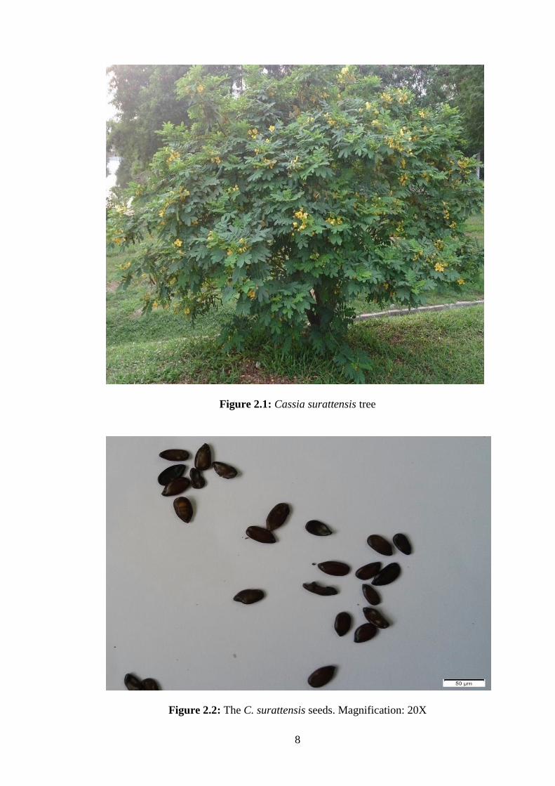
Cassia surattensis is a medium to large flowering tree (Figure 2.1). It is a native of
India, Southeast Asia and Tropical part of Australia. C. surattensis is a fast growing
tree with 12-15 feet tall, likes full sun and well-drained soil and bloom profusely
during dry season. The leaves are even-pinnate, alternate, dark green, to about 7
inches long and usually have 6-9 opposite pairs of ovate to oblong leaflets. It has
masses of bright yellow flower clusters and appears on almost every branch. The
pea-like pod about 7 inches long, containing 3 to 5 seeds (Figure 2.2) and green in
color initially, turning black once ripe (Hou et al., 1996; Randell and Barlow, 1998;
Wagner et al., 2014). C. surattensis is commonly planted as a street or ornamental
tree for its pretty yellow flowers (Hanelt et al., 2001).
2.1.2 Taxonomy
C. surattensis is also known as Senna surattensis, Cassia glauca or Senna glauca. In
Malay language, it is called kembang kuning or gelenggang. Table 2.1 shows the
scientific classification of C. surattensis plant.
2.1.3 Medicinal Uses
In traditional medicine, medicinal plants are used for therapeutic purposes due to the
presence of phytochemical with various medicinal properties. C. surattensis is a very
popular herb amongst practitioners of traditional medicine, widely used as a
decoction or infusion to treat various ailments. In Chinese traditions, leaves are used
to treat constipation problem, sore throat and cough by consuming the infusion of
boiled leaves orally (Hanelt et al., 2001). C. surattensis is also used in folk medicine
7
as antihyperglycemic (Chopra et al., 1956; El-Sawi and Sleem, 2009). Bark and
leaves of this plant are believed to be antihemorrhagic. It is also believed that
Balinese rub the leaves of C. surattensis into both external and internal cooling
medicines (Perry, 1980; Sangetha et al., 2008b).
Table 2.1: Scientific classification of C. surattensis (Irwin and Barneby, 1982;
Zhengyi and Raven, 2010)
CATEGORY CLASSIFICATION
Kingdom Plantae
Subkingdom Tracheobionta
Superdivision Spermatophyta
Division Magnoliophyta
Class Magnoliopsida
Subclass Rosidae
Order Fabales
Family Fabaceae / Leguminosae
Subfamily Caesalpinioideae
Tribe Cassieae
Subtribe Cassiinae
Genus Cassia
Species Cassia surattensis
Common names Golden Senna, Scrambled Egg Tree, Glossy
Shower, Bush Senna, Scrambled egg tree bush and
Scrambled egg tree
8
Figure 2.1: Cassia surattensis tree
Figure 2.2: The C. surattensis seeds. Magnification: 20X
9
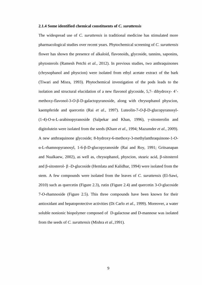
2.1.4 Some identified chemical constituents of C. surattensis
The widespread use of C. surattensis in traditional medicine has stimulated more
pharmacological studies over recent years. Phytochemical screening of C. surattensis
flower has shown the presence of alkaloid, flavonoids, glycoside, tannins, saponins,
phytosterols (Ramesh Petchi et al., 2012). In previous studies, two anthraquinones
(chrysophanol and physcion) were isolated from ethyl acetate extract of the bark
(Tiwari and Misra, 1993). Phytochemical investigation of the pods leads to the
isolation and structural elucidation of a new flavonol glycoside, 5,7- dihydroxy- 4’-
methoxy-flavonol-3-O-β-D-galactopyranoside, along with chrysophanol physcion,
kaempferide and quercetin (Rai et al., 1997). Luteolin-7-O-β-D-glucopyranosyl-
(1-4)-O-α-L-arabinopyranoside (Salpekar and Khan, 1996), γ-sitosterolin and
digitolutein were isolated from the seeds (Khare et al., 1994; Mazumder et al., 2009).
A new anthraquinone glycoside; 8-hydroxy-6-methoxy-3-methylanthraquinone-1-O-
α-L-rhamnopyranosyl, 1-6-β-D-glucopyranoside (Rai and Roy, 1991; Gritsanapan
and Nualkaew, 2002), as well as, chrysophanol, physcion, stearic acid, β-sitosterol
and β-sitosterol- β -D-glucoside (Hemlata and Kalidhar, 1994) were isolated from the
stem. A few compounds were isolated from the leaves of C. surattensis (El-Sawi,
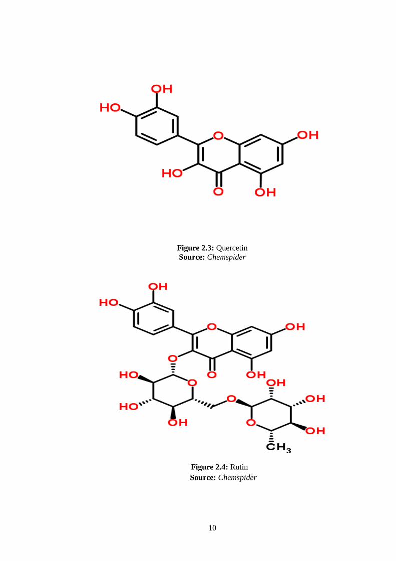
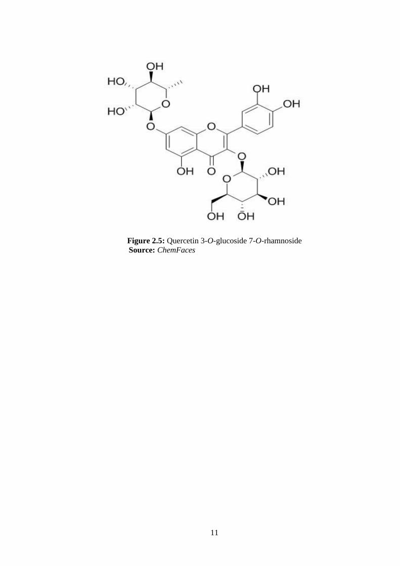
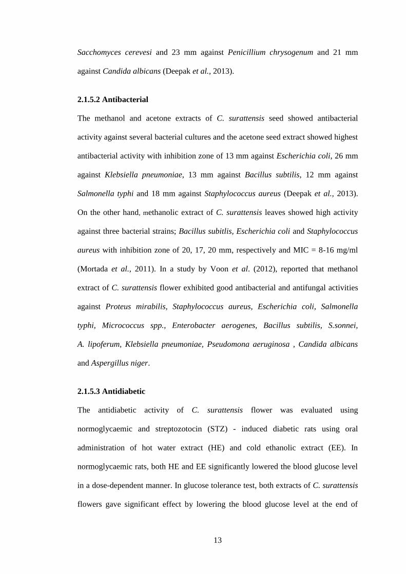
2010) such as quercetin (Figure 2.3), rutin (Figure 2.4) and quercetin 3-O-glucoside
7-O-rhamnoside (Figure 2.5). This three compounds have been known for their
antioxidant and hepatoprotective activities (Di Carlo et al., 1999). Moreover, a water
soluble nonionic biopolymer composed of D-galactose and D-mannose was isolated
from the seeds of C. surattensis (Mishra et al.,1991).
10
Figure 2.3: Quercetin
Source: Chemspider
Figure 2.4: Rutin
Source: Chemspider
11
Figure 2.5: Quercetin 3-O-glucoside 7-O-rhamnoside
Source: ChemFaces

12
2.1.5 Biological activities of C. surattensis
2.1.5.1 Antifungal
The methanolic flower extract of C. surattensis has been evaluated for their
antifungal activity against human pathogenic microorganism Aspergillus niger by
using the agar disc diffusion method, broth dilution method, the percentage of hyphal
growth inhibition and scanning electron microscopy (SEM) observation
(Sumathy et al., 2013). The extract exhibited good antifungal activity with zone of
inhibition 15 mm and minimum inhibitory concentration (MIC) 6.25 mg/ml. The
flower extract of C. surattensis exhibited considerable antifungal activity against A.
niger with an IC50 of 2.49 mg/ml on the hyphal growth. Percentage of hyphal growth
inhibition of this fungus was determined at concentrations 0.195, 0.39, 0.78,1.562,
3.125, 6.25, 12.5, 25, 50 and 100 mg/ml. For minimum fungicidal concentration
(MFC) the flower extract showed only moderate reduction (42%) at 1.562 mg/ml of
hyphal growth, while inhibition was substantial at 12.5 mg/ml with 100%. The
percentage reduced from 62% at 6.25 mg/ml to 0 at 0.195 mg/ml. In SEM squashed,
collapsed, empty and deformation of hyphae were the major changes observed, while
shrunken conidiophores were the obvious alteration. Morphological alterations
observed on A. niger caused by the flower extract could be the contribution of
chemical compounds present in the C. surattensis flower.
The seed extracts showed antifungal activity against all fungal cultures. The
acetone C. surattensis seed extract showed inhibition zone 22 mm against
Aspergillus niger, 27 mm against Penicillum chrysogenum, 25 mm against
Sacchomyces cerevesi and 27 mm against Candida albicans. While the methanolic
seed extract showed inhibition zone 24 mm against Aspergillus niger, 22 mm against
13
Sacchomyces cerevesi and 23 mm against Penicillium chrysogenum and 21 mm
against Candida albicans (Deepak et al., 2013).
2.1.5.2 Antibacterial
The methanol and acetone extracts of C. surattensis seed showed antibacterial
activity against several bacterial cultures and the acetone seed extract showed highest
antibacterial activity with inhibition zone of 13 mm against Escherichia coli, 26 mm
against Klebsiella pneumoniae, 13 mm against Bacillus subtilis, 12 mm against
Salmonella typhi and 18 mm against Staphylococcus aureus (Deepak et al., 2013).
On the other hand, methanolic extract of C. surattensis leaves showed high activity
against three bacterial strains; Bacillus subitlis, Escherichia coli and Staphylococcus
aureus with inhibition zone of 20, 17, 20 mm, respectively and MIC = 8-16 mg/ml
(Mortada et al., 2011). In a study by Voon et al. (2012), reported that methanol
extract of C. surattensis flower exhibited good antibacterial and antifungal activities
against Proteus mirabilis, Staphylococcus aureus, Escherichia coli, Salmonella
typhi, Micrococcus spp., Enterobacter aerogenes, Bacillus subtilis, S.sonnei,
A. lipoferum, Klebsiella pneumoniae, Pseudomona aeruginosa , Candida albicans
and Aspergillus niger.
2.1.5.3 Antidiabetic
The antidiabetic activity of C. surattensis flower was evaluated using
normoglycaemic and streptozotocin (STZ) - induced diabetic rats using oral
administration of hot water extract (HE) and cold ethanolic extract (EE). In
normoglycaemic rats, both HE and EE significantly lowered the blood glucose level
in a dose-dependent manner. In glucose tolerance test, both extracts of C. surattensis
flowers gave significant effect by lowering the blood glucose level at the end of
14
60 minutes after glucose loaded and even lower level at the end of 120 minutes and
180 minutes. For HE extract, at the dose of 400 mg/kg body weight (bw), the glucose
concentration at 60 minutes was 136.5 ± 3.2 mg/dl, 126.2 ± 4.2 mg/dl at 120 minutes
and 118.2 ± 3.4 mg/dl at 180 minutes. For EE extract (dose 400 mg/kg bw), the
glucose concentration at 60 minutes was 139.4 ± 3.4 mg/dl, 128.9 ± 2.3 mg/dl at 120
minutes and 119.2 ± 3.3 mg/dl at 180 minutes. The HE and EE of C. surattensis
flower enhanced glucose utilization, so the blood glucose level was significantly
decreased in glucose loaded rats. The efficacy of the antidiabetic activity of both HE
and EE extracts was almost comparable to standard drug, Glybenclamide (Ramesh
Petchi et al., 2012).
2.1.5.4 Antioxidant
The evaluation of antioxidant activity through 2,2-diphenyl-1-picrylhydrazyl (DPPH)
radical scavenging activity by Deepak et al. (2013), showed that acetone extract of
C. surattensis seed exhibited maximum antioxidant activity with 76.11% inhibition
than other extracts in comparison to standard drug ascorbic acid. A study on
antioxidant assessment on C. surattensis flowers (Chew et al., 2009) and leaves
(Thilagam et al., 2012) proved that they have good antioxidant activities. In another
study by Sangetha et al. (2008), the antioxidant properties of C. surattensis (flowers,
leaves, stem and pod) extracts were evaluated through DPPH radical scavenging
activity and xanthine oxidase inhibition assay. C.surattensis flowers revealed the best
antioxidant property, presenting much lower IC50 values (423.32 μg/ml for DPPH
assay and 11.1 μg/ml for xanthine oxidase assay). Furthermore, the highest
antioxidant contents (polyphenols) were found in these extracts (657.2392 + 2.0321
mg GAEs/g extract). The scavenging effects of flowers, stem and leaves extracts on
DPPH radicals were excellent (P < 0.05), especially in the case of C. surattensis

15
flower (93.54% at 1.0 mg/ml). The inhibition values were also remarkably good for
stem (81.97% at 1.0 mg/ml) and leaves (66.28% at 1.0 mg/ml), but C. surattensis
pod (45.72% at 1.0 mg/ml) revealed a low value of antioxidant activity compared
with BHT (66.23% at 1.0 mg/ml). Mortada et al. (2011) revealed that among the
methanol extracts of the tested Cassia species, the total antioxidant activity of C.
surattensis was the most active (556.65 ± 2.45 mg equivalent to ascorbic acid/g
extract). In a previous study, there appeared to be a positive correlation between the
total antioxidant capacity of the methanolic extracts of the tested Cassia species and
their total phenolic contents (El-Hashasa, 2010). It is also reported that the phenolic
compounds are responsible for antimicrobial properties and antioxidant activity
(Vasudevan, 2009; Panda, 2010).
2.1.6 Toxicological assessment
The cytotoxic activity of methanolic extract of C. surattensis flower was evaluated
based on primary brine shrimp assay and in vivo toxicity study. The extract was
found not toxic in brine shrimp assay with lethal concentration 50 (LC50) value of
3.32 mg/ml. In the oral acute toxicity study, mice were administrated orally with a
single dose of 5000 mg/ml extract and observed for 14 days for any toxicity sign.
Based on the body weight and histopathological examination, the C. surattensis
flower extract was found to be nontoxic as there were no significant differences in
the body weight of the mice. Histopathology analysis on subjected organs did not
reveal any pathological condition in the treatment group. Doses of C. surattensis
flower up to 5000 mg/kg bw appear to be safe in a mouse model. In short, the
methanolic flower extract of C. surattensis did not show any toxicity signs based on
the brine shrimp assay and oral acute toxicity findings (Sumathy et al., 2011). In
another study, the toxicity of C. surattensis flower was tested using chronic
16
administration of hot water extract (HE) and cold ethanolic extract (EE). Both
extracts was found to be non-toxic and well tolerated, as there are no overt signs of
toxicity, hepatotoxicity or renotoxicity following chronic oral administration
(Ramesh Petchi et al., 2012). The leaf extract of C. surattensis seems to have no
toxicity since the value of LD50 (8.6 gm/kg bw) was up to 80 times of effective dose
(El-Sawi and Sleem, 2009).
2.2 Free radicals
Free radicals are atoms or groups of atoms with an odd (unpaired) number of
electrons in its outer shell and can be formed when oxygen interacts with certain
molecules. They are highly reactive molecules capable of independent existence
(Halliwell and Gutteridge, 2007). Free radicals are derived either from normal
essential metabolic processes in the body or from external sources such as exposure
to X-rays, cigarette smoking, air pollutants and industrial chemicals.
Bi-radical nature of oxygen readily accepts unpaired electrons to form a series
of partially reduced species collectively known as ROS including O2•-, H2O2, OH
•,
peroxy (ROO), alkoxy (RO) and nitric oxide (NO). Generation of ROS is an integral
feature of normal cellular function like mitochondrial respiratory chain,
phagocytosis, ovulation and fertilization (Valko et al., 2004; Genestra, 2007). Their
production, however multiplies several folds during pathological conditions. The
superoxide radicals generated by the mitochondrial electron transport chain
participate in several reactions yielding various ROS such as H2O2 and OH• (Valko et
al., 2007). These ROS are capable of damaging biologically relevant molecules in
cells and tissues such as DNA, proteins, carbohydrates and lipids that lead to cell
damage and homeostatic disruption (Young and Woodside, 2001). They induce
undesirable oxidation, causing membrane damage, protein modification, DNA

17
damage, and cell death induced by DNA fragmentation and lipid peroxidation (Frei,
1997; Willcox et al., 2004; Pacher et al., 2007).
This oxidative damage, associated with ROS is believed to be involved not
only in the toxicity of xenobiotics but also in the pathophysiological role in the aging
of skin and several diseases like heart disease (atherosclerosis), cataract, cognitive
dysfunction, cancer, critical illness such as sepsis and acute respiratory distress
syndrome, chronic inflammatory diseases of the gastrointestinal tract, organ
dysfunction and disseminated intravascular coagulation.
2.3 Antioxidant
An antioxidant is a molecule stable enough to donate an electron to a rampaging free
radical and neutralize it, thus reducing its capacity to damage. These antioxidants
scavenge free radicals which delay or inhibit cellular damage (Halliwell, 1995;
Kensler, 2007). These low-molecular-weight antioxidants can safely interact with
free radicals and terminate the chain reaction, to protect the cells against free radicals
toxic effects (Jeong, 2006).
2.3.1 Mechanism of antioxidant action
Antioxidants act as radical scavenger, hydrogen donor, electron donor, peroxide
decomposer, singlet oxygen quencher, enzyme inhibitor and metal-chelating agents
(Krinsky, 1992). The antioxidant can function in one of the two principles
mechanisms of action (Rice-Evans and Diplock, 1993). The first is a chain- breaking
mechanism by which the primary antioxidant donates an electron to the free radical
present in the systems. The second mechanism involves removal of ROS initiators by
inhibiting chain-initiating catalyst. For the first mechanism, when a radical release or
steals an electron, a second radical is formed. The last one exerts the same action on

18
another molecule and continues until either the free radical formed is stabilized by a
chain-breaking antioxidant such as vitamin C and vitamin E, or it simply
disintegrates into an inoffensive product. For the second mechanism, an antioxidant
enzyme like superoxide dismutase and catalase can prevent oxidation by reducing the
rate of chain initiation, for example by scavenging initiating free radicals (Young and
Woodside, 2001).
2.3.2 Antioxidant classification
The major antioxidant enzymes directly involved in direct elimination of ROS are
superoxide dismutase (SOD), catalase (CAT) and glutathione peroxidase (GPx)
(Pacher et al., 2007; Valko et al., 2007). The non-enzymatic antioxidants that belong
to endogenous antioxidants such as glutathione, coenzyme Q10, melatonin, uric acid
and bilirubin are produced during normal metabolism in the body (Droge, 2002;
Willcox et al., 2004). Although there are several enzyme systems within the body
that scavenge free radicals, the principle micronutrient (vitamins) antioxidants are
vitamin E, vitamin C and B-carotene (Levine et al., 1991). The body cannot produce
these micronutrients and must be supplied in the diet or supplements. Phytonutrients
are being increasingly appreciated for their antioxidant activity, one example is
phenolic compounds (Gale, 2001).
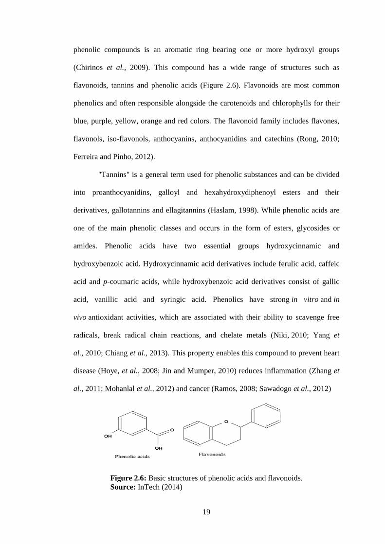
2.3.3 Phenolic compounds
Phenolic compounds are a large and diverse group of secondary plant metabolites
that are widespread in the plant kingdom (Strube et al., 1993). Phenolic compounds
are synthesized in plants partly as a response to ecological and physiological
pressures such as pathogen attack, UV radiation and wounding (Zulak et al., 2006;
Diaz et al., 2010; Kennedy and Wightman, 2011). The basic structural feature of
19
phenolic compounds is an aromatic ring bearing one or more hydroxyl groups
(Chirinos et al., 2009). This compound has a wide range of structures such as
flavonoids, tannins and phenolic acids (Figure 2.6). Flavonoids are most common
phenolics and often responsible alongside the carotenoids and chlorophylls for their
blue, purple, yellow, orange and red colors. The flavonoid family includes flavones,
flavonols, iso-flavonols, anthocyanins, anthocyanidins and catechins (Rong, 2010;
Ferreira and Pinho, 2012).
"Tannins" is a general term used for phenolic substances and can be divided
into proanthocyanidins, galloyl and hexahydroxydiphenoyl esters and their
derivatives, gallotannins and ellagitannins (Haslam, 1998). While phenolic acids are
one of the main phenolic classes and occurs in the form of esters, glycosides or
amides. Phenolic acids have two essential groups hydroxycinnamic and
hydroxybenzoic acid. Hydroxycinnamic acid derivatives include ferulic acid, caffeic
acid and p-coumaric acids, while hydroxybenzoic acid derivatives consist of gallic
acid, vanillic acid and syringic acid. Phenolics have strong in vitro and in
vivo antioxidant activities, which are associated with their ability to scavenge free
radicals, break radical chain reactions, and chelate metals (Niki, 2010; Yang et
al., 2010; Chiang et al., 2013). This property enables this compound to prevent heart
disease (Hoye, et al., 2008; Jin and Mumper, 2010) reduces inflammation (Zhang et
al., 2011; Mohanlal et al., 2012) and cancer (Ramos, 2008; Sawadogo et al., 2012)
Figure 2.6: Basic structures of phenolic acids and flavonoids.
Source: InTech (2014)
20
2.3.4 Evaluation of antioxidant activity
There are many in vitro methods that have been developed to evaluate the
antioxidant capacities of natural antioxidants either as pure compounds or as plant
extracts. The in vitro methods are divided into two main groups based on the
reactions; i) Hydrogen atom transfer reactions like Oxygen Radical Absorbance
Capacity (ORAC), Total radical trapping antioxidant potential (TRAP) and β
carotene bleaching; and ii) Electron transfer reactions like DPPH radical scavenging
assay, superoxide anion radical scavenging assay, hydroxyl radical scavenging assay,
nitric oxide radical scavenging assay and total phenolic content assay (Niki, 1993;
Rice-Evans and Diplock,1993). These methods are popular due to their high speed
and sensitivity. However, it is important to use more than one method to evaluate the
antioxidant capacity of plant materials because of the complex nature of
phytochemicals (Chanda and Dave, 2009).
2.4 Liver
2.4.1 Functions of the Liver
The liver is the largest metabolic organ of the body and is positioned beneath the
diaphragm in the right hypochondrium of the abdominal cavity (Ellis, 2011). The
liver has a wide range of functions in the body, including the synthesis and
breakdown of small and complex molecules, many of which are necessary for normal
vital functions. It is the major drug-metabolizing and drug-detoxifying organ of the
body. It is a unique organ where it can regenerate the significant loss of liver cells
due to drug toxicity or other damages (Mehendale, 2005). The liver helps in the
maintenance, performance and regulating homeostasis of the body. It is involved
with almost all the biochemical pathways to growth, fight against disease, nutrient
supply, energy provision and reproduction. In addition, it aids metabolism of
21
carbohydrate, protein and fat, secretion of bile and storage of vitamins (Ahsan et al.,
2009). The liver performs a variety of important host defense and metabolic
functions that include synthesis of acute-phase proteins, gluconeogenesis,
detoxification, and clearance of endogenous mediators, as well as secretion of pro-
inflammatory cytokines (Pastor et al., 1995).
2.4.2 Drug Metabolism in liver
The liver is the primary site for metabolism of almost all drugs because it is
relatively rich in a large variety of metabolizing enzymes. Drug metabolism is a
process where drug molecules are chemically altered, usually to more polar
metabolites that exhibit increased water solubility to allow excretion in urine or bile.
Drug metabolism is likely to be a byproduct of metabolic pathways that metabolize
endogenously synthesized compounds (endobiotics) such as steroids, sterols, bile
acids and eicosanoids (Nebert and Russell, 2002).
Drug metabolism is often divided into two phases of biochemical reaction -
phase 1 and phase 2. Some drugs may undergo only phase 1 or only phase 2
metabolism, but more often, the drug will undergo phase 1 and then phase 2
sequentially. In phase 1 reaction, the enzymes carry out oxidation, reduction, or
hydrolytic reactions. This prepares the drug for phase 2 in which enzymes form a
conjugate of the substrate (the phase 1 product). These processes tend to increase
water solubility of the drug and this facilitates excretion as well as decreasing
pharmacological activity. Most of phase 2 reactions occur in the hepatocyte
cytoplasm and involve conjugation with endogenous substance (example glucuronic
acid, sulfate, glycine) via transferase enzymes. Chemically active products from
phase 1 are made relatively inert and suitable for elimination by the phase 2 step
22
(Liston et al., 2001). The most important family of metabolizing enzymes in the liver
is cytochrome P450.
2.4.2.1 Role of cytochrome P450 enzymes
The predominant catalysts of phase I metabolism in the liver is cytochrome P450
(CYP450), a microsomal superfamily of isoenzymes that catalyzes the oxidation of
many drugs. Cytochrome P450 isoenzymes are a group of heme-containing enzymes
primarily located in the lipid bilayer of the smooth endoplasmic reticulum (ER) of
hepatocytes ranging from bacteria to humans (Nelson et al., 1996; Denisov et al.,
2012).
CYP450 plays an important role in oxidative metabolism of many exogenous
compounds from a variety of drug classes (example, thiazolidinediones and
meglitinides), environmental chemicals and pollutants, and natural plant products.
CYP450 acts as a catalyst in oxidative reactions in organs such as the liver, intestine,
kidney, lung, and brain (Jefferson, 1998; Schoch et al., 2008). They develop
gradually to allow organisms to metabolize foreign chemicals (Nemeroff et al., 1996;
Jefferson, 1998). A major function of CYP450 is to metabolize lipid soluble
chemicals into water-soluble compounds for excretion in bile or urine. Cytochrome
P450 enzymes are essential for the production of cholesterol, bile-acid biosynthesis,
steroids, vitamin D3 synthesis and metabolism, retinoic acid hydroxylation
prostacyclins, and thromboxane A2 (Nebert and Russell, 2002; Lynch and Price,
2007). There are more than 50 CYP450 enzymes, but these enzymes CYP1A2,
CYP2C9, CYP2C19, CYP2D6, CYP3A4, and CYP3A5 metabolize 90 percent of
drugs (Slaughter, 1995; Wilkinson, 2005). Cytochrome P450 enzymes can be
inhibited or induced by drugs, causing drug-drug interactions. This can lead to
unexpected adverse reactions or therapeutic failures. Cytochrome P450 enzymes
23
often interact with warfarin, antidepressants and antiepileptic drugs (Lynch and
Price, 2007)
2.5 Hepatotoxicity
The liver is an important target organ of the toxic drugs, xenobiotics and oxidative
stress because of its unique metabolism and relationship to gastrointestinal tract.
Hepatotoxicity is defined as hepatic toxicity or damage to the liver cells caused by
exposure to a drug or other chemical agents called hepatotoxins. This results the liver
to function irregularly (Vichitra et al., 2013). Drug induced liver injury (DILI) is one
of the most frequent cause of liver injury that poses a major clinical problem and
challenge to drug regulators (Russman et al., 2009). Drug induced liver injury makes
up a total of 5% of all hospital admissions and 50% of all acute liver failures
(McNally, 2006). The idiosyncratic drug reactions with more than 75 percent of
cases cause liver transplantation or death (Ostapowicz et al., 2002). One of the most
frequent reason a drug withdrawn from the market because of its hepatic adverse
effect and it is estimated more than 900 drugs cause the liver damage (Rajamani et
al., 2010).
2.6 Mechanism of liver injury
There are a number of mechanisms that initiate hepatic injury or aggravate the
ongoing injury processes. Many chemicals damage the prominent intracellular
organelle mitochondria. The impaired organelle releases excessive amounts of ROS
that leads to hepatic cell injury. Liver injury also significantly aggravated by the
accumulation of inflammatory cells such as neutrophils and activation of sinusoidal
lining cells, especially Kupffer cells (Jaeschke, 2000). Activated Kupffer cells
release ROS, cytokines and chemokines, which promote oxidative stress in injuries
24
caused by toxicants. The activation of some enzymes in the cytochrome P-450
system such as CYP2E1 involves production of injurious metabolites that promote
oxidative stress (Jaeschke et al., 2002). Injury to hepatocyte and bile duct cells lead
to accumulation of bile acid in the liver and this promotes further liver damage or
injury (Patel et al., 1998; Davit-Spraul et al., 2009).
2.7 Types of Hepatotoxicity
2.7.1 Acetaminophen - induced hepatotoxicity
Acetaminophen, also known as paracetamol or N-acetyl-p-aminophenol (APAP).
This drug is most commonly used as analgesics to reduce fever and mild to moderate
pain. Acetaminophen induced hepatotoxicity has been linked with a number of
cirrhosis, hepatitis and suicide attempts cases. Acetaminophen if taken in overdose,
can cause severe hepatotoxicity that leads to liver failure and nephrotoxicity
depletion (Masubuchi et al., 2005). In therapeutic dose, acetaminophen is converted
by drug metabolizing enzymes to water-soluble metabolites and eliminated in the
urine (Ramachandran and Kakar, 2009; Hinson et al., 2010)
Paracetamol induced liver injury is a classic case of drug - induced liver
injury (DILI-1). This case can be tested in more than one strain of rodents
(Mehendale, 2005) and show a significant dose-dependency in both animals and
humans. Oxidative stress plays a significant role in the hepatotoxicity caused by
paracetamol and research have been done on antioxidants as an alternative treatment
against paracetamol toxicity (Avila et al., 2011).
Figure 2.7 shows that acetaminophen is directly conjugated with glucuronic
acid or sulfate by glucuronyl transferases or sulfonyltransferases. The conjugated
compound is eliminated into the bile by multidrug resistance-associated protein
(Mrp2) or into the blood by Mrp3 (Chen et al., 2003). The remaining unconjugated