Embed Size (px)
Citation preview

JOURNAL OF BACTERIOLOGY, Oct. 2007, p. 7290–7301 Vol. 189, No. 200021-9193/07/$08.00�0 doi:10.1128/JB.00731-07Copyright © 2007, American Society for Microbiology. All Rights Reserved.
Binary Toxin Production in Clostridium difficile Is Regulated by CdtR,a LytTR Family Response Regulator�
Glen P. Carter, Dena Lyras, David L. Allen, Kate E. Mackin, Pauline M. Howarth,Jennifer R. O’Connor,† and Julian I. Rood*
Australian Bacterial Pathogenesis Program, Department of Microbiology, Monash University, Victoria 3800, Australia
Received 9 May 2007/Accepted 1 August 2007
Clostridium difficile binary toxin (CDT) is an actin-specific ADP-ribosyltransferase that is produced byvarious C. difficile isolates, including the “hypervirulent” NAP1/027 epidemic strains. In contrast to the twomajor toxins from C. difficile, toxin A and toxin B, little is known about the role of CDT in virulence or how C.difficile regulates its production. In this study we have shown that in addition to the cdtA and cdtB toxinstructural genes, a functional cdt locus contains a third gene, here designated cdtR, which is predicted to encodea response regulator. By introducing functional binary toxin genes into cdtR� and cdtR-negative strains of C.difficile, it was established that the CdtR protein was required for optimal expression of binary toxin. Signif-icantly increased expression of functional binary toxin was observed in the presence of a functional cdtR gene;an internal deletion within cdtR resulted in a reduction in binary toxin production to basal levels. Strains thatdid not carry intact cdtAB genes or cdtAB pseudogenes also did not have cdtR, with the entire cdt locus, orCdtLoc, being replaced by a conserved 68-bp sequence. These studies have shown for the first time that binarytoxin production is subject to strict regulatory control by the response regulator CdtR, which is a member ofthe LytTR family of response regulators and is related to the AgrA protein from Staphylococcus aureus.
Clostridium difficile is an anaerobic, gram-positive spore-forming bacterium that is recognized as the causative agent ofa broad spectrum of intestinal diseases ranging from mild self-limiting diarrhea to more serious and potentially life threaten-ing manifestations such as pseudomembranous colitis and toxicmegacolon (4). Collectively, these diseases are referred to as C.difficile-associated disease (CDAD). The onset of CDAD istypically associated with the use of antibiotics or other thera-peutics such as cytotoxic anticancer drugs, which disrupt thenormal intestinal microflora (50). Once this colonization resis-tance (51) is removed, it is proposed that ingested C. difficilespores germinate, colonize the gastrointestinal tract, and elab-orate toxins, which results in an acute inflammatory responseand severe damage to the intestinal epithelium.
C. difficile is a significant and increasing source of morbidityand mortality and is a very significant financial burden in hos-pitals throughout the developed world (9, 22, 41, 59). Morerecently, a new, highly virulent group of strains, known asNAP1/027, has emerged. These hypervirulent strains are re-ported to produce significantly more toxin than other strainsand are associated with more severe disease and high mortalityrates (59). The hypervirulent strains are highly resistant tofluoroquinolones and are responsible for epidemics seen inCanada, the United States, and the United Kingdom and sev-eral other European countries (21, 25, 28, 48).
The major virulence factors of C. difficile are two membersof the large clostridial cytotoxin family, toxin A and toxin B,
which are encoded by the genes tcdA and tcdB, respectively,and are located within a five-gene locus known as the PaLoc(6). Both toxins A and B are potent monoglucosyltransferasesthat have been shown to target small GTP-binding proteins,including Rho, Rac, and Cdc42, which are involved in theregulation and formation of the actin cytoskeleton in eukary-otic cells (18, 19).
In addition to producing toxins A and B, some C. difficilestrains, including the NAP1/027 epidemic strains, produce athird toxin, known as C. difficile binary toxin (CDT) (12, 38,53). This toxin is an actin-specific ADP-ribosyltransferase thatconsists of two subunits, namely, CDTa, which is the enzymaticcomponent, and CDTb, the binding component (3, 39). It hasa high degree of amino acid sequence similarity to iota toxinfrom Clostridium perfringens type E strains and Clostridiumspiroforme toxin, the subunits of which are reportedly inter-changeable (38). CDTb binds to an as-yet-unidentified cellularreceptor, leading to the internalization of CDTa into the cy-tosol, where it catalyzes the ADP-ribosylation of monomericactin and the resultant disruption of the actin cytoskeleton (1,47). Since most human C. difficile strains do not produce func-tional CDT, its role in the pathogenesis of CDAD is yet to bedetermined. Recent studies have shown that CDT productionis much more prevalent in strains isolated from animals, withthe CDT structural genes being found in approximately 23% to100% of these isolates (43). However, the significance of thisfinding for C. difficile-mediated disease in animals is also un-known.
CDT is encoded by two genes, cdtA and cdtB, which aretranscriptionally linked and are located on the C. difficile chro-mosome at a locus separate from the PaLoc (38). In contrast tothe situation with toxin A and toxin B, very little research hasbeen carried out to examine how the expression of this poten-tially important virulence factor is regulated. Here we report
* Corresponding author. Mailing address: Department of Micro-biology, Monash University, Victoria 3800, Australia. Phone: (613)9905 4825. Fax: (613) 9905 4811. E-mail: [email protected].
† Present address: Hines VA Hospital, Fifth Avenue and RooseveltRoad, Hines, IL 60141.
� Published ahead of print on 10 August 2007.
7290
on June 9, 2018 by guesthttp://jb.asm
.org/D
ownloaded from

the identification of a functional LytTR family response regu-lator, which we have named CdtR, and we show that it is ableto activate CDT production in C. difficile. Furthermore, ourresults reveal that the cdtR and the cdtAB genes are geneticallylinked as part of the CDT locus (CdtLoc). Finally, in CDT-negative isolates we have identified a novel 68-bp nucleotidesequence that is absent in isolates carrying the CdtLoc.
MATERIALS AND METHODS
Bacterial strains and growth conditions. The characteristics and origins of allrecombinant strains and plasmids are shown in Table 1. All bacteriologicalculture media were obtained from Oxoid. For mating procedures and extraction
of plasmid and genomic DNAs, C. difficile strains were cultured in BHIS broth(49) supplemented with 0.1% L-cysteine HCl and 0.5% glucose or on BHIS agarwith the additional supplement of 0.09% FeSO4. Cycloserine (250 �g · ml�1),cefoxitin (8 �g · ml�1), thiamphenicol (10 �g · ml�1), or erythromycin (10 �g ·ml�1) (all from Sigma-Aldrich) were used for selection of recombinant strains, asrequired. C. difficile cultures were grown in an atmosphere of 10% H2, 10% CO2,and 80% N2 at 37°C in a Coy anaerobic chamber. Escherichia coli strains werederivatives of either DH5�, Top10, or HB101(pVS520) and were cultured aer-obically at 37°C in 2� YT agar or broth (30). Recombinant E. coli cultures weresupplemented with chloramphenicol (25 �g · ml�1), erythromycin (400 �g ·ml�1), or tetracycline (10 �g · ml�1), as required.
Isolation and manipulation of nucleic acids. Plasmid DNA was isolated fromE. coli strains grown overnight in 5 ml of 2 �YT broth or from C. difficile strainsgrown in 5 ml of BHIS broth, with appropriate antibiotic selection, using the
TABLE 1. Bacterial strains and plasmids
Strain or plasmid Characteristics and/or origina Source or reference
StrainsC. difficile
630 Clinical isolate, Tcr Emr, Switzerland 13CD196 Binary toxin-producing reference strain, France 39VPI 10463 Toxinotype 0 reference strain, United States 56L253 Toxigenic isolate 14CD37 Nontoxigenic isolate, United States 49JIR8094 630 Ems; has a spontaneous deletion of an erm(B) gene from Tn5398 35KZ1623 Nontoxigenic isolate, Japan 32662 Nontoxigenic isolate, Switzerland 60M7404 NAP1/027 Canadian isolate, 2005 J. Pepin661 Toxigenic isolate, France, 1992 M. Sebald660/2 Toxigenic isolate, France, 1992 M. Sebald685 Toxigenic isolate, France, 1992 M. SebaldAM421 Nontoxigenic isolate; Fairfield Hospital, Australia; 1980 R. WilkinsonAM662 Nontoxigenic isolate; St. Vincents Hospital, Australia; 1982 R. WilkinsonAM1180 Toxigenic isolate; Latrobe Valley Regional Hospital, Australia; 1985 R. WilkinsonM334581 Toxigenic isolate; Austin Hospital, Australia; 2002 L. GraysonM336632 Toxigenic isolate; Austin Hospital, Australia; 2002 L. GraysonM336948 Nontoxigenic isolate; Austin Hospital, Australia; 2002 L. GraysonM345206 Nontoxigenic isolate; Austin Hospital, Australia; 2002 L. GraysonM345300 Toxigenic isolate; Austin Hospital, Australia; 2002 L. GraysonM361441 Nontoxigenic isolate; Austin Hospital, Australia; 2002 L. GraysonWA7 Toxigenic isolate; Rockingham Kwinana District Hospital, Australia; 2006 T. RileyWA9 Toxigenic isolate; Katanning Hospital, Australia; 2006 T. RileyMMC CD1 Toxigenic isolate; Monash Medical Centre, Australia; 2006 G. JenkinMMC CD2 Toxigenic isolate; Monash Medical Centre, Australia; 2006 G. JenkinMMC CD8 Toxigenic isolate; Monash Medical Centre, Australia; 2006 G. Jenkin
E. coliDH5� F� �80dlacZM15 (lacZYA-argF)U169 deoR recA1 endA1 hsdR17(rK
� mK�) phoA supE44
thi-1 gyrA96 relA1 ��Life Technologies
Top10 F� mcrA �(mrr-hsdRMS-mcrBC) �80lacZ�M15 �lacX74 deoR recA1 araD139�(ara-leu)7697 galU galK rpsL (Strr) endA1 nupG
Invitrogen
HB101 thi-1 hsdS20(rB� mB
�) supE44 recAB ara-14 leuB5 proA2 lacY1 galK rpsL20 (Smr)xyl-5 mtl-1
5
PlasmidspVS520 Tra� Mob� RP4 derivative, Tcr 37pMTL9301 E. coli-C. difficile mobilizable shuttle vector, carries pCD6 replication region, Emr 40pMTL500E E. coli-Clostridium shuttle vector (pAM1 replicon), Emr 36pMTL9361Cm E. coli-C. difficile mobilizable shuttle vector, carries pCD6 replication region, Tmr G. Carter, N. Minton
(unpublished)pCR2.1-TOPO E. coli PCR cloning vector, ColE1, Apr Kmr InvitrogenpT7Blue3 E. coli PCR cloning vector, ColE1, Apr NovagenpJIR3107 pMTL9361Cm CD196 4.5-kb cdtAB fragment (SacI/SphI), CDT binary toxin expression
plasmid, TmrRecombinant
pJIR3394 pMTL9301 (FspI) JIR8094 cdtR 1.13 kb (EcoRV/5maI), CdtR expression plasmid, Emr RecombinantpJIR3395 pMTL3394 �cdtR (BamHI/PstI), internal deletion in cdtR, Emr Recombinant
a Tmr, thiamphenicol (chloramphenicol) resistance; Emr, erythromycin resistance; Apr, ampicillin resistance; Kmr, kanamycin resistance; Tcr, tetracycline resistance;Smr, streptomycin resistance.
VOL. 189, 2007 REGULATION OF BINARY TOXIN PRODUCTION IN C. DIFFICILE 7291
on June 9, 2018 by guesthttp://jb.asm
.org/D
ownloaded from

QIAprep spin miniprep kit (QIAGEN) according to the manufacturer’s instruc-tions. For C. difficile plasmid preparations, cell pellets were incubated in bufferP1 (supplied with the kit) supplemented with 30 �g · ml�1 lysozyme (Sigma-Aldrich) for 30 min at 37°C, before extraction as per the manufacturer’s instruc-tions. C. difficile genomic DNA was prepared using the DNeasy tissue kit(QIAGEN) according to the manufacturer’s instructions. DNA was quantifiedusing a NanoDrop spectrophotometer (NanoDrop Technologies) and stored at�20°C. Standard methods for the digestion, modification, ligation, and analysisof plasmid and genomic DNA and PCR products were used (44). Nucleotidesequence analysis was carried out using a PRISM BigDye Terminator cyclesequencing kit (Applied Biosystems) according to the manufacturer’s instruc-tions. Sequence detection was carried out on an Applied Biosystems 3730Sgenetic analyzer and was performed by Micromon at Monash University. Se-quences were analyzed using ContigExpress (Invitrogen).
PCR. Oligonucleotide primer sequences are listed in Table 2. Unless otherwisestated, all PCR experiments were carried out with Taq DNA polymerase and thesupplied 10� PCR buffer with magnesium according to the manufacturer’sinstructions (Roche). The following parameters were utilized for all PCR exper-iments: initial denaturation was performed at 94°C for 4 min, followed by 30cycles consisting of a denaturation step at 94°C for 30 s, an oligonucleotideprimer annealing step at 50°C for 30 s, and an extension step at 72°C. Theextension time was 1 min per 1 kb of amplified DNA. A final extension step wasperformed at 72°C for 12 min.
Construction of recombinant plasmids. For the construction of the CDTexpression plasmid, PCR was performed using primers JRP2423 and JRP2424(Table 2), chromosomal DNA from strain CD196, and the Triple MastermixPCR system (Roche). The resultant 4.5-kb fragment containing the CDT regionwas cloned into the EcoRV site of pT7Blue3 using the Perfectly Blunt cloning kitaccording to the manufacturer’s instructions (Novagen). After sequencing, the4.5-kb fragment carrying both the cdtA and cdtB genes was excised from thepT7Blue3 derivatives with SphI and SacI before cloning into the equivalent sitesof the E. coli-C. difficile shuttle plasmid pMTL9361Cm, resulting in pJIR3107.
For construction of the cdtR-plasmid pJIR3394, PCR was performed usingprimers JRP3720 and JRP3721 (Table 2). Chromosomal DNA from strainJIR8094 was used in the Failsafe PCR system with buffer E (Epicenter Biotech-nologies) to amplify the cdtR region. The PCR product was then TA cloned intopCR2.1-TOPO, according to the manufacturer’s instructions (Invitrogen). The
1.13-kb cdtR fragment was excised from this plasmid with XhoI and BamHI andsubcloned into the equivalent sites of pMTL500E. The insert was subsequentlyexcised from this intermediate vector using EcoRV and SmaI and inserted intothe FspI site of pMTL9301, resulting in pJIR3394.
The deletant cdtR allele was generated using splice overlap extension PCR(SOE-PCR) (15, 16). PCR was used to amplify the left and right sides of the cdtRregion using primers JRP3631 with JRP3632 and JRP3633 with JRP3634, re-spectively (Table 2). This reaction used pJIR3394 as the template together withthe Failsafe PCR system enzyme mix and buffer E. Each fragment was purifiedby extraction from a 1% agarose gel using the Qiaquick gel extraction kit(QIAGEN). An aliquot of each of the purified PCR products was mixed in anequimolar ratio and used as the DNA template in a second PCR with primersJRP3631 and JRP3634, which resulted in a DNA fragment with a 561-bp deletionwithin cdtR, which was then TA cloned into pCR2.1-TOPO (Invitrogen). Thefragment was excised from pCR2.1-TOPO using BamHI and PstI, and clonedinto the equivalent sites of pJIR3394, resulting in the final construct, pJIR3395.
Transfer of plasmid DNA into C. difficile by conjugation. The conjugationprocedure was carried out as previously described (40), except thatHB101(pVS520) (37) was used as the E. coli donor. Bacterial pellets collectedfrom 1-ml aliquots of an overnight E. coli donor culture were resuspended in200-�l aliquots of the C. difficile recipient strain, which had been collected from20-ml overnight cultures incubated at 37°C in an anaerobic chamber. This matingmixture was then spread onto a thick BHIS agar plate and incubated for 6 h orovernight at 37°C under anaerobic conditions. Bacterial growth was harvested byflooding the agar three times with 500 �l of sterile phosphate-buffered saline(137 mM NaCl, 2.7 mM KCl, 1.4 mM KH2PO4, 4.3 mM Na2HPO4). Aliquots(100 �l) of the combined cell suspension were then spread onto BHIS agarsupplemented with cycloserine and cefoxitin to prevent growth of the E. colidonor and with either thiamphenicol or erythromycin to select for C. difficiletransconjugants carrying the appropriate plasmids. The plates were incubatedunder anaerobic conditions for 24 to 72 h.
Determination of the integration site of the CdtLoc. The region flanked byCD2602 and trpS from C. difficile strain CD37 was amplified by PCR usingprimers JRP3722 and JRP3725 (Table 2) and the Failsafe PCR system buffer G.The sequence of the amplified product was then determined using primersJRP3722, JRP3725, JRP3728, and JRP3729 (Table 2). The CDT integration sitesof all other nontoxigenic C. difficile strains were determined by PCR amplifica-
TABLE 2. Oligonucleotide primers
Primer Sequence (5�33�) Usea
JRP1744 GGGAAAGAAAAGAAGCAGAAAG Amplify internal fragment of cdtA (�)JRP1745 TGTATTTTCATTGTTTCTCCTC Amplify internal fragment of cdtA (�)JRP2342 CCGGAATTCGCTCTATTGGACTAGACCGTTG Amplify internal fragment of tcdA (�)JRP2343 CCGGAATTCTTAAAGTTTTTTCTATAGTATT Amplify internal fragment of tcdA (�)JRP2344 CCGGAATTCGTTCAGTAACAGTGGATTTTTG Amplify internal fragment of tcdB (�)JRP2345 CCGGAATTCTCAAATGGATATTCATACTGCC Amplify internal fragment of tcdB (�)JRP2423 CCGGAATTCGGCCATATATGTCGTATATCTA Amplify C. difficile CD196 cdtA and cdtB genes and
upstream, region introduces EcoRI site (�)JRP2424 CCGGAATTCTTGTGGGGACAAATTTAAATCA Amplify C. difficile CD196 cdtA and cdtB genes and
upstream region, introduces EcoRI site (�)JRP2523 AGTTCTAGAGGAATGAAACTAAAAGAAAAAAT Amplify internal fragment of cdtR (�)JRP2524 AGTTGCGCAGCATGCATCTAAATCTGGTAAAG Amplify internal fragment of cdtR (�)JRP3631 GTTGCGGGGATCCTCTAGTAACGG Amplify �cdtR by SOE-PCR (�)JRP3632 CTCATTTTAATATTTCTTACAACATCATTATCAAAACCTTC Amplify �cdtR by SOE-PCR (�)JRP3633 TAAGAAATATTAAAATGAGTAATGGCCATATATG Amplify �cdtR by SOE-PCR (�)JRP3634 GGTCAGGCTGCATCTGCAGAATTCG Amplify �cdtR by SOE-PCR (�)JRP3720 CTTCTATAATTATTGTTAAATAATTCTTC Amplify cdtR gene and upstream region (�)JRP3721 GAGACATCTCCTTTTTCTATTTATTATG Amplify cdtR gene and upstream region (�)JRP3722 GTTGATAAATTTATTATTAACTCTCTAG Amplify internal fragment of CD2602 (�)JRP3723 GTAAACACTAGAATAATATCTATAG Amplify internal fragment of CD2602 (�)JRP3724 CATGTTTACAATCTCTTCTACACTC Amplify internal fragment of trpS (�)JRP3725 CAATCACATGTCAAAGAGCATGTTGAAC Amplify internal fragment of trpS (�)JRP3726 CTACTATTAGATATTATAACAATTGTTCC Amplify putative cdtA promoter region (�)JRP3727 CCCTACAAATATTAGTTTAGTTAAC Amplify putative cdtA promoter region (�)JRP3728 GCCACCTGATACTATAGATATTATTC Amplify/sequence CD2602/trpS intervening region (�)JRP3729 AAGTTGGATTTATAGAGAGAAG Amplify/sequence CD2602/trpS intervening region (�)JRP3845 TGCAATACTACTTACAAGGCTCCTATAGA Amplify internal cdtA fragment in QRT-PCR (�)JRP3846 TCTTTCCCATTCTTTAGCCTTTTC Amplify internal cdtA fragment in QRT-PCR (�)
a �, leading-strand-specific oligonucleotide primer; �, lagging-strand-specific oligonucleotide primer.
7292 CARTER ET AL. J. BACTERIOL.
on June 9, 2018 by guesthttp://jb.asm
.org/D
ownloaded from

tion of this region using primers JRP3728 and JRP3729 and Failsafe buffer E,followed by sequencing with primers JRP3728 and JRP3729.
Detection of CDTa and CDTb proteins by Western blotting. Isolation of CDTaand CDTb and Western blotting were carried out as previously described (38).Proteins were precipitated at a concentration of 70% ammonium sulfate fromcell-free supernatants of C. difficile cultures grown overnight in 100 ml of BHISbroth. The precipitate was collected, dissolved in 2 ml of distilled water, anddialyzed against 10 mM Tris-HCl buffer, pH 7.0, for 24 h with three changes ofbuffer. Proteins were then subjected to sodium dodecyl sulfate-polyacrylamidegel electrophoresis (23) and transferred by electrophoresis (44) to nitrocellulosemembranes (Whatman). The membranes were incubated for 1 h in phosphate-buffered saline containing 5% dry milk powder and then incubated overnightwith immunopurified antibodies specific for C. perfringens iota toxin componentsA (Ia) and B (Ib), which are cross-reactive with the C. difficile CDTa and CDTbproteins, respectively (38). CDTa- and CDTb-bound antibodies were then de-tected with peroxidase-conjugated anti-rabbit sheep antibodies (Chemicon) andthe Western lightning chemiluminescence reagent kit (Perkin Elmer), accordingto the manufacturer’s instructions.
ADP-ribosyltransferase assays. ADP-ribosyltransferase assays were carriedout essentially as before (55). In brief, precipitated supernatant protein wasincubated for 1 h in a reaction solution containing 50 mM triethanolamine-HClbuffer (pH 7.5), 5 mM MgCl2, 10 mM dithiothreitol, 10 mM thymidine, and 10�g rabbit skeletal muscle actin (Sigma-Aldrich), together with [32P]NAD (3�Ci). The entire reaction volume was then applied to a glass fiber filter (Milli-pore) and allowed to dry. Protein was then precipitated onto the filter by washingwith ice-cold 10% (wt/vol) trichloroacetic acid, followed by three washes inice-cold 5% (wt/vol) trichloroacetic acid and a final wash using 95% ethanol. Thefilters were allowed to dry before being placed in Optiphase Hisafe 3 scintillationfluid (Perkin-Elmer), and radioactivity was counted using a Tri-Carb 2100TRliquid scintillation analyzer (Packard Biosciences).
Quantitative real-time PCR (QRT-PCR) analysis of cdtA expression. C. diffi-cile cultures (10 ml) were grown to a turbidity of approximately 1.0, and RNAwas extracted using the Ribopure Bacteria kit (Ambion Inc.) according to themanufacturer’s instructions. To remove contaminating genomic DNA, the puri-fied RNA was treated with the DNA-free kit (Ambion Inc.), according to themanufacturer’s instructions. Reverse transcription reactions were then per-formed using 2 �g of purified RNA, 0.5 �M primer, and the Omniscript reversetranscription kit (QIAGEN) according to the manufacturer’s instructions. Thereaction mixtures were diluted twofold, and real-time PCR analysis was per-formed in a final volume of 25 �l with SYBR green PCR master mix (AppliedBiosystems), 2 �l of diluted reverse transcription reaction mixture, and 120 nMprimers using an Eppendorf Realplex 4 Mastercycler. Triplicate reactions wereperformed in multiple experiments using RNAs from three biological replicates.The data were normalized to C. difficile rpoA mRNA levels.
Nucleotide sequence accession number. The GenBank accession number ofthe CD196-derived sequence determined in this study is EF581852.
RESULTS
More binary toxin is produced in a strain 630 backgroundthan in a strain CD37 background. To facilitate the functionalanalysis of CDT in C. difficile, the E. coli-C. difficile shuttleplasmid pJIR3107 (Fig. 1A) was constructed. This plasmidencoded the complete binary toxin region of C. difficile strainCD196. The nucleotide sequence of the cloned 4.5-kb frag-ment was determined to see if any PCR-derived mutations hadbeen introduced. When this sequence was compared to theCD196-derived binary toxin region in the GenBank database(accession number L76081), several differences were observed.Sequencing of two independent CDT-specific PCR productsrevealed that these changes were not the result of PCR-derivederrors.
The most significant difference occurred within cdtA, afternucleotide position 267, where the residues ACCAGAAGAwere present in the sequence determined here but not in thedatabase sequence. The addition of these residues would leadto the insertion of three amino acids, Thr Arg Arg. Within thesequenced region, there were two other changes that would
lead to a change to the resultant protein sequence. Within cdtAthere was a G at position 871, not an A, resulting in a changefrom Asn to Ser. Within cdtB, the T at position 1720 was an A,changing the encoded amino acid from Ser to Thr. It is notknown if errors in the previous CD196-derived sequence orstrain variation is the reason for the differences that wereobserved. However, comparison of the cdtAB sequence that weobtained from CD196 with those of the sequenced NAP1/027strain QCD-32g58 (http://www.ncbi.nlm.nih.gov/entrez/viewer.fcgi?val�120673853&view�gbwithparts) and of an additionalcdtAB positive isolate, CCUG 20309 (accession numberAF271719) (7), revealed that the differences observed herewere present in both of these strains. This analysis showed thatthe CDT region that had been cloned into pJIR3107 was iden-tical to that from the chromosome of strain CD196, as well as
FIG. 1. Relevant shuttle plasmids. Schematic representations ofthe CDT binary toxin expression plasmid pJIR3107 (A) and the cdtRcomplementation plasmid pJIR3394 (B) are shown. The position andorientation of the pCD6 plasmid replication genes (repA and orfB) areshown, as is the transfer origin (oriT), the selectable markers (ermB orcatP), the CDT binary toxin genes (cdtA and cdtB), and the cdtR gene.
VOL. 189, 2007 REGULATION OF BINARY TOXIN PRODUCTION IN C. DIFFICILE 7293
on June 9, 2018 by guesthttp://jb.asm
.org/D
ownloaded from

strains QCD-32g58 and CCUG 20309, validating the use of thisplasmid in subsequent studies.
The CDT� plasmid pJIR3107 was introduced by conjuga-tion into JIR8094, an erythromycin-susceptible derivative ofthe tcdA� tcdB� strain 630, which does not have intact cdtgenes and therefore produces toxins A and B but not binarytoxin. The plasmid was also introduced into the nontoxigenicstrain CD37. The integrity of the plasmids from several inde-pendently derived transconjugants was confirmed by introduc-ing plasmid DNA into E. coli DH5� and carrying out restric-tion analysis.
To determine if the C. difficile transconjugants produced theCDTa and CDTb proteins, Western blotting was performed.Neither JIR8094, CD37, nor derivatives of these strains carry-ing the vector plasmid pMTL9361Cm produced any detectableCDTa or CDTb protein (Fig. 2A and B). As expected, bothCDT components could be detected in the supernatant of thereference binary toxin-producing strain CD196. The JIR8094derivative that carried pJIR3107 produced substantial quanti-ties of CDTa and CDTb, much more than was produced byCD196. Unexpectedly, the CD37 derivative carrying the sameplasmid produced very low levels of the CDT proteins (Fig. 2Aand B).
To quantitatively measure CDT production and to confirmthat functional CDT binary toxin was being produced, ADP-ribosyltransferase assays were performed using ammonium sul-fate-precipitated culture supernatants prepared from JIR8094and CD37 derivatives carrying pJIR3107, together with theappropriate controls (Fig. 2C). In agreement with the Westernblots, the level of ADP-ribosyltransferase activity associatedwith JIR8094(pJIR3107) was more than 15-fold higher thanthat associated with CD37(pJIR3107) and more than 4-foldhigher than that associated with the CD196 reference strain(Fig. 2C). No significant ADP-ribosyltransferase activity wasdetected in extracts from strain JIR8094(pMTL9361Cm), asexpected (Fig. 2C).
A LytTR family response regulator gene is adjacent to theCDT-encoding genes. To determine if a regulatory gene wasresponsible for the increased levels of CDT production byJIR8094(pJIR3107), bioinformatic analysis of the CdtLoc ofthe strain 630 genome sequence (45) and of strain QCD-32g58was undertaken. Note that although strain 630 does not pro-duce CDT or have an intact cdtAB operon, it does have cdtABpseudogenes (Fig. 3), which have accumulated several frame-shift mutations and premature stop codons (45).
Our analysis revealed the presence of a gene (CD2603)encoding a putative LytTR family response regulator, whichwas located immediately upstream of the predicted cdtA pseu-dogene (Fig. 3). Due to its close proximity to the cdtAB genes,and in view of the results reported in this paper, we havenamed CD2603 the “Clostridium difficile binary toxin regula-tory gene,” or cdtR. The cdtR gene was 747 bp in length andencoded a putative 30-kDa CdtR protein that had approxi-mately 28% amino acid sequence identity to the accessory generegulator A (AgrA) protein from Staphylococcus aureus andthat had similarity to several other AgrA orthologs (Fig. 4).Unlike AgrA from S. aureus, cdtR was an orphan responseregulator gene, since it did not appear to have a cognate sensorhistidine kinase gene associated with it.
The similar genetic organizations of the cdt locus in strains
630 and QCD-32g58 (Fig. 3) suggested that cdtR might encodea regulator of binary toxin production. The low level of CDTproduction by CD37(pJIR3107) might be due to the absence ofcdtR or to a mutation within this gene in CD37. To investigatethis hypothesis, primers JRP3720 and JRP3721 (Table 2) wereused in an attempt to PCR amplify the cdtR gene region fromstrains JIR8094 and CD37. While a PCR product specific tocdtR was amplified using chromosomal DNA from JIR8094, nosuch product could be generated when CD37-derived DNAwas used (data not shown). This result indicated either thatcdtR was absent from CD37 or that the nucleotide sequence ofthe gene was significantly different from that of the gene instrain 630. PCR analysis using other primers that anneal withinthe cdtR coding region yielded similar results (data not shown),indicating that strain CD37 does not carry the cdtR gene.
The cdt locus was found only in toxigenic C. difficile isolates.The cdt loci from 26 C. difficile strains, isolated over a period ofat least 27 years, were investigated in more detail. GenomicDNA from each strain was subjected to PCR amplificationusing primers specific for the cdtR gene, the predicted pro-moter region of cdtA, and CD2602 and trpS, which are locatedimmediately upstream and downstream, respectively (Fig. 3).The cdtA promoter region was analyzed in preference to aninternal cdtA fragment since there are a number of isolates thathave large deletions within the coding regions of cdtAB (53).Any such strains would have appeared as false negatives incdtA-specific analysis. In addition, the major toxin genes, tcdAand tcdB, were also PCR amplified. Of these 26 strains, eightisolates did not yield amplification products for either tcdA ortcdB and therefore were classed as nontoxigenic isolates. Boththe cdtR gene and the predicted cdtA promoter region werefound exclusively in the remaining 18 isolates, which had bothtcdA and tcdB, although all 26 strains carried both CD2602and trpS.
To define the cdt locus, the region located between theCD2602 and trpS genes of strain CD37 was PCR amplified. Aproduct of only 1.8 kb in size was generated (data not shown),indicating that a substantial portion of the equivalent 4.2-kband 6.2-kb regions present in strains 630 and QCD-32g58,respectively, was absent from strain CD37. The nucleotidesequence of this 1.8-kb fragment was determined, allowing theboundaries of the cdt locus to be defined. The cdtR, cdtA, andcdtB genes, as well as some flanking sequences, were all absentin strain CD37. Instead, a unique 68-bp nucleotide sequencewas found at this location (Fig. 3). This region did not have anysimilarity with sequences within the cdt locus of strain 630 orQCD-32g58 or with any other sequences found in the data-base.
To determine whether the same 68-bp region was presentin the remaining seven nontoxigenic strains, PCR was usedto amplify a smaller internal fragment located between theCD2602 and trpS genes. In every strain, a product of ap-proximately 700 bp was generated, which was identical insize to that from CD37 (data not shown). Subsequent se-quence analysis of each PCR product revealed the presenceof the same 68-bp sequence in every nontoxigenic isolateexamined (Fig. 3).
CdtR positively regulates binary toxin production in C. dif-ficile. To determine if CdtR regulates the expression of thecdtAB operon, the cdtR gene and a 300-bp upstream region
7294 CARTER ET AL. J. BACTERIOL.
on June 9, 2018 by guesthttp://jb.asm
.org/D
ownloaded from
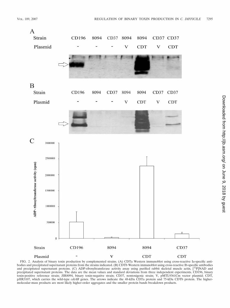
FIG. 2. Analysis of binary toxin production by complemented strains. (A) CDTa Western immunoblot using cross-reactive Ia-specific anti-bodies and precipitated supernatant proteins from the strains indicated. (B) CDTb Western immunoblot using cross-reactive Ib-specific antibodiesand precipitated supernatant proteins. (C) ADP-ribosyltransferase activity assay using purified rabbit skeletal muscle actin, [32P]NAD andprecipitated supernatant proteins. The data are the mean values and standard deviations from three independent experiments. CD196, binarytoxin-positive reference strain; JIR8094, binary toxin-negative strain; CD37, nontoxigenic strain; V, pMTL9361Cm vector plasmid; CDT,pJIR3107, which carries the wild-type cdtAB genes. The arrows indicate the 48-kDa CDTa protein and 75-kDa CDTb protein. The higher-molecular-mass products are most likely higher-order aggregates and the smaller protein bands breakdown products.
VOL. 189, 2007 REGULATION OF BINARY TOXIN PRODUCTION IN C. DIFFICILE 7295
on June 9, 2018 by guesthttp://jb.asm
.org/D
ownloaded from

were PCR amplified from strain JIR8094 and cloned into theshuttle plasmid pMTL9301, generating pJIR3394 (Fig. 1B),which encodes erythromycin resistance. This plasmid wastransferred by conjugation into the CD37 derivative that car-ries the cdtA�B� plasmid, pJIR3107. The transconjugantswere isolated and maintained on medium supplemented withboth thiamphenicol and erythromycin to ensure the continuedpresence of both plasmids, each of which was derived from apCD6 replicon. PCR analysis of several transconjugants con-firmed the presence of both cdtA and cdtR (data not shown),verifying the presence of both pJIR3107 and pJIR3394 in theresultant strain.
To provide the appropriate control for this experiment,pJIR3395 was constructed. This plasmid was a derivative ofpJIR3394 that contained a large in-frame internal deletion of
cdtR, spanning codons 36 to 222, designated �cdtR. This plas-mid was transferred by conjugation into strain CD37(pJIR3107), and thiamphenicol and erythromycin resistanttransconjugants were selected, maintained, and analyzed asbefore.
Western blot analysis was then performed on the isogenicCD37 derivatives carrying pJIR3107 (cdtA�B�) alone orpJIR3107 together with pJIR3394 (cdtR�) or pJIR3395(�cdtR), as well as CD37 carrying the shuttle vectorpMTL9361Cm and pJIR3394. Other controls included theCD196 binary toxin reference strain and a CDT� NAP1/027strain, M7404. The results showed that the presence ofpJIR3394 in CD37(pJIR3107) led to a dramatic increase in theproduction of both CDTa and CDTb (Fig. 5A and B). Bycontrast, pJIR3395 had no effect on CDTa or CDTb produc-
FIG. 3. Schematic representation of the CDT region and flanking genes. The regions from the nontoxigenic isolate CD37 (A), the binarytoxin-negative isolate strain 630 (B), and the binary toxin-positive isolate QCD-32g58 (C) are shown. The positions of the 5� flanking genes CD2601and CD2602, the 3� flanking gene trpS, the response regulator gene cdtR, and the CDT binary toxin-encoding genes cdtAB, or their pseudogenes,are shown. For each variant of the CDT region the positions of the 5� and 3� conserved boundaries are shown, and the size of the entire CdtLocis indicated. The unique 68-bp sequence that is present in CD37 and other nontoxigenic isolates in place of the CdtLoc is shown in bold, and thenucleotide boundaries of the CDT region that are conserved in all three variants are underlined.
7296 CARTER ET AL. J. BACTERIOL.
on June 9, 2018 by guesthttp://jb.asm
.org/D
ownloaded from

tion. As expected, a CD37 derivative carrying pMTL9361Cmand pJIR3394 did not produce any detectable CDTa or CDTb,since it did not contain the cdtA or cdtB gene. Note that CDTaand CDTb production by CD37(pJIR3107) carrying the cdtR�
plasmid appeared to be greater than that by either of thebinary toxin-producing positive controls.
To confirm the data obtained by Western blot analysis,ADP-ribosytransferase assays were performed on the samestrains (Fig. 5C). The results showed that the functional cdtRgene results in a 17-fold increase in CDT activity. The CD37(pJIR3107)(pJIR3394) strain had approximately fivefold moreactivity than the positive control strains CD196 and M7404.The introduction of the internal deletion into cdtR led to anapproximately ninefold reduction in ADP-ribosytransferaseactivity, to a level that was not significantly different from thatof the isogenic negative controls. These results provide goodexperimental evidence that the cdtR gene encodes a positiveactivator of binary toxin production in C. difficile.
To verify that CdtR regulates the expression of the binarytoxin genes at the transcriptional level, cdtA-specific QRT-PCRanalysis was performed on RNAs extracted from strains CD37(pJIR3107)(pJIR3394) and CD37(pJIR3107)(pJIR3395). Theresults showed that there was a 90-fold 10.5-fold increase inthe levels of cdtA-specific mRNA in the strain carrying theintact cdtR gene compared to the strain carrying the �cdtRplasmid (P � 0.0002). These data confirm that cdtR is a tran-scriptional regulator of the binary toxin genes.
DISCUSSION
The results presented in this paper clearly show that binarytoxin production in C. difficile is positively regulated by theLytTR family response regulator, CdtR. The evidence for thisconclusion comes from the observation that strain 630-derivedrecombinant strains carrying the binary toxin structural genescdtA and cdtB produced large amounts of binary toxin, but theequivalent CD37 derivatives, which do not have a cdtR gene,produced approximately 15-fold less binary toxin, despite thesestrains carrying the same CDT-encoding plasmid. In addition,when a recombinant CD37 derivative carrying cdtA and cdtBwas complemented with a functional cdtR gene, binary toxinproduction was activated and greater than 17-fold more binarytoxin was produced.
To rule out the possibility that another plasmid-encodedfactor was responsible for the increase in binary toxin produc-tion seen in the complemented strains, the intact cdtR gene wasreplaced with a mutant cdtR allele. This �cdtR gene containedan in-frame deletion that spans codons 36 to 222, resulting inthe removal of most of the cdtR sequence. When comple-mented with �cdtR, CDT production was not statistically dif-ferent from that of the noncomplemented parental strain,indicating that CdtR is indeed responsible for the observedup-regulation of binary toxin production.
Compared to the NAP1/027 strain M7404 and the referenceCDT� strain CD196, which both produced similar levels of
FIG. 4. Alignment of CdtR orthologs. The predicted CdtR protein from C. difficile strain 630 and predicted AgrA orthologs of S. aureus(ABB17535), Staphylococcus epidermidis (AAO25552) and Clostridium acetobutylicum (AAK78066) were aligned using CLUSTALW (58). Iden-tical amino acid residues are shaded with black, and strongly similar amino acid residues are shaded with gray.
VOL. 189, 2007 REGULATION OF BINARY TOXIN PRODUCTION IN C. DIFFICILE 7297
on June 9, 2018 by guesthttp://jb.asm
.org/D
ownloaded from

FIG. 5. Analysis of binary toxin production in derivatives of CD37. (A) CDTa Western immunoblot using cross-reactive Ia specific antibodiesand precipitated supernatant proteins from the strains indicated. (B) CDTb Western immunoblot using cross-reactive Ib specific antibodies and
7298 CARTER ET AL. J. BACTERIOL.
on June 9, 2018 by guesthttp://jb.asm
.org/D
ownloaded from

binary toxin, the cdtAB-complemented strain 630 derivativeand the cdtAB- and cdtR-complemented CD37 derivative hadsignificantly more ADP-ribosyltransferase activity. This resultmost likely reflects the higher copy number of the plasmid-carried cdtAB genes in the recombinant strains.
The comparative genomic studies showed that there was anabsolute correlation between the presence of the cdtR geneand the upstream cdtA region. Strains that did not have cdtA ora cdtA pseudogene did not carry cdtR. The term PaLoc iswidely used to describe the cytotoxin gene locus of C. difficile,a locus that includes both toxin structural genes and regulatorygenes. Based on the findings presented here, and in keepingwith the PaLoc nomenclature, it is proposed that the termCdtLoc be used to describe the cdt locus, which is herebydefined as the region containing cdtR, cdtA, and cdtB (as shownin Fig. 3), irrespective of whether in a particular isolate cdtAand cdtB are functional genes or pseudogenes.
Of the strains examined here, only those isolates that hadthe tcdA and tcdB genes carried cdtR and therefore had theCdtLoc. Recently, other workers performed genomic microar-ray analysis that compared diverse C. difficile isolates (52), veryfew of which were included in our study. Of the 75 strainsanalyzed, 68 provided definitive data with regard to the pres-ence or absence of cdtR. Our analysis of the microarray dataappears to reinforce the findings reported here, since approx-imately 96% of cdtR-positive isolates also carried the cdtABgenes or, at the very least, fragments of these genes, indicatingthat cdtR and the cdtAB genes are genetically linked. Interest-ingly, we found that approximately 82% of the cdtR-negativeisolates were clustered within the A�B� clade. The remaining18% were located in the HA1 clade and were not from diseaseoutbreaks. Upon closer inspection of the entire A�B� clade,we discovered that only four of these 22 isolates carried theCdtLoc. Remarkably, each of these four strains was actuallygenotypically classified as A�B� and not A�B�, and they wereall of animal origin (52). Analysis of the seven A�B� isolatesincluded in the microarray study revealed that only two con-tained the CdtLoc. These strains were located within the HA1and HA2 clades, respectively. The remaining five genotypicallyA�B� isolates were cdtR negative and were clustered withinthe A�B� clade.
Other studies have identified CDT-positive but toxin A- andtoxin B-negative strains, providing evidence that these loci arenot always linked (10, 11). However, the observations de-scribed above, together with the results presented in this study,suggest that, with the exception of strains found within theA�B� clade, there is a correlation between the presence of thePaLoc and the CdtLoc. More than 98% of CdtLoc-positivestrains also have the PaLoc. Furthermore, it is apparent, againwith the exception of A�B� strains, that the CdtLoc is widely
disseminated, with greater than 90% of the non-A�B� cladeisolates analyzed with the microarrays containing this region.
Further analysis of the site of insertion/deletion of theCdtLoc in CD37 and seven other PaLoc-negative isolatesshowed that the entire CdtLoc was absent in these isolates andhad been replaced by a novel 68-bp sequence of unknownfunction. This finding is reminiscent of the analysis of thePaLoc, where nontoxigenic strains were found to harbor aunique and highly conserved 115-bp sequence instead of thePaLoc (6). As with the PaLoc, there is no evidence of trans-poson-, plasmid-, or bacteriophage-related genes in close prox-imity to the CdtLoc. Since both of these toxin loci share someimportant characteristics, namely, the loss of an apparentlyinnocuous nucleotide sequence upon their acquisition and theabsence of any obvious genes or nucleotide sequences relatedto mobile genetic elements, it is postulated that the integrationof the PaLoc and the CdtLoc may have occurred throughsimilar, but as yet unknown, mechanisms.
It is estimated that between 1.6% and 6% of all C. difficileisolates produce a functional binary toxin, including the highlyvirulent toxinotype III NAP1/027 epidemic strains (53). How-ever, the role of this toxin in the pathogenesis of C. difficileinfections remains to be determined. Several studies have sug-gested an association between binary toxin-producing strainsand more severe disease and community-acquired infections(2, 29, 57). Furthermore, CDT is known to be cytotoxic to Verocells (38) and causes fluid accumulation in the rabbit ileal loopmodel (10). Indirect evidence therefore does suggest that CDTplays a role in C. difficile-mediated disease. However, a recentstudy showed that toxin A- and toxin B-negative, CDT-positiveisolates were unable to cause disease in the hamster model(10).
Many bacteria tightly control their ability to produce toxins.C. difficile regulates the expression of its major toxins, toxin Aand toxin B, in response to several environmental cues, includ-ing nutritional signals, cell density, and temperature (8, 17, 20,27). This process involves the tcdR and tcdC genes, which areencoded within the PaLoc. However, the PaLoc does not carrygenes encoding a two-component signal transduction system.
Members of the LytTR response regulator family, namedafter the LytT and LytR response regulators from Bacillussubtilis and S. aureus, respectively, are characterized by thepresence of a novel DNA-binding domain, which shows littlesimilarity to the helix-turn-helix or winged-helix domains thatare more typical of other response regulators (33). They arewidely distributed in low-G�C gram-positive bacteria, such asthe clostridia, and many members have a well-documentedassociation with bacterial virulence. Some play an integral rolein quorum sensing, such as the AgrA response regulator, whichis the master regulator of the quorum-sensing-controlled vir-
precipitated supernatant proteins. (C) ADP-ribosyltransferase activity assayed using purified rabbit skeletal muscle actin, [32P]NAD and precip-itated supernatant proteins. The data are the mean values and standard deviations from three independent experiments CD196, binary toxin-positive reference strain; M7404, binary toxin-positive NAP1/027 strain; CD37, nontoxigenic strain; pJIR3107, encodes the wild-type cdtAB genes(shown as � in the CDT rows); V, pMTL9361Cm vector plasmid (shown as � in the CDT rows); pJIR3394, encodes the wild-type cdtR gene(shown as � in the cdtR rows); pJIR3395, encodes the �cdtR gene (shown as �cdtR in the cdtR rows). The arrows indicate the 48-kDa CDTaprotein and 75-kDa CDTb protein.
VOL. 189, 2007 REGULATION OF BINARY TOXIN PRODUCTION IN C. DIFFICILE 7299
on June 9, 2018 by guesthttp://jb.asm
.org/D
ownloaded from

ulence response of S. aureus (31). Others are important in theregulation of toxin production, as demonstrated by the VirRresponse regulator of C. perfringens, which controls the synthe-sis of several toxins, including perfringolysin O (26, 42, 46),while others are involved in the production of extracellularpolysaccharides, such as alginate in Pseudomonas aeruginosa,which is regulated by the AlgR response regulator (24). CdtRis now another virulence-related member of this family, con-trolling the production of CDT in C. difficile. CdtR may acti-vate cdtAB gene expression either directly or by a secondaryregulator.
LytTR response regulators are activated by the transfer of aphosphoryl group to a conserved aspartate residue located intheir receiver domain, through the action of a cognate sensorhistidine kinase (54). The genes encoding these proteins oftencomprise an operon, as observed for both virR and agrA, whichare adjacent to virS (26) and agrC (34), respectively. By con-trast, the cdtR gene is an orphan response regulator; there is nosensor kinase gene located in close proximity, and the histidinekinase that presumably is responsible for the activation ofCdtR is unknown. However, the integration site of the CdtLocpositions cdtR only 11 bp from the predicted start site of asensor histidine kinase gene (CD2602), although this gene istranscribed on the opposite DNA strand (Fig. 3). CD2602appears to be associated with another downstream LytTR re-sponse regulator (CD2601). It is possible that the acquisitionof the CdtLoc might result in the differential regulation ofCD2602. Current studies in this laboratory are aimed at deter-mining whether CD2601 and CD2602 are involved in the reg-ulation of binary toxin expression.
In conclusion, we have identified a LytTR family responseregulator that activates binary toxin production in C. difficileand together with the cdtAB operon comprises a newly definedlocus, the CdtLoc. The genetic boundaries of this region weredefined by the comparison of different CDT� and CDT� iso-lates. Key questions that remain to be resolved but which arethe subject of current studies in this laboratory involve thedetermination of the environmental signals and signal trans-duction pathways that lead to the activation of CdtR and bi-nary toxin production and the determination of the role ofbinary toxin production in diseases caused by this increasinglyimportant human pathogen.
ACKNOWLEDGMENTS
We thank R. Poon for technical assistance; J. Cheung for helpfuldiscussions; P. Wright for assistance with the ADP-ribosyltransferaseassay; G. Jenkin, J. Pepin, L. Grayson, and T. Riley for providing C.difficile isolates; N. Minton for kindly providing pMTL9301 andpMTL9361Cm; M. Popoff for the kind gift of Ia- and Ib-specific anti-bodies; and R. Stabler for providing access to the C. difficile genomicmicroarray data.
This research was funded by grant AI057637 from the NationalInstitute of Allergy and Infectious Diseases and by Program Grant284214 from the Australian National Health and Medical ResearchCouncil.
REFERENCES
1. Aktories, K., and A. Wegner. 1992. Mechanisms of the cytopathic action ofactin-ADP-ribosylating toxins. Mol. Microbiol. 6:2905–2918.
2. Barbut, F., B. Gariazzo, L. Bonne, V. Lalande, B. Burghoffer, R. Luiuz, andJ. C. Petit. 2007. Clinical features of Clostridium difficile-associated infectionsand molecular characterization of strains: results of a retrospective study,2000–2004. Infect. Control Hosp. Epidemiol. 28:131–139.
3. Barth, H., K. Aktories, M. R. Popoff, and B. G. Stiles. 2004. Binary bacterialtoxins: biochemistry, biology, and applications of common Clostridium andBacillus proteins. Microbiol. Mol. Biol. Rev. 68:373–402.
4. Borriello, S. P. 1998. Pathogenesis of Clostridium difficile infection. J. Anti-microb. Chemother. 41(Suppl. C):13–19.
5. Boyer, H. W., and D. Roulland-Dussoix. 1969. A complementation analysisof the restriction and modification of DNA in Escherichia coli. J. Mol. Biol.41:459–472.
6. Braun, V., T. Hundsberger, P. Leukel, M. Sauerborn, and C. von Eichel-Streiber. 1996. Definition of the single integration site of the pathogenicitylocus in Clostridium difficile. Gene 181:29–38.
7. Chang, S. Y., and K. P. Song. 2001. ADP-ribosylating binary toxin genes ofClostridium difficile strain CCUG 20309. DNA Seq. 12:115–120.
8. Dupuy, B., and A. L. Sonenshein. 1998. Regulated transcription of Clostrid-ium difficile toxin genes. Mol. Microbiol. 27:107–120.
9. Eggertson, L. 2004. Quebec strikes committee on Clostridium difficile. Can.Med. Assoc. J. 171:123.
10. Geric, B., R. J. Carman, M. Rupnik, C. W. Genheimer, S. P. Sambol, D. M.Lyerly, D. N. Gerding, and S. Johnson. 2006. Binary toxin-producing, largeclostridial toxin-negative Clostridium difficile strains are enterotoxic but donot cause disease in hamsters. J. Infect. Dis. 193:1143–1150.
11. Geric, B., S. Johnson, D. N. Gerding, M. Grabnar, and M. Rupnik. 2003.Frequency of binary toxin genes among Clostridium difficile strains that donot produce large clostridial toxins. J. Clin. Microbiol. 41:5227–5232.
12. Goncalves, C., D. Decre, F. Barbut, B. Burghoffer, and J. C. Petit. 2004.Prevalence and characterization of a binary toxin (actin-specific ADP-ribo-syltransferase) from Clostridium difficile. J. Clin. Microbiol. 42:1933–1939.
13. Hachler, H., B. Berger-Bachi, and F. H. Kayser. 1987. Genetic character-ization of a Clostridium difficile erythromycin-clindamycin resistance deter-minant that is transferable to Staphylococcus aureus. Antimicrob. Agents.Chemother. 31:1039–1045.
14. Hayter, P. M., and J. W. Dale. 1984. Detection of plasmids in clinical isolatesof Clostridium difficile. Microbios Lett. 27:151–156.
15. Ho, S. N., H. D. Hunt, R. M. Horton, J. K. Pullen, and L. R. Pease. 1989.Site-directed mutagenesis by overlap extension using the polymerase chainreaction. Gene 77:51–59.
16. Horton, R. M., H. D. Hunt, S. N. Ho, J. K. Pullen, and L. R. Pease. 1989.Engineering hybrid genes without the use of restriction enzymes: gene splic-ing by overlap extension. Gene 77:61–68.
17. Hundsberger, T., V. Braun, M. Weidmann, P. Leukel, M. Sauerborn, and C.von Eichel-Streiber. 1997. Transcription analysis of the genes tcdA-E of thepathogenicity locus of Clostridium difficile. Eur. J. Biochem. 244:735–742.
18. Just, I., J. Selzer, M. Wilm, C. von Eichel-Streiber, M. Mann, and K.Aktories. 1995. Glucosylation of Rho proteins by Clostridium difficile toxin B.Nature 375:500–503.
19. Just, I., M. Wilm, J. Selzer, G. Rex, C. von Eichel-Streiber, M. Mann, and K.Aktories. 1995. The enterotoxin from Clostridium difficile (ToxA) monoglu-cosylates the Rho proteins. J. Biol. Chem. 270:13932–13936.
20. Karlsson, S., B. Dupuy, K. Mukherjee, E. Norin, L. G. Burman, and T.Akerlund. 2003. Expression of Clostridium difficile toxins A and B and theirsigma factor TcdD is controlled by temperature. Infect. Immun. 71:1784–1793.
21. Kuijper, E. J., B. Coignard, and P. Tull. 2006. Emergence of Clostridiumdifficile-associated disease in North America and Europe. Clin. Microbiol.Infect. 12(Suppl. 6):2–18.
22. Kyne, L., M. B. Hamel, R. Polavaram, and C. P. Kelly. 2002. Health carecosts and mortality associated with nosocomial diarrhea due to Clostridiumdifficile. Clin. Infect. Dis. 34:346–353.
23. Laemmli, U. K. 1970. Cleavage of structural proteins during the assembly ofthe head of bacteriophage T4. Nature 227:680–685.
24. Lizewski, S. E., D. S. Lundberg, and M. J. Schurr. 2002. The transcriptionalregulator AlgR is essential for Pseudomonas aeruginosa pathogenesis. Infect.Immun. 70:6083–6093.
25. Loo, V. G., L. Poirier, M. A. Miller, M. Oughton, M. D. Libman, S. Michaud,A. M. Bourgault, T. Nguyen, C. Frenette, M. Kelly, A. Vibien, P. Brassard,S. Fenn, K. Dewar, T. J. Hudson, R. Horn, P. Rene, Y. Monczak, and A.Dascal. 2005. A predominantly clonal multi-institutional outbreak of Clos-tridium difficile-associated diarrhea with high morbidity and mortality.N. Engl. J. Med. 353:2442–2449.
26. Lyristis, M., A. E. Bryant, J. Sloan, M. M. Awad, I. T. Nisbet, D. L. Stevens,and J. I. Rood. 1994. Identification and molecular analysis of a locus thatregulates extracellular toxin production in Clostridium perfringens. Mol. Mi-crobiol. 12:761–777.
27. Mani, N., D. Lyras, L. Barroso, P. Howarth, T. Wilkins, J. I. Rood, A. L.Sonenshein, and B. Dupuy. 2002. Environmental response and autoregula-tion of Clostridium difficile TxeR, a sigma factor for toxin gene expression. J.Bacteriol. 184:5971–5978.
28. McDonald, L. C. 2005. Clostridium difficile: responding to a new threat froman old enemy. Infect. Control Hosp. Epidemiol. 26:672–675.
29. McEllistrem, M. C., R. J. Carman, D. N. Gerding, C. W. Genheimer, and L.Zheng. 2005. A hospital outbreak of Clostridium difficile disease associatedwith isolates carrying binary toxin genes. Clin. Infect. Dis. 40:265–272.
7300 CARTER ET AL. J. BACTERIOL.
on June 9, 2018 by guesthttp://jb.asm
.org/D
ownloaded from

30. Miller, J. H. 1972. Experiments in molecular genetics. Cold Spring HarborLaboratory, Cold Spring Harbor, NY.
31. Morfeldt, E., K. Tegmark, and S. Arvidson. 1996. Transcriptional control ofthe agr-dependent virulence gene regulator, RNAIII, in Staphylococcus au-reus. Mol. Microbiol. 21:1227–1237.
32. Nakamura, S., K. Yamakawa, S. Nakashio, S. Kamiya, and S. Nishida. 1987.Correlation between susceptibility to chloramphenicol, tetracycline and clin-damycin, and serogroups of Clostridium difficile. Med. Microbiol. Immunol.176:79–82.
33. Nikolskaya, A. N., and M. Y. Galperin. 2002. A novel type of conservedDNA-binding domain in the transcriptional regulators of the AlgR/AgrA/LytR family. Nucleic Acids Res. 30:2453–2459.
34. Novick, R. P., S. J. Projan, J. Kornblum, H. F. Ross, G. Ji, B. Kreiswirth, F.Vandenesch, and S. Moghazeh. 1995. The agr P2 operon: an autocatalyticsensory transduction system in Staphylococcus aureus. Mol. Gen. Genet.248:446–458.
35. O’Connor, J. R., D. Lyras, K. A. Farrow, V. Adams, D. R. Powell, J. Hinds,J. K. Cheung, and J. I. Rood. 2006. Construction and analysis of chromo-somal Clostridium difficile mutants. Mol. Microbiol. 61:1335–1351.
36. Oultram, J. D., H. Peck, J. K. Brehm, D. E. Thompson, T. J. Swinfield, andN. P. Minton. 1988. Introduction of genes for leucine biosynthesis fromClostridium pasteurianum into C. acetobutylicum by cointegrate conjugaltransfer. Mol. Gen. Genet. 214:177–179.
37. Palombo, E. A., K. Yusoff, V. A. Stanisich, V. Krishnapillai, and N. S.Willetts. 1989. Cloning and genetic analysis of tra cistrons of the Tra 2/Tra 3region of plasmid RP1. Plasmid 22:59–69.
38. Perelle, S., M. Gibert, P. Bourlioux, G. Corthier, and M. R. Popoff. 1997.Production of a complete binary toxin (actin-specific ADP-ribosyltrans-ferase) by Clostridium difficile CD196. Infect. Immun. 65:1402–1407.
39. Popoff, M. R., E. J. Rubin, D. M. Gill, and P. Boquet. 1988. Actin-specificADP-ribosyltransferase produced by a Clostridium difficile strain. Infect. Im-mun. 56:2299–2306.
40. Purdy, D., T. A. O’Keeffe, M. Elmore, M. Herbert, A. McLeod, M. Bokori-Brown, A. Ostrowski, and N. P. Minton. 2002. Conjugative transfer of clos-tridial shuttle vectors from Escherichia coli to Clostridium difficile throughcircumvention of the restriction barrier. Mol. Microbiol. 46:439–452.
41. Riley, T. V., J. P. Codde, and I. L. Rouse. 1995. Increased length of hospitalstay due to Clostridium difficile associated diarrhoea. Lancet 345:455–456.
42. Rood, J. I. 1998. Virulence genes of Clostridium perfringens. Annu. Rev.Microbiol. 52:333–360.
43. Rupnik, M. 2007. Is Clostridium difficile-associated infection a potentiallyzoonotic and foodborne disease? Clin. Microbiol. Infect. 13:457–459.
44. Sambrook, J., and D. W. Russell. 2001. Molecular cloning: a laboratorymanual, 3rd. ed. Cold Spring Harbor Laboratory, Cold Spring Harbor, NY.
45. Sebaihia, M., B. W. Wren, P. Mullany, N. F. Fairweather, N. Minton, R.Stabler, N. R. Thomson, A. P. Roberts, A. M. Cerdeno-Tarraga, H. Wang,M. T. Holden, A. Wright, C. Churcher, M. A. Quail, S. Baker, N. Bason, K.Brooks, T. Chillingworth, A. Cronin, P. Davis, L. Dowd, A. Fraser, T.Feltwell, Z. Hance, S. Holroyd, K. Jagels, S. Moule, K. Mungall, C. Price, E.Rabbinowitsch, S. Sharp, M. Simmonds, K. Stevens, L. Unwin, S. Whithead,
B. Dupuy, G. Dougan, B. Barrell, and J. Parkhill. 2006. The multidrug-resistant human pathogen Clostridium difficile has a highly mobile, mosaicgenome. Nat. Genet. 38:779–786.
46. Shimizu, T., W. Ba-Thein, M. Tamaki, and H. Hayashi. 1994. The virR gene,a member of a class of two-component response regulators, regulates theproduction of perfringolysin O, collagenase, and hemagglutinin in Clostrid-ium perfringens. J. Bacteriol. 176:1616–1623.
47. Simpson, L. L., H. Zepeda, and I. Ohishi. 1988. Partial characterization ofthe enzymatic activity associated with the binary toxin (type C2) produced byClostridium botulinum. Infect. Immun. 56:24–27.
48. Smith, A. 2005. Outbreak of Clostridium difficile infection in an Englishhospital linked to hypertoxin-producing strains in Canada and the US. Eur.Surveill. 10:E050630 2.
49. Smith, C. J., S. M. Markowitz, and F. L. Macrina. 1981. Transferabletetracycline resistance in Clostridium difficile. Antimicrob. Agents Che-mother. 19:997–1003.
50. Spencer, R. C. 1998. Clinical impact and associated costs of Clostridiumdifficile-associated disease. J. Antimicrob. Chemother. 41(Suppl. C):5–12.
51. Spencer, R. C. 1998. The role of antimicrobial agents in the aetiology ofClostridium difficile-associated disease. J. Antimicrob. Chemother. 41(Suppl.C):21–27.
52. Stabler, R. A., D. N. Gerding, J. G. Songer, D. Drudy, J. S. Brazier, H. T.Trinh, A. A. Witney, J. Hinds, and B. W. Wren. 2006. Comparative phylo-genomics of Clostridium difficile reveals clade specificity and microevolutionof hypervirulent strains. J. Bacteriol. 188:7297–7305.
53. Stare, B. G., M. Delmee, and M. Rupnik. 2007. Variant forms of the binarytoxin CDT locus and tcdC gene in Clostridium difficile strains. J. Med. Mi-crobiol. 56:329–335.
54. Stock, A. M., V. L. Robinson, and P. N. Goudreau. 2000. Two-componentsignal transduction. Annu. Rev. Biochem. 69:183–215.
55. Stubbs, S., M. Rupnik, M. Gibert, J. Brazier, B. Duerden, and M. Popoff.2000. Production of actin-specific ADP-ribosyltransferase (binary toxin) bystrains of Clostridium difficile. FEMS Microbiol. Lett. 186:307–312.
56. Sullivan, N. M., S. Pellett, and T. D. Wilkins. 1982. Purification and char-acterization of toxins A and B of Clostridium difficile. Infect. Immun. 35:1032–1040.
57. Terhes, G., E. Urban, J. Soki, K. A. Hamid, and E. Nagy. 2004. Community-acquired Clostridium difficile diarrhea caused by binary toxin, toxin A, andtoxin B gene-positive isolates in Hungary. J. Clin. Microbiol. 42:4316–4318.
58. Thompson, J. D., D. G. Higgins, and T. J. Gibson. 1994. CLUSTAL W:improving the sensitivity of progressive multiple sequence alignment throughsequence weighting, position-specific gap penalties and weight matrix choice.Nucleic Acids Res. 22:4673–4680.
59. Warny, M., J. Pepin, A. Fang, G. Killgore, A. Thompson, J. Brazier, E. Frost,and L. C. McDonald. 2005. Toxin production by an emerging strain ofClostridium difficile associated with outbreaks of severe disease in NorthAmerica and Europe. Lancet 366:1079–1084.
60. Wust, J., and U. Hardegger. 1983. Transferable resistance to clindamycin,erythromycin, and tetracycline in Clostridium difficile. Antimicrob. AgentsChemother. 23:784–786.
VOL. 189, 2007 REGULATION OF BINARY TOXIN PRODUCTION IN C. DIFFICILE 7301
on June 9, 2018 by guesthttp://jb.asm
.org/D
ownloaded from