Embed Size (px)
Citation preview
Biliary Esophagitis: A Case ReportNicholas J Demos, Ioana Agams and Tara J Becker
Department of Surgery, Hoboken University Medical Center, Hoboken, New Jersey, USA
*Corresponding author: Demos NJ, Department of Surgery, Hoboken University Medical Center, Hoboken, New Jersey, USA, Tel: +12014181000; E-mail:[email protected]
Received date: September 04, 2017; Accepted date: September 25, 2017; Published date: September 29, 2017
Copyright: © 2017 Demos NJ, et al. This is an open-access article distributed under the terms of the Creative Commons Attribution License, which permits unrestricteduse, distribution, and reproduction in any medium, provided the original author and source are credited.
Abstract
Bile reflux esophagitis has been reported and discussed in the literature for many years. What makes our caseunusual are the gross and microscopic photos of bile in the esophageal mucosa. The pertinent literature isdiscussed especially the deleterious and oncogenic effects of bile in the esophageal mucosa.
Keywords: Mucosa; Esophageal mucosa; Hiatal hernia; Biliary tract
Case ReportA 76 year old woman was admitted to Christ Hospital, Jersey City,
New Jersey on March 27, 2004, with intractable bilious vomiting. Onexamination she was found to have severe dehydration, prostrationsand atrial fibrillation with hypertension. She appeared to havecontinuous bilious vomiting, spilling on her bed.
Rales were present on the left lower chest. Past history revealed 20prior hospitalizations with malnutrition, vomiting, anaemia and urgentand chronic diabetic care. Four months previously, she was admittedfor removal of infected right pectoral pacemaker and replacement witha new one in the left pectoral area.
Figure 1: Barium esophagram; sliding hiatal hernia.
Work-up revealed a gastrointestinal series, oropharyngealdysphagia, esophagogastric paresis, and hiatal hernia with severegastroesophageal reflux (Figure 1). Other tests revealed severe anemia,
fundal esophagitis, severe malnutrition, left lower lobe atelectasis andpneumonia. Electrocardiogram revealed atrial fibrillation, sick sinussyndrome and a functioning pacemaker. Echocardiogram showednormal left ventricular size and contractility and mild left atrialenlargement. Bacterial culture revealed gram positive bacterium andurinary infection.
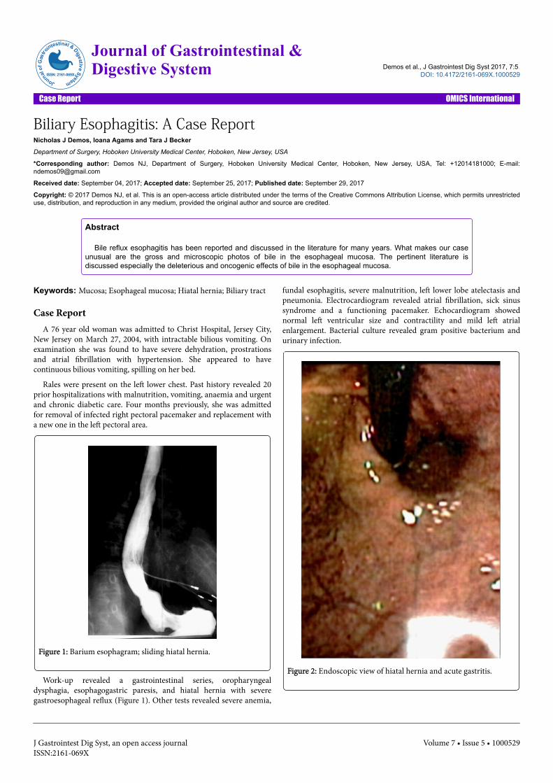
Figure 2: Endoscopic view of hiatal hernia and acute gastritis.
Journal
of G
astro
intestinal & Digestive System
ISSN: 2161-069X
Journal of Gastrointestinal &Digestive System Demos et al., J Gastrointest Dig Syst 2017, 7:5
DOI: 10.4172/2161-069X.1000529
Case Report OMICS International
J Gastrointest Dig Syst, an open access journalISSN:2161-069X
Volume 7 • Issue 5 • 1000529

A left subclavian catheter and a nasogastric catheter were inserted.Nevertheless, she kept vomiting bile. On April 22, 2004, I was called onconsultation and performed an upper gastrointestinal fiberopticendoscopy. Photograph revealed pronounces gastritis and a large hiatalhernia (Figure 2).
The esophagogastric area revealed mucosal biliary staining andBarret’s esophagus (Figure 3). Biopsies confirmed the Barret’s ulcers ofesophagus (Figures 4 and 5).
The black spots in these pictures were negative for melanin andhemosiderin. No chemical tests for bile were available. The patient’srelatives refused surgical correction at this time and for the next twomonths.
Figure 3: Esophagogastric area bile stains, Barrett’s changes.
Figure 4: Low power biopsy of Barrett’s area.
Figure 5: High power view: see bile particles in the esophagealmucosa.
The patient was treated for two months. A percutaneous fiberopticgastrostomy was performed. Diabetic management, antibiotics, andantacids were given. Vomiting continued even though to a lesserextent. Two months after admission, repeat upper endoscopy wasperformed revealing clearing of bile staining but confirming theBarrett’s metaplasia (Figure 6). She was discharged and two monthslater she was readmitted to a different hospital for a repeatpercutaneous gastrostomy. On November 6, 2004, she died at thenursing home.
Figure 6: Barrett’s metaplasia 2 months later: no bile stains.
Citation: Demos NJ, Agams I, Becker TJ (2017) Biliary Esophagitis: A Case Report. J Gastrointest Dig Syst 7: 529. doi:10.4172/2161-069X.1000529
Page 2 of 3
J Gastrointest Dig Syst, an open access journalISSN:2161-069X
Volume 7 • Issue 5 • 1000529

DiscussionThis case report emphasizes the destructive and fatal results of long
standing gastroesophageal reflux. Bile esophagitis was present in ourpatient. The additional effects of bile on the esophageal mucosa areseen in Figures 3-5. Jiang has described esophagitis in children causedby bile and acid [1]. Eros described bile induced ATP depletion, vastcell degranulation and tissue damage in dogs, all of which could beprevented by choline antagonists. Severe inflammation in thesubmucosa but no bile was seen [2]. Aiyer also studied the bile changesin esophageal mucosa but no bile was seen in biopsies [3]. Mitros’sAtlas of extensive study of gastrointestinal pathology mentions bilegastritis but no bile esophagitis [4].
Bile plays a role with refluxed acid in the development of Barrett’sesophagus and its malignant transformation, according to Peters andcolleagues [5]. In our case, visual evidence of bile staining haddisappeared two months later (Figure 6). Perhaps the injury to theesophageal mucosa had occurred or it may have continued, sinceBarrett’s metaplasia continued in the second endoscopy (Figure 6).
Refusal of operation was unfortunate. The patient’s aspirationpneumonia cleared. The diabetes was under control. Echocardiogramrevealed normal ventricular size and contractibility. The intendedsurgical correction carried no formidable risk. We had ampleexperience of surgical correction. In 1999, we published a 24 yearfollow-up of our stapled uncut gastroplasty in 161 patients. Only one
fatality occurred in an emergency operation [6,7]. Our patient expiredseven months after the first endoscopy in a nursing facility.
References1. Jiang M, Chen J, Chen F, Yu J, Liang J, et al. (2009) Bile and acid reflux in
the pathogenesis of reflux oesophagitis in children. J Paediatr ChildHealth 45: 64-67.
2. Eros G, Kaszaki J, Czobel M, Boros M (2006) Systemicphosphatidylcholine pretreatment protects canine esophageal mucosaduring acute experimental biliary reflux. World J Gastroenterol 12:271-279.
3. Aiyer HS, Li Y, Harper N, Myers SR, Martin RC (2011) Molecularchanges in the esophageal epithelium after a subchronic exposure tocigarette smoke in the presence of bile-acid reflux. Inhal Toxicol 23:304-311.
4. Mitros FA, Winawer SJ, Lu CC (1988) Atlas of gastrointestinal pathology.Philadelphia, Lippincott.
5. Peters JH, Reveiller M, Ghatak S, Toia L, Kalatskaya I, et al. (2012) Bileexposure inhibits expression of squamous dfferentiation genes in humanesophageal epithelial cells. Ann Surg 255: 1113-1120.
6. Demos NJ, Ahmad I, Scalia J, Wu J, Zaklama S, et al. (1998) Theesophagogastric junction: 420 questions, 420 answers. J Libbey Eurotext,France pp: 884-891.
7. Demos NJ (1999) The stapled, uncut gastroplasty for hiatal hernia: 24years’ follow-up. Dis Esophagus 12: 14-21.
Citation: Demos NJ, Agams I, Becker TJ (2017) Biliary Esophagitis: A Case Report. J Gastrointest Dig Syst 7: 529. doi:10.4172/2161-069X.1000529
Page 3 of 3
J Gastrointest Dig Syst, an open access journalISSN:2161-069X
Volume 7 • Issue 5 • 1000529