Embed Size (px)
Citation preview

BILATERAL EMPYEMA OF THE PLEURAL CAVITIESBY E. LAWRENCE KEYES, M.D.
OF NEW YoRK, N. Y.FROM THE SECOND SURGICAL DIVISION OF THE NEW YORK HOSPITAL
MEDICAL progress since the late war has been remarkable for the triumphsof the thoracic surgeon. Profiting by a rare circumstance which combinedthe necessity of treating large numbers of severe war wounds of the chestwith that of treating event greater numbers of empyemas, he has producedan abundance of excellent work which has revolutionized and radicallyimproved, among other things, the treatment of empyema. His successesare reflected in practice by the more intelligent care of empyema, and in theliterature by the appearance of excellent articles upon the subject.
One phase of the subject of empyema, however, has stood apart fromthe stream of investigation and discussion like some small hamlet untouchedamid the changes of a great world. This phase is bilateral empyema. Itsoccurrence is much less rare than many text-books would indicate. Its sever-ity tends to be exaggerated. Its treatment is sometimes undertaken with ahesitancy that is the complement of unfamiliarity, while the principles oftreatment are not always clearly understood. Hence any attempt which maystimulate renewed interest in this subject is not likely to be futile.
Such an attempt has been made here in the form of an outline of thenatural history and treatment of the disease based upon forty-one casereports and guided by a review of the literature. The forty-one casesinclude one case* personally observed at The New York Hospital and twoother unpublished casest from the same hospital; also thirty-eight casescollected from the literature of the past ten years. The review of theliterature briefly summarizes the partial reviews of Scanlan' in I928 andBozzotti2 in I9I3, and brings up-to-date and abstracts the more thoroughmonographs of Mackenzie3 in I9I4, Fabrikant4 in I9I I, and Hellin in19055 and I907.6
Natural History of Bilateral Empyema. Definitiont.-The term "bilateralempyema of the pleural cavities" denotes a condition in which pus is foundin both pleural cavities at or about the same time.
Bilateral empyema is usually considered a complication of unilateralempyema. It is almost always acute, very rarely chronic.
* The author takes pleasure in an expression of his indebtedness to Dr. Eugene H.Pool for a suggestion which inspired this work.
t One of these cases is from the First Surgical (Cornell) Division of The NewYork Hospital and is included by permission of Dr. James Morley Hitzrot.
1050

BILATERAL PLEURAL EMPYEMA
Incidence.---About 7.7 per cent. of all empyema cases are bilateral accord-ing to Hellin5 who found that II3 of I,448 empyema cases showed involve-ment of both sides. Lower figures are usually given. Thus, Lord7 inOsler's "Modern Medicine" quotes 2.0 per cent., or 5 out of 248 cases;Geitz8 records o.8 per cent., or 5 out of 588 cases from a review of twenty-three Swedish hospitals; while my figure is o.6 per cent., or 3 out of 495 casesat The New York Hospital from I9I4 to I930. Standard text-books, ifthey mention the subject at all, usually agree with Da Costa9 in saying"in civil practice I have seldom seen it." Graham,10 however, observesthat empyema "is bilateral in probably less than 5 per cent. of . . . non-fatal cases."
For children, Holt and Howland1" call empyema "bilateral in about 3per cent. of all cases, but oftener in infants."
The incidence during the epidemics of influenza of I9I7 to I919 wasvery high. Dunham12 found it to be 42 per cent. in the United StatesArmy Camps in I9I7, or 253 out of 603 empyemas. Stone's13 figure wasI9 per cent. at Fort Riley for the winter of I9I7-I9I8; he observed I9out of I00 empyema cases. Likewise with other epidemics of influenza theincidence may be high; thus Mackenzie3 saw many cases during an epidemicnear Portland, Oregon, in I899.
Age.-The average age is eleven and one-half years. Extremes of eightmonths and sixty years were reported by Fabrikant.4 About 75 per cent.of cases occur before the twelfth year.
Sex.-Males predominate over females by 3: 2.Primary Disease.-The most frequent cause is pneumonia; it preceded
75 per cent. of cases. Three-quarters of these pneumonias were broncho-pneumonias. "Primary" bilateral empyemas accounted for 20 per cent. ofFabrikant's cases.4 One of the cases of this series might so be classified(Case 26).
The rarer causes are pyaemia, tuberculosis, actinomycosis and the exan-themata. Surgical operation or its complications may also precede. Veryinteresting is the epidemic of puerperal sepsis which was followed by manycases of bilateral empyema described by Charrier14 in i855; the occasionalcases recorded since have been collected by Hellin5 and Fabrikant.4
Bacteriology.-The pneumococcus is the usual causative organism. It wascultured from the pus of twelve patients of this series. The haemolyticstreptococcus, however, almost invariably accompanied the post-epidemicinfluenzal cases. A variety of other organisms may be present.
Rarely a different organism is obtained in the two sides, as with Case 39.Pathology.-The pathology does not differ from that of unilateral
empyema.Pathogenesis.-The involvement of the two sides rather than one may
be accounted for in part by a preceding pneumonia; of the two lungs, andthis was true of twenty-one cases of this series. An increase of virulence of
1051

E. LAWRENCE KEYES
the infective organism may also be a factor as was indicated by the higlincidence accompanying the wartime influenza epidemic. On the other hand,there are patients whose lowered resistance is a predisposing cause, asillustrated by Cases 4O and 4I. Occasionally empyema of both sides followspneumonia of one lung (Cases 30, 3I and 40). Here the empyema of theother side possibly arose from a pneumonia so slight as to be overlooked,or possibly by metastatic infection. The cases of "primary" empyema arevery difficult to explain.
Why should not bilateral pneumonia more often be followed by empyemaof both sides? Certainly the majority of the pneumonias of both lungswhich are succeeded by empyemas are succeeded by an empyema confinedto one side. It may be that most pneumonias mature with unequal rapidityon the two sides so that the antibodies from the first empyema are sufficientto prevent a contralateral empyema.
The time of appearance of the two empyemas may be simultaneous, asshown by Cases 31, 32 and 4I; or not, as Cases 33 and 39 illustrate. InCase 33 almost two months intervened.
Diagnosis.-Diagnosis presents no unusual features, and it will becorrectly made more often if the condition be borne in mind.
Coinplications.-These are similar to those of unilateral empyema.Prognosis.-The mortality was 37 per cent. for Fabrikant's series of ii8
cases.4 It is I9.5 per cent. for this series, but this figure is of little significanceas it omits all the numerous deaths of I917 to I9I9.
The prognosis should be influenced greatly by the type of the infection.Thus many patients with the pneumococcic type recover, whereas there isno record of recovery of a single individual with the streptococcus haemolytictype. Failure to make this distinction may have influenced certain authors,Heuer15 for example, to classify all bilateral empyema "among the fatalcases of empyema." However, cases following influenza are by no meansnecessarily fatal. Thus, Mackenzie3 saw at least three during the Portlandepidemic of I899 (Cases 6, 7 and 8), while Cafritz16 and Beck17 successfullytreated influenza cases, the latter that of a pregnant woman (Case 25).
Treatmnent. (A) General Principles.-The general principles of thetreatment do not differ from those of unilateral empyema, and may befound thoroughly discussed in any of the principal works on chest surgery18written since the late war. A brief note on current practice must suffice here.
(a) Maintenance of the nutrition of the patient by high-fluid andcalorific intake is very important; for empyema is sometimes like typhoid, along wearing disease.
(b) Obliteration of the empyema cavities is to be accomplished bydrainage and reexpansion of the lungs. Thoracotomy with rib resection isoften necessary, though intercostal drainage alone may suffice. The patientmay hasten reexpansion by using blow bottles. Secondary operations topromote drainage or to prevent chronicity may be required.
1052

BILATERAL PLEURAL EMPYEMA
The drainage ttubes should not be permanently removed until the empyemacavities have become obliterated as shown by physical signs and by r6ntgeno-grams, and until discharge from the wound sinuses has become slight inamount and sterile.
Drainage should be deferred if possible until the pus within the pleuralcavity has become thick, thoracenteses being employed meanwhile. Thusintrapleural adhesions will have time to form, and the pneumonic processto subside. Streptococcic empyemas must not be drained as early as pneu-moccocic and other types of empyema.
Rarely, repeated thoracenteses may cure a bilateral empyema.(c) Avoidance of open pneumothorax is a measure of great importance.
It should be attempted by using drainage of the closed type; in other words,by drainage which maintains a negative intrapleural pressure. After somedays the closed may be changed to the open type.
(d) Sterilization of the empyema cavity may best be accomplished byfrequent irrigations with some solution, such as solution of chlorinated soda(Dakin's). The solution if kept warm will avoid heat loss from the lung.
(B) Particular Applicationi.-For discussion, let us assume that there isa patient having bilateral empyema of similar extent and duration on thetwo sides. Let us further assume that by application of the principles out-lined above a decision has been reached that it is time for drainage. Thequestion immediately arises as to whether to drain both sides simultaneouslyor not.
To this question many writers have in the past answered "No" becauseof the fear of bilateral open pneumothorax. They have based their fear uponthe assumption, prevalent up to the ending of the late war, that an openpneumothorax of both sides would be followed by collapse of both lungs andconsequently by death.
Investigations carried on during and since the late war, particularlythose of Graham and Bell,19 have shed new light on the pneumothorax prob-lem and on the treatment of empyema. They have shown that the medias-tinum of normal individuals is so flexible that it will readily transmit smallerpressure changes from one pleural cavity to the other; in other words thatthe chest may be considered practically as one instead of two cavities inso far as intrapleural pressures are concerned.
They have also determined that there is a certain area of opening intothe pleural cavity, 5I.5 square centimetres, which is the maximum com-patible with life if the thorax is normal and if the individual's vital capacityequals 3,700 cubic centimetres (a common average figure for the vitalcapacity). This is so because of a definite relationship between the amountof air entering the lunigs and that enterilng the pleural cavity. Openilngsinto the pleural cavity smaller than 51.5 square centimetres are increasinglywell borne. It is to b)e understood, however, that the above deductions weremade with reference to the normal thorax. If the mediastinum is stabilized
1053

E. LAWRENCE KEYES
by adhesions or by induration, then a unilateral opening of much largersize than the one mentioned may be made. On the contrary, if no suchstabilization has occurred and if the patient's vital capacity is low (e.g.,1,OOO cubic centimetres) because of pneumonia, or other reasons, then amuch smaller pleural opening may be fatal. Since it is the area of openingand not the number or position of the openings that is of importance, similaropenings into each pleural cavity will have no more ill effect than a singleopening into one pleural cavity of twice the area.
Experience has justified these theoretical conclusions. Many soldierswith surprisingly large open wounds of both chest cavities survived. More-over, eleven patients with bilateral empyema20 have been operated upon bysimultaneous thoracotomies on both sides. No patient died as a result ofthe operation; one patient collapsed temporarily but was resuscitated, andanother died twelve days later from a cause not attributable to the operation.Furthermore, forty-two' other patients2' were operated upon by almostsimultaneous thoracotomies, that is to say by thoracotomies performed onthe two sides within twenty-four hours of each other. None of thesepatients died, and all eventually recovered. Thus, a total of fifty-threesimultaneous, or nearly simultaneous, thoracotomies has been performedwithout a single operative death, and with eventual recovery of all but onepatient.
Bilateral simultaneous thoracotomy for bilateral empyema, then, hasproved itself an excellent method of treatment. This will be more apparentwhen it is observed that all the above simultaneous thoracotomies were per-formed under the open method, hence under conditions less favorable thanwould have prevailed under the safer closed' method. Certainly simul-taneous or nearly simultaneous closed thoracotomies should be moreextensively used in future. It is also notable that many patients are verylittle relieved after the first thoracotomy; they tend to remain very littlechanged until after the second thoracotomy when they often show suddenand remarkable improvement, as was the experience with Case 39 and othersof this series. Delay in opening the second side, therefore, seems toaccomplish little, but rather to weaken the powers of resistance; anotherreason for the simultaneous method.
If for some reason it is decided not to drain simultaneously, there arisesthe question as to which side is best for the initial operation. The left sidehas usually been preferred because of the belief that the heart would therebygain greater freedom of action, and that pericarditis would be less apt to occur.These are worthy considerations. Concerning the likelihood of pericarditisDunham22 says that "there is no marked difference in the relations of thepericardium to the pleura on the two sides of the chest, and pericarditishas occurred at least as frequently with an empyema on the right side as withone on the left."
If the empyemas are quite unequal in extent, the larger should be drained1054
BILATERAL PLEURAL EMPYEMA
first; but again, let it be repeated that the second side should be drained verysoon thereafter. In the interval thoracenteses are usually beneficial.
CASE I.- (Personal observation.) A girl of two years, Jewish, was admitted toThe New York Hospital, December i8, i928. Family History.-Three of the tenbrothers and sisters were dead before the age of four years of pneumonia, meningitis,and diphtheria. Previous history.-One year before admission there had been an opera-tion for abscess of the right thigh, the operation being complicated by pneumonia
.... ...
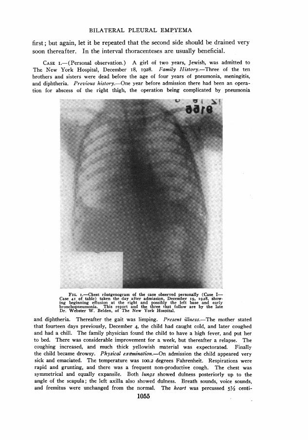
FIG. i.-Chest r6ntgenogram of the case observed personally (Case I-Case 41 of table) taken the day after admission, -December i9g I928 'show-ing beginning effusion at the right and possibly the left base and earlybronchopneumonia. This report and the three that follow are by the lateDr. Webster W. Belden, of The New York Hospital.
and diphtheria. Thereafter the gait was limping. Present illn.essThe mother statedthat fourteen days previously, December 4, the child had caught cold, and later coughedand had a chill. The family physician found the child to have a high fever, and put herto bed. There was considerable improvement for a week, but thereafter a relapse. Thecoughing increased, and much thick yellowish material was expectorated. Finallythe child became drowsy. Physical exchmination.-On admission the child appeared verysick and emaciated. The temperature was 1oo.2 degrees Fahrenheit. Respirations wererapid and grunting, and there was a frequent non-productive cough. The chest wassymmetrical and equally expansile. Both lungs showed dulness posteriorly up to theangle of the scapula; the left axilla also showed dulness. Breath sounds, voice sounds,and fremitus were unchanged from the normal. The hear-t was percussed 5Y2 centi-
1055

E. LAWRENCE KEYES
metres to the left of the midsternal line in the fifth interspace. The pulse rate wasII0 to the minute, the rhythm regular. The leucocytes were 12,400 per cubic millimetrewith 83 per cent. polymorphonuclear leucocytes and 17 per cent. lymphocytes. Theurine was normal, and other findings normal.
A diagnosis of bilateral bronchopneumonia and bilateral acute empyema was made.It was confirmed by rontgenogram on December I9 (Fig. i) which revealed pleuraleffusion at both bases and bronchopneumonia. Thoracenteses three days after admis-sion (December 2I) yielded thick greenish pus from both pleural cavities. Culturesfrom both sides showed a pneumococcus type I. A total of 8o cubic centimetres ofpus was withdrawn from the right side, and an amount unrecorded from the left.Seventy cubic centimetres of pus were withdrawn from the left side the next day.Little improvement followed. The child lay propped up in bed, semicomatose andcyanotic, struggling to breathle and to cough. It was now or never for drainage;moreover, six or more days of waiting after onset of the empyema seemed time enoughto render opening the pleural cavity relatively safe.' It was decided to drain the rightside first because of the preponderance of pus in the, right chest. Physical signs showeddisplacement to the left of the heart and trachea, while the signs of fluid extended onthe right as high as the spine of the scapula posteriorly, and to the third rib anteriorlyin the midclavicular line; whereas on the left they extended only to the angle of thescapula posteriorly, and were absent anteriorly. Meanwhile it was hoped that repeate(dthoracenteses would take care of the left side.
Accordingly operation for closed drainage of the right pleural cavity was per-formed December 24. With the patient seated the operative field was prepared byapplication of a half-strength solution of tincture of iodine. A preliminary thora-centesis at the operative site yielded thick pus. After infiltrating the overlying skinwith I per cent. procaine a small incision was made in the eighth right interspacein the posterior axillary line and carried down to the parietal pieura. A No. 22French catheter was then seized in a Kelly clamp and plunged through the unopenedpleura into the pleural cavity. Thick pus immediately flowed through the catheterwhich was clamped off in order to maintain closed drainage. The soft tissues and skinwere then approximated closely about the catheter by silkworm sutures passed throughan encircling cuff. The operation lasted ten minutes and caused no reaction.
With the patient removed to the ward a drainage-irrigation apparatus wasarranged. The end of the catheter, which protruded from the wound, was con-nected by a glass Y-tube to two other tubes, a drainage tube and an irrigation tube.The drainage tube was carried under the bed to a glass receptacle and its end keptunder water. The irrigation tube led up to a bottle of irrigating solution hung atthe head of the bed, and was kept clamped off except when in use. Before removingthe clamp from; the catheter which protruded from the wound all the tubing was filledwith a solution of chlorinated soda (Dakin's), and all air excluded. After removingthe clamp negative pressure was thus immediately exerted on the empyema cavity.
The tubes were irrigated three times a day by allowing to flow through them aboutI0 cubic centimetres of the solution of chlorinated soda from the bottle at the head ofthe bed. When clogged by pieces of exudate, as often happened, the apparatus wasdisconnected, and the protruding catheter irrigated by manual suction with a syringeuntil all obstructions were removed. During this procedure the protruding catheterwas never 'for a moment left open except while being subjected to negative pressure.Then, after cleaning the apparatus it was reconnected. Responsibility for the apparatusand its proper working rested with one house officer.
The amounts of pus drained day by day varied greatly. No accurate measurementswere made, but some days at least I00 cubic centimetres were returned. On one occa-
sion 70 cubic centimetres of pus were withdrawn through a syringe within a few minutes.As a rule manual drainage proved more efficient than mechanical arrangements for the
1056

BILATERAL PLEURAL EMPYEMA
removal of pus and exudate. Large clumps of whitish-yellow fibrinous matter wereoften obtained; the pus or exudate bore some resemblance to curdled milk.
Slight improvement in the condition of the patient followed operation. Thetemperature ranged between ioo and IOI degrees, sometimes reaching I03 degrees.The nutrition remained poor. Fluids were forced. A transfusion of 200 cubic centi-metres of whole blood was given (December 26). The left pleural cavity was tappedrepeatedly; December 26, forty cubic centimetres of pus were obtained; January i, forty,and January 3, only eight.
The right pleural cavity was reported free of fluid by a rontgenogram of December3', seven days post-operatively, but the left chest still showed haziness and mottling.
Closed drainage was replaced by open on the eighth day after operation (Jan-uary i ). The catheter was removed and a soft open rubber tube put in its place.
The patient's condition was becoming steadily worse. While not so sick as justbefore the first operation she still did not appear to be very far from death. Examina-tion revealed a rather extensive bronchopneumonia of both sides added to the leftempyema. The wound on the right continued to drain moderately.
Since the right lung was well expanded it was decided to open the left side. Thetwelfth day post-operative, therefore (January 5), operation for closed drainage ofthe left pleural cavity was performed. The technic employed and the after care weresimilar to that already described.
Immediate and continued improvement followed operation. The temperature gradu-ally subsided to normal. The child was allowed up after the twenty-seventh day post-operative, the temperature never exceeding normal limits thereafter.
The closed drainage of the left side was changed to open on the fourth day post-operative by removal of the catheter and insertion of a soft rubber tube into the wound.
The drainage tubes were retained in both sides until the thirty-third day after thesecond operation (February 7) when they were finally removed.
R6ntgenograms of the chest after the third day following the second operation(January 8 and i6) showed both pleural cavities free of fluid and both lungs com-pletely expanded and clear.
The patient was discharged cured April 4, I929, about four months after the onsetof illness. She had gained much weight and was in excellent physical condition. Bothwounds were completely healed. There were no abnormal physical signs in the chest,and no cough.
The condition was entirely satisfactory when seen in the Follow-Up ClinicJuly 7, I929.
CASE II.-A boy of seventeen was admitted to The New York Hospital Mayi8, I9I8. The family history and previous history were irrelevant. Present illness.-The patient was taken with pneumonia of the right lung April 29, I9I8, which spreadto the left lung on May 4. The family physician made a diagnosis of pleurisy ofboth sides, the right more marked. Physical examination.-The day of admission thepatient appeared emaciated and acutely sick. The temperature was 102.4 degrees,Fahrenheit, pulse I26, and respirations 34. The right chest lagged during inspiration.The right lung showed flatness below the angle of the scapula with diminished breathsounds; anteriorly there was flatness below the level of the nipple and diminishedbreath sounds with aegophony and pectoriloquy at the level of the nipple. The leftlung showed impairment of percussion, and a few coarse riles below the angle of thescapula. The urine was normal and the examination was otherwise normal. A pro-visional diagnosis of empyema of the right pleural cavity was made.
At operation the next day open drainage of the right pleural cavity was performed.Gas oxygen anaesthesia was employed. A needle was inserted in the eighth interspace,posterior axillary line, and pus obtained. One and one-half inch of the eighth rib was
67 1057

E. LAWRENCE KEYES
then resected. After opening the pleura a large empyema cavity was revealed. Tworubber tubes were inserted for drainage. Culture from the pus revealed strepto-coccus viridans.
Following operation there was moderate improvement. A r6ntgenogram sevendays later (May 26) showed a thickened right pleura and right pneumothorax, but nofluid in the right chest. The wound did not drain well so exploration of the woundwas done ten days after the first operation (May 29). Under gas-oxygen anaesthesia afinger was passed into the cavity through the wound, but no pocketing was found. Adrainage tube with suction, closed drainage, was applied, and following operation the cav-ity was regularly irrigated according to Carrel-Dakin technic. The condition then showedslight improvement. Cultures from the wound showed a mixed culture of pneumococcustype II and staphylococcus aureus.
Improvement was such that the patient was up and about from June i8 to June 25.He was then returned to bed because of accumulation of fluid in the left chest as shownby physical signs and r6ntgenogram.
Operation for open drainage of the left pleural cavity was performed fifty-one daysafter the first operation (July 3). The technic was the same as for the first operation,except that the ninth instead of the eighth rib was resected. Culture from this sideshowed a pure growth of pneumococcus which was not typed. Rapid improvementfollowed this operation and the temperature remained within normal limits after thefifth day post-operative.
The patient was discharged in excellent condition July I3. He left against advice,but the right wound had completely healed, and the left was closed and granulating.
A follow-up' a year later, July 25, I9I9, showed the patient in good health.CASE III.-A boy of two and three-quarter years was admitted to The New York
Hospital September I9, 19I7. The family history and previous history were irrelevant.Present illness.-The child was taken with cough and fever September i. He grewworse and developed severe dyspnea on September 12. Physical examination onadmission revealed a very sick white boy coughing frequently. The left lung showeddulness, bronchial breathing, and a few coarse riles below the angle of the scapulaposteriorly, and dulness between the third and sixth ribs anteriorly. The right lungwas normal. The heart was displaced, slightly, to the right. The leucocytes were38,600 per cubic millimetre of blood; 86 per cent. were polymorphonuclear leucocytes.The urine was normal, and the examination otherwise normal.
A provisional diagnosis of lobar pneumonia, left, and empyema (?) was made.Suspicious signs of empyema were reported by r6ntgenogram taken the next day.
The patient's condition became progressively worse. Nine days after admission(September 28) a left thoracentesis was done and yielded about 5 cubic centimetres ofthick pus. Culture of the pus returned a pneumococcus.
The operation of open drainage of the left pleural cavity was performed eightdays after thoracentesis (October 6). Without anaesthesia an intercostal incision wasmade in the eighth left interspace and scissors were forced through the pleura intoa large empyema cavity, from which much thick pus ran out. Two drains were placedin the cavity.
The post-operative condition was better for about a week, but was progressivelyworse thereafter. A r6ntgenogram taken October 14 showed no signs of fluid, however.
November ii fluid was discovered in the right chest. Marked emaciation wasalso present. The next day, thirty-five days after the first operation, an operationwas performed for open drainage of the right chest using the same technic as on theleft. Pus was obtained; it was sterile.
Following operation there was no improvement, and the chil(d died tight days afterthe second operation (November 20).
1058

BI3LATERAL PLEURAL EMPYEMA
CASE REPORTS OF BILATERAL EMPYEMA, I910 TO I930
I 2 3 4 s 6 7 8 9
Patient Days from Days______- diagnosis of Organism be-
Author and Age Sex empyema Primary disease cultured Type of drainage tween Re-year until first from opera- sult
operation empyema tions
Edmond 5 Pertussis; ? '-Right, open 7 CI910 28 broncho-pneu- 2-Left, open
monia
2.Corner and 29 F o ? ? i-Right, open; re- I CGrant section of ribI91I 24 2-Left, open; re-
section of rib
3.Gand and 4 M "Grippe"; pneu- ? Open, simultaneous o CPoissonier monia
4.Bozzotti 25 F II ? ? I-Left, open; rib 5 C1913 2 resection
2-Right, open; ribresection
5.Zingher 6.5 M o Bilateral Pneumo- i-Left, open; re- 26 C
bronchopneu- coccus section 8th rib1913 26 monia 2-Right, open; in-
tercostal incisionand drainage
6. 34 M " Influenza " ? Open, simultaneous o CMackenzie19143 and1924 277. ? M "Influenza" ? i-Right, open I C
(adult) 2-Left, open
8. ? M " Influenza " ? i-Right, open I C(adult) 2-Left, open
9. 7 o ? ? Open, simultaneous o C
10.Bunts 13 ? ? Acute appendi- ? i- ? open ? C
citis, operation; 2- ? openI9I428 pleural pneu-
monia
Lund and i6 F 7 Left lower lobar ? i-Left, open; gth I CMorrison pneumonia; rib resectedI9I6 29 right middle 2-Right, open
and lower lobarpneumonia
I2.Cafritz ? F ? "Grippe'" ? i-Left, open; 8th 23 C
Pus was thin rib resectionI9I8 26 2-Right, open'3.Norrlin 8 F II Pneumonia Diplococcus I-Right, open; 8th 28 C
pneumo- intercostal inci-1g1g 30 coccus? sion
2-Left, open; sametechnic
I4.Gundrum 26 M 22 Left lower lobar Pneumococ- i-Right, closed; 25 C
pneumonia; cus #i8 F. catheter inI920 31 right lower type IV ioth right inter-
lobar space2-Left, closed;same technic
I5.Durham 14 M 8 Influenza; otitis Staphylococ- I-Left, open; gth 42 C
media; bilat. cus aureus rib resected; re-1920 32 mastoiditis; peated aspirations
bilat. parotitis; 2-Right, open; 8thpneumonia rib resected
1059

E. LAWRENCE KEYES
CASE REPORTS (Continued)
I 2 3 4 5 6 7 8 9
Patient Days from Daysdiagnosis of Organism be-
Author and Age Sex empyema Primary disease cultured Type of drainage tween Re-year until first from opera- sult
operation empyema tions
i6.Glenn Child ? ? Left empyema fol- ? ? ? D1920 33 lowed by right
I7. Child ? ? Left empyema fol- ? ? ? Dlowed by right
i8.Jehn 24 F I4 Pneumonia, left ? i-Left, closed; rib II D *
resection1921 34 2-Right, closed; rib
resection
I9. 50 F ? Bilateral encap- ? Both sides drained; ? Csulated empy- methods notema stated
20.Ladd and Child ? ? ? ? Simultaneois bilat- o CCutler eral open drainageI921 35
21. Child ? ? ? ? Simultaneous bilat- o Ceral open drainage
22. Child ? ? ? ? ? ? D
23. ? ? ? ? ? ? 25 CStenius1920 36
24.Schweizer 5I ? ? A chronic empy- ? ? ? D
ema, probablyI921 37 of tuberculous
origin
25.Beck 36 F ? Influenza; bilat- ? i-Right, closed in- 24 C
eral broncho- tercostal,followed192I 17 pneumonia; 2I days later by
pregnancy, 8th resection of 8thmonth rib
2-Left, closed
26.Andrenelli 43 M ? Abdominal symp- ? No operation; re- ? C
toms; retention Exudate was peated aspirationsI923 38 of urine serofibrinous
27.Auer 8 F 2 (?) Acute respiratory Pneumococ- i-Right, open (?) I6 C
infection; cus with rib resection;1925 39 pneumonia repeated aspira-
tions2-Left, closed
28.Hedblom 19 M ? Bronchopneu- ? i-Right, closed (?! ? C
mos. monia with rib resection1925 40 2-Left, closed (?)
with intercostaldrainage
29.Mackey 29 M I (?) Lobar pneumo- Pneumococ- I-Left, closed; 22 C
nia rt. lung and cus and costal drainageI925 41 left lower lobe pneumo- 2-Right, closed; in-
bacillus tercostal drainage
30.Matthews 5 M ? Pneumonia, left Streptococ- i-Left, closed (in- I7 C
(lobar) cus viridans tercostal drainage)I927 42 2-Right, closed
(intercostal drain-age)
1060

BILATERAL PLEURAL EMPYEMA
CASE REPORTS (Continued)
I 2 3 4 5 6 7 8 9
Patient Days from DaysAuthor and diagnosis of Organism be-
year Age Sex empyema Primary disease cultured Type of drainage tween Re-until first from opera- sultoperation empyema tions
3'1.Graves 6 F 4 Acute appendi- Pneumococ- I--Right,open,with 5 C
citis; appendec- cus rib resectiong9284 tomy; (tertian 2-Left, open, with
malaria); lobar rib -esectionpneumonia,right upper
32.Scanlan 21 F II Lobar pneumonia, B. Coli I-Left, closed (in- 3 C
both lower lobes tercostal drainaqe)1928 1 2-Right, closed (in-
tercostal drainage)
33.Tixier and 8 M 2 Lobar pneumonia. Pneumococ- I-Left, open, with 63 Cde Seze left cus rib resection
Lobar pneumonia. 2-Right, closed1929 44 right
34.Ravnitzky 5 F ? Pneumonia ? i-Right, intercos- 33 Cand Bogin tal
2-Left, rib resec-I930 tion35. 17 mo. F ? Pneumonia, both ? No operation; re- D
sides peated aspira-tions; brain ab-scess
36. 2% M 5 Otitis media; Pneumococ- I-Left, intercostal 37 Cacute lobar- cus drainagepneumo nia, 2-Right, rih resec-both bases and tionright middle
37. 8 F Bronchopneumo- Pneumococ- I-Right, rib re- 26 Cnia cus section
2-Left, rib resec-tion
38. s F Pneumonia Streptococ- Left, chest tapped; D(lobar) left up- cus no operationper and rightbase
39. I7 M ? Bronchopneumo- Pneumococ- I-Right,open,with 45 CKeyes nia cus,type II rib resection
(Right side 2-Left, open, withCase 2 also showed rib resectionabove streptococ-
cus viridansand staphyl-ococcus au-reus)
40. 2 j M ? Lobar pneumo- Pneumococ- I-Left, open; in- 37 Dnia, left cus tercostal drainage
Case 3 2-Right, open; in-above tercostal drainage
4I. 2 F 6 Bronchopneumo- Pneumococ- I-Right,closed;in- 12 Cnia cus, type I tercostal drainage
Case 4 2-Left, closed; in-above tercostal drainage
* Death from intercurrent infection. Autopsy showed caseous tuberculosis of left side.
1061

E. LAWRENCE KEYES
SUMMARY AND CONCLUSIONS
I. Forty-one cases of bilateral empyema occurring from I9IO to 1930,three previously unreported, are summarized.
2. The natural history and treatment of the disease are outlined.3. The literature since the last complete reports is reviewed.4. Attention is called to simultaneous bilateral thoracotomies as an advis-
able method of treatment under certain circumstances.5. It is recommended that the second side be drained very soon after
the first.6. The initial drainage should be of the closed type.
REFERENCESlScanlan, D. W.: Bilateral Acute Pleural Empyema. Jour. Med. Soc. of N. J.,
vol. xxv, p. 488, 1928.'Bozzotti, L.: A Case of Bilateral Empyema of the Pleura Cured by Double Thora-
cotomy. Gazz. d. Osp., vol. xxxiv, p. i29, Milan, I9I3.'Mackenzie, K. A. J.: Double and Anomalous Forms of Empyema. Trans. Am. Surg.
Ass'n., vol. xxxii, p. 659, I914.Fabrikant, M. B.: Bilateral Purulent Pleurisy. Deut. Ztschrft. f. Chir., vol. cviii,
p. 584, I911.6Hellin, D.: Bilateral Empyema. Berliner Klin. Wschn., vol. xlii, p. I415, 1905.6 Hell-n, D.: Bilateral Empyema. Arch. f. Klin. Chir., vol. lxxxii, p. 866, I9o7.'Lord, F. T.: Osler's Modern Medicine, vol. iv, p. 260, Lea and Febiger Company, Phila-
delphia, 1927.'Geitz; Surgical Treatment of Pulmonary Conditions, ioth Congress of the Society of
the North, Copenhagen, I9I3. Quoted by Norrlin, L., reference 30 infra.'Da Costa, J. C.: Modern Surgery, gth edition, p. 878, W. B. Saunders and Company,
Philadelphia, I925.1Graham, E. A.: Surgical Diagnosis, vol. i, p. I27, W. B. Saunders and Company,
Philadelphia and London, I930."Holt, L. E., and Howland, J.: Diseases of Infancy and Childhood, 8th edition, p. 444,
D. Appleton and Company, New York, I923."Duiiham, E. K.: Empyema, p. i63, Government Printing Office, Washington, D. C.,
I924.1Stone, W. J.: The Management of Postpneumonic Empyema Based upon 310 Cases.
Am. J. Med. Sc., vol. clviii, p. i, I9I9.14 Charrier: These de Paris, i855, quoted by Hellin, reference 5 supra.Heuer, G. J.: Surgery of the Thorax, Nelson's Loose Leaf Surgery, vol. iv, p. 503s,
Thos. Nelson and Sons, New York, 1927.6 Cafritz, E. A.: A Case of Bilateral Empyema Treated Successfully by Operation.
Bull. Univ. Maryland School Med., vol. iii, p. 2I6, I9I8-I9I9.7 Beck, E. G.: Bilateral Empyenia Complicated by Childbirth during an Attack of
Influenza. Int. Clinics, vol. i, p. 27, I92I."8Graham, E. A.: Some Fundamental Cons-derations of the Treatment of Empyema
Thoracis. C. V. Mosby Co., St. Louis, I925. Also reference I5 supra, etc.9 Graham, E. A., and Bell, R. D.: Open Pneumothorax: Its Relation to the Treat-
ment of Empyetna. Am. J. Med. Sc., vol. clvi, p. 839, Ic98." Includes Fabrikant's cases, reference 4 supra, also Cases 3, 6, 9, 20 and 21 of this series.'Includes Fabrikant's cases, reference 4 supra, also Cases 2, 7, 8 and ii of this series." Dunham, E. K.: Reference I2 sutpra, p. I46." Edmond, W.: Double Empyema in a Child; Operation; Recovery. Brit. M. J., vol. i,
p. I052, I9I1.
1062

BILATERAL PLEURAL EMPYEMA
" Corner, E. M., and Grant, L.: A Case of Healed Double Empyema. Proc. Royal Soc.Med., vol. v, Clin. Section, p. go, I9II-1912.
" Gand, and Poissonnier: A Case of Double Enpyema in a Child. Rev. Gen. de Clin. et.de Therap., vol. xxv, p. I84, 19II.
"Zingher, A.: A Case of Bilateral Empyema Thoracis Treated by Successive RibResection. Am. J. Obst., vol. lxviii, p. 599, 19I3.
Mackenzie, K. A. J.: Double and Anomalous Forms of Empyema. Boston M. andS. J., vol. clxxi, p. 424, I924.
'Bunts, F. E.: Discussion of Mackenzie's paper, reference 27 supra.Lund, and Morrison: Double Empyema. Boston M. and S. J., vol. clxxv, p. 6o6, i9i6.
3' Norrlin, L.: Double Metapneumonic Empyema, Cured. Acta Chir. Scandin., vol. i,part 2, p. 55, I9I9-I920.
3Gundrum, F. F.: Triple Empyema. Ann. Med., vol. i, p. 420, 1920.3'Durham, R.: Bilateral Empyema. J. A. M. A., vol. lxxiv, p. I5i6, 1920.Glenn, E.: Empyema in Children; Report of 64 Consecutive Cases. N. Y. M. J.,
vol. cxii, p. 987, I920.'Jehn, W.: The Treatment of Bilateral Pleural Empyema. Munchen Med. Wochnschr.,
vol. lxviii, p. III4, I921.3'Ladd, W. E., and Cutler, G. D.: Empyema in Children with Analysis of I72 Cases.
Am. J. Dis. Child., vol. xxi, p. 546, I921.M Stenius, F.:, Bilateral Pleural Empyema. Abstracted in J. A. M. A., vol. lxxv, p.
I302, 1920.
3Schw'eizer, R.: Latent Bilateral Empyema. Schweiz. Med. Wochnschr., vol. ii, p.1264, I92I.
"Andrenelli, L.: Bilateral Acute Empyema. Riforma Med., vol. xxxix, p. 705, 1923.39Auer, C.: Bilateral Empyema. Med. Jour. and Record, vol. cxxi, p. 269, I925.4Hedblom, C. A.: Bilateral Empyema. J. Indiana M. A., vol. xviii, p. 209, I925.41Mackey: L. G. J.: Three Simultaneous Empyemata Following Pneumonia. Brit.
Med. J., vol. i, p. I I24, 1925."Matthews, A. A.: Double Empyema. S. Clin. N. A., vol. vii, p. 1237, 1927.'3Graves, J. Q.: Double Empyema of the Pleural Cavities, New Orleans M. and S. J.,
vol. lxxx, p. 420, 1928.4Tixier, L., and de Seze, S.: Surgical Treatment of Bilateral Pneumococcic Empyema.
Arch. de Med. d. Enf., vol. xxxii, p. 20, 1929.'3Raunitsky, N., and Bogin, M.: Treatment of Empyema in Children; Study of I00
Cases; Report of 5 Cases of Bilateral Empyema. L. I. M. J., vol. xxiv, p. I9I, I930.
1063