Embed Size (px)
Citation preview

The Journal of Neuroscience, December 1994, 74(12): 7393-7403
Bidirectional Promoter of Human Monoamine Oxidase A (MAO A) Controlled by Transcription Factor Sp 1
Qin-shi Zhu, Kevin Chen, and Jean Chen Shih
Department of Molecular Pharmacology and Toxicology, School of Pharmacy, University of Southern California, Los Angeles, California 90033
The core promoter region of human monoamine oxidase (MAO) A has been identified in the two 90 bp repeat se- quences, which can be further divided into four imperfect tandem repeats, each containing an Spl binding site in the reversed orientation. Gel retardation and DNase 1 footprint- ing assays identified Spl to be the major transcription factor binding to MAO A core promoter. In addition, positive as- sociation has been observed between cellular Spl concen- tration and MAO A promoter or catalytic activity, indicating that Spl is a controlling factor for human MAO A expression. DNA fragments from MAO A core promoter exhibit promoter activity in both orientations in a transient transfection assay, using human growth hormone as the reporter gene. A DNA probe isolated from upstream of the core promoter detected positive signals in a Northern analysis, suggesting that the reverse promoter activity may endogenously transcribe a new gene located upstream of MAO A.
[Key words: human monoamine oxidase A, bidirectional promoter, Spl, human growth hormone reporter gene, gel retardation, DNase 1 footprinting, Northern analysis]
Monoamine oxidase A and B (MAO A and MAO B; amine : oxygen oxidoreductase, EC 1.4.3.4) catalyze the oxidative de- amination of neurotransmitters (von Korff, 1979; Tipton et al., 1987), dietary amines, and the parkinsonism-producing neu- rotoxin 1 -methyl-4-phenyl- 1,2,3,6-tetrahydropyridine (MPTP) (Chiba et al., 1984; Fritz et al., 1985). MAO A preferentially oxidizes serotonin and is sensitive to inhibitor clorgyline (John- ston, 1968) whereas MAO B preferentially oxidizes phenylethyl- amine and benzylamine and is sensitive to inhibitors pargyline and deprenyl (Knoll and Magyar, 1972). Dopamine, tyramine, and tryptamine are common substrates for the two forms of MAO.
The cloning of cDNAs for human liver MAO A and B (Bach et al., 1988) has clearly demonstrated that the two types of the enzyme are made of different primary structure. Further, the deduced amino acid sequences of human brain, platelet, and liver MAO B are identical (Chen et al., 1993). Both MAO A
Received Dec. 27, 1993; revised May 3, 1994; accepted May 16, 1994.
We thank Joseuh Grimsbv for the ureliminarv Northern analvsis in detectine reverse transcripts and Ms. Hui Xu for her technical assistance. This work was supported by Grant ROl MH37020, R37 MH39085 (Merit Award), and Research Scientist Award K05 MH00796, from the National Institute of Mental Health. Support from the Boyd and Elsie Welin Professorship is also appreciated.
Correspondence should be addressed to Jean Chen Shih, Department of Mo- lecular Pharmacology and Toxicology, School of Pharmacy, University of Southern California, 1985 Zonal Avenue, Los Angeles, CA 90033. Copyright 0 1994 Society for Neuroscience 0270-6474/94/147393-l 1$05.00/O
and B genes consist of 15 exons and exhibit identical exon- intron organizations, suggesting that these two genes are derived from duplication of a common ancestral gene (Grimsby et al., 1991). These genes reside closely between bands Xpll.23 to Xp22.1 of the X chromosome (Ozelius et al., 1988; Lan et al., 1989). Recently, MAO A deficiency was found in males with aggressive behavior in a Dutch family (Brunner et al., 1993a,b).
MAO A and B can be detected in most human tissues ex- amined (Grimsby et al., 1990); however, there are tissue- and cell-specific distributions (Yu and Hertz, 1982; Westlund et al., 1985; Youdim et al., 1986). Human placenta expresses predom- inantly MAO A (Egashira and Yamanaka, 1981; Bach et al., 1988; Grimsby et al., 1990); human platelets and lymphocytes contain only MAO B (Donnelly and Murphy, 1977). In human brain, MAO B is found in astrocytes and serotonergic neurons, while MAO A is expressed in catecholaminergic neurons (Fow- ler et al., 1987; Thorpe et al., 1987). Furthermore, MAO A and B expression are also different during development. MAO A activity appears before MAO B activity in fetal brain (Lewin- sohn et al., 1980) whereas MAO B activity is higher than MAO A in adult human brain (Garrick and Murphy, 1982).
In order to understand the tissue- and cell-specific expression of MAO A and B, we have characterized the promoter of both MAO A and B genes (Zhu et al., 1992). Sequence homology (60% identity) between MAO A and B promoter indicates that these two promoters may also be derived from duplication. However, none of the transcription factor binding sites are con- served between the two promoters, suggesting that the expres- sion of these two forms of the enzyme have diverged in evo- lution. The promoter activity of MAO A gene is found in two 90 bp repeats, each containing two Spl sites in the reversed orientation. Similar findings were reported recently (Denney et al., 1993). MAO B promoter activity resides in a 0.15 kb PstI/ NaeI fragment, which contains two groups of overlapping Spl binding sites separated by a CACCC element and followed by a downstream TAATATA box. The different structure of MAO A and B promoter may provide the basis for the observed dif- ferential MAO A and B expression.
Data is presented in this report to demonstrate that Spl is the major transcription factor interacting with MAO A core promoter and the concentration of Spl in cells appears to be a controlling factor for MAO A expression. In addition, the MAO A core promoter promotes transcription in both orientations and the reverse promoter activity may transcribe endogenously a new gene.
Materials and Methods
Mapping of transcription initiation site by 5’ RACE. mRNAs from hu- man frontal cortex, cerebellum, liver, small intestine, and aorta smooth

7394 Zhu et al. * MAO A Bidirectional Promoter
muscle were purchased from Clontech (Palo Alto, CA). They were re- verse transcribed with a polyT primer. The 3’ end of the cDNA mol- ecules was C-tailed through terminal transferase as instructed by the manufacturer (GIBCO-Bethesda Research Labs) of a 5’ RACE kit. Two consecutive PCR were performed with a primer targeted to the C tail and two nested primers specific for MAO A. The primers used for the first round of PCR primers were E7A: 5’ GCTCACTTGACCA- GATCCACC 3’, which bound to a sequence in exon 7 and anchor primer (AP), which contained polyG sequence for the C tail and an adaptor sequence. The product was diluted 10 times for the second round of PCR. The primers used were E3A: 5’ CAUCAUCAUCAUGACA- TATTGAACGAGACGC 3’. which bound to a seauence in MAO A exon 3 and contained four CAU repeats at its 5’ end, and universal anchor primer (UAP), which contained the adaptor sequence in AP and four CAU repeats. The product was cloned into the vector pAMP1 through the four CAU repeats at both ends in the presence of uracil DNA glycosolase supplied by another kit from GIBCO-BRL. The se- quence of the inserts was determined and compared with genomic DNA to determine the 5’ end of MAO A mRNA.
DNA cloning and promoter activity measurement. Most DNA frag- ments tested for promoter activity were isolated from the subcloned 0.24 kb PvuII/DraII fragment, which contains the core promoter se- quences for human MAO A gene (Zhu et al., 1992). To determine both the forward and the reverse promoter activity, these fragments were linked, in both orientations, to the human growth hormone reporter gene in the promoterless expression vector pOGH (Nichols Diagnosis Institute). This was achieved by first cloning these fragments into pUC 19. They are then isolated from the pUC 19 constructs in HindIII/ BamHI fragments and cloned into the vector pOGH digested with Hind111 and BamHI. To insert these fragments conveniently into pOGH in the reversed orientation, a new pOGH vector (named pOGHR) was con- structed in which the position of the Hind111 site and the BamHI site are reversed. The 0.2 kb Rsa/DraII fragment, which contains all four Spl sites but without the upstream TAATAA sequence, was resistant to subcloning, possibly due to certain secondary structure at their 5’ end. This fragments was cloned into pOGH by using DNA fragments obtained by polymerase chain reaction (PCR) with introduced EcoRI and BamHI site at their ends. The oligonucleotide used as the 5’ primer of the 0.20 kb fragment is 5’ GGAATTCTCGCCGAGTGTCAG 3’, which binds to -267 to -25 1 of the MAO A 5’ flanking sequence, at the 5’ end of the upstream 90 bp repeat (Fig. 1B). This primer will produce a cleavableEcoR1 site at ihe 5’ end‘ oftheir PCR pioduct. The 3’ mimer used is 5’ CCGGATCCTTCTATCAACTCCC 3’. which binds to MAO A sequence -85 to -64. This region contains the DraII site previously used as 3’ end of A0.24. Therefore, the 0.2 kb fragment produced will have the same 3’ end as A0.24 and have a cleavable BamHI site for cloning. The 5’ restriction enzyme site was changed to Hind111 by cloning the EcoRI/BamHI fragment in pBSK (bluescript) and then isolating the fragment by Hind111 + BamHI digestion. Pro- moter activity was assayed by measuring the human growth hormone secreted into medium by transfected SHSY-5Y (human neuroblastoma) cells as described previously (Zhu et al., 1992).
Site-directed mutagenesis ofSp1 binding sites. The mutagenesis of the Sp 1 binding sites in A0.24 was carried out with Amersham oligonucle- otide-directed in vitro mutagenesis system (version 2.1). Oligonucleo- tides with desired mutation were annealed to single-stranded A0.24 fragment in M 13, and a complementary strand was synthesized which incorporated the thionucleotide dCTP& instead of normal dCTP. This substitution will protect the mutant strand from being nicked by NciI; therefore, only the wild-type strand was nicked. Exonuclease III was then used to digest part of the nonmutant strand, including the region to be mutated. The remaining part of the nonmutant strand was used as a primer to synthesize a complete double-stranded DNA with both strands mutated. In practice, however, mutagenesis experiments were difficult, possibly due to the repetitive structure of the A0.24 fragment. For example, the oligonucleotide designed to mutate Spl 2 mutated both site 2 and 4 (site 2 CCCCGCCCA was mutated to CCCCGAACA and site 4 CCCCGCTCT to CCCCGAACT). The A0.14 fragment in which Spl site 2 was mutated was isolated from the A0.24 double mutant and cloned into pOGH in both orientations for promoter activity measurement. The mutated fragments was sequenced both in M 13 and after their cloning into pOGH to ensure that the right mutations were achieved.
Nuclear extracts. Nuclear extracts from SHSY-SY (human neuroblas-
toma), NIH3T3 (mouse fibroblast) and Caco2 (human colon carcinoma) cells were prepared essentially according to published procedures (Dig- nam et al., 1983) except that the cells were ruptured in buffer A in a polytron (Brinkman Homogenizer, model pcu 11) at position 3. Mi- croscopic examination was performed to check cell breakage. The con- centration of the nuclear proteins was determined by the bicinchoninic acid-Cu+ method (Pierce), using BSA as standard. The extracts were stored in 0.1 ml aliquots at - 80°C until use.
Gel retardation assay. The 0.24 kb PvuII/DraII fragment and 0.14 kb PvuII/DraII fragments were end-labeled with 32P-dNTP and Klenow large fragment of DNA polymerase. Five micrograms of poly[dI-dC][dI- dC] (Sigma) were preincubated with 1.6 pg of nuclear protein or 30-60 ng bf purified human Spl (Promega) for 10 min at room temperature in a mixture of 19 ul containing 20 rnM HEPES-NaOH, pH 7.8, 1 mM MgCl,, 50 mM NaCl, 0.5 mM CDTA, 0.5 mM DTT, a&l-l 0% glycerol. One microliter of end-labeled DNA (about 2 fmol) was added, and incubation was continued for another 20 min at room temperature. After mixing with 2.5 pl of loading buffer (250 mM Tris-HCl, pH 7.8, 0.2% xylene cyanol, 0.2% bromophenyl blue, 40% glycerol), the mixture was loaded on a 5% nondenaturing acrylamide gel (acrylamide : bisacryl- amide = 37.5: 1) in 90 mM Tris, 90 mM boric acid, 0.1 mM EDTA prerun at 10 mA for 1 hr. Gel electrophoresis was at 10 mA in the same buffer for the gel for 1.5 hr. The bands were visualized by autoradiography.
DNase I footprinting analysis. The 0.24 kb PvuII/DraII fragment, which contains all four Spl sites, the GA-rich sequence, and the TAA- TAA-containing sequence, was used for footprinting analysis. It was excised from their pOGH plasmids using the linker sites Hind111 and XbaI. The sense strand and the antisense strand of these two fragments were selectively labeled by using 32P-dCTP for the 3’ XbaI site and 32P- dATP for the 5’ Hind111 site, respectively, in a fill-in reaction in the presence of a mutant Klenow large fragment of DNA polymerase that lacks exonuclease activity (Stratagene); 16.5 pg of nuclear protein was first incubated with 5 fig of poly[dA-dT][dA-dT] for 10 min at room temperature in 20 rnht HEPES&aOH, pH 7.9, 50 mM KCl, 10 mM MeCl,. 0.5 mM DTT. 0.5 mM EDTA, and 10% elvcerol in a total volume of”l9 5. One microliter of labeled DNA (3-5 f;r;ol) was added, and the mixture was incubated for another 20 min followed by DNase 1 diges- tion. The partial enzymatic cleavage was carried out at room temper- ature for 60 seconds. The amount of DNase 1 (Promega) used was determined empirically. The digestion was terminated by the addition of 5 ~1 of 3 M ammonium acetate and 0.1 M EDTA, 125 ~1 TE buffer (10 mM Tris-HCl, pH 8.0 and 1 mM EDTA) was added, and the mixture was phenolized with 75 ~1 of chloroform/phenol/isoamyl alcohol (24: 24: 1, v/v/v). The phenol phase was extracted with 50 ~1 of TE. The two aqueous phases were combined and DNA was ethanol precipitated and washed twice with -20°C 70% ethanol. The pellet was dried in air and dissolved in 12 ~1 of 5 M urea, 5 mM EDTA, 50% formamide, 0.3% xylene cyanol, and 0.3% bromophenyl blue. The samples were heated at 90°C for 2 min, and then loaded immediately on a 7.5% acrylamide (acrylamide : bisacrylamide = 29: l), 8 M urea gel. Both a short run and a long run were performed to examine the proximal (relative to the labeled end) and the distal part of the fragment. MspI-digested pBR322 fragments were end-labeled using 32P-dCTP and Klenow polymerase and were used as molecular weight standards. DNA bands were visu- alized by autoradiography.
MAO A catalytic activity assay. MAO A activity was measured in a mixture containing 50 mM sodium phosphate buffer, pH 7.4, cell ho- mogenate and 14C serotonin as previously described (Chen et al., 1984).
Northern analysis. Total RNA from SHSY-SY, Caco2 cells and hu- man placenta were isolated by guanidinium isothiocyanate extraction as described by Ausubel et al. (1989); 20 pg of total RNA from SHSY- 5Y and Caco2 cells was fractionated on an agarose/formaldehyde gel and transferred to a nylon membrane (Bio-Rad), and then hybridized with V-labeled MAO A cDNA probe A8, which contains nucleotides l-552 ofMA A cDNA (Bach et al., 1988). The membrane was washed at high stringency as described previously (Grimsby et al., 1990) and exposed for 3 d. For detection of transcripts from upstream of MAO A core promoter, PolyA+ RNA was isolated from the placenta and SHSY- 5Y total RNA with polyT columns supplied in the Micro-Fast Track mRNA Isolation Kit (version 1.2) from Invitrogen. A 1.2 kb PstI/PvuII fragment, immediately upstream of MAO A core promoter, was 32P- labeled and hybridized to the polyA+ RNAs blotted on a nylon mem- brane as described above.

The Journal of Neuroscience, December 1994, 14(12) 7395
A P R SP H S SS D ATG
I I I Ivl MAO A core promoter region (+I)
II (90 bp) , I (90 bp)
4-3, 2-l t
100 lop
(48) (42) (48) (42) t
P D A0.24 (-300 to -65)
P D A0.14 (-203 to -65)
B P
-310 TCCTTTCCAGCTGCCGACA TTcTi+AAlx@Fd
&:r * t::: *t** * ****** ******** * ******** ::::*::::s::::::: 1:: 1:: ::::::::::S::*
-178 C~TcCC~TA~A~~~~~C~~~C~~CCCC~TA~~~~~C~~ 1 :::S** ‘V : ::::::D: :::2: * * *
-88 CTCCC ~~CTTCCCACCC~CG~CC
C
GA-rich
- I I: I * &:::r * 1.
-268 CTC'IrCGCCGA13TGTC~T~~~C~~~~~C~ t:: cs::: $1 *::rP. I$.. ** ::4::::.: *
-220 CTCCCCCCGGGTATCAGCTA~CGCC@~ *II: 111.1 ******* . I:.. I. *:3::*::: *
-178 CGCTCCC~TATCA~~CC~C~~~CC~ 1.: :::S: :**::: * 1::: I. *:2:::::: s *
-130 CTCCCCCCGGG@CCGCCC!@C~
$11 rrs I * :v I :::t ::D I:: :I: :
-88 CTCCC~TlGAT~CTTCCCACCC-CGTCCC
-269
-221 4 (48 bp) II -179 3 (42 w 1 -131 2 (48 bp)
I -89 1 (42 m) 1 -41
-269
-179 II (90 bp)
-89 I (90 bp)
-41
GA-rich
Figure 1. Direct repeats in MAO A core promoter region. A is a restriction enzyme map of MAO A core promoter region; ATG represents translation initiation codon. The location of the two 90 bp repeats (repeats Z and ZZ) and the four shorter repeats (repeats 1.2, 3, and 4) are indicated by arrows. The positions of the MAO A 0.24 kb PvuWDraII fragment (A0.24) and the 0.14 kb PvuWDraII fragment (AO. 14) are also shown. The location of the transcription initiation site mapped in human tissues is marked by a triangle (also in B and C). The restriction enzyme sites are represented by a single letter: D, DraII; H, HinpI; P, PvuII; R, RsaI; S, SmaI. B shows sequence comparison of the two 90 bp repeats, which are from -268 to - 179 and from - 178 to -89, respectively (the A of MAO A translation start codon ATG is defined as + 1). The base pairs conserved between the two repeats are double dotted (:). The homology of the upstream (from -310 to -269) and the downstream (from -88 to -41) sequences with both repeats is also double dotted. The four Spl binding sites are boxed and numbered sequentially from 3’ to 5’ as in our previous report (Zhu et al., 1992). The upstream TAATAA sequence and the downstream GA-rich sequences are also boxed. Restriction enzyme sites are overlined and marked by single letters as in A. C shows repeats 1-4, which are derived from the two 90 bp repeats I and II and numbered according to the number of the Spl site they contain. The base pairs conserved in at least three of the four repeats are double dotted. The singIe dots represent the conservation interrupted by a nonconserved base pair. For the upstream and downstream sequences, only the base pairs common to the conserved basepairs in repeats 14 are marked. The Spl sites, the TAATAA sequence, the GA-rich sequence, and the restriction enzyme sites are marked as in B.
Results have been shown to be essential for MAO A promoter activity (Zhu et al., 1992).
The core promoter region of human MAO A gene is composed Further analysis of the two 90 bp repeats showed that each of four repeats, each containing an Spl binding site repeat can be further divided into two repeats of 42 and 48 bp Our previous work (Zhu et al., 1992) has shown that a MAO in length, each containing an Spl site (Fig. lC, repeats l-4). A promoter activity is located in two 90 bp repeats, which are Thus, the two 90 bp regions can be divided into four imperfect contained in a 0.24 kb PvuII/DraII fragment (Fig. lA,B; repeat repeats, with the four Spl sites positioned at corresponding I is from nucleotide -268 to - 179, and repeat II is from nu- locations in each repeat. cleotide - 17 8 to - 89; the A of the translation start codon ATG As shown in Figure 1, B and C, the upstream (- 3 10 to - 269) was defined as + 1; the A0.24 fragment is from nucleotide -300 and downstream (- 88 to - 4 1) sequences showed certain degree to -65). There is 81% identity between these two repeats, and of sequence homology with the two repeats. However, these each repeat contains two potential Spl binding sites in the re- sequences contain different potential c&elements: instead of versed orientation (Fig. lB, boxed and sequentially numbered the Spl sites seen in repeats l-4, they contain a TAATAA from 3’ to 5’ as sites 1, 2 and 3,4, respectively). These Spl sites sequence and a GA-rich sequence, respectively (also boxed).

7396 Zhu et al. l MAO A Bidirectional Promoter
A A0.14 - - 125 25 3 - - - - - - - 5 .r,
A0.24 - - - - - 2.5 50 - - 13.5 3-5 50 - -
:- 3-
1 2 3 4 5 6 7 8 91011121314 labeled AO. 14 labeled A0.24
1 2 3 4 5 6 7 8 9 1011 labeled AO. I4 labeled A0.24
Figure 2. Gel retardation experiments showing Spl binding to h4AO A core promoter fragments. A shows the specificity of nuclear protein binding. The 0.14 kb PvuIIDraII fragment (A0.14) and the 0.24 kb PvuII/DraII fragment (A0.24) were ‘*P-end labeled and incubated with nuclear proteins under conditions described in Materials and Methods. Lanes 1 and 8 show free DNA of 32P-labeled AO. 14 and A0.24 fragment, respectively, without addition of nuclear proteins. Lane 2, Nuclear pro- teins from SHSY-5Y cells were incubated with the labeled AO. 14 frag- ment. The retarded bands are numbered. Lanes 3-7, The nuclear protein binding was competed with 12.5 (Zane 3), 25- (lane 4), and 50- (lane 5) fold excess unlabeled A0.14 fragment and 25- (lane 6) and 50- (lane 7) fold excess of unlabeled A0.24 fragment. Lanes 9-14, Nuclear pro- teins from SHSY-5Y cells were incubated with labeled A0.24 fragment then resolved on a polyacrylamide gel. In lane 9, no specific competitive DNA was added. The nuclear protein binding was competed with 12.5- (lane IO), 25- (lane II), and 50- (lane 12) fold excess unlabeled A0.24 fragment and 25-/(lane 13) and 50- (lane 14) fold excess unlabeled AO. 14 fragment. B demonstrates binding of Spl to the AO. 14 (lanes 1-5) and A0.24 (lanes 6-11) fragments. Lane I shows binding of SHSY-5Y nu- clear proteins to the AO. 14 fragment in the absence of specific compet- itive DNA. The retarded bands are numbered as in A. In lanes 2 and 3, 30 ng and 60 ng purified Spl @omega) instead of SHSY-5Y nuclear proteins were interacted with labeled AO. 14 fragment. Lane 4, The binding of SHSY-5Y nuclear proteins to the labeled A0.14 fragment was competed with 0.5 pmol of Spl consensus oligonucleotides. Lane 5, The same binding was competed with 50-fold excess of unlabeled
In a previous study (Zhu et al., 1992), we mapped multiple transcription initiation sites in a human neuroblastoma cell line (SHSY-5Y) that expresses high levels of MAO A mRNA. With the availability of more sensitive methods like 5’ RACE, we further investigated MAO A transcription initiation site in var- ious human tissues. In this experiment, a polyC tail was added to the 3’ end of cDNA molecules with terminal transferase and PCR was performed using a primer for the C tail and nested primers specific for MAO A cDNA. With this method, we found that the MAO A initiation site was the same in all human tissues we tested, including frontal cortex, cerebellum, liver, intestine, and aorta smooth muscle, and was identical to the cDNA start site we published before (see Zhu et al., 1992, their Fig. 1, marked by an open arrow). This site was at -73, downstream of all four Spl sites and right in the middle of the GA-rich sequence (Fig. lA-C, marked by a triangle).
Spl is the major transcription factor interacting with the MAO A core promoter
To identify the transcription factor(s) interacting with the MAO A core promoter, gel retardation and DNase 1 footprinting ex- periments were performed. Two end-labeled DNA fragments from MAO A core promoter region were used. One was the A0.24 fragment, which contains all four Spl sites and the flank- ing TAATAA and GA-rich sequences. The other was the AO. 14 fragment, in which the TAATAA-containing sequence and Sp 1 site 4 were deleted (Fig. 1). Since AO. 14 shows the highest pro- moter activity in transfected cells, comparative study with A0.24 will demonstrate if there are any cis-elements in the extra se- quence in A0.24 that may downregulate the promoter activity. Nuclear proteins were extracted from SHSY-SY cells.
The gel retardation experiments shows that there are two strong (bands 1, 2) and one weaker (band 3) retarded bands when AO. 14 fragment was used (Fig. 2A, lane 2, bands marked by arrows). A concentration-dependent competition was ob- served in all three bands when 12.5 (lane 3) 25- (lane 4), and 50- (lane 5) fold excess unlabeled AO. 14 DNA was used. The highest amount of the unlabeled AO. 14 competitive DNA (50- fold) was less than 1% of the nonspecific DNA (5 pg of poly[dI.dC][dI.dC]; see Materials and Methods), indicating that the three bands are specific to AO. 14 fragment.
Similar results were observed with A0.24 fragment: two strong bands and a much weaker band were seen in gel retardation experiment (Fig. 2A, lane 9). Unlabeled A0.24 DNA competed efficiently (12.5-fold excess in lane 10, 25-fold in lane 11, and 50-fold in lane 12), indicating that the binding is specific. Fur- thermore, A0.14 and A0.24 were good competitors for each other: 25- (lane 6) and 50- (lane 7) fold of A0.24 effectively removed the bands produced by labeled AO. 14, and vice versa (Fig. 2A, lanes 13, 14). These results suggest that the extra se-
c
0.15 kb PstvNaeI MAO B core promoter fragment (B0.15). Lane 6 shows the free DNA of labeled A0.24 fragment. Lane 7 shows binding of SHSY-5Y nuclear proteins to the labeled A0.24 fiagrnent in the absence of specific competitive DNA. In lanes 8 and 9, 30 ng and 60 na nurified SD~ nrotein instead of SHSY-5Y nuclear nroteins were in- c&Ied with the-labeled A0.24 fragment, respectively. Lane 10, The binding of SHSY-5Y nuclear proteins to the labeled A0.24 fragment was competed with 0.5 pmol of Spl consensus oligonucleotides. In lane 11, the same binding was competed with 50-fold excess of unlabeled BO. 15 fragment mentioned above.
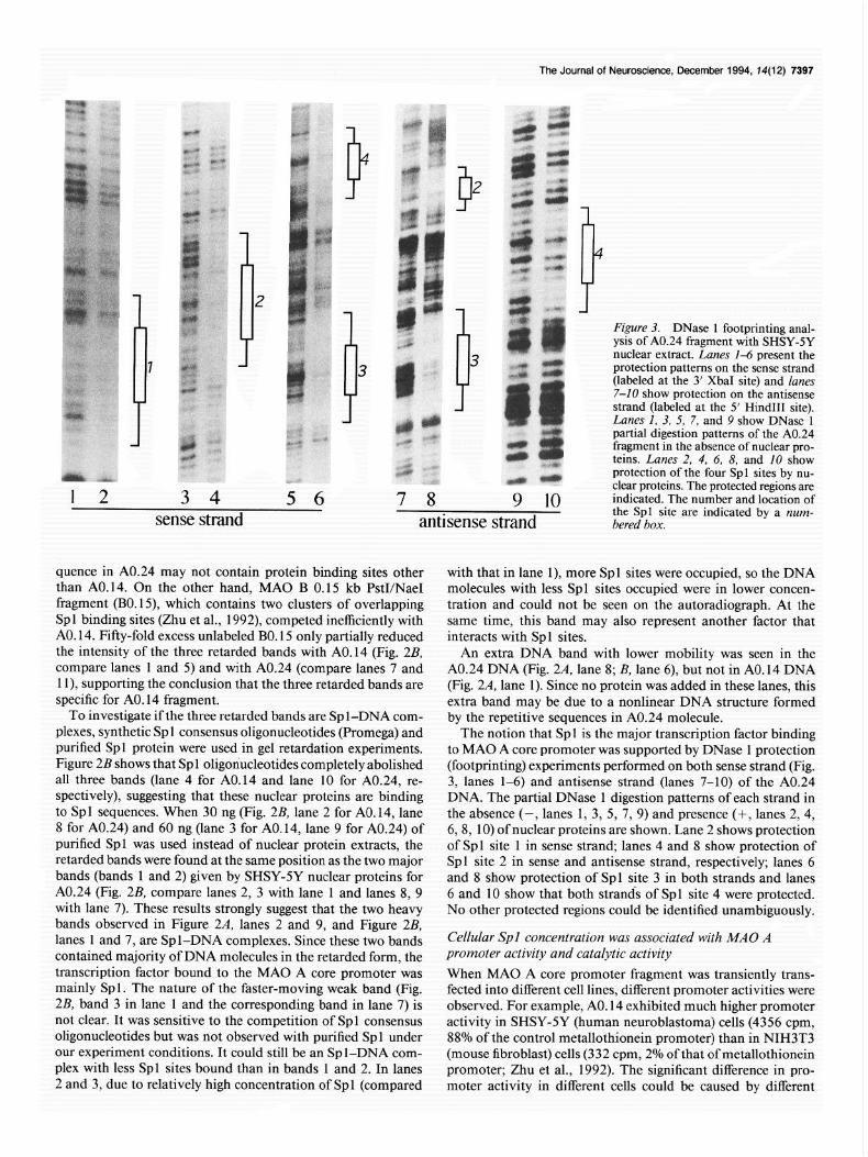
The Journal of Neuroscience, December 1994, 14(12) 7397
P
1
Figure 3. DNase 1 footprinting anal- ysis of A0.24 fragment with SHSY-SY nuclear extract. Lanes 1-6 present the protection patterns on the sense strand (labeled at the 3’ XbaI site) and lanes 7-10 show protection on the antisense strand (labeled at the 5’ Hind111 site). Lanes I, 3, 5, 7, and 9 show DNase 1 partial digestion patterns of the A0.24 fragment in the absence of nuclear pro- teins. Lanes 2, 4, 6, 8, and 10 show
r protection of the four Spl sites by nu-
3 4 5 6 7 8 clear proteins. The protected regions are
9 10 indicated. The number and location of
sense strand the Spl site are indicated by a num- antisense strand bered box.
quence in A0.24 may not contain protein binding sites other with that in lane I), more Spl sites were occupied, so the DNA than A0.14. On the other hand, MAO B 0.15 kb PstI/NaeI molecules with less Spl sites occupied were in lower concen- fragment (B0.15) which contains two clusters of overlapping tration and could not be seen on the autoradiograph. At the Spl binding sites (Zhu et al., 1992) competed inefficiently with same time, this band may also represent another factor that AO. 14. Fifty-fold excess unlabeled BO. 15 only partially reduced interacts with Spl sites. the intensity of the three retarded bands with A0.14 (Fig. 2B, An extra DNA band with lower mobility was seen in the compare lanes 1 and 5) and with A0.24 (compare lanes 7 and A0.24 DNA (Fig. 2A, lane 8; B, lane 6), but not in AO. 14 DNA 1 l), supporting the conclusion that the three retarded bands are (Fig. 2A, lane 1). Since no protein was added in these lanes, this specific for AO. 14 fragment. extra band may be due to a nonlinear DNA structure formed
To investigate if the three retarded bands are Sp l-DNA com- by the repetitive sequences in A0.24 molecule. plexes, synthetic Spl consensus oligonucleotides (Promega) and The notion that Spl is the major transcription factor binding purified Spl protein were used in gel retardation experiments. to MAO A core promoter was supported by DNase 1 protection Figure 2B shows that Spl oligonucleotides completely abolished (footprinting) experiments performed on both sense strand (Fig. all three bands (lane 4 for AO. 14 and lane 10 for A0.24, re- 3, lanes l-6) and antisense strand (lanes 7-10) of the A0.24 spectively), suggesting that these nuclear proteins are binding DNA. The partial DNase 1 digestion patterns of each strand in to Spl sequences. When 30 ng (Fig. 2B, lane 2 for AO. 14, lane the absence (-, lanes 1, 3, 5, 7, 9) and presence (-t, lanes 2, 4, 8 for A0.24) and 60 ng (lane 3 for A0.14, lane 9 for A0.24) of 68, 10) of nuclear proteins are shown. Lane 2 shows protection purified Spl was used instead of nuclear protein extracts, the of Spl site 1 in sense strand, lanes 4 and 8 show protection of retarded bands were found at the same position as the two major Spl site 2 in sense and antisense strand, respectively; lanes 6 bands (bands 1 and 2) given by SHSY-5Y nuclear proteins for and 8 show protection of Spl site 3 in both strands and lanes A0.24 (Fig. 2B, compare lanes 2, 3 with lane 1 and lanes 8, 9 6 and 10 show that both strands of Spl site 4 were protected. with lane 7). These results strongly suggest that the two heavy No other protected regions could be identified unambiguously. bands observed in Figure 2A, lanes 2 and 9, and Figure 2B, lanes 1 and 7, are Spl-DNA complexes. Since these two bands Cellular Spl concentration was associated with MAO A contained majority of DNA molecules in the retarded form, the promoter activity and catalytic activity transcription factor bound to the MAO A core promoter was When MAO A core promoter fragment was transiently trans- mainly Spl. The nature of the faster-moving weak band (Fig. fected into different cell lines, different promoter activities were 2B, band 3 in lane 1 and the corresponding band in lane 7) is observed. For example, AO. 14 exhibited much higher promoter not clear. It was sensitive to the competition of Spl consensus activity in SHSY-5Y (human neuroblastoma) cells (4356 cpm, oligonucleotides but was not observed with purified Spl under 88% of the control metallothionein promoter) than in NIH3T3 our experiment conditions. It could still be an Spl-DNA com- (mouse fibroblast) cells (332 cpm, 2% of that of metallothionein plex with less Spl sites bound than in bands 1 and 2. In lanes promoter; Zhu et al., 1992). The significant difference in pro- 2 and 3, due to relatively high concentration of Spl (compared moter activity in different cells could be caused by different

7399 Zhu et al. l MAO A Bidirectional Promoter
1 2 3 4 5 6 7 8 9 10 11 labeled AO. 14 labeled A024 labeled Spl oligo
1 2
Figure 4. Sp 1 and MAO A mRNA concentration in different cells. A, Comparison of Spl concentration in different cells-gel retardation as- say. Lane 1 shows free DNA of ‘*P-end labeled AO. 14 fragment. Same amount (1.6 pg of total protein) nuclear extracts from SHSY-SY cells (lane 2), NIH3T3 cells (lane 3), and Caco2 cells (lane 4) was incubated with the labeled AO. 14 fragment. Lane 5 shows free DNA of 3ZP-end labeled A0.24 fragment. Same amount (1.6 pg oftotal protein) ofnuclear extracts from SHSY-SY cells (lane 6), NIH3T3 cells (lane 7), and Caco2 cell (lane 8) was incubated with the labeled A0.24 fragment. Lanes 9- 11 show Spl concentration measured by T4 polynucleotide kinase- labeled Spl consensus oligonucleotides in 1.6 pg of nuclear extracts from SHSY-5Y cells (lane 9), NIH3T3 cells (he IO), and Caco2 cells (lane I I). B, Comparison of MAO A mRNA concentration in SHSY- 5Y (lane I) and Caco2 (lane 2) cells determined by Northern analysis as described in Materials and Methods. The blot was exposed for 3 d at - 80°C with an intensifying screen. 18s rRNA was used as a control.
concentration of transcription factors, either activators or re- pressors, in different cells.
To study the effect of transcription factor concentration on MAO A promoter activity, nuclear extracts were made from SHSY-SY and NIH3T3 cells and were used to perform gel re- tardation experiments with A0.14 fragment. When equal amounts of nuclear proteins from these cells were incubated with radiolabeled AO. 14 fragment, the intensity of Sp 1 binding
in SHSY-SY cells (Fig. 4,4, lane 2) was much stronger than that in NIH3T3 cells (lane 3). This results suggests that the concen- tration of Sp 1 is associated with the promoter activities of AO. 14 measured in these two cell lines. No additional band was ob- served with NIH3T3 nuclear proteins, indicating that the lower promoter activity of AO. 14 observed in NIH3T3 cells may not be due to the presence of an inhibiting transcription factor.
To study if the concentration of different transcription factors is associated with MAO A mRNA and catalytic activity, nuclear proteins were extracted from SHSY-SY cells and Caco2 (human colon carcinoma) cells, which exhibit different MAO A catalytic activities (in SHSY-SY cells, 3.36 nmol/20 min/mg protein; in Caco2 cells, 0.26 nmoV20 min/mg protein). Northern analysis showed that MAO A mRNA level in SHSY-5Y cells (Fig. 4B, lane 1) was higher than in Caco2 cells (lane 2), indicating that MAO A regulation is mainly at the transcription level. Gel retardation showed that the intensity of Spl binding was higher in SHSY-5Y cells (Fig. 4A, lane 2) than in Caco2 cells (lane 4). These results again suggest that MAO A expression may be controlled by the concentration of Spl . No additional band was seen with Caco2 nuclear proteins, indicating that the lower MAO A catalytic activity exhibited by this cell line may not be due to the presence of an inhibiting transcription factor.
In our previous article (Zhu et al., 1992), we reported that the mouse cell line NIH3T3 expresses very high MAO A cat- alytic activity (15.59 nmol/mg/20 min) but showed very low promoter activity for transfected human MAO A promoter frag- ments (332 cpm for A0.14), suggesting that mouse MAO A promoter is different from human. Accordingly, the observed low Spl concentration in NIH3T3 cells, although associated nicely with the low promoter activity of transfected human MAO A promoter fragments, was not associated with mouse MAO A catalytic activity.
The same results were obtained with A0.24 fragment (Fig. 4A, lanes 5-8). The intensity of the Spl bands was stronger in SHSY-5Y cells (lane 6) than in NIH3T3 cells (lane 7), which is associated with higher promoter activity of A0.24 fragment measured in SHSY-5Y cells (2318 cpm) than in NIH3T3 cells (286 cpm). The stronger Spl bands observed in SHSY-5Y cells (lane 6) than in Caco2 cells (lane 8) are also associated with the higher MAO A mRNA level and catalytic activity in SHSY-5Y cells than in Caco2 cells.
Furthermore, the concentration of Spl in these cell lines was measured by using radiolabeled Sp 1 consensus oligonucleotides (lanes 9-l 1). The intensity of the Spl-DNA complex was the highest with SHSY-5Y nuclear proteins (lane 9) compared with that with NIH3T3 (lane 10) and Caco2 (lane 11) nuclear pro- teins. These results indicate that the intensity of the Spl bands indeed represents Spl concentration in these cells.
These results show clearly that there is a positive association between Sp 1 concentration and MAO A promoter and catalytic activity. Thus, cellular Spl concentration seems to be an im- portant controlling factor for MAO A expression.
MAO A core promoter exhibited bidirectional promoter activities that were dlxerentially controlled by four Spl sites The A0.24 fragment, which contains four Spl binding sites, was found to exhibit promoter activity in both orientations. To de- fine the role of each c&element (the four Spl binding sites and GA-rich and TAATAA-containing sequences; see Fig. 1 for the sequences) in promoter activities in two opposite directions, serial 5’ and 3’ deletions were made for the A0.24 fragment (Fig.

The Journal of Neuroscience, December 1994, 14(12) 7399
5' 3'
SS D MAO A core promoter region
u u ++
II(90bp) _ I(gObp) ,
4-3-2 -1
(48 bp) '(42 WI (48 bp) (42 d
s m m S m
s 0 m s
I 100 bp
I
promoter actiuity (74. + SD1 f reverse forward +
(ml 0.24 34 f 5 26 f 4
0.20 190 f 53 21 f 1
0.14 47 f 6 100
0.12 174 f 40 40 f 1
0.09 115 f 5 104 f 9
0.14M 24 f 3 62 f 8
Figure 5. Bidirectional promoter activity of the MAO A core promoter. At the top is the restriction map of the 0.24 kb PvuII/DraII core promoter fragment similar to the shown in Figure 1. The restriction enzyme sites are marked by a single letter as in Figure 1. Ovals represent the location of the four Spl sites (numbered sequentially from 3’ to 5’): Spl sites 2 and 3 are on the same side of the DNA while Spl sites 1 and 4 are on the other side of the DNA (see text for explanations). The open oval represents a mutated Spl site. The TAATAA sequence (TA) is shown by the crosshatched box and the GA-rich sequences (GA) are shown by checkered boxes. The positions of the two 90 bp repeats (I and ZI) and the four shorter repeats of 48 and 42 bp in length (1-4) are indicated by thin arrows below the map. The direction of thick arrow under its corresponding cis-element [Spl sites l-4 and the GA-rich (GA) sequences] indicates the favorable transcription direction of the element. Theforward arrow under the element GA means the element favors the forward promoter activity; the rearward arrows under Spl sites 1 and 4 mean these elements favor the reverse promoter activity. The double-headed arrows under Spl sites 2 and 3 indicate that these two sites promote both forward and reverse transcription. The sizes of the DNA fragments (kb) used in promoter activity measurements are shown at their 3’ end. Fragment 0.144 (the fragment at the bottom) is the same as fragment 0.14 except that Spl site 2 was mutated from CCCGCCC to CCCGAAC. The symbol PCR at the 5’ end of fragment 0.20 indicates that the 5’ end of this fragment was produced by PCR. The forward and the reverse promoter activity are expressed as percentage of the forward promoter activity of the fragment 0.14, which is defined as lOO%, with SD given.
5). Each fragment was then linked, in both orientations, to the human growth hormone reporter gene in promoterless vector pOGH and pOGHR (see Materials and Methods). These con- structs were transfected into SHSY-SY cells. The activities mea- sured in both orientations were presented as the percentage of the forward activity of AO. 14 fragment, which was defined as 100%.
As shown in Figure 5, the A0.24 fragment exhibited similar promoter activity in both orientations (26% and 34% activity for forward and reverse activity, respectively). A 5’ deletion that removed the TAATAA-containing sequences 5’ of the two 90 bp repeats (construct 0.20) had little effect on the forward pro- moter activity (from 26% to 27%), but increased significantly the reverse promoter activity (from 34% to 190%), suggesting that the TAATAA-containing sequence inhibits the reverse ac- tivity. Further deletion of Spl site 4 increased the forward ac- tivity about four times (from 27% to 100%; see fragments 0.20 and 0.14) but decreased the reversed activity about fourfold (from 190% to 47%), indicating that site 4 is important for the reverse activity but downregulates the forward activity.
A 3’ deletion that removed the GA-rich sequence 3’ of the two 90 bp repeats decreased forward activity of AO. 14 to 40% (compare fragments 0.12 and 0.14) but increased the reverse activity by more than threefold (from 47% to 174%), suggesting that the GA-rich sequence enhanced transcription toward the forward direction but inhibited the reverse promoter activity. Since the transcription initiation site mapped with 5 RACE is located at the center of this sequence, it is possible that this
sequence enhances transcription and at the same positions ini- tiation site, like the reported transcription element Initiator (Smale and Baltimore, 1989) and HIP1 (Means and Farnham, 1990). Further removal of Sp 1 site 1 from fragment 0.12 dou- bled the forward activity (from 40% to 104%; see fragments 0.12 and 0.09) but decreased the reverse activity significantly (from 174% to 1 15%).
Mutation of site 2 from CCCGCCC to CCCGAAC decreased both the forward activity (from 100% to 62%; compare frag- ments 0.14 and 0.14M) and the reverse activity (from 47% to 24%) of the AO. 14 fragment, indicating that Spl site 2 promotes transcription in both orientations. Spl sites 2 and 3 contained in a 90,bp SmaI/SmaI fragment (0.09) promoted transcription in both orientations with approximately equal efficiency (115% and 104% for the reverse and forward activity, respectively).
These results are summarized with thick arrows under each c&element in the restriction enzyme map in Figure 5. The two central Spl sites (2 and 3) drive transcription in both orienta- tions with similar efficiency. The forward activity, which is for the expression of the MAO A gene, is downregulated by Spl sites 1 and 4, but is enhanced by the GA-rich sequence proximal to the MAO A gene. Their reverse promoter activity, which might drive expression of an unidentified gene, is increased by Spl sites 1 and 4, but downregulated by the GA-rich sequence at the 3’ end and the TAATAA-containing sequence at the 5’ end. Thus, the promoter activity of Spl sites 2 and 3 for each orientation has its own means for positive and negative regu- lation. The balanced effects of all these elements are the nearly

7400 Zhu et al. l MAO A Bidirectional Promoter
Figure 6. Northern analysis showing that the sequence upstream of MAO core promoter is expressed. A shows that two genes, the MAO A gene and a pos- sible new gene, are transcribed by the bidirectional MAO A core promoter (the box labeled 0.24). The opposite arrows from the core promoter represent the opposite promoter activities. A 1.2 kb PstI (Ps)/PvuII (P) fragment 5’ of the core promoter was used as a DNA probe to detect the reverse transcripts. B, Northern analysis. PolyA+ RNA from SHSY-5Y (lane I) and human placenta (lane 2) cells was hybridized with the 32P-labeled DNA probe shown in A. The 10 kb, 6 kb, and 3 kb bands in placenta RNA are marked. The residue 28s and 18s rRNAs in both RNA samples are also indicated. The blot was exposed for 20 d at -80°C with an intensifying screen.
A F?3 PP D
5’
B
equal forward (26%) and reverse (34%) promoter activities by fragment A0.24, which contains all these elements.
Transient transfection assay in SHSY-SY and NIH3T3 cells showed that the reverse promoter activity of the AO. 14 fragment was higher in SHSY-SY cells (688 cpm, 14% of the control metallothionein promoter) than in NIH3T3 cells (58 cpm, 1% of the control metallothionein promoter). Thus, the reverse pro- moter activity is also dependent on the concentration of Spl .
The sequence upstream of MAO A core promoter was expressed endogenously To study the possible physiological significance of the reverse promoter activity, a 1.2 kb PstI/PvuII fragment, immediately 5’ of the core promoter (0.24 kb PvuII/DraII fragment; see Fig. 6A), was isolated, 32P-labeled, and hybridized to a human North- em blot. Positive signals were seen in placenta RNA (not shown). Since our probe also hybridized with ribosome RNAs, to get a clearer picture; polyA+ RNA from placenta and SHSY-5Y cells were used. Figure 68 shows three bands of 3, 6, and 10 kb in placenta polyA+ RNA (lane 2), but not in SHSY-5Y RNA (lane 1). These bands demonstrate that the DNA sequence upstream of MAO A core promoter is expressed endogenously. Very likely these upstream transcripts were from the other strand of DNA (relative to MAO A transcripts) by the reverse promoter activity of MAO A core promoter. In other words, MAO A core pro- moter may endogenously transcribe a new gene upstream of MAO A.
Previously we reported that it is composed of two 90 bp repeats, each containing two Spl binding sites (Zhu et al., 1992). It is interesting that the core promoter sequences can be further di- vided into four imperfect repeats, each containing a Spl site at corresponding position. In addition, the sequence 3’ of the four repeats, from nucleotide -88 to -52 (the 3’ C of the sequence CCCACCC, see Fig. lC), exhibits lower but still significant sequence homology with the four repeats. Due to these repeating sequences, the boundary of the two 90 bp repeats is somewhat arbitrary. In fact, the two 90 bp repeats shown in this report are shifted to the 5’ direction by 9 bp from the ones we published previously in order to coincide with the five shorter repeats presented in this report. The base pair -268 seems to be the 5’ limit of the repeating structure; from here down, any segment of DNA will be homologous to the DNA segment 90 bp down- stream, until the 3’ limit at -52 is reached.
Discussion
With the more sensitive 5’ RACE techniques, we have in- vestigated MAO A transcription initiation sites in various hu- man tissues. The transcription initiation site was found at - 73 and was the same as the cDNA start sites in all human tissues tested. This initiation site is downstream of the Spl binding sequences in MAO A core promoter and seems to be the ini- tiation site for the promoter we detected in the two 90 bp repeat region. The upstream initiation sites we mapped in SHSY-5Y cells with primer extension experiments may be derived from an upstream CCAAT-TATA type promoter that operates only under certain conditions, as we suggested previously (Zhu et al., 1992). In that experiment, the -73 initiation site was in the low-molecular-weight region, thus could not be seen clearly.
This report describes our further studies on human MAO A With similar anchor-PCR techniques, Denney’s group mapped core promoter and provides a number of interesting new results. multiple initiation sites between - 30 and - 140 in human brain The structure of MAO A core promoter is highly organized. and colon tumor cells, with the major initiation sites between
+- 10kb
6 6kb
+- 3kb

The Journal of Neuroscience, December 1994, 74(12) 7401
-30 and -40 (Denney et al., 1993). Thus, the initiation sites mapped by the two laboratories with similar techniques are in the same broad region at the 3’ end of MAO A core promoter, supporting the key role of the Spl sites in the core promoter for MAO A expression.
Sequence homology data show that the similarity between the two 90 bp repeats (81%) is higher than that between the four shorter repeats (74% if the residues present in at least three of the four repeats are considered conserved; 50% if the residues present in all four repeats are considered conserved; the extra 6 bp in the 48 repeats were not included in homology calcula- tion). It is tempting to postulate that the present MAO A core promoter was formed by a recent duplication of a 90 bp segment in the prototype of MAO A promoter. Such duplication in- creased the number of Spl sites in the promoter, thus rendering the present promoter more resistant to the effect of mutational destruction ofits Spl sites. This redundant structure ofthe MAO A core promoter implies that MAO A expression may not be easily abolished completely by a few point mutations in the promoter. Therefore, complete loss of MAO A catalytic activity may be caused by mutations or deletions of the coding sequenc- es, such as a point mutation resulted in the premature termi- nation of MAO A protein found in the Dutch kindred (Brunner et al., 1993b).
The four Sp 1 sites created by duplication process are arranged in special ways: the central two Spl sites (sites 2 and 3) are 42 bp apart, so they are on the same side of the DNA (each turn of B-DNA contains 10.5 bp; Wang, 1979) and the two flanking Spl sites (sites 1 and 4) are 48 bp from site 2 or 3; thus, they are on the other side of the DNA (see Fig. 5). These special arrangements, together with other c&elements (like the 3’ GA- rich and 5’ TAATAA-containing sequences), provide additional means for regulation, which is demonstrated in deletion analysis shown in Figure 5. Therefore, different levels of MAO A ex- pression may be caused by the alteration of the promoter se- quence, or the change of concentration of the relevant tran- scription factor (Spl) in the cells.
The following evidence obtained from gel retardation, DNase 1 footprinting experiments, and promoter activity measure- ments strongly suggests that Spl is the major transcription factor interacting with MAO A core promoter: (1) the binding of nu- clear proteins to MAO A promoter fragments was effectively competed by Spl consensus oligonucleotides, (2) only the Spl sites were clearly protected from DNase 1 digestion, (3) purified Spl gave identical retarded bands as nuclear proteins, (4) the promoter activity of the A0.14 (or A0.24) fragment, MAO A mRNA level, and catalytic activity are associated with the cel- lular concentration of Spl (also see below).
It has been reported that several other transcription factors also bind to Spl sites. However, these transcription factors can be excluded to be the binding factor in our experiments. For example, AP-2 consensus oligonucleotides did not compete with the labeled A0.14 or A0.24 fragment (figure not shown), indi- cating that the binding factor is not AP-2. In addition, AP-2 has a molecular weight of 52 kDa (Mitchell et al., 1987) different from that of Spl (95 and 105 kDa; Michael et al., 1986). If AP-2 were the binding factor, then the mobility of the retarded bands would be different from Spl-DNA complexes we ob- served. The same reasoning can be used to exclude ETF (120 kDa; Kageyama et al., 1988) EGR-1 (80 kDa; Cao et al., 1990) EGR-2 (43 kDa; Joseph et al., 1988) and WTl (49-51 kDa; Telerman et al., 1992). Furthermore, AP2, ETF, and EGR-1
are induced by phorbol esters, but the activity of MAO A frag- ments from the core promoter region was not (data not shown). The molecular weight of GCF (100 kDa; Kageyama and Pastan, 1989) is very similar to Sp 1. However, the observed positive association between MAO activity and the amount of bound nuclear proteins in gel retardation experiments may argue against GCF to be the major bound species, because GCF has been shown to be a negative regulator. On the other hand, the present report does not rule out the possibility that other transcription factors may bind to these Spl sites in MAO A core promoter and give the same binding patterns as Sp 1, so are indistinguish- able in gel retardation and footprinting experiments. Since we have tested only a limited number of cell lines, the possibility cannot be excluded that in other cells or tissues, different tran- scription factors may interact with MAO A core promoter and regulate MAO A expression. The sequences outside of the core promoter may also play a role. Nonetheless, in the cell lines we so far studied, Spl seems to be the major transcription factor interacting with MAO A core promoter.
Sp 1 is a general transcription factor found in all cells of higher animals and is required for GC box containing promoters. Since many housekeeping gene promoters are GC rich and contain Spl binding sites, the existence of Spl should be universal. On the other hand, Spl concentrations changes dramatically (as high as 1 OO-fold) from tissue to tissue and during development (Saffer et al., 1991), suggesting that Spl has a regulatory role. In other words, Spl might be a limiting factor for many pro- moters. This hypothesis is supported by the elevated transcrip- tion of Spl-depending genes when cellular Spl concentration becomes higher (Saffer et al., 1990). The positive association between cellular Spl concentration and MAO A promoter ac- tivity (both forward and reverse), mRNA level, and catalytic activity we observed in cultured cell also suggests that cellular Spl concentration is an important controlling factor for the expression of human MAO A gene. It will be interesting to investigate further whether the different MAO A catalytic ac- tivities in different tissues, developmental stages, and diseased states are also regulated by Spl concentration.
Our transient transfection assays demonstrate that the MAO A core promoter has bidirectional activities. Promoters func- tioning in both orientations are found initially in microorgan- isms and mitochondrial DNA. In recent years, bidirectional promoters in higher eukaryotic organisms have been reported. Some of these bidirectional promoters are housekeeping genes and contain Spl binding sites, such as the promoters of mouse dihydrofolate reductase (dhfr) gene, mouse thymidine kinase (TK) gene (Weichselbraun et al., 1990) and human hypoxan- thine phosphoribosyl transferase (HPRT) gene (Johnson and Friedman, 1990). However, bidirectional promoter is not lim- ited to the Sp 1 type. In mouse heavy chain immunoglobin pro- moter VH441, the bidirectional transcription is driven by a octomer element (ATGCAAAT) and controlled by a TATA- like element at each side of the element (Nguyen et al., 1991). For some of such bidirectional promoters, in addition to their “normal” transcripts, the endogenous reverse transcripts have also been detected, like the mouse heavy chain immunoglobin promoter VH441 (Nguyen et al., 199 l), murine cy- 1 and-2 (IV) collagen transcripts (Burbello et al., 1988) human proliferating cell nuclear antigen (PCNA) gene promoter (Rizzo et al., 1990) chicken skeletal cu-actin gene promoter (Grichnik et al., 1988) mouse HTF9 housekeeping promoter (Somma et al., 199 l), and the most intensively studied dihydrofolate reductase promoter

7402 Zhu et al. l MAO A Bidirectional Promoter
(Crouse et al., 1985; Famham et al., 1985; Linton et al., 1989; Shimada et al., 1989; Smith et al., 1990; Ciudad et al., 1992; Fujii et al., 1992). The increasing list of physiologically func- tional eukaryotic bidirectional promoters indicates that pro- moters transcribing both strands of DNA really work in higher animals, including human being. The difficulties in detecting some reverse transcripts at least in some cases can be due to the fast turnover of these transcripts (Grichnik et al., 1988), or the reverse activity works only under special circumstances. On the other hand, the bidirectional promoters are often detected with isolated DNA fragments; thus, the DNA elements and endogenous promoter environments that prevent the reverse transcription (which may not always be beneficial) in the cells may be lost, so the reverse promoter activity detected in trans- fection assay may not function similarly in the intact genome in the cells. For example, human HPRT promoter exhibits bi- directional promoter activities in transfection assay, but no en- dogenous reverse promoter activity has been detected (Johnson and Friedman, 1990).
Our data show that the sequence upstream of MAO A core promoter is expressed endogenously, implying that the bidirec- tional promoter activities exhibited by the core promoter might transcribe another gene in addition to MAO A. So far, the up- stream transcripts have been detected in placenta only, sug- gesting that there may be mechanisms controlling the relative expression level of the two genes. Thus, in addition to the role of Sp 1 in MAO A expression, the study on how the two opposite promoter activities are regulated might provide further insight for the mechanism of MAO A regulation.
Analysis of the mouse dhfr promoter region: existence of a divergently transcribed gene. Mol Cell Biol 5: 1847-l 858.
Denney RM, Dave SK, Waguespack A (1993) Identification of likely transcription initiation sites for the human MAO A gene: new evi- dence from PCR-assisted primer extension and RNase protection assays. Slide presentation at the 23rd annual meeting of Society for Neuroscience, Washington, DC, November.
Dignam JD, Lebovites RM, Roeder RG (1983) Accurate transcription initiation by RNA polymerase II in a soluble extract from isolated mammalian nuclei. Nucleic Acids Res 11: 1475-1489.
Donnelly CH, Murphy DL (1977) Substrate- and inhibitor-related characteristics of human platelet monoamine oxidase. Biochem Phar- macol 26:853-858.
Egashira T, Yamanaka Y (198 1) Further studies on the synthesis of A-form of MAO. Jpn J Pharmacol 31:763-770.
Famham PJ, Abrams JM, Schimke RT (1985) Opposite-strand RNAs from the 5’ flanking region ofthe mouse dihydrofolate reductase gene. Proc Nat1 Acad Sci USA 82:3978-3982.
Fowler JB, Macgregor RR, Wolf AP, Amett CD, Dewey SL, Schlyer D, Christman D, Logan J, Smith M, Sachs H, Aquilonius SM, Bjjurling P, Halldin C, Hartvig P, Leenders KL, Lundqvist H, Oreland L, Stalnacke C-G, Langstrom B (1987) Mapping human brain mono- amine oxidase A and B with “C-labelled suicide inactivators and PET. Science 235148 l-485.
Fritz RR, Abel1 CW, Pate1 NT, Gessner W, Brossi A (1985) Metab- olism of the neurotoxin in MPTP by human liver monoamine oxidase B. FEBS Lett 186:224-228.
Fujii H, Shinya E, Shimada T (1992) A CC box in the bidirectional promoter is essential for expression of the human dihydrofolate re- ductase and mismatch repair protein genes. FEBS Lett 3 14:33-36.
Garrick NA, Murphy DL (1982) Monoamine oxidase type A: differ- ences in selecti& towards norepinephrine compared-to serotonin. Biochem Pharmacol31:40614066.
Grichnik JM, French BA, Schwartz RJ (1988) The chicken skeletal alpha-actin gene promoter region exhibits partial dyad symmetry and a capacitv to drive bidirectional transcriution. Mol Cell Biol 8:4587- 45917. .
References Ausubel FM, Brent R, Kingston RE, Moore DD, Seidman JG, Smith
JA, Struhl K, eds (1989) Current protocols in molecular biology, pp 4.2.14.2.5. New York: Wiley.
Bach AWJ, Lan NC, Johnson DL, Abel1 CW, Bembenek ME, Kwan S-W, Seeberg PH, Shih JC (1988) cDNA cloning of human mono- amine oxidase A and B: molecular basis of differences in enzymatic properties. Proc Nat1 Acad Sci USA 85:4934-4938.
Brunner HG, Nelen MR, van Zandvoort P, Abeling NGGM, van Gen- nip AH, Wolters EC, Kuiper MA, Ropers HH, van Oost BA (1993a) X-linked borderline mental retardation with prominent behavioral disturbance: phenotype, genetic localization, and evidence for dis- turbed monoamine oxidase metabolism. Am J Hum Genet 52:1032- 1039.
Brunner HG, Nelen M, Breakefield X0, Ropers HH, van Oost BA (199313) Abnormal behavior associated with a ooint mutation in the &m&al gene for monoamine oxidase A. Science 262:578-580.
Burbelo PD, Martin GR, Yamada Y (1988) Alpha 1 (IV) and alpha 2 (IV) collagen genes are regulated by a bidirectional promoter and a shared enhancer. Proc Nat1 Acad Sci USA 85:9679-9682.
Cao X, Koski RA, Gashler A, McKieman M, Morris CF, Gaffney R, Hay RV, Sukhatme VP (1990) Identification and characterization ofthe EGR- 1 gene product, a DNA binding zinc finger protein induced by differentiation and growth signals. Mol Cell Biol 10: 193 l-l 939.
Chen K, Wu HF, Shih JC (1993) The deduced amino acid sequences of human platelet and frontal cortex monoamine oxidase B are iden- tical. J Neurochem 6 1: 187-l 90.
Chen S, Shih JC, Xu QP (1984) Interaction of N-(2-nitro-4-azido- phenyl)-serotonin with two types of monoamine oxidase in rat brain. J Neurochem 43:1680-1687.
Chiba K, Trevor A, Castagnoli N (1984) Metabolism of the neurotoxic tertiary amine, MPTP by brain monoamine oxidase. Biochem Bio- phys Res Commun 120:574-578.
Ciudad CJ, Morris AE, Jeng C, Chasin LA (1992) Point mutation analysis of the hamster dihydrofolate reductase minimum promoter. J Biol Chem 267:3650-3656.
Crouse GF, Leys EJ, McEwan RN, Frayne EG, Kellems RE (1985)
Grimsby J, Lan NC, Neve R, Chen K, Shih JC (1990) Tissue distri- bution of human monoamine oxidase A and B mRNA. J Neurochem 55:1166-l 169.
Grimsby J, Chen K, Wang LJ, Lan NC, Shih JC (199 1) Human mono- amine oxidase A and B genes exhibit identical exon-in&on organi- zation. Proc Nat1 Acad Sci USA 88:3637-3641.
Johnston JP (1968) Some observations upon a new inhibitor of mono- amine oxidase in brain tissue. Biochem Pharmacol 17: 1285-l 297.
Johnson P, Friedmann T (1990) Limited bidirectional activity of two house keeping gene promoters: human HPRT and PGK. Gene 88: 207-213.
Joseph LJ, LeBeau MM, Jammison GA, Acharya S, Shows TB, Rowley JD, Sukhatme VP (1988) Molecular cloning. seauencine. and mao- ping of EGR-2, a human karly growth response gene encoding a pro- tein with “zinc-binding finger” structure. Proc Nat1 Acad Sci USA 85:7164-7168.
Kageyama R, Pastan I (1989) Molecular cloning and characterization of a human DNA binding factor that represses transcription. Cell 59: 815-825.
Kageyama R, Merlin0 GT, Pastan I (1988) A transcription factor active on the epidermal growth factor receptor gene. Proc Nat1 Acad Sci USA 85:5016-5020.
Knoll J, Magyar K (1972) Some puzzling pharmacological effects of monoamine oxidase inhibitors. In: Advances in biochemical psycho- pharmacology, Vo15, Monoamine oxidase-new vista (Costa E, San- dler M, eds), pp 393408. New York: Raven.
Lan NC, Heinzmann C, Gal A, Klisak I, Orth U, Lai E, Grimsby J, Sparkes R, Mohandas T, Shih JC (1989) Human monoamine ox- idase A and B genes map to Xpll.23 and are deleted in a patient with Norrie disease. Genbmics &552-559.
Lewinsohn R. Glover W. Sandler M (1980) Develoomenr of benzvl- amine oxidase and monoamine oxidase A and B in man. Biochem Pharmacol29:1221-1230.
Linton JP, Yen J-YJ, Selby E, Chen Z, Chinsky JM, Liu K, Kellems RE, Crouse GF (1989) Dual bidirectional promoters at the mouse dhfr locus: cloning and characterization of two mRNA classes of the divergently transcribed Rep-l gene. Mol Cell Biol 9:3058-3072.
Means AL, Famham PJ (1990) Transcription initiation from the dihy-

The Journal of Neuroscience, December 1994, 14(12) 7403
drofolate reductase promoter is positioned by HIP1 binding at the initiation site. Mol Cell Biol l&653-661.
Michael RB, Kadonaga JT, Bell SP, Tjian R (1986) Purification and biochemical characterization of the promoter-specific transcription factor, Spl. Science 23447-52.
Mitchell PJ, Wang C, Tjian R (1987) Positive and negative regulation of transcription in vitro: enhancer-binding protein AP-2 is inhibited by SV40 T antigen. Cell 50:847-861.
Nguyen QT, Doyen N, d-Andon MF, Rougeon F (199 1) Demonstra- tion of a divergent transcript from the bidirectional heavy chain im- munodobin aromoter VH44 1 in B-cells. Nucleic Acids Res 19:5339- 5344.- -
Ozelius L, Hsu Y-PP, Bruns G, Powell JF, Chen S, Weyler W, Utterback M, Zucker D. Haines J, Trofalter JA, Conneally PM, Gusella JF, Breakefield XG (1988) ‘Human monoamine oxidase gene (MAOA)i chromosome position (Xp2 1 -p 11) and DNA polymorphism. Geno- mics 3:53-58.
Rizzo MG, Ottavio L, Travali S, Chang CD, Kaminska B, Baseraa R (1990) The promoter of human proliferating cell nuclear antigen (PCNK) acne is bidirectional. Exn Cell Res 188:286-293.
Sailer JD,‘J&kson SJ, Thurston SJ (1990) SV40 stimulates expression of the trans-act factor Spl at the mRNA level. Genes Dev 4:659- 666.
Saffer JD, Jackson SP, Annarella MB (199 1) Developmental expres- sion of Spl in the mouse. Mol Cell Biol 11:2189-2 199.
Shimada T, Fujii H, Lin H (1989) A 165-base pair sequence between the dihydrofolate reductase gene and the diversely transcribed up- stream gene is sufficient for bidirectional transcription. Mol Cell Biol 8:45874597.
Smale ST, Baltimore D (1989) The “Initiator” as a transcription con- trol element. Cell 57: 103-l 13.
Smith ML, Mitchell PJ, Grouse GF (1990) Analysis of the mouse Dhfr/Rep-3 major promoter region by using linker scanning and in- ternal deletion mutations and DNase 1 footprinting. Mol Cell Biol 10:6003-6012.
Somma MP, Gambino I, Lavia P (199 1) Transcription factors binding to the mouse HTF9 housekeeping promoter differ between cell types. Nucleic Acids Res 19:445 14458.
Telerman A, Dodemont H, Degraef C, Galand P, Bauwens S, Van- Oostveldt P, Amson RB (1992) Identification of the cellular protein encoded by the human Wilms’ tumor (WTl) gene. Oncogene 7:2545- 2548.
Thorpe LW, Westlund KN, Kochersperger LM, Abel1 CW, Denney RM (1987) Immunocytochemical localization of monoamine oxidase A and B in human peripheral tissues and brain. J Histochem Cytochem 35:217-236.
Tipton KF, O’Carroll AM, McCrodden JM (1987) The catalytic be- havior of monoamine oxidase. J Neural Transm [Suppl] 23:25-35.
von KorII’RW (1979) Monoamine oxidase: unanswered questions. In: Monoamine oxidase: structure, function, and altered functions (Single TP, von Korff RW, Murphy DL, eds), pp l-7. New York: Academic.
Wang JC (1979) Helical reneat of DNA in solution. Proc Nat1 Acad Sc;USA‘76:2&203. -
Weichselbraun I, Ogris E, Wintersberger E (1990) Bidirectional pro- moter activity of the 5’ flanking region of the mouse thymidine kinase gene. FEBS Lett 275:49-52.
Westlund KN, Denney RM, Kochersperger LM, Rose RM, Abel1 CW (1985) Distinct monoamine oxidase A and B populations in primate brain. Science 230:18 1-183.
Youdim MBH, Heldman E, Pollard HB, Fleming P, McHugh E (1986) Contrasting monoamine oxidase activity and tyramine induced cat- echolamine release in PC12 and chromaffin cells. Neuroscience 19: 1311-1318.
Yu PH, Hertz L (1982) Differential expression of type A and B mono- amine oxidase of mouse astrocvtes in primarv cultures. J Neurochem 39:1492-1495.
Zhu QS, Grimsby J, Chen K, Shih JC (1992) Promoter organization and activity of human monoamine oxidase (MAO) A and B genes. J Neurosci 1214437-4446.