Embed Size (px)
Citation preview
1
Benign Paroxysmal Positional Vertigo
Cupulolithiasis and Atypical BPPV
Tonya Fuller, [email protected]
Provider Disclaimer• Allied Health Education and the presenter of this
webinar do not have any financial or other
associations with the manufacturers of any products
or suppliers of commercial services that may be
discussed or displayed in this presentation.
• There was no commercial support for this
presentation.
• The views expressed in this presentation are the
views and opinions of the presenter.
• Participants must use discretion when using the
information contained in this presentation.
Review
2
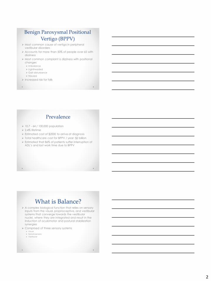
Benign Paroxysmal Positional
Vertigo (BPPV) Most common cause of vertigo in peripheral
vestibular disorders
Accounts for more than 50% of people over 65 with
dizziness
Most common complaint is dizziness with positional
changes
Imbalance
Lightheaded
Gait disturbance
Nausea
Increased risk for falls
Prevalence
10.7 - 64 / 100,000 population
2.4% lifetime
Estimated cost of $2000 to arrive at diagnosis
Total healthcare cost for BPPV / year: $2 billion
Estimated that 86% of patients suffer interruption of
ADL’s and lost work time due to BPPV
What is Balance? A complex biological function that relies on sensory
inputs from the visual, proprioceptive, and vestibular
systems that converge towards the vestibular
nuclei, where they are integrated and result in the
induction of oculomotor and postural stabilization
synergies
Comprised of three sensory systems Visual
Somatosensory
Vestibular
3
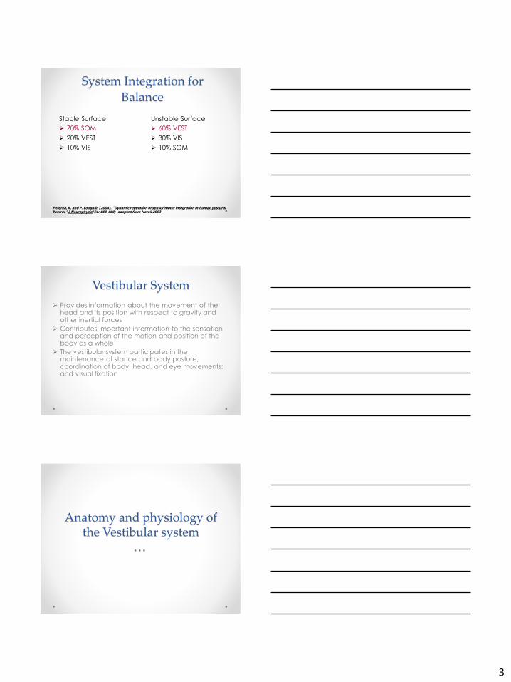
System Integration for
Balance
Stable Surface
70% SOM
20% VEST
10% VIS
Unstable Surface
60% VEST
30% VIS
10% SOM
Peterka, R. and P. Loughlin (2004). "Dynamic regulation of sensorimotor integration in human postural control." J Neurophysiol 91: 000-000; adopted from Horak 2003
Vestibular System
Provides information about the movement of the head and its position with respect to gravity and other inertial forces
Contributes important information to the sensation and perception of the motion and position of the body as a whole
The vestibular system participates in the maintenance of stance and body posture; coordination of body, head, and eye movements; and visual fixation
Anatomy and physiology of the Vestibular system
4
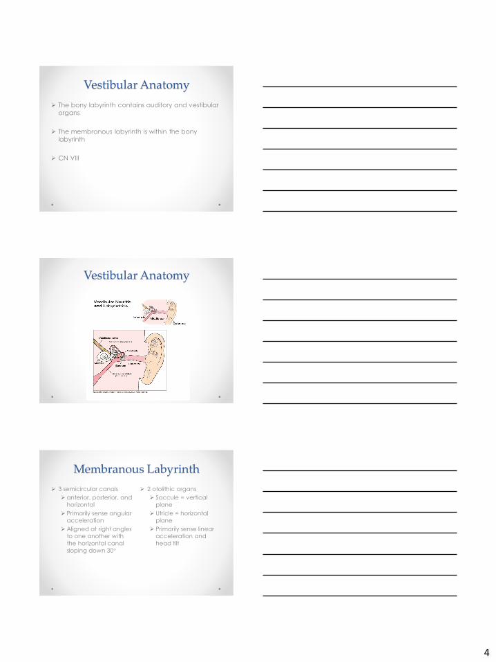
Vestibular Anatomy
The bony labyrinth contains auditory and vestibular
organs
The membranous labyrinth is within the bony
labyrinth
CN VIII
Vestibular Anatomy
Membranous Labyrinth
3 semicircular canals
anterior, posterior, and
horizontal
Primarily sense angular
acceleration
Aligned at right angles
to one another with
the horizontal canal
sloping down 30°
2 otolithic organs
Saccule = vertical
plane
Utricle = horizontal
plane
Primarily sense linear
acceleration and
head tilt
5
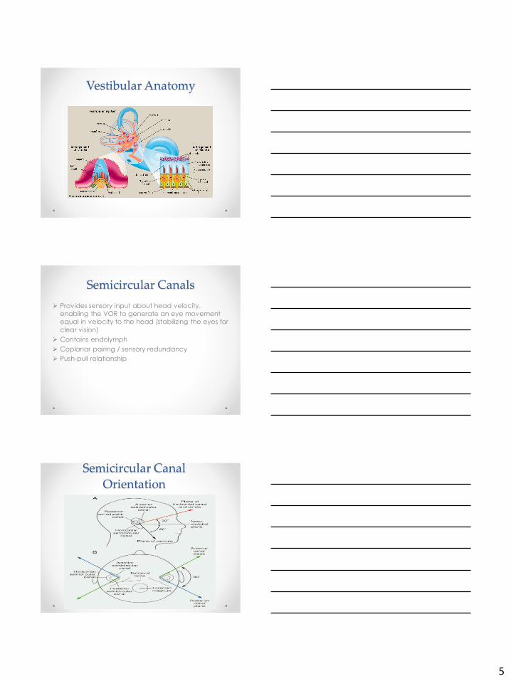
Vestibular Anatomy
Semicircular Canals
Provides sensory input about head velocity,
enabling the VOR to generate an eye movement
equal in velocity to the head (stabilizing the eyes for
clear vision)
Contains endolymph
Coplanar pairing / sensory redundancy
Push-pull relationship
Semicircular Canal
Orientation
6
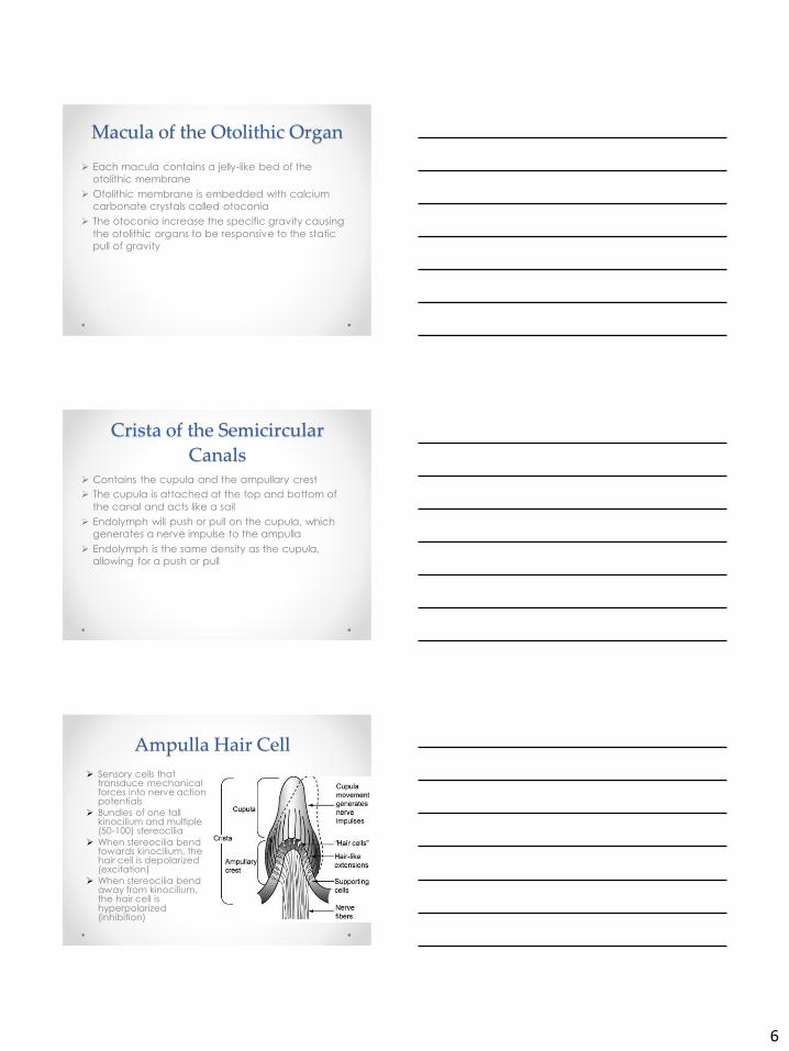
Macula of the Otolithic Organ
Each macula contains a jelly-like bed of the
otolithic membrane
Otolithic membrane is embedded with calcium
carbonate crystals called otoconia
The otoconia increase the specific gravity causing
the otolithic organs to be responsive to the static
pull of gravity
Crista of the Semicircular
Canals Contains the cupula and the ampullary crest
The cupula is attached at the top and bottom of
the canal and acts like a sail
Endolymph will push or pull on the cupula, which
generates a nerve impulse to the ampulla
Endolymph is the same density as the cupula,
allowing for a push or pull
Ampulla Hair Cell
Sensory cells that transduce mechanical forces into nerve action potentials
Bundles of one tall kinocilium and multiple (50-100) stereocilia
When stereocilia bend towards kinocilium, the hair cell is depolarized (excitation)
When stereocilia bend away from kinocilium, the hair cell is hyperpolarized (inhibition)
7
BENIGN PAROXYSMAL
POSITIONAL VERTIGO (BPPV)
Cupulolithiasis
Causes of BPPV
Under the age of 50
Head injury is most common cause
Over the age of 50
Idiopathic
Most common onset between 50 – 70 years
Other potential causes
Degeneration
Prolonged Positioning
Viral
Signs & Symptoms of BPPV
Dizziness with positional changes
rolling over in bed
quick head turns
bending over
Nausea due to excessive dizziness
Loss of balance with gait
Sense of “floating” or “swimming”
Initial onset may produce severe spinning
dizziness lasting hours and nausea / vomiting
8
Forms of BPPV
Canalithiasis
Cupulolithiasis
Cupulolithiasis
Least common
Onset of vertigo/nystagmus is immediate (1 – 2
seconds)
Symptoms persist > 60 seconds or as long as the
patient is in the provoking position
Cupulolithiasis
Mechanism
Otoconia from the utricle adhere to the cupula
and increase the density of the cupula. This
causes an inappropriate deflection of the cupula
when the head and affected ear are positioned
below the horizon. This sends an abnormal signal
resulting in dizziness.
Otoconia can be attached to canal side or
utricle side of the cupula
9
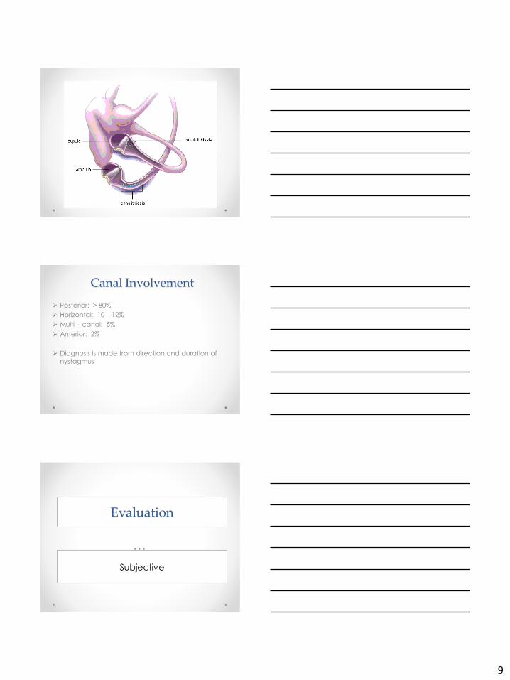
Canal Involvement
Posterior: > 80%
Horizontal: 10 – 12%
Multi – canal: 5%
Anterior: 2%
Diagnosis is made from direction and duration of
nystagmus
Evaluation
Subjective
10
Evaluation
• Subjective
o Chief Complaint / Hx of Illness
o Onset
o Conditions provoking symptoms
o Duration / Severity (of initial onset and of subsequent
episodes)
o Fall History
o Limitations in ADL’s
o Previous Medical History
o Previous Medical / Vestibular Testing
• Information that you get from your subjective evaluation
should drive the objective evaluation
Evaluation
Objective
BPPV Testing
• Dix – Hallpike
• Roll Test
• Head Hanging
11
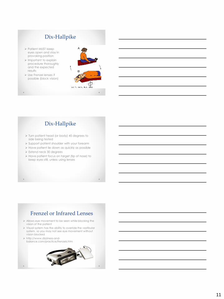
Dix-Hallpike
Patient MUST keep
eyes open and stay in
provoking position
Important to explain
procedure thoroughly
and the expected
results
Use Frenzel lenses if
possible (block vision)
Dix-Hallpike
Turn patient head (or body) 45 degrees to
side being tested
Support patient shoulder with your forearm
Have patient lie down as quickly as possible
Extend neck 30 degrees
Have patient focus on target (tip of nose) to
keep eyes still, unless using lenses
Frenzel or Infrared Lenses Allows eye movement to be seen while blocking the
vision of the patient
Visual system has the ability to override the vestibular
system, so you may not see eye movement without
vision blocked
http://www.dizziness-and-balance.com/practice/frenzels.htm

12
Results
Posterior Canal
Upbeating, torsional nystagmus to affected
side
Anterior Canal
Downbeating, torsional nystagmus to
affected side OR downbeating
Horizontal Canal
If horizontal nystagmus is seen, should
confirm with a roll test
Torsional Nystagmus
Roll Test
Patient MUST keep
eyes open and stay in
provoking position
Patient will
experience vertigo
and nystagmus in
both directions due to
debris moving back
and forth within the
canal
13
Roll Test
Patient lies supine with neck flexed 30 degrees to position horizontal canal perpendicular to horizontal plane
Turn head 90 degrees to affected side as quickly as possible
Have patient focus on target (tip of nose) to keep eyes still
Repeat to other side
If patient is unable to turn head 90 degrees, have them turn body to achieve 90 degrees
Affected side is the side with the strongest nystagmus
Testing Results
Canalithiasis
Geotropic nystagmus (beating towards the
ground)
Cupulolithiasis
Ageotropic nystagmus (beating away from
the ground)
Ageotropic Nystagmus
14
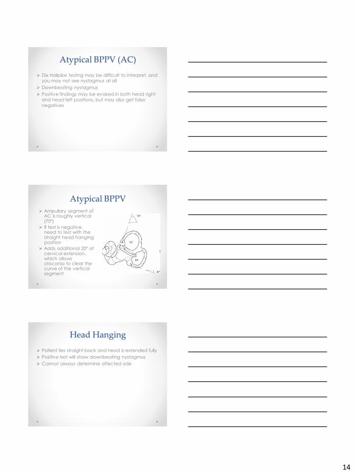
Atypical BPPV (AC)
Dix Hallpike testing may be difficult to interpret, and
you may not see nystagmus at all
Downbeating nystagmus
Positive findings may be evoked in both head right
and head left positions, but may also get false
negatives
Atypical BPPV
Ampullary segment of AC is roughly vertical (70º)
If test is negative, need to test with the straight head hanging position
Adds additional 20º of cervical extension, which allows otoconia to clear the curve of the vertical segment
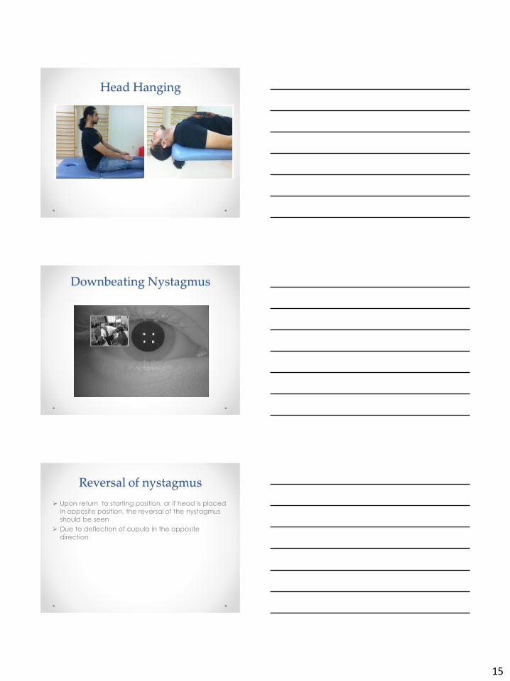
Head Hanging
Patient lies straight back and head is extended fully
Positive test will show downbeating nystagmus
Cannot always determine affected side
15
Head Hanging
Downbeating Nystagmus
Reversal of nystagmus
Upon return to starting position, or if head is placed
in opposite position, the reversal of the nystagmus
should be seen
Due to deflection of cupula in the opposite
direction
16
Treatment
BPPV
Cupulolithiasis
Semont Maneuver (Liberatory)
Modified Semont by Casani for horizontal cupulolithiasis
Cupulolith Repositioning Maneuver (CuRM)
Brandt-Daroff
Other treatments
Head tilt Hopping
Cranial Oscillation
Cupulolithiasis
Semont Maneuver
(Liberatory
Maneuver)
Works by
floating the
debris through
the canal, or
dislodging
debris from the
cupula
17
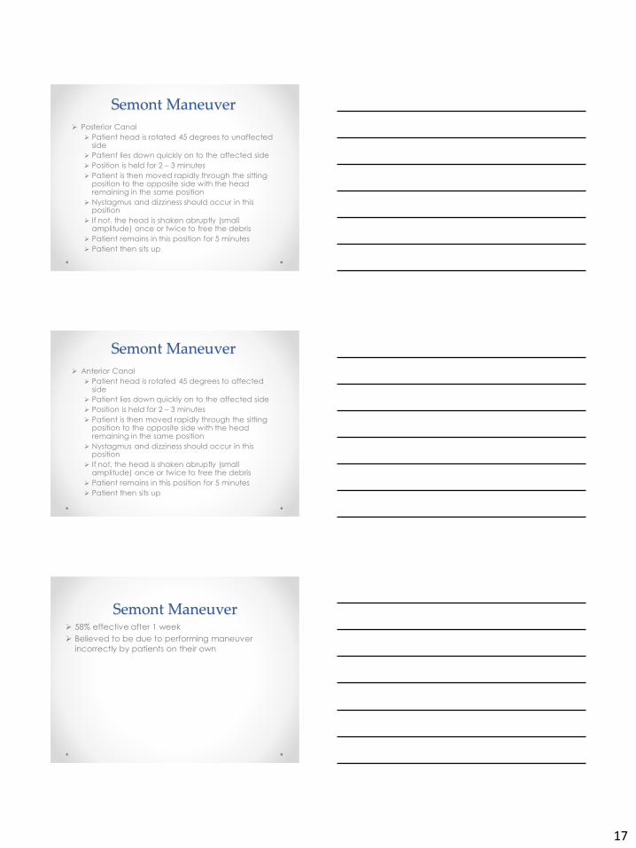
Semont Maneuver
Posterior Canal
Patient head is rotated 45 degrees to unaffected side
Patient lies down quickly on to the affected side
Position is held for 2 – 3 minutes
Patient is then moved rapidly through the sitting position to the opposite side with the head remaining in the same position
Nystagmus and dizziness should occur in this position
If not, the head is shaken abruptly (small amplitude) once or twice to free the debris
Patient remains in this position for 5 minutes
Patient then sits up
Semont Maneuver
Anterior Canal
Patient head is rotated 45 degrees to affected side
Patient lies down quickly on to the affected side
Position is held for 2 – 3 minutes
Patient is then moved rapidly through the sitting position to the opposite side with the head remaining in the same position
Nystagmus and dizziness should occur in this position
If not, the head is shaken abruptly (small amplitude) once or twice to free the debris
Patient remains in this position for 5 minutes
Patient then sits up
Semont Maneuver 58% effective after 1 week
Believed to be due to performing maneuver
incorrectly by patients on their own
18
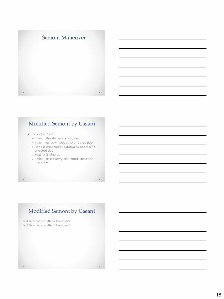
Semont Maneuver
Modified Semont by Casani
Horizontal Canal
Patient sits with head in midline
Patient lies down quickly to affected side
Head is immediately rotated 45 degrees to
affected side
Hold for 2 minutes
Patient sits up slowly and head is returned
to midline
Modified Semont by Casani
80% effective after 2 treatments
90% effective after 3 treatments

19
Modified Semont by Casani
Modified Semont by Casani
Cupulolith Repositioning
Maneuver (CuRM) Treats when otoconia are attached to either canal
side or utricle side
Patient begins in supine position
Head and body are rotated to affected side 135
degrees and oscillation is performed to mastoid
bone for 30 seconds
Head is rotated 45 degrees to the unaffected side
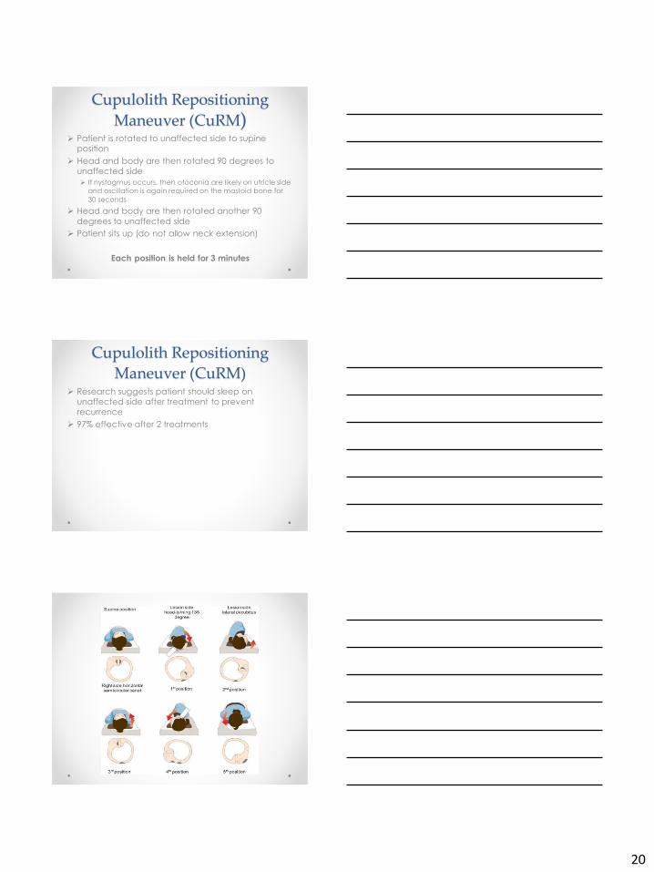
20
Cupulolith Repositioning
Maneuver (CuRM) Patient is rotated to unaffected side to supine
position
Head and body are then rotated 90 degrees to
unaffected side
If nystagmus occurs, then otoconia are likely on utricle side
and oscillation is again required on the mastoid bone for
30 seconds
Head and body are then rotated another 90
degrees to unaffected side
Patient sits up (do not allow neck extension)
Each position is held for 3 minutes
Cupulolith Repositioning
Maneuver (CuRM) Research suggests patient should sleep on
unaffected side after treatment to prevent
recurrence
97% effective after 2 treatments
21
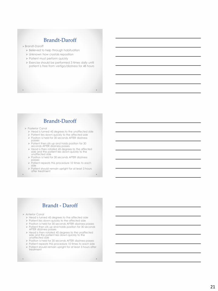
Brandt-Daroff Brandt-Daroff
Believed to help through habituation
Unknown how crystals reposition
Patient must perform quickly
Exercise should be performed 3 times daily until
patient is free from vertigo/dizziness for 48 hours
Brandt-Daroff Posterior Canal
Head is turned 45 degrees to the unaffected side
Patient lies down quickly to the affected side
Position is held for 30 seconds AFTER dizziness passes
Patient then sits up and holds position for 30 seconds AFTER dizziness passes
Head is then rotated 45 degrees to the affected side and the patient lies down quickly to the unaffected side
Position is held for 30 seconds AFTER dizziness passes
Patient repeats this procedure 10 times to each side
Patient should remain upright for at least 3 hours after treatment
Brandt - Daroff
Anterior Canal
Head is turned 45 degrees to the affected side
Patient lies down quickly to the affected side
Position is held for 30 seconds AFTER dizziness passes Patient then sits up and holds position for 30 seconds
AFTER dizziness passes
Head is then rotated 45 degrees to the unaffected side and the patient lies down quickly to the unaffected side
Position is held for 30 seconds AFTER dizziness passes
Patient repeats this procedure 10 times to each side
Patient should remain upright for at least 3 hours after treatment
22
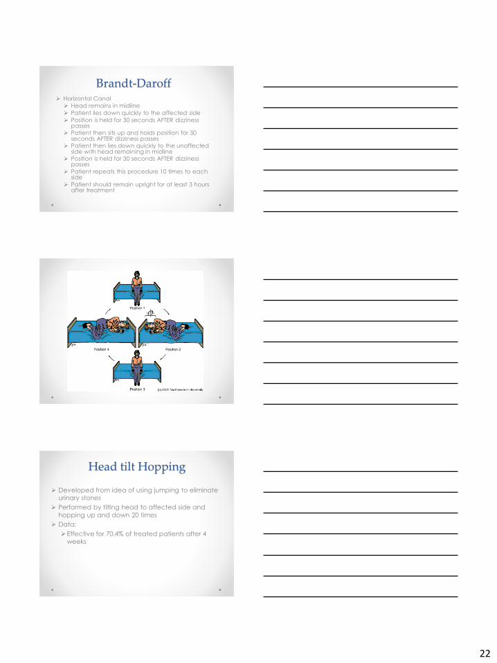
Brandt-Daroff Horizontal Canal
Head remains in midline
Patient lies down quickly to the affected side
Position is held for 30 seconds AFTER dizziness passes
Patient then sits up and holds position for 30 seconds AFTER dizziness passes
Patient then lies down quickly to the unaffected side with head remaining in midline
Position is held for 30 seconds AFTER dizziness passes
Patient repeats this procedure 10 times to each side
Patient should remain upright for at least 3 hours after treatment
Head tilt Hopping
Developed from idea of using jumping to eliminate
urinary stones
Performed by tilting head to affected side and
hopping up and down 20 times
Data:
Effective for 70.4% of treated patients after 4
weeks
23
Cranial Oscillation
Research on patients with Meniere’s Disease
Additionally shown to detach otoconia from cupula
Performed with patient sidelying on unaffected side
Vibration placed on mastoid bone for 20 minutes
Patient lies in same position for an additional 30
minutes after treatment
Used when all other methods are ineffective
Key things to remember
Positions must be performed quickly to dislodge
otoconia from cupula
May take more than one treatment
Use of vibration can help to dislodge otoconia
Need to retest after treatment
May convert to canalithiasis
Cervical Restrictions
Patients with cervical restrictions can be turned on
their side and tilted with a table or laid back on a
wedge to achieve the testing and treating positions
Canals just need to be positioned relative to gravity
24
Precautions / Contraindications
History of neck surgery (P)
Recent neck trauma (C unless cleared)
Severe Rheumatoid Arthritis (C)
Atlantoaxial / Occipitoatlantal (C1 – C2) instability
(C)
Cervical myelopathy / radiculopathy (P)
Chiari Malformation (C)
Vascular Dissection Syndromes (C unless cleared)
Post Maneuver Restrictions
Previous
Sit / sleep upright for 48 hours
Avoid any provoking positions for 48 hours
Current
Sit upright for 15 minutes to avoid canal
conversion
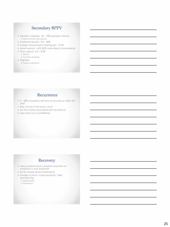
Surgical Options
BPPV
Singular Neurectomy:
removal of all or part
of the nerve
Surgical Blockade
(canal “plugging”): bone plugs placed in
affected canal to
block function without
affecting other canals
25
Secondary BPPV
Meniere’s Disease: 65 – 70% between attacks
More common recurrence
Vestibular Neuritis: 9.8 – 20%
Sudden sensorineural hearing loss: 12.7%
Head trauma: 6.6% (25% more likely to be bilateral)
Post-surgical: 6.3 – 8.5%
Dental
Cochlear Implants
Migraine
Poorly understood
Recurrence
9 – 30% of patients will have a recurrence within first
year
May not be in the same canal
No time frame associated with recurrence
May return as a canalithiasis
Recovery
Many patients have complete resolution of
symptoms in one treatment
Some require several treatments
Possible to have “canal sensitivity” after
repositioning
Lightheaded
Imbalance
26
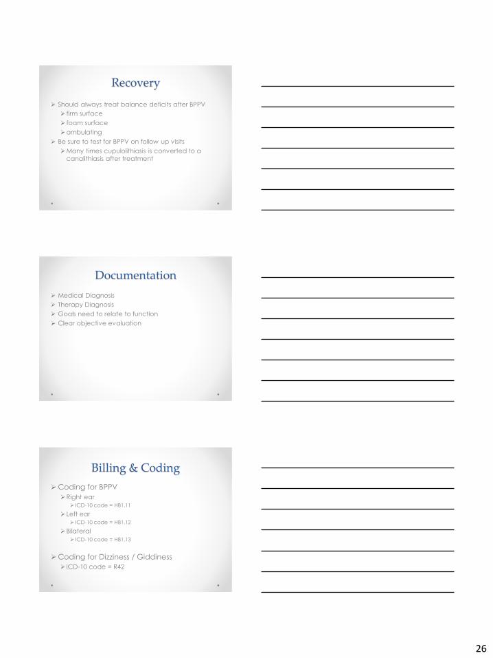
Recovery
Should always treat balance deficits after BPPV
firm surface
foam surface
ambulating
Be sure to test for BPPV on follow up visits
Many times cupulolithiasis is converted to a
canalithiasis after treatment
Documentation
Medical Diagnosis
Therapy Diagnosis
Goals need to relate to function
Clear objective evaluation
Billing & Coding
Coding for BPPV
Right ear
ICD-10 code = H81.11
Left ear
ICD-10 code = H81.12
Bilateral
ICD-10 code = H81.13
Coding for Dizziness / Giddiness
ICD-10 code = R42
27
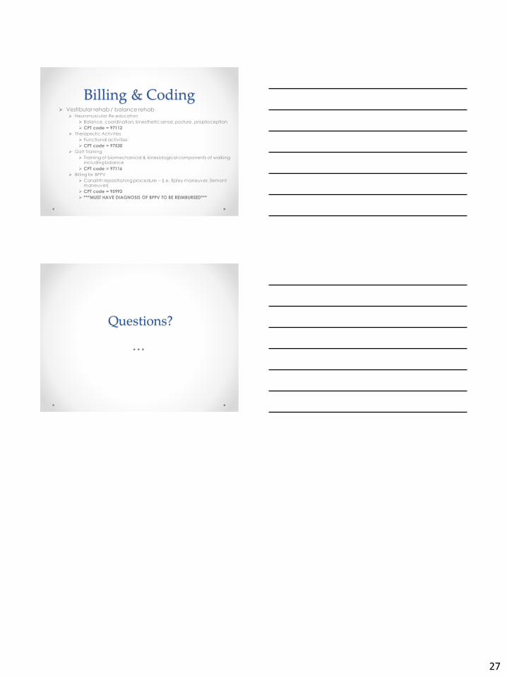
Billing & Coding Vestibular rehab / balance rehab
Neuromuscular Re-education
Balance, coordination, kinesthetic sense, posture, proprioception
CPT code = 97112
Therapeutic Activ ities
Functional activities
CPT code = 97530
Gait Training
Training of biomechanical & kinesiological components of walking including balance
CPT code = 97116
Billing for BPPV
Canalith repositioning procedure – (i.e. Epley maneuver, Semontmaneuver)
CPT code = 95992
***MUST HAVE DIAGNOSIS OF BPPV TO BE REIMBURSED***
Questions?