Embed Size (px)
Citation preview

217
Benign epithelioid peripheral nerve sheath tumour resembling schwannoma
Thejasvi KRISHNAMURTHY MD and SR NIVEDITHA MD, DNB
Department of Pathology, Kempegowda Institute of Medical Sciences, Bangalore. India
Abstract
Peripheral nerve sheath tumours (PNST) with epithelial appearing cells compromise a heterogeneous group of neoplasms that are rare and diagnostically challenging. Of these, malignant PNSTs with epithelioid features (epithelioid MPNST) are commonly described in literature. However benign epithelioid PNSTs are rare and till date about 38 cases have been described in the literature. We report a benign epithelioid PNST with light microscopical and immunohistochemical features suggestive of schwannoma, presenting as a thigh mass in a 23-year-old female. The tumour was encapsulated, showed epithelioid cells in aggregates, and expressed vimentin and S-100 positivity. There was no expression of CD34, CK, EMA, CD99, p63 and HMB 45. Typical Antoni A and Antoni B areas were absent. At 18 months follow-up, the patient was well.
Keywords: schwannoma, epithelioid, benign epithelioid peripheral nerve sheath tumour.
Address for correspondence: Dr Thejasvi Krishnamurthy, Department of Pathology, Kempegowda Institute of Medical Sciences, Bangalore – 560070, India. Email: [email protected]
CASE REPORT
INTRODUCTION
The epithelioid variants of peripheral nerve sheath tumour (PNST) are rare but a well-known entities, especially the malignant variants. Benign peripheral nerve sheath tumours are a group of heterogeneous tumours composed of schwannoma, neurofibroma and perineurioma. Schwannomas have varying morphological subtypes1 such as cellular, ancient, plexiform and the epithelioid subtypes.2 Epithelioid schwannoma is an unusual variant, posing difficulties in diagnosis due to its increased cellularity and epithelioid morphology.3 We report a case of epithelioid schwannoma arising in the calf and discuss the differential diagnosis.
CASE REPORT
A 23 year-old female, developed a mass in the calf region which was gradually progressing with tenderness on palpation. The mass was excised. Post surgery, the patient was on follow-up for the past 18 months and is doing well.
Gross pathologyThe mass was nodular, measuring 2.5 x 2.0 x 1.5cm. The outer surface was tan-brown in colour and showed some adherent fascia. The
cut surface appeared yellow to grey white. A thin capsule was evident.
MicroscopyThe tumour appeared circumscribed and showed a thin capsule (Fig. 1). The tumour cells were small and round, arranged singly, in small aggregates or in cords amidst a collagenous stroma. At places, the stroma appeared myxoid (Fig.1). Individual cells showed sharp cytoplasmic borders and a good number of them had intranuclear inclusion-like nucleoli (Fig. 2). Focal areas showed dense collagen cores resembling irregular rosettes, which appeared more prominent with the Masson trichrome stain (Fig 2, inset). The collagen was seen encircling individual cells (Fig. 2, inset). Although focally few cells showed cytological atypia in the form of mild to moderate nuclear pleomorphism, no mitotic figures were observed. Typical Antoni A and Antoni B areas were also not seen. Immunohistochemistry was performed for cytokeratin (Envision, 1:200 dilution), CD 99 (Ho36.1.1), CD 34 (Envision, prediluted), EMA (Biogenex – E 29, prediluted), HMB 45 (Biogenex, prediluted), p63 (Biogenex 4A 4, prediluted), S-100 (Envision, 1:1000) and Vimentin (Envision, 1:1000). There was strong
Malaysian J Pathol 2014; 36(3) : 217 – 221
Malaysian J Pathol December 2014
218
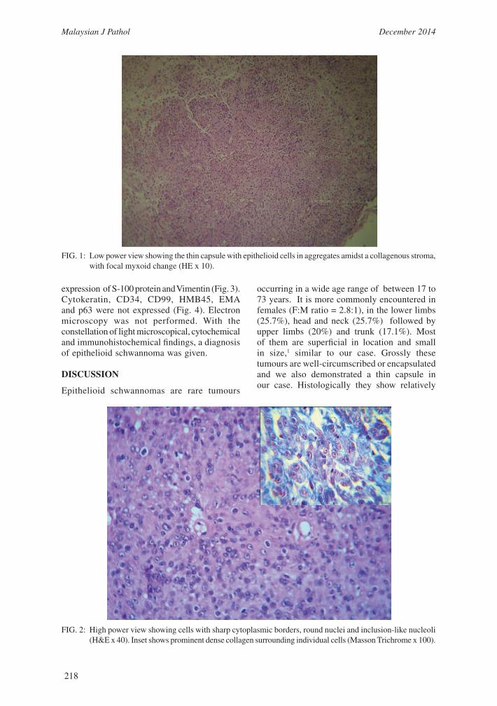
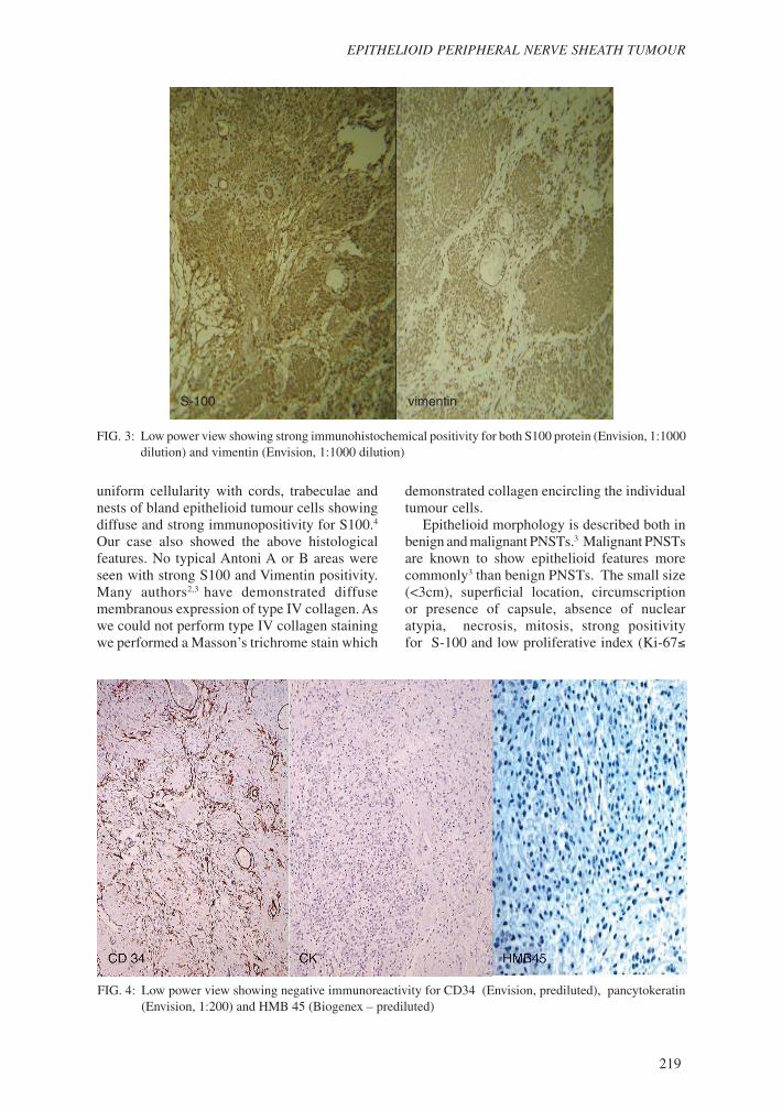
expression of S-100 protein and Vimentin (Fig. 3).Cytokeratin, CD34, CD99, HMB45, EMA and p63 were not expressed (Fig. 4). Electron microscopy was not performed. With the constellation of light microscopical, cytochemical and immunohistochemical findings, a diagnosis of epithelioid schwannoma was given. DISCUSSION
Epithelioid schwannomas are rare tumours
occurring in a wide age range of between 17 to 73 years. It is more commonly encountered in females (F:M ratio = 2.8:1), in the lower limbs (25.7%), head and neck (25.7%) followed by upper limbs (20%) and trunk (17.1%). Most of them are superficial in location and small in size,1 similar to our case. Grossly these tumours are well-circumscribed or encapsulated and we also demonstrated a thin capsule in our case. Histologically they show relatively
FIG. 1: Low power view showing the thin capsule with epithelioid cells in aggregates amidst a collagenous stroma, with focal myxoid change (HE x 10).
FIG. 2: High power view showing cells with sharp cytoplasmic borders, round nuclei and inclusion-like nucleoli (H&E x 40). Inset shows prominent dense collagen surrounding individual cells (Masson Trichrome x 100).
219
EPITHELIOID PERIPHERAL NERVE SHEATH TUMOUR
uniform cellularity with cords, trabeculae and nests of bland epithelioid tumour cells showing diffuse and strong immunopositivity for S100.4
Our case also showed the above histological features. No typical Antoni A or B areas were seen with strong S100 and Vimentin positivity. Many authors2,3 have demonstrated diffuse membranous expression of type IV collagen. As we could not perform type IV collagen staining we performed a Masson’s trichrome stain which
demonstrated collagen encircling the individual tumour cells. Epithelioid morphology is described both in benign and malignant PNSTs.3 Malignant PNSTs are known to show epithelioid features more commonly3 than benign PNSTs. The small size (<3cm), superficial location, circumscription or presence of capsule, absence of nuclear atypia, necrosis, mitosis, strong positivity for S-100 and low proliferative index (Ki-67≤
FIG. 3: Low power view showing strong immunohistochemical positivity for both S100 protein (Envision, 1:1000 dilution) and vimentin (Envision, 1:1000 dilution)
S-100 vimentin
FIG. 4: Low power view showing negative immunoreactivity for CD34 (Envision, prediluted), pancytokeratin (Envision, 1:200) and HMB 45 (Biogenex – prediluted)

Malaysian J Pathol December 2014
220
Table 1: Differential diagnoses of benign soft tissue tumours with epithelioid features:1-4,9
Tumour Light microscopy Immunohistochemistry
Epithelioid Encapsulated S100: intensely positiveschwannoma/benign Epithelioid cells in cords and nests, Vimentin: positiveepithelioid PNST amidst a collagenous stroma, GFAP: positive NGFR: positive
Cellular variant of Spindled and epithelioid cells in S100: Negativeneurothekeoma3 tight whorls and fascicles. GFAP: negative Diffuse myxoid areas. NGFR: negative
Nevomelanocytic tumours3 No capsule S100: positivity less(cellular blue nevus, Cells in close proximity to intense than schwannoma.pigmented epithelioid epidermis. HMB 45: positivemelanocytoma, melanocytic elements of neurocristic hamartoma)
Myoepithelioma of soft Epithelioid cells in cords and S100: positive tissue nests Cytokeratin: positive P63: positive GFAP: positive SMA: positive Calponin: positive
Ossifying fibromyxoid Pseudocapsuletumour3,8 Superficial location S100: positive Lamellar or woven bone in septae in 80%, GFAP: positive
Desmin: focally positive
Glomus tumour3 Nucleus more central with S100: negative intact cytoplasmic borders3 SMA: positive No syncytial aggregates3 Calponin: positive
Caldesmon: positive
Palisaded encapsulated Commonly seen on face. Not describedneuroma2,3 Encapsulated with club like extensions into soft tissue. Solid proliferation of schwann cells. Absence of myxoid areas and hyalinization.2
NGFR- Neural growth factor receptor, SMA- Smooth muscle actin, GFAP- Glial fibrillary acid protein,
1%) are highly suggestive of benign nature in epithelioid schwannomas.1,3 Though we have not performed Ki-67 assessment in our case, the rest of the features are suggestive of benign epithelioid schwannoma. Till date 38 cases have been reported in the literature.3,4
Taxy and Battifora5 documented the first
two cases in 1981 of epithelioid tumours showing schwannonian features on light and electron microscopy. In 1985 Franks et al6 reported the ultrastructural features of Schwann cells in an epithelioid neurilemmoma of the trigeminal nerve. The existence of epithelioid schwannoma was first suggested by Orosz et

221
EPITHELIOID PERIPHERAL NERVE SHEATH TUMOUR
al7 in 1993. Kindblom et al described the first series of epithelioid schwannomas in 1998, which comprised of 5 cases predominantly in the subcutis and one in a rare site like the submucosa of the urinary bladder.8
The latest and the largest series is reported by Laskin et al3 in 2005. They reviewed 33 cases of benign epithelioid PNSTs from the AFIP between 1970 and 2002. All PNSTs lacking equivocal features of malignancy were reviewed by three pathologists with immunohistochemistry and cytogenetic analysis. They were of the opinion that a non-committal term “benign epithelioid peripheral nerve sheath tumour” (BEPNST) be used for these lesions as tumours called schwannomas on conventional microscopy showed CD34 positivity (in fibroblasts) and ultrastructural evidence of neurofibromas. Hence, they suggested to classify these as benign epithelioid peripheral nerve sheath tumour of indeterminate histiogenesis.3 Though the present case showed more features of benign epithelioid schwannoma, in retrospect we would agree with Laskin et al3 and prefer to call these tumours as benign epithelioid peripheral nerve sheath tumours unless we can demonstrate ultrastructural features of Schwannonian nature. As recurrence and malignant transformation is more common in neurofibromas rather than schwannomas, follow up of all benign epithelioid peripheral nerve sheath tumours appears reasonable as electron microscopy is not widely available. The differential diagnoses of various benign tumours in soft tissue showing epithelioid features have been summarized in Table 1. Epithelioid features in a soft tissue tumour should always be appropriately worked up to differentiate these lesions and avoid misinterpretation.
REFERENCES
1. Rezanko T, Sari AA, Tunakan M, Calli AO, Altin-boga AA. Epithelioid schwannoma of soft tissue: unusual morphological variant causing a diagnostic dilemma. Ann Diagn Pathol. 2012; 16(6): 521-6.
2. Weiss SW, Goldblum JR. Benign tumors of periph-eral nerves. In: Weiss SW, Goldblum JR: Enzinger and Weiss’s soft tissue tumors. 5th ed. St Louis, Mo: Mosby; 2008. p. 853-72.
3. Laskin WB, Fetsch JF, Lasota J, Miettinen M. Benign epithelioid peripheral nerve sheath tumors of the soft tissues: clinicopathologic spectrum of 33 cases. Am J Surg Pathol. 2005; 29(1): 39-51.
4. Chan AW, Mak SM, Chan GP. Benign epithelioid schwannoma of intraparotid facial nerve. Pathology. 2011; 43(3): 280-2.
5. Taxy JB, Battifora H. Epithelioid schwannoma: diagnosis by electron microscopy. Ultrastruct Pathol. 1981; 2(1): 19-24.
6. Franks AJ. Epithelioid neurilemmoma of the trigeminal nerve: an immunohistochemical and ultrastructural study. Histopathology. 1985; 9(12): 1339-50.
7. Orosz Z, Sápi Z, Szentirmay Z. Unusual benign neurogenic soft tissue tumor. Epithelioid schwan-noma or an ossifying fibromyxoid tumour? Pathol Res Pract. 1993: 189(5): 601-5; discussion 605-7.
8. Kindblom LG, Meis-Kindblom JM, Havel G, Busch C. Benign epithelioid schwannoma. Am J Surg Pathol. 1998: 22(6): 762-70.
9. Saad AG, Mutema GK, Mutasim DF. Benign cu-taneous epithelioid schwannoma: case report and review of the literature. Am J Dermatopathol. 2005; 27(1): 45-7.