Embed Size (px)
Citation preview
n engl j med
353;3
www.nejm.org july
21, 2005
229
The
new england
journal
of
medicine
established in 1812
july
21
,
2005
vol. 353 no. 3
Benign Breast Disease and the Risk of Breast Cancer
Lynn C. Hartmann, M.D., Thomas A. Sellers, Ph.D., Marlene H. Frost, Ph.D., Wilma L. Lingle, Ph.D.,Amy C. Degnim, M.D., Karthik Ghosh, M.D., Robert A. Vierkant, M.A.S., Shaun D. Maloney, B.A.,
V. Shane Pankratz, Ph.D., David W. Hillman, M.S., Vera J. Suman, Ph.D., Jo Johnson, R.N.,Cassann Blake, M.D., Thea Tlsty, Ph.D., Celine M. Vachon, Ph.D.,
L. Joseph Melton III, M.D., and Daniel W. Visscher, M.D.
abstract
From the Divisions of Medical Oncology(L.C.H., M.H.F., J.J.), Experimental Pathol-ogy (W.L.L.), General Surgery (A.C.D.), Gen-eral Internal Medicine (K.G.), Biostatistics(R.A.V., S.D.M., V.S.P., D.W.H., V.J.S.), Ep-idemiology (C.M.V., L.J.M.), and AnatomicPathology (D.W.V.), Mayo Clinic College ofMedicine, Rochester, Minn.; H. Lee Mof-fitt Cancer Center and Research Institute,Tampa, Fla. (T.A.S.); Wayne State Univer-sity, Detroit (C.B.); and the University ofCalifornia, San Francisco, San Francisco(T.T.). Address reprint requests to Dr.Hartmann at Mayo Clinic College of Med-icine, Rochester, MN 55905.
N Engl J Med 2005;353:229-37.
Copyright © 2005 Massachusetts Medical Society.
background
Benign breast disease is an important risk factor for breast cancer. We studied a largegroup of women with benign breast disease to obtain reliable estimates of this risk.
methods
We identified all women who received a diagnosis of benign breast disease at the MayoClinic between 1967 and 1991. Breast-cancer events were obtained from medical recordsand questionnaires. To estimate relative risks, we compared the number of observedbreast cancers with the number expected on the basis of the rates of breast cancer in theIowa Surveillance,
Epidemiology, and End Results registry.
results
We followed 9087 women for a median of 15 years. The histologic findings were non-proliferative lesions in 67 percent of women, proliferative lesions without atypia in 30percent, and atypical hyperplasia in 4 percent. To date, 707 breast cancers have devel-oped. The relative risk of breast cancer for the cohort was 1.56 (95 percent confidenceinterval, 1.45 to 1.68), and this increased risk persisted for at least 25 years after biopsy.The relative risk associated with atypia was 4.24 (95 percent confidence interval, 3.26to 5.41), as compared with a relative risk of 1.88 (95 percent confidence interval, 1.66to 2.12) for proliferative changes without atypia and of 1.27 (95 percent confidence in-terval, 1.15 to 1.41) for nonproliferative lesions. The strength of the family history ofbreast cancer, available for 4808 women, was a risk factor that was independent of his-tologic findings. No increased risk was found among women with no family historyand nonproliferative findings. In the first 10 years after the initial biopsy, an excess ofcancers occurred in the same breast, especially in women with atypia.
conclusions
Risk factors for breast cancer after the diagnosis of benign breast disease include thehistologic classification of a benign breast lesion and a family history of breast cancer.
The New England Journal of Medicine Downloaded from nejm.org at NORTH DAKOTA STATE UNIV on October 28, 2014. For personal use only. No other uses without permission.
Copyright © 2005 Massachusetts Medical Society. All rights reserved.

n engl j med
353;3
www.nejm.org july
21
,
2005
The
new england journal
of
medicine
230
enign breast disease is an impor-
tant risk factor for a later breast cancer,which can develop in either breast.
1
It en-compasses a spectrum of histologic entities, usuallysubdivided into nonproliferative lesions, prolifera-tive lesions without atypia, and atypical hyperpla-sias, with an increased risk of breast cancer associ-ated with proliferative or atypical lesions.
2-4
Theidentification of benign breast disease has becomemore common as the use of mammography has in-creased, and thus, having accurate risk estimates forwomen who receive this diagnosis is imperative.
Important questions remain, however, about thedegree of risk associated with the common nonpro-liferative benign entities and the extent to whichfamily history influences the risk of breast cancer inwomen with proliferative or atypical lesions. Du-pont and Page found that women with nonprolifer-ative disease did not have an increased risk of a lat-er breast cancer.
2
By contrast, a companion studyto the National Surgical Adjuvant Breast and BowelProject (NSABP) Breast Cancer Prevention Trial (P1)found a relative risk of 1.6 for women who receiveda diagnosis of a “lower category” of benign breastdisease.
5
A limitation of the NSABP study, howev-er, was the lack of central pathological review.
Another major question concerns the possibleinterplay between atypia and a family history ofbreast cancer. The Dupont and Page study foundthat women with atypia and a family history had 11times the risk of those with nonproliferative lesionsand no family history.
2
However, two other majorstudies of benign breast disease
6,7
did not find a sig-nificant interaction between atypia and family his-tory. The duration of increased risk after a findingof benign disease on biopsy is also uncertain.
2,4,8
Studies of benign breast disease can also clarifywhether there is a continuum of breast alterationsthat culminates in breast cancer. However, it re-mains unclear which of the benign entities are ac-tual precursors and which reflect a background ofincreased risk involving all breast tissue in a wom-an. Determining the extent of agreement betweenthe side (right or left) of the benign lesion and thesubsequent breast cancer is one means of assess-ing these issues.
To investigate these questions, we studied 9087women with benign breast disease for whom we hadfollow-up data on breast-cancer events. This cohorthas been followed for a median of 15 years, and 707breast cancers have developed, making this, to ourknowledge, one of the largest such studies of its
kind. We report on the risk of breast cancer accord-ing to histologic findings, the age at diagnosis of be-nign breast disease, and the strength of the familyhistory. We also recorded the side of the cancer (ip-silateral or contralateral) and the time to the diag-nosis of cancer.
study population
We accessed data from the Mayo Clinic Surgical In-dex and Pathology Index to identify all women 18 to85 years of age who had undergone surgical excisionof a benign breast lesion during the 25-year periodfrom January 1, 1967, through December 31, 1991.For women who had more than one biopsy duringthis period, we used the first sample. The originallist contained 12,132 women, but we excluded 1,047women for any of the following: a diagnosis ofbreast cancer or lobular carcinoma in situ at, before,or within six months after the biopsy of the benignlesion; mastectomy (unilateral or bilateral) or breastreduction at or before biopsy; or refusal to allowuse of their medical records for research.
9
This left11,085 women. Of these, 1053 (9.5 percent) had nofollow-up information after the biopsy. Thus, a totalof 10,032 women met our criteria for study entryand had follow-up information. Of these, 945 wom-en had unusable or unavailable biopsy specimensof the benign lesion. The remaining group of 9087women constitutes our study cohort. The relativerisks of breast cancer (described below) did not dif-fer significantly between the 10,032 women whomet our criteria and the 9087 women who made upthe study cohort (1.59 and 1.56, respectively).
family history and follow-up
A questionnaire designed for this study was used toobtain information about family history and otherpossible risk factors for breast cancer. Thus, ourfamily-history data were obtained at the time of fol-low-up contact. We categorized family history asnone, weak, or strong. The criteria for a strong fam-ily history were as follows: at least one first-degreerelative with breast cancer before the age of 50 yearsor two or more relatives with breast cancer, with atleast one being a first-degree relative. Any lesser de-gree of family history of breast cancer was catego-rized as weak. The questionnaire also asked aboutbreast-cancer occurrences. Follow-up for breast-cancer events was also obtained through the com-prehensive (inpatient and outpatient) Mayo medical
b
methods
The New England Journal of Medicine Downloaded from nejm.org at NORTH DAKOTA STATE UNIV on October 28, 2014. For personal use only. No other uses without permission.
Copyright © 2005 Massachusetts Medical Society. All rights reserved.
n engl j med
353;3
www.nejm.org july
21, 2005
benign breast disease and the risk of breast cancer
231
record. Questionnaire information was available for5619 women (61.8 percent). Of the questionnaires,604 (10.7 percent) were completed by proxy (thenext of kin of a deceased patient). As of August 1,2004, 7260 (79.9 percent) members of the cohortwere still alive. All protocol procedures and patient-contact materials were reviewed and approved bythe institutional review board of the Mayo Clinic;returning the contact materials was considered im-plied consent.
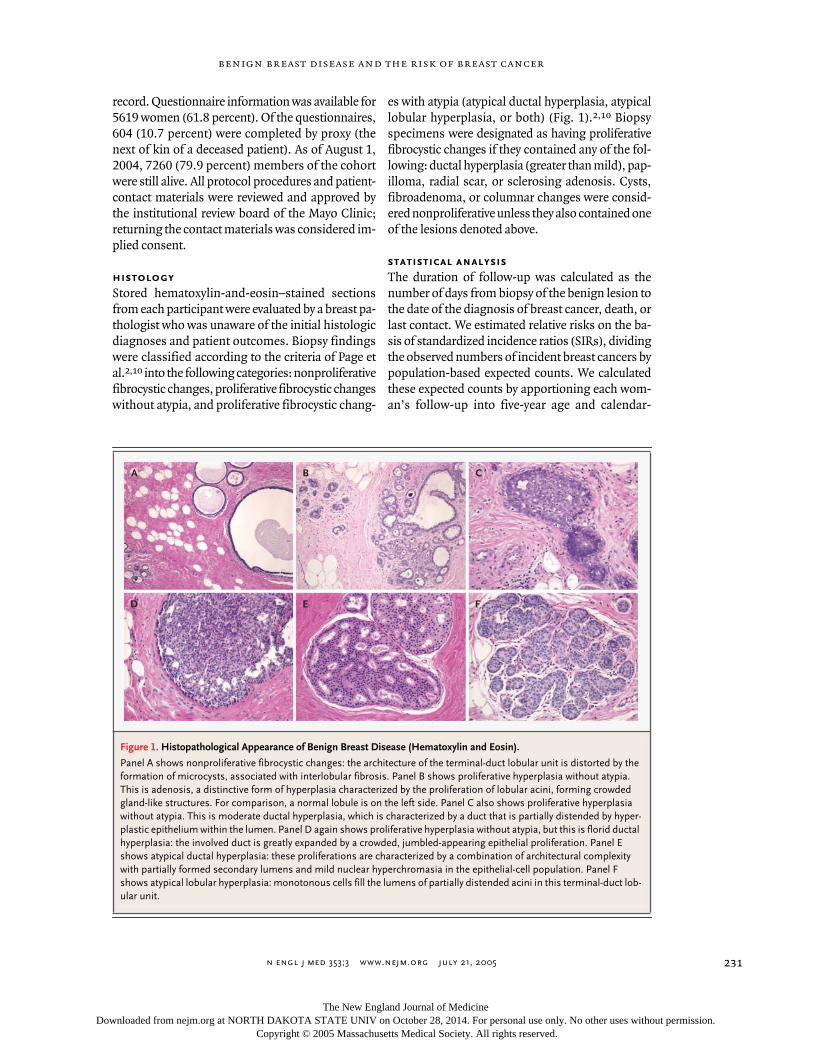
histology
Stored hematoxylin-and-eosin–stained sectionsfrom each participant were evaluated by a breast pa-thologist who was unaware of the initial histologicdiagnoses and patient outcomes. Biopsy findingswere classified according to the criteria of Page etal.
2,10
into the following categories: nonproliferativefibrocystic changes, proliferative fibrocystic changeswithout atypia, and proliferative fibrocystic chang-
es with atypia (atypical ductal hyperplasia, atypicallobular hyperplasia, or both) (Fig. 1).
2,10
Biopsyspecimens were designated as having proliferativefibrocystic changes if they contained any of the fol-lowing: ductal hyperplasia (greater than mild), pap-illoma, radial scar, or sclerosing adenosis. Cysts,fibroadenoma, or columnar changes were consid-ered nonproliferative unless they also contained oneof the lesions denoted above.
statistical analysis
The duration of follow-up was calculated as thenumber of days from biopsy of the benign lesion tothe date of the diagnosis of breast cancer, death, orlast contact. We estimated relative risks on the ba-sis of standardized incidence ratios (SIRs), dividingthe observed numbers of incident breast cancers bypopulation-based expected counts. We calculatedthese expected counts by apportioning each wom-an’s follow-up into five-year age and calendar-
Figure 1. Histopathological Appearance of Benign Breast Disease (Hematoxylin and Eosin).
Panel A shows nonproliferative fibrocystic changes: the architecture of the terminal-duct lobular unit is distorted by the formation of microcysts, associated with interlobular fibrosis. Panel B shows proliferative hyperplasia without atypia. This is adenosis, a distinctive form of hyperplasia characterized by the proliferation of lobular acini, forming crowded gland-like structures. For comparison, a normal lobule is on the left side. Panel C also shows proliferative hyperplasia without atypia. This is moderate ductal hyperplasia, which is characterized by a duct that is partially distended by hyper-plastic epithelium within the lumen. Panel D again shows proliferative hyperplasia without atypia, but this is florid ductal hyperplasia: the involved duct is greatly expanded by a crowded, jumbled-appearing epithelial proliferation. Panel E shows atypical ductal hyperplasia: these proliferations are characterized by a combination of architectural complexity with partially formed secondary lumens and mild nuclear hyperchromasia in the epithelial-cell population. Panel F shows atypical lobular hyperplasia: monotonous cells fill the lumens of partially distended acini in this terminal-duct lob-ular unit.
A B C
D E F
The New England Journal of Medicine Downloaded from nejm.org at NORTH DAKOTA STATE UNIV on October 28, 2014. For personal use only. No other uses without permission.
Copyright © 2005 Massachusetts Medical Society. All rights reserved.

n engl j med
353;3
www.nejm.org july
21
,
2005
The
new england journal
of
medicine
232
period categories, thereby accounting for differ-ences associated with these variables. We used theIowa Surveillance,
Epidemiology, and End Results(SEER) registry as the reference population be-cause of its demographic similarities to the MayoClinic population (80 percent of cohort membersreside in the upper Midwest). Over 95 percent ofour cohort was white, equivalent to that reported inIowa census data during the study period.
11
In theSIR analyses, we considered the time since theoriginal biopsy as a time-dependent variable andall other factors as fixed.
Associations between the risk of breast cancerand histologic findings, the age at diagnosis of be-
nign breast disease, and the strength of the familyhistory of cancer, as well as pairwise combinationsof these variables, were examined with the use ofCox proportional-hazards regression analysis. Themain effects for each categorized variable and thecorresponding interaction terms were included ineach model, and the statistical significance of eachinteraction was evaluated with the use of a multiple-degree-of-freedom likelihood-ratio test.
We studied ipsilateral and contralateral breastcancer as a function of the time since biopsy by es-timating the relative risk of cancer in the same ascompared with the opposite breast for five-year in-tervals. When calculating the incidence of ipsilat-
* Plus–minus values are means ±SD.
† Menopausal status was categorized according to the age at breast biopsy.
Table 1. Characteristics of the Women According to the Histologic Category of Benign Breast Disease.*
CharacteristicAll Women(N=9087)
NonproliferativeDisease
(N=6061)
ProliferativeDisease
without Atypia(N=2690)
AtypicalHyperplasia
(N=336)
Percentage of total 100.0 66.7 29.6 3.7
Age at biopsy — no. of women (%)
<40 yr 1841 (20.3) 1500 (24.7) 323 (12.0) 18 (5.4)
40–49 yr 2474 (27.2) 1621 (26.7) 770 (28.6) 83 (24.7)
50–59 yr 2145 (23.6) 1297 (21.4) 759 (28.2) 89 (26.5)
60–69 yr 1639 (18.0) 1034 (17.1) 522 (19.4) 83 (24.7)
≥70 yr 988 (10.9) 609 (10.0) 316 (11.7) 63 (18.8)
Mean age at biopsy — yr 51.4±14.3 49.9±14.8 53.9±12.6 57.8±12.3
Menopausal status at biopsy — no. of women (%)†
Premenopausal (<45 yr) 2948 (32.4) 2246 (37.1) 652 (24.2) 50 (14.9)
Perimenopausal (45–55 yr) 2583 (28.4) 1610 (26.6) 871 (32.4) 102 (30.4)
Postmenopausal (>55 yr) 3556 (39.1) 2205 (36.4) 1167 (43.4) 184 (54.8)
Family history of breast cancer— no. of women (%)
Unknown 4279 (47.1) 2970 (49.0) 1170 (43.5) 139 (41.4)
Known 4808 (52.9) 3091 (51.0) 1520 (56.5) 197 (58.6)
None 2668 (55.5) 1735 (56.1) 831 (54.7) 102 (51.8)
Weak 1174 (24.4) 756 (24.5) 378 (24.9) 40 (20.3)
Strong 966 (20.1) 600 (19.4) 311 (20.5) 55 (27.9)
Breast-cancer status as of August 2004 — no. of women (%)
Negative 8380 (92.2) 5682 (93.7) 2426 (90.2) 272 (81.0)
Positive 707 (7.8) 379 (6.3) 264 (9.8) 64 (19.0)
Vital status — no. of women (%)
Deceased 1827 (20.1) 1172 (19.3) 566 (21.0) 89 (26.5)
Alive 7260 (79.9) 4889 (80.7) 2124 (79.0) 247 (73.5)
The New England Journal of Medicine Downloaded from nejm.org at NORTH DAKOTA STATE UNIV on October 28, 2014. For personal use only. No other uses without permission.
Copyright © 2005 Massachusetts Medical Society. All rights reserved.
n engl j med
353;3
www.nejm.org july
21, 2005
benign breast disease and the risk of breast cancer
233
eral cancer, we censored follow-up on women withcontralateral cancer after the date of diagnosis. Sim-ilarly, when calculating the incidence of contralat-eral cancer, we censored follow-up on women withipsilateral cancer after the date of diagnosis. Dataon women missing information on the side of thecancer or women who had bilateral biopsies or can-cer were not included in these analyses. This ap-proach yields identical numbers of person-years foreach type of event. As a result, the length of follow-up is no longer a factor in the analysis and the rela-tive risks are equivalent to simple ratios of eventcounts. We therefore used properties of the binomi-al distribution to obtain exact P values and 95 per-cent confidence intervals for these relative risks.
12
Statistical tests were two-sided, and analyses wereconducted with the use of SAS (SAS) and Splus (In-sightful) software.
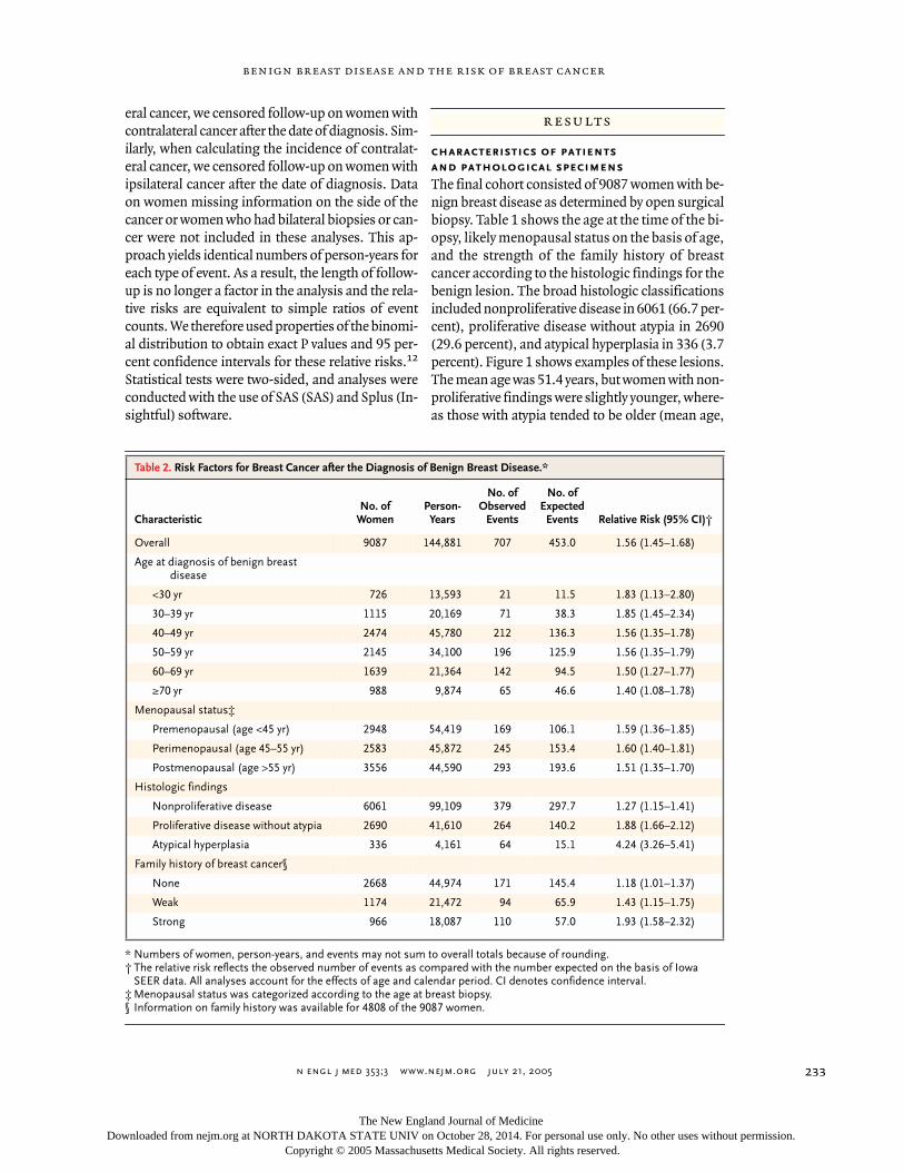
characteristics of patientsand pathological specimens
The final cohort consisted of 9087 women with be-nign breast disease as determined by open surgicalbiopsy. Table 1 shows the age at the time of the bi-opsy, likely menopausal status on the basis of age,and the strength of the family history of breastcancer according to the histologic findings for thebenign lesion. The broad histologic classificationsincluded nonproliferative disease in 6061 (66.7 per-cent), proliferative disease without atypia in 2690(29.6 percent), and atypical hyperplasia in 336 (3.7percent). Figure 1 shows examples of these lesions.The mean age was 51.4 years, but women with non-proliferative findings were slightly younger, where-as those with atypia tended to be older (mean age,
results
* Numbers of women, person-years, and events may not sum to overall totals because of rounding.† The relative risk reflects the observed number of events as compared with the number expected on the basis of Iowa
SEER data. All analyses account for the effects of age and calendar period. CI denotes confidence interval.‡ Menopausal status was categorized according to the age at breast biopsy.
§ Information on family history was available for 4808 of the 9087 women.
Table 2. Risk Factors for Breast Cancer after the Diagnosis of Benign Breast Disease.*
CharacteristicNo. of
WomenPerson-Years
No. of Observed
Events
No. ofExpected
Events Relative Risk (95% CI)†
Overall 9087 144,881 707 453.0 1.56 (1.45–1.68)
Age at diagnosis of benign breastdisease
<30 yr 726 13,593 21 11.5 1.83 (1.13–2.80)
30–39 yr 1115 20,169 71 38.3 1.85 (1.45–2.34)
40–49 yr 2474 45,780 212 136.3 1.56 (1.35–1.78)
50–59 yr 2145 34,100 196 125.9 1.56 (1.35–1.79)
60–69 yr 1639 21,364 142 94.5 1.50 (1.27–1.77)
≥70 yr 988 9,874 65 46.6 1.40 (1.08–1.78)
Menopausal status‡
Premenopausal (age <45 yr) 2948 54,419 169 106.1 1.59 (1.36–1.85)
Perimenopausal (age 45–55 yr) 2583 45,872 245 153.4 1.60 (1.40–1.81)
Postmenopausal (age >55 yr) 3556 44,590 293 193.6 1.51 (1.35–1.70)
Histologic findings
Nonproliferative disease 6061 99,109 379 297.7 1.27 (1.15–1.41)
Proliferative disease without atypia 2690 41,610 264 140.2 1.88 (1.66–2.12)
Atypical hyperplasia 336 4,161 64 15.1 4.24 (3.26–5.41)
Family history of breast cancer§
None 2668 44,974 171 145.4 1.18 (1.01–1.37)
Weak 1174 21,472 94 65.9 1.43 (1.15–1.75)
Strong 966 18,087 110 57.0 1.93 (1.58–2.32)
The New England Journal of Medicine Downloaded from nejm.org at NORTH DAKOTA STATE UNIV on October 28, 2014. For personal use only. No other uses without permission.
Copyright © 2005 Massachusetts Medical Society. All rights reserved.
n engl j med
353;3
www.nejm.org july
21
,
2005
The
new england journal
of
medicine
234
49.9 and 57.8 years, respectively; P<0.001). Infor-mation on family history was available for 4808women and was negative in 2668 (55.5 percent),
weakly positive in 1174 (24.4 percent), and strong-ly positive in 966 (20.1 percent). More women withatypia than without atypia had a strong family his-tory of breast cancer (27.9 percent vs. 19.8 percent,P=0.06). The risk of cancer was highest in the groupwith atypia: breast cancer developed in 64 of the336 women (19.0 percent).
features of benign breast diseaseand subsequent risk of breast cancer
Patients in the cohort were followed for a medianof 15 years. A total of 1827 women (20.1 percent)had died and 7260 (79.9 percent) were alive as ofAugust 2004. We have documented 707 breast can-cers to date. The median time from the original bi-opsy to the diagnosis of breast cancer was 10.7years. Table 2 shows the estimated relative risks ofbreast cancer associated with the age at the initialbiopsy, the strength of the family history, meno-pausal status, and histologic findings of the biop-sy, as compared with expected population-basedincidence. The estimated relative risk of breast can-cer in the cohort was 1.56 (95 percent confidenceinterval, 1.45 to 1.68). The risk was inversely asso-ciated with the age at biopsy, with younger womenhaving a greater risk than older women. The type ofbenign breast disease identified at biopsy was amajor predictor of risk. Atypical hyperplasia had arelative risk of 4.24 (95 percent confidence interval,3.26 to 5.41), proliferative disease without atypiahad a relative risk of 1.88 (95 percent confidenceinterval, 1.66 to 2.12), and nonproliferative lesionshad a relative risk of 1.27 (95 percent confidenceinterval, 1.15 to 1.41). Family history was an inde-pendent risk factor. For women with no knownfamily history of breast cancer, the relative risk wasonly 1.18 (95 percent confidence interval, 1.01 to1.37), as compared with 1.43 (95 percent confi-dence interval, 1.15 to 1.75) for women with a weakfamily history and 1.93 (95 percent confidence in-terval, 1.58 to 2.32) for those with a strong familyhistory.
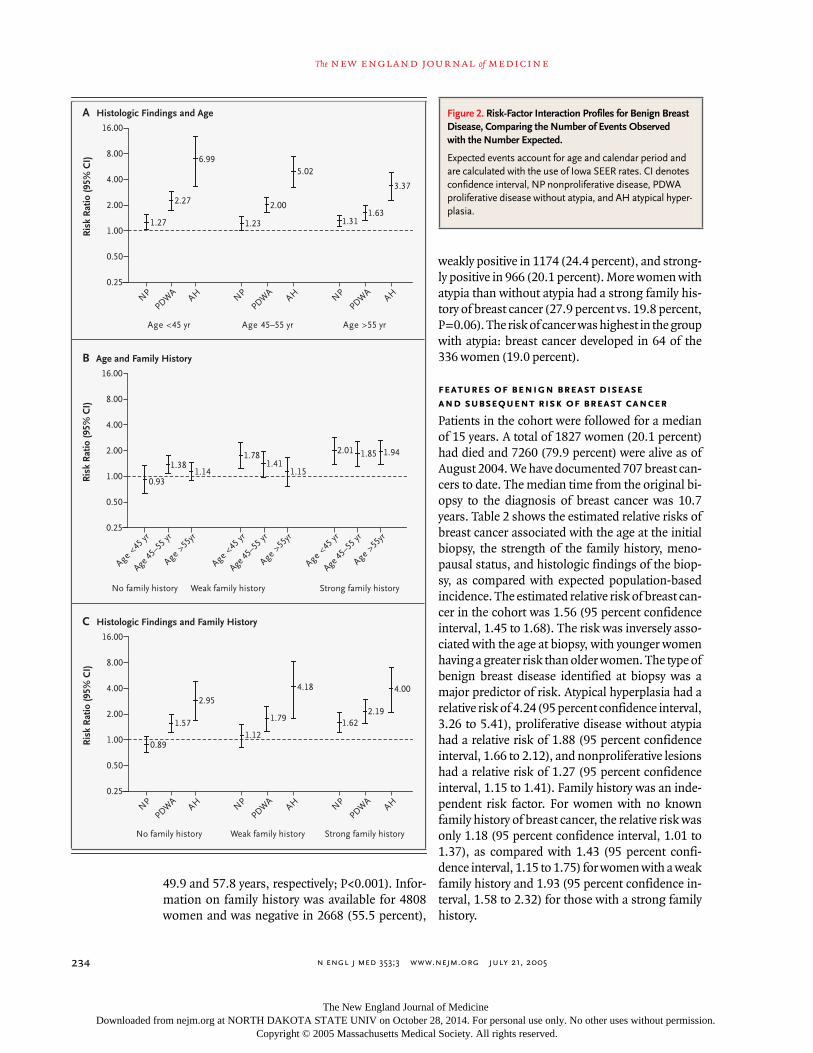
Figure 2. Risk-Factor Interaction Profiles for Benign Breast Disease, Comparing the Number of Events Observed with the Number Expected.
Expected events account for age and calendar period and are calculated with the use of Iowa SEER rates. CI denotes confidence interval, NP nonproliferative disease, PDWA proliferative disease without atypia, and AH atypical hyper-plasia.
Ris
k R
atio
(95%
CI)
8.00
4.00
2.00
1.00
0.50
0.25
16.00
Age <45
yr
Age 45
–55 y
r
Age >55
yr
Age <45
yr
Age <45 yr Age >55 yrAge 45–55 yr
Age 45
–55 y
r
Age >55
yr
Ris
k R
atio
(95%
CI)
8.00
4.00
2.00
1.00
0.50
0.25
16.00
NP
1.27 1.23
0.93
1.38
2.01 1.85 1.94
Age <45
yr
Age 45
–55 y
r
Age >55
yr
1.781.41
1.151.14
2.00
5.02
1.311.63
3.37
2.27
6.99
PDWA
No family history Weak family history Strong family history
AH NP
PDWA
AH NP
PDWA
AH
No family history Strong family historyWeak family history
Ris
k R
atio
(95%
CI)
8.00
4.00
2.00
1.00
0.50
0.25
16.00
NP
0.891.12
1.79
4.18
1.622.19
4.00
1.57
2.95
PDWA
AH NP
PDWA
AH NP
PDWA
AH
A Histologic Findings and Age
C Histologic Findings and Family History
B Age and Family History
The New England Journal of Medicine Downloaded from nejm.org at NORTH DAKOTA STATE UNIV on October 28, 2014. For personal use only. No other uses without permission.
Copyright © 2005 Massachusetts Medical Society. All rights reserved.
n engl j med
353;3
www.nejm.org july
21, 2005
benign breast disease and the risk of breast cancer
235
Figure 2 shows possible interactions betweenpairs of the major risk factors of age, histologicfindings, and family history. No significant inter-actions were observed between age and family his-tory or between histologic findings and family his-tory, including atypia and family history. However,there was a significant interaction between age andhistologic findings (P=0.05): the risk of breast can-cer was 6.99 times the expected risk among womenwho received a diagnosis of atypia before the age of45 years; the risk was 5.02 times the expected riskwhen the atypia was diagnosed between the ages of45 and 55 years and 3.37 times the expected riskwhen it was diagnosed after the age of 55 years. Animportant finding was that for women with non-proliferative disease and no family history or a weakfamily history, there was no increase in the risk ofbreast cancer.
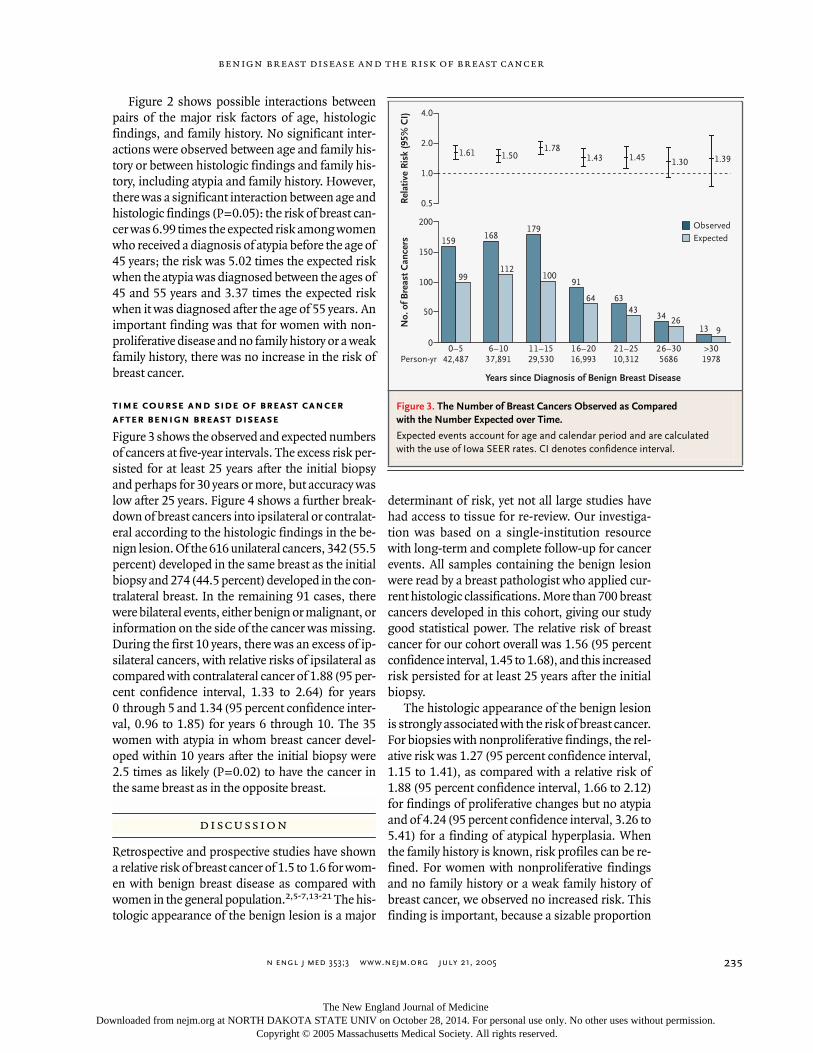
time course and side of breast cancerafter benign breast disease
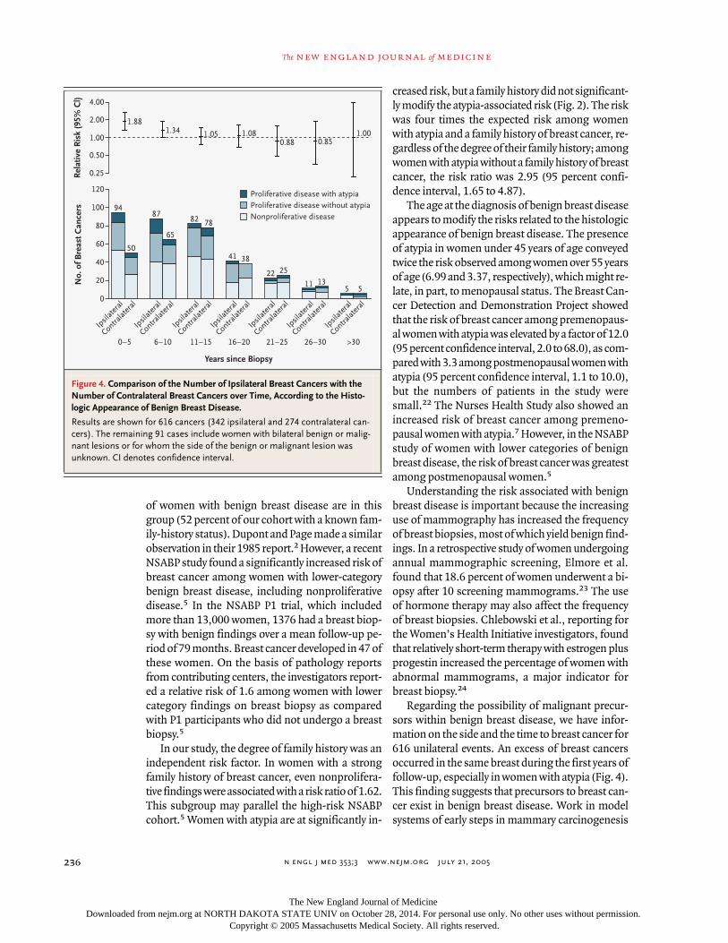
Figure 3 shows the observed and expected numbersof cancers at five-year intervals. The excess risk per-sisted for at least 25 years after the initial biopsyand perhaps for 30 years or more, but accuracy waslow after 25 years. Figure 4 shows a further break-down of breast cancers into ipsilateral or contralat-eral according to the histologic findings in the be-nign lesion. Of the 616 unilateral cancers, 342 (55.5percent) developed in the same breast as the initialbiopsy and 274 (44.5 percent) developed in the con-tralateral breast. In the remaining 91 cases, therewere bilateral events, either benign or malignant, orinformation on the side of the cancer was missing.During the first 10 years, there was an excess of ip-silateral cancers, with relative risks of ipsilateral ascompared with contralateral cancer of 1.88 (95 per-cent confidence interval, 1.33 to 2.64) for years0 through 5 and 1.34 (95 percent confidence inter-val, 0.96 to 1.85) for years 6 through 10. The 35women with atypia in whom breast cancer devel-oped within 10 years after the initial biopsy were2.5 times as likely (P=0.02) to have the cancer inthe same breast as in the opposite breast.
Retrospective and prospective studies have showna relative risk of breast cancer of 1.5 to 1.6 for wom-en with benign breast disease as compared withwomen in the general population.
2,5-7,13-21
The his-tologic appearance of the benign lesion is a major
determinant of risk, yet not all large studies havehad access to tissue for re-review. Our investiga-tion was based on a single-institution resourcewith long-term and complete follow-up for cancerevents. All samples containing the benign lesionwere read by a breast pathologist who applied cur-rent histologic classifications. More than 700 breastcancers developed in this cohort, giving our studygood statistical power. The relative risk of breastcancer for our cohort overall was 1.56 (95 percentconfidence interval, 1.45 to 1.68), and this increasedrisk persisted for at least 25 years after the initialbiopsy.
The histologic appearance of the benign lesionis strongly associated with the risk of breast cancer.For biopsies with nonproliferative findings, the rel-ative risk was 1.27 (95 percent confidence interval,1.15 to 1.41), as compared with a relative risk of1.88 (95 percent confidence interval, 1.66 to 2.12)for findings of proliferative changes but no atypiaand of 4.24 (95 percent confidence interval, 3.26 to5.41) for a finding of atypical hyperplasia. Whenthe family history is known, risk profiles can be re-fined. For women with nonproliferative findingsand no family history or a weak family history ofbreast cancer, we observed no increased risk. Thisfinding is important, because a sizable proportion
discussion
Figure 3. The Number of Breast Cancers Observed as Compared with the Number Expected over Time.
Expected events account for age and calendar period and are calculated with the use of Iowa SEER rates. CI denotes confidence interval.
No.
of B
reas
t Can
cers
Rel
ativ
e R
isk
(95%
CI)
100
150
50
0
200
159
99
168
112
179
10091
64 6343
34 2613 9
1.391.301.451.431.78
1.501.61
0.5
4.0
2.0
1.0
ObservedExpected
Years since Diagnosis of Benign Breast Disease
0–542,487Person-yr
6–1037,891
11–1529,530
16–2016,993
21–2510,312
26–305686
>301978
The New England Journal of Medicine Downloaded from nejm.org at NORTH DAKOTA STATE UNIV on October 28, 2014. For personal use only. No other uses without permission.
Copyright © 2005 Massachusetts Medical Society. All rights reserved.
n engl j med
353;3
www.nejm.org july
21
,
2005
The
new england journal
of
medicine
236
of women with benign breast disease are in thisgroup (52 percent of our cohort with a known fam-ily-history status). Dupont and Page made a similarobservation in their 1985 report.
2
However, a recentNSABP study found a significantly increased risk ofbreast cancer among women with lower-categorybenign breast disease, including nonproliferativedisease.
5
In the NSABP P1 trial, which includedmore than 13,000 women, 1376 had a breast biop-sy with benign findings over a mean follow-up pe-riod of 79 months. Breast cancer developed in 47 ofthese women. On the basis of pathology reportsfrom contributing centers, the investigators report-ed a relative risk of 1.6 among women with lowercategory findings on breast biopsy as comparedwith P1 participants who did not undergo a breastbiopsy.
5
In our study, the degree of family history was anindependent risk factor. In women with a strongfamily history of breast cancer, even nonprolifera-tive findings were associated with a risk ratio of 1.62.This subgroup may parallel the high-risk NSABPcohort.
5
Women with atypia are at significantly in-
creased risk, but a family history did not significant-ly modify the atypia-associated risk (Fig. 2). The riskwas four times the expected risk among womenwith atypia and a family history of breast cancer, re-gardless of the degree of their family history; amongwomen with atypia without a family history of breastcancer, the risk ratio was 2.95 (95 percent confi-dence interval, 1.65 to 4.87).
The age at the diagnosis of benign breast diseaseappears to modify the risks related to the histologicappearance of benign breast disease. The presenceof atypia in women under 45 years of age conveyedtwice the risk observed among women over 55 yearsof age (6.99 and 3.37, respectively), which might re-late, in part, to menopausal status. The Breast Can-cer Detection and Demonstration Project showedthat the risk of breast cancer among premenopaus-al women with atypia was elevated by a factor of 12.0(95 percent confidence interval, 2.0 to 68.0), as com-pared with 3.3 among postmenopausal women withatypia (95 percent confidence interval, 1.1 to 10.0),but the numbers of patients in the study weresmall.
22
The Nurses Health Study also showed anincreased risk of breast cancer among premeno-pausal women with atypia.
7
However, in the NSABPstudy of women with lower categories of benignbreast disease, the risk of breast cancer was greatestamong postmenopausal women.
5
Understanding the risk associated with benignbreast disease is important because the increasinguse of mammography has increased the frequencyof breast biopsies, most of which yield benign find-ings. In a retrospective study of women undergoingannual mammographic screening, Elmore et al.found that 18.6 percent of women underwent a bi-opsy after 10 screening mammograms.
23
The useof hormone therapy may also affect the frequencyof breast biopsies. Chlebowski et al., reporting forthe Women’s Health Initiative investigators, foundthat relatively short-term therapy with estrogen plusprogestin increased the percentage of women withabnormal mammograms, a major indicator forbreast biopsy.
24
Regarding the possibility of malignant precur-sors within benign breast disease, we have infor-mation on the side and the time to breast cancer for616 unilateral events. An excess of breast cancersoccurred in the same breast during the first years offollow-up, especially in women with atypia (Fig. 4).This finding suggests that precursors to breast can-cer exist in benign breast disease. Work in modelsystems of early steps in mammary carcinogenesis
Figure 4. Comparison of the Number of Ipsilateral Breast Cancers with the Number of Contralateral Breast Cancers over Time, According to the Histo-logic Appearance of Benign Breast Disease.
Results are shown for 616 cancers (342 ipsilateral and 274 contralateral can-cers). The remaining 91 cases include women with bilateral benign or malig-nant lesions or for whom the side of the benign or malignant lesion was unknown. CI denotes confidence interval.
No.
of B
reas
t Can
cers
Rel
ativ
e R
isk
(95%
Cl)
40
60
80
100
20
0
Years since Biopsy
120
Ipsil
atera
l
Contralat
eral
Ipsil
atera
l
Contralat
eral
Ipsil
atera
l
Contralat
eral
Ipsil
atera
l
Contralat
eral
Ipsil
atera
l
Contralat
eral
Ipsil
atera
l
Contralat
eral
Ipsil
atera
l
Contralat
eral
94
50
87
65
82 78
41 38
22 25
11 135 5
1.000.850.88
1.081.051.34
0–5 6–10 11–15 16–20 21–25 26–30 >30
1.88
0.25
4.00
2.00
1.00
0.50
Nonproliferative diseaseProliferative disease without atypiaProliferative disease with atypia
The New England Journal of Medicine Downloaded from nejm.org at NORTH DAKOTA STATE UNIV on October 28, 2014. For personal use only. No other uses without permission.
Copyright © 2005 Massachusetts Medical Society. All rights reserved.
n engl j med
353;3
www.nejm.org july
21, 2005
benign breast disease and the risk of breast cancer
237
has identified alterations in key regulatory indica-tors that can be studied in selected benign breastlesions.
25,26
In summary, our study shows that histologicfeatures, the age at biopsy, and the degree of familyhistory are major determinants of the risk of breastcancer after the diagnosis of benign breast disease.We found no increased risk among women withnonproliferative lesions, unless a strong family his-tory was present. No significant interaction betweenatypia and family history was apparent. The excess
risk of cancer in the ipsilateral breast in the first 10years after the diagnosis of benign breast disease,especially in women with atypia, points to the pres-ence of precursors in some women.
Supported by a Department of Defense Center of ExcellenceGrant (FEDDAMD17-02-1-0473-1), a grant (R01 CA46332) fromthe National Institutes of Health, a grant (BCTR99-3152) from theSusan G. Komen Breast Cancer Foundation, the Breast Cancer Re-search Foundation, and the Andersen Foundation.
We are indebted to Joel Worra and Dr. Piet de Groen for databasedevelopment; to Teresa Allers, Mary Amundsen, Mary Campion,Lois Penheiter, and Romayne Thompson for data collection; and toAnn Harris and the Survey Research Center for patient follow-up.
references
1.
Connolly JL, Schnitt SJ. Benign breastdisease: resolved and unresolved issues.Cancer 1993;71:1187-9.
2.
Dupont WD, Page DL. Risk factors forbreast cancer in women with proliferativebreast disease. N Engl J Med 1985;312:146-51.
3.
Gail MH, Brinton LA, Byar DP, et al. Pro-jecting individualized probabilities of devel-oping breast cancer for white females whoare being examined annually. J Natl CancerInst 1989;81:1879-86.
4.
Fitzgibbons PL, Henson DE, Hutter RV.Benign breast changes and the risk for sub-sequent breast cancer: an update of the 1985consensus statement, Cancer Committee ofthe College of American Pathologists. ArchPathol Lab Med 1998;122:1053-5.
5.
Wang J, Costantino JP, Tan-Chiu E,Wickerham DL, Paik S, Wolmark N. Lower-category benign breast disease and the riskof invasive breast cancer. J Natl Cancer Inst2004;96:616-20.
6.
Carter CL, Corle DK, Micozzi MS,Schatzkin A, Taylor PR. A prospective studyof the development of breast cancer in16,692 women with benign breast disease.Am J Epidemiol 1988;128:467-77.
7.
London SJ, Connolly JL, Schnitt SJ,Colditz GA. A prospective study of benignbreast disease and the risk of breast cancer.JAMA 1992;267:941-4. [Erratum, JAMA1992;267:1780.]
8.
Schnitt SJ. Benign breast disease andbreast cancer risk: potential role for anties-trogens. Clin Cancer Res. 2001;7:Suppl:4411s-4422s.
9.
Melton LJ III. The threat to medical-records research. N Engl J Med 1997;337:1466-70.
10.
Page DL, Dupont WD, Rogers LW, Ra-dos MS. Atypical hyperplastic lesions of thefemale breast: a long-term follow-up study.Cancer 1985;55:2698-708.
11.
Surveillance, Epidemiology, and EndResults (SEER) Program. SEER statistics da-tabase. (Accessed June 27, 2005, at http://www.seer.cancer.gov.)
12.
Bain LJ, Englehardt M. Introduction toprobability and mathematical statistics. 2nded. Boston: PWS-Kent Publishing, 1992:369-77.
13.
Page DL, Schuyler PA, Dupont WD,Jensen RA, Plummer WD Jr, Simpson JF.Atypical lobular hyperplasia as a unilateralpredictor of breast cancer risk: a retrospec-tive cohort study. Lancet 2003;361:125-9.[Erratum, Lancet 2003;361:1994.]
14.
Dupont WD, Page DL. Breast cancer riskassociated with proliferative disease, age atfirst birth, and a family history of breast can-cer. Am J Epidemiol 1987;125:769-79.
15.
Jensen RA, Page DL, Dupont WD, Rog-ers LW. Invasive breast cancer risk in womenwith sclerosing adenosis. Cancer 1989;64:1977-83.
16.
Dupont WD, Page DL, Parl FF, et al.Long-term risk of breast cancer in womenwith fibroadenoma. N Engl J Med 1994;331:10-5.
17.
Marshall LM, Hunter DJ, Connolly JL, etal. Risk of breast cancer associated withatypical hyperplasia of lobular and ductaltypes. Cancer Epidemiol Biomarkers Prev1997;6:297-301.
18.
Rohan TE, Hartwick W, Miller AB, Kan-del RA. Immunohistochemical detection ofc-erbB-2 and p53 in benign breast diseaseand breast cancer risk. J Natl Cancer Inst1998;90:1262-9.
19.
Kandel R, Li SQ, Ozcelik H, Rohan T.p53 Protein accumulation and mutations innormal and benign breast tissue. Int J Can-cer 2000;87:73-8.
20.
Kandel R, Zhu XL, Li SQ, Rohan T. CyclinD1 protein overexpression and gene amplifi-cation in benign breast tissue and breast can-cer risk. Eur J Cancer Prev 2001;10:43-51.
21.
Krieger N, Hiatt RA. Risk of breast can-cer after benign breast diseases: variation byhistologic type, degree of atypia, age at biop-sy, and length of follow-up. Am J Epidemiol1992;135:619-31.
22.
Dupont WD, Parl FF, Hartmann WH, etal. Breast cancer risk associated with prolif-erative breast disease and atypical hyperpla-sia. Cancer 1993;71:1258-65.
23.
Elmore JG, Barton MB, Moceri VM, PolkS, Arena PJ, Fletcher SW. Ten-year risk offalse positive screening mammograms andclinical breast examinations. N Engl J Med1998;338:1089-96.
24.
Chlebowski RT, Hendrix SL, Langer RD,et al. Influence of estrogen plus progestinon breast cancer and mammography inhealthy postmenopausal women: the Wom-en's Health Initiative Randomized Trial.JAMA 2003;289:3243-53.
25.
Crawford YG, Gauthier ML, Joubel A, etal. Histologically normal human mammaryepithelia with silenced p16(INK4a) overex-press COX-2, promoting a premalignantprogram. Cancer Cell 2004;5:263-73.
26.
Li JJ, Weroha SJ, Lingle WL, Papa D, Sal-isbury JL, Li SA. Estrogen mediates Aurora-A overexpression, centrosome amplifica-tion, chromosomal instability, and breastcancer in female ACI rats. Proc Natl Acad SciU S A 2004;101:18123-8.
Copyright © 2005 Massachusetts Medical Society.
The New England Journal of Medicine Downloaded from nejm.org at NORTH DAKOTA STATE UNIV on October 28, 2014. For personal use only. No other uses without permission.
Copyright © 2005 Massachusetts Medical Society. All rights reserved.


![Benign Breast Disease[1]](https://img.dokumen.tips/doc/110x75/5571f7b649795991698bd982/benign-breast-disease1.jpg)