Embed Size (px)
Citation preview
fmicb-08-00177 February 11, 2017 Time: 14:46 # 1
PERSPECTIVEpublished: 14 February 2017
doi: 10.3389/fmicb.2017.00177
Edited by:Michael Sauer,
University of Natural Resourcesand Life Sciences, Vienna, Austria
Reviewed by:Linda Christine DeVeaux,
South Dakota School of Minesand Technology, USA
Alexandros G. Georgakilas,National Technical University
of Athens, GreeceMario Xavier Ruiz-González,
Secretaría de Educación Superior,Ciencia, Tecnología e Innovación and
Universidad Técnica Particularde Loja, Ecuador
*Correspondence:Hugo Castillo
Specialty section:This article was submitted to
Microbial Physiology and Metabolism,a section of the journal
Frontiers in Microbiology
Received: 24 October 2016Accepted: 24 January 2017
Published: 14 February 2017
Citation:Castillo H and Smith GB (2017)
Below-Background Ionizing Radiationas an Environmental Cue for Bacteria.
Front. Microbiol. 8:177.doi: 10.3389/fmicb.2017.00177
Below-Background IonizingRadiation as an Environmental Cuefor BacteriaHugo Castillo* and Geoffrey B. Smith
Department of Biology, New Mexico State University, Las Cruces, NM, USA
All organisms on earth grow under the influence of a natural and relatively constantdose of ionizing radiation referred to as background radiation, and so cells havedifferent mechanisms to prevent the accumulation of damage caused by its differentcomponents. However, current knowledge of the deleterious effects of radiation on cellsis based on the exposure to acute and high or to chronic, above background dosesof radiation and therefore is not appropriate to explain the cellular and biochemicalmechanisms that cells employ to sense and respond to chronic below-backgroundlevels. Studies at below-background radiation doses can provide insight into thebiological role of radiation, as suggested by several examples of what appears to bea stress response in cells grown at doses that range from 10 to 79 times lower thanbackground. Here, we discuss some of the technical constraints to shield cells fromradiation to below-background levels, as well as different approaches used to detectand measure responses to such unusual environmental conditions. Then, we presentdata from Shewanella oneidensis and Deinococcus radiodurans experiments that showhow two taxonomically distant bacterial species sense and respond to unnaturally lowlevels of radiation. In brief, we grew S. oneidensis and D. radiodurans in liquid culture atdose rates of 72.05 (control) and 0.91 (treatment) nGy hr−1 (including radon) for up to72 h and measured cell density and the expression of stress-related genes. Our resultssuggest that a stress response is triggered in the absence of normal levels of radiation.
Keywords: below-background radiation, stress response, relative gene expression, radiation biology, Shewanellaoneidensis, Deinococcus radiodurans
INTRODUCTION
Ionizing radiation is a ubiquitous environmental stress that has been present throughout the lifehistory of all organisms on earth (Karam and Leslie, 2005). It would be expected that adaptivemolecular mechanisms have been selected in response to the constant exposure to this naturalbackground radiation dose. Although this background dose varies significantly across geographicalareas (Brooks, 2012), the worldwide average dose has been estimated to be 2400 µGy yr−1 (Thorne,2003) and only the proximity to natural and human activity-derived sources such as radioactiveminerals, medical exposure or nuclear energy-related events significantly increase this number. Itshould be noted, however, that the background dose range has been established solely for humanexposure protection purposes and that radon, one of its main components, is not relevant forbacterial models. Also, the variability of background radiation occurs only in one direction (above
Frontiers in Microbiology | www.frontiersin.org 1 February 2017 | Volume 8 | Article 177

fmicb-08-00177 February 11, 2017 Time: 14:46 # 2
Castillo and Smith Stress Induction by Below-Background Radiation
background), as there are no instances of a natural environmentwith the levels we have imposed in our experiment.
Ionizing radiation, such as gamma rays, deposit energy asthey traverse cells causing DNA lesions, membrane damage,and the induction of pro-apoptotic genes (Todorovic et al.,2008). Ionizing radiation can cause significant increases in theconcentration of intracellular reactive oxygen species, ROS,(Ghosal et al., 2005) and in bacteria, the SOS response (Brena-Valle and Serment-Guerrero, 1998). These reactive species triggerthe expression of enzymes that scavenge for ROS in orderto protect against protein and DNA damage (Daly, 2009).Background radiation is part of the physical environment thatbacteria may sense and react to in order to regulate homeostasis.If bacteria have adapted to thrive in the presence of radiation, howwould they react to a significant decrease in radiation dose rate?Could cells translate the “absence” of radiation into a modulatingsignal to initiate a specific response? Could such a reaction bedefined as a stress response? We propose that research on thebiological responses to reduced-radiation environments, “fromthe other side of background” (Smith et al., 2011), can yieldunique information that will inform both the underlying scienceand policy applications of radiation protection. It is necessaryto consider that natural background radiation is made up ofboth low and high LET (Linear Energy Transfer) sources andthat the latter tend to have stronger biological effects (Goodhead,1999). However, researchers at Italy’s Gran Sasso lab and IstitutoSuperiore di Sanita (Belli et al., 1989; Goodhead et al., 2009)have shown that the biological effects on Chinese hamster V79cells also depends on particle type, with protons causing morecell inactivation than α-particles at the same LET. In this report,because of the experimental set-up (shielding from the saltoverburden and steel vault), the observed effects are largely dueto low-LET sources.
Traditional radiation biology experiments have resulted in animpressive amount of data on the effects of acute exposure todifferent sources of ionizing radiation at different dose rates, alladditional to natural background radiation. The study of theeffects of radiation at doses below background, however, remainsrelatively unexplored due to the difficulties of data acquisition.The first evidence of a deleterious biological response to radiationdoses lower than background was reported by Planel et al. (1987),who observed a longer generation time in Paramecium tetraureliaand a lower cell number in Synechococcus lividus cultures whenthese organisms were grown shielded from radiation at doserates 17 and 6 times lower than background, respectively. Sincethen, experiments with other organisms have shown similardeleterious effects when exposed to below background levels ofradiation, such as lower protection to mutational damage inSaccharomyces cerevisiae (Satta et al., 1995); higher sensitivity toapoptosis, higher intracellular concentration of oxidative stress-related enzymes, and higher mutation rate induced by gammarays (after conditioning under below background conditions) inCricetulus griseus (Satta et al., 2002); growth rate retardation inMus musculus L5178Y cells (Kawanishi et al., 2012), changesin the concentration of oxidative stress-related enzymes (Fratiniet al., 2015), and lower cell number and/or up regulation ofDNA repair and oxidative stress genes in Shewanella oneidensis
and Deinococcus radiodurans (Smith et al., 2011; Castillo et al.,2015).
Below background radiation biology experiments have beenhampered by the enormous technical constrains to effectivelyachieve radiation doses below background levels. Here, wepresent the Low Background Radiation Experiment (LBRE) atthe Waste Isolation Pilot Plant (WIPP) in Carlsbad, NM., asan effective way to achieve an estimated 79-fold reduction inionizing radiation dose (relative to gamma) and propose twobacterial models as reference points to study the detectionof ultra-low levels of ionizing radiation and the use of geneexpression analysis to describe their molecular response to thedeprivation of natural levels of radiation.
THE CHALLENGE OFBELOW-BACKGROUND RADIATIONBIOLOGY WORK
Shielding organisms from the different components ofbackground radiation down to levels that are significantlybelow-background is not an easy task. Protection from cosmicrays and cosmic rays-derived components can be achieved atsites located deep underground or protected by a significantoverburden, common for numerous physics experiments thatrequire radiation “free” conditions, while protection fromemissions derived from the decay of naturally occurringradioisotopes depends on the abundance of these elements in theimmediate surroundings of the experiment. To our knowledge,only two laboratories that meet the first condition are currentlyrunning biology experiments to explore the effects of radiationdeprivation on different organismal models: (1) The LaboratoriNazionali del Gran Sasso in L’Aquila, Italy, has established the“Cosmic Silence Program” to study the contribution of naturalradiation to different molecular processes leading to the defenseagainst oxidative stress (dps) using yeast, rodent, and human cellsas models, at a dose rate 10 times lower than background1; and(2) the Laboratoire Souterrain de Modane, in Modane, France,who initiated the IRIS Project (Impacts of Ionizing Radiation onLife) to study the effect of ionizing radiation on the evolution ofEscherichia coli exposed to chronic doses 30 times lower thantheir reference background2.
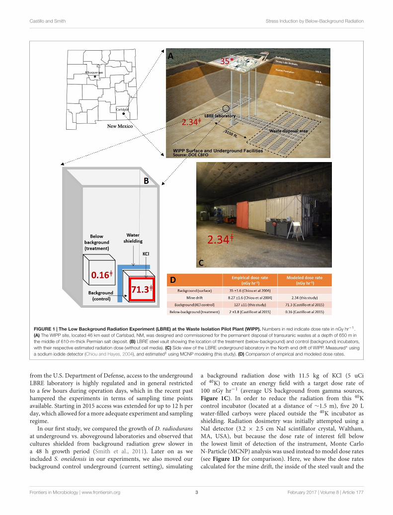
The LBRE laboratory, hosted by the WIPP in Carlsbad, NM.,lays at a depth of 660 m within a 610 m thick halite layer(Salado formation) with an isotope composition of 29, 78, and169 ppb for 238U, 232Th, and 40K, respectively (Department ofEnergy, 1996)3. This NaCl-based salt deposit emits significantlyless radiation than other rock formations like limestone andgranite (Figure 1A). Additionally, part of the LBRE experimentsare conducted inside a 15 cm-thick vault made from pre-WorldWar II, low-activity steel to further reduce exposure to radiation(Figure 1B). As the main mission of the WIPP site is thepermanent disposal of low-level radioactive transuranic waste
1https://www.lngs.infn.it/en/pulex-cosmic-silence2http://www-lsm.in2p3.fr/activites/autres_activ/Bio.htm3http://wipp.energy.gov/science/biology/biology2.html
Frontiers in Microbiology | www.frontiersin.org 2 February 2017 | Volume 8 | Article 177
fmicb-08-00177 February 11, 2017 Time: 14:46 # 3
Castillo and Smith Stress Induction by Below-Background Radiation
FIGURE 1 | The Low Background Radiation Experiment (LBRE) at the Waste Isolation Pilot Plant (WIPP). Numbers in red indicate dose rate in nGy hr−1.(A) The WIPP site, located 46 km east of Carlsbad, NM, was designed and commissioned for the permanent disposal of transuranic wastes at a depth of 650 m inthe middle of 610-m-thick Permian salt deposit. (B) LBRE steel vault showing the location of the treatment (below-background) and control (background) incubators,with their respective estimated radiation dose (without cell media). (C) Side view of the LBRE underground laboratory in the North end drift of WIPP. Measured∗ usinga sodium iodide detector (Chiou and Hayes, 2004), and estimated‡ using MCNP modeling (this study). (D) Comparison of empirical and modeled dose rates.
from the U.S. Department of Defense, access to the undergroundLBRE laboratory is highly regulated and in general restrictedto a few hours during operation days, which in the recent pasthampered the experiments in terms of sampling time pointsavailable. Starting in 2015 access was extended for up to 12 h perday, which allowed for a more adequate experiment and samplingregime.
In our first study, we compared the growth of D. radioduransat underground vs. aboveground laboratories and observed thatcultures shielded from background radiation grew slower ina 48 h growth period (Smith et al., 2011). Later on as weincluded S. oneidensis in our experiments, we also moved ourbackground control underground (current setting), simulating
a background radiation dose with 11.5 kg of KCl (5 uCiof 40K) to create an energy field with a target dose rate of100 nGy hr−1 (average US background from gamma sources,Figure 1C). In order to reduce the radiation from this 40Kcontrol incubator (located at a distance of ∼1.5 m), five 20 Lwater-filled carboys were placed outside the 40K incubator asshielding. Radiation dosimetry was initially attempted using aNaI detector (3.2 × 2.5 cm NaI scintillator crystal, Waltham,MA, USA), but because the dose rate of interest fell belowthe lowest limit of detection of the instrument, Monte CarloN-Particle (MCNP) analysis was used instead to model dose rates(see Figure 1D for comparison). Here, we show the dose ratescalculated for the mine drift, the inside of the steel vault and the
Frontiers in Microbiology | www.frontiersin.org 3 February 2017 | Volume 8 | Article 177

fmicb-08-00177 February 11, 2017 Time: 14:46 # 4
Castillo and Smith Stress Induction by Below-Background Radiation
inside of both the treatment and control incubators, using themass fraction and the specific activity for 238U, 232Th, 40K, and222Rn.
Independent of cell-type and growth media, the calculateddose rate values represent a 445-fold difference in the twoexperimental treatment locations (Figure 1B). However, whenthe contribution to dose rate from the 40K in the growth mediawas calculated based on the total potassium content in thecomponents of 1.5 mL of TGY [24.45 and 143.77 µg K intryptone and yeast extract, respectively; BD Becton Dickinson(2006)], this adds 0.75 nGy hr−1 to both treatments. Therefore,when both environmental and media radiation sources areconsidered, the cells’ dose in the treatment (0.91 nGy hr−1)represents a 79-fold reduction from that in the control (72.05 nGyhr−1).
Shewanella oneidensis and D. radiodurans cultures were grownas previously described (Castillo et al., 2015). In summary,both bacteria were grown in a shielded treatment incubatorand a radiation-supplemented control incubator for 24 h asa conditioning period, after which they were transferred intofresh broth to initiate the experiment. At this point, radiation-deprived cultures were also transferred back into the KClcontrol incubator in order to start a reciprocal control by“rescuing” cells and restoring them to normal levels of radiation.Optical density at 630 nm was measured at different timepoints (5, 8, 13, 24, and 29 for S. oneidensis and 10, 24,29, 34, and 48 for D. radiodurans) to quantify and comparethe growth of the cultures using a Student’s t-test (n = 12).In parallel, cells were sampled, treated with RNA protect(QIAGEN, Valencia, CA, USA) and kept frozen for furtheranalysis. Total RNA was extracted using the RNeasy Mini kit(QIAGEN, Valencia, CA, USA) and used as template in RT-quantitative PCR to measure the differential expression of stress-related genes: katB, recA, oxyR, lexA, dnaK, and SOA0154 forS. oneidensis and DR1998 (katB), DR0615 (oxyR), DRA0344(lexA), recA, dnaK, DR2263 (dps), and DR1343 (gapdH) forD. radiodurans, using an efficiency-corrected approach (Pfafflet al., 2002).
BACTERIA RESPONSE TO RADIATIONDEPRIVATION
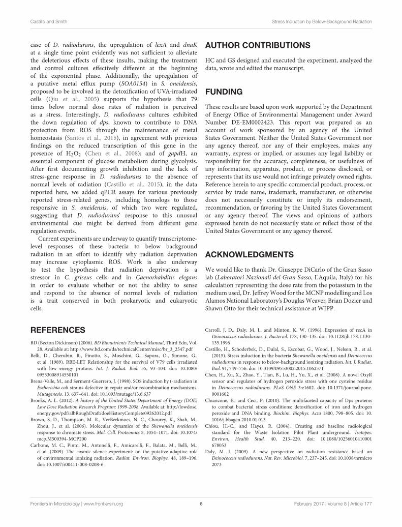
In our previous report (Castillo et al., 2015), we showedgrowth inhibition and/or the upregulation of stress-related genesin experiments with limited time points. In the experimentreported here with more site access, S. oneidensis cultures didnot show a significant difference in growth in response to thereduced radiation dose (Figure 2A), whereas D. radioduransgrowth became inhibited at the beginning of its exponentialgrowth phase and remained significantly different throughout theduration of the experiment (Figure 2B). As reported previously,when reciprocal controls were carried out in which radiation-deprived cells were restored to radiation-sufficient conditions,growth of D. radiodurans recovered and approximated thecell density of the control (dotted line, Figure 2B). However,using growth as the only endpoint to measure the effect of
a particular environmental variable might be misleading sincecells employ multiple physiological mechanisms in responseto changing environments to maintain homeostasis. In orderto circumvent this limitation, similar low-radiation doseexperiments have used different approaches for a more in-depth analysis of the cellular response, such as micronucleusassays (Carbone et al., 2009), enzymatic activity (Satta et al.,2002; Carbone et al., 2009; Fratini et al., 2015), mutation assays(Satta et al., 1995, 2002; Fratini et al., 2015), and differentialgene expression (Castillo et al., 2015; Fratini et al., 2015).In the present study, we measured the expression of genesrelated to some of these types of stress, on cells collectedat various time points in order to compare their differentialexpression.
First it should be noted the different growth kinetics ofS. oneidensis (doubling time = 1.1 h) and D. radiodurans(doubling time = 3 h) under our culture conditions (1.5 mLTGY broth, 30◦C, 200 rpm in 24-well plate) and so the samegrowth stage in the two species are represented by offset times.In S. oneidensis, 33% of the genes were significantly upregulatedin the minus-radiation treatment (as defined by greater thantwofold change, p < 0.10), whereas in D. radiodurans only 4.5and 6.8% were up or downregulated, respectively (Figures 2A,B).The gene expression data in Figure 2A shows S. oneidensis tohave upregulated five of the six stress genes tested during midlog phase and then Shewanella tapered expression back duringlate-log and early stationary phase. In contrast, D. radioduransonly upregulated 2 of 11 genes during a single time point atearly log phase; at 24 h D. radiodurans also down regulateddps and gapdH (Figure 2C), and at 34 h downregulateddnaK. The upregulated genes in S. oneidensis are indicativeof a stress response, being involved in activities such as ROSscavenging (katB, oxyR), DNA repair (recA, lexA), protein folding(dnaK), and metal efflux (SOA0154). However, D. radioduransupregulated lexA and dnaK at 24 h but at the same time,also downregulated two other genes involved in dps and ATPsynthesis (gapdH).
During mid-exponential phase (8 h in S. oneidensis), sixgenes related to oxidative stress response (katB, oxyR), DNArepair (recA, lexA), protein folding (dnaK), and a putative effluxpump (SOA0154) were again significantly upregulated as wereported previously (Castillo et al., 2015). Oxidative stress andefflux pump activity was initiated and maintained during late-exponential (13 h) but dissipated by early stationary phase(17 h). Growth inhibition in D. radiodurans cultures becamesignificant (p < 0.05) during mid exponential phase (34 h)and was maintained throughout the duration of the experiment.The difference in gene expression, however, was only observedat 24 h when lexA and dnaK were significantly upregulated,suggesting an increase in DNA repair and protein foldingactivities, while dps, and gapdH were downregulated, suggestinga diminished capacity to fend off oxidative stress (Chiancone andCeci, 2010) and synthesize ATP, respectively. Moreover, whenwe transferred cells from the reduced radiation treatment to thecontrol incubator (orange bars in Figure 2C), the regulation ofthe genes dissipated supporting our hypothesis that these effectswere due to the absence of normal levels of radiation.
Frontiers in Microbiology | www.frontiersin.org 4 February 2017 | Volume 8 | Article 177

fmicb-08-00177 February 11, 2017 Time: 14:46 # 5
Castillo and Smith Stress Induction by Below-Background Radiation
FIGURE 2 | Upregulation of stress genes in Shewanella evidently protects cells from growth inhibition by radiation deprivation. Line graphs showbacterial growth under radiation-sufficient (background) and radiation-deprived (below background) conditions with p-values shown above timepoints (the dottedreciprocal control was shown to be statistically the same as the background control). Both S. oneidensis and D. radiodurans were grown in 1.5 mL of TGY broth(0.5% Bacto tryptone, 0.3% yeast extract, and 0.1% glucose) in 24-wells plates, shaken at 200 rpm and at 30◦ C. Bar graphs show the percentage of genes testedthat were upregulated (as defined by > 2-fold change relative to rad-sufficient controls, p < 0.10) in (A) S. oneidensis and (B) D. radiodurans. (C) Differential geneexpression in S. oneidensis and D. radiodurans calculated using the efficiency-corrected 1Cp equation in the software tool REST2009 (Pfaffl et al., 2002). RT-qPCRon S. oneidensis RNA samples was performed with the iScript One-Step RT-PCR kit (BioRad, Hercules, CA, USA) and on D. radiodurans RNA samples with theiScript Reverse Transcription Supermix for RT-qPCR (BioRad, Hercules, CA, USA) followed by the SsoAdvanced Universal SYBR Green Supermix (BioRad, Hercules,CA, USA) according to the manufacturer’s instructions. The expression of target genes was normalized using rRNA (D. radiodurans) or rRNA and rpoA(S. oneidensis) as reference. For D. radiodurans, the expression of groEL, sufB, DR1838 (stringent factor) and DR2422 (large conductance mechanosensitivechannel) was not regulated, and therefore not shown in the figure.
A BIOLOGICAL ROLE FORBACKGROUND IONIZING RADIATION?
We propose that S. oneidensis has mounted a classic stress-response to this unusual environmental cue of the deprivationof natural levels of environmental radiation (and its growth wasthus not inhibited) whereas D. radiodurans has not sensed thisstress (and thus its growth was inhibited). A basic element ofthe biological stress response at the cellular level is to return theorganism back to homeostasis (Kultz, 2005) and those organismsnot capable of sensing and responding to stressors would beat a distinct fitness disadvantage. For example, in mammaliansystems, deletion of the chemical and radiation stress sensorprotein, INrf2 leads to adverse effects on cell survival (Lee et al.,2007). We propose here that the different response to the absenceof radiation by our two model organisms led to a fitness cost inthe organism (D. radiodurans) that did not mount an effectivestress response.
The upregulation of katB in S. oneidensis has also beenobserved upon exposure to other stressors, such as H2O2,chromate and ionizing radiation (Brown et al., 2006; Qiu et al.,2006; Jiang et al., 2014), stressors known to induce oxidativestress. Interestingly, the upregulation of recA and lexA has alsobeen reported as a direct effect of exposure to ionizing radiationand chromate in S. oneidensis (Brown et al., 2006; Qiu et al., 2006)and of recA in D. radiodurans after gamma irradiation (Carrollet al., 1996). The upregulation of dnaK has been associated withchromate shock in S. oneidensis (Brown et al., 2006), oxidative,heat and acid stresses in L. lactis (Smith et al., 2010), and ionicsilver in E. coli (Salou-Berion et al., 2015).
Our differential gene expression analysis shows that whengrown under radiation-reduced conditions, S. oneidensisincreases the transcription of this suite of genes, suggestingan effective response to an increase of intracellular ROS, therepair of DNA breakage, and the need of re-folding damagedproteins, allowing for the resumption of normal growth. In the
Frontiers in Microbiology | www.frontiersin.org 5 February 2017 | Volume 8 | Article 177
fmicb-08-00177 February 11, 2017 Time: 14:46 # 6
Castillo and Smith Stress Induction by Below-Background Radiation
case of D. radiodurans, the upregulation of lexA and dnaKat a single time point evidently was not sufficient to alleviatethe deleterious effects of these insults, making the treatmentand control cultures effectively different at the beginningof the exponential phase. Additionally, the upregulation ofa putative metal efflux pump (SOA0154) in S. oneidensis,proposed to be involved in the detoxification of UVA-irradiatedcells (Qiu et al., 2005) supports the hypothesis that 79times below normal dose rates of radiation is perceivedas a stress. Interestingly, D. radiodurans cultures exhibitedthe down regulation of dps, known to contribute to DNAprotection from ROS through the maintenance of metalhomeostasis (Santos et al., 2015), in agreement with previousfindings on the reduced transcription of this gene in thepresence of H2O2 (Chen et al., 2008); and of gapdH, anessential component of glucose metabolism during glycolysis.After first documenting growth inhibition and the lack ofstress-gene response in D. radiodurans to the absence ofnormal levels of radiation (Castillo et al., 2015), in the datareported here, we added qPCR assays for various previouslyreported stress-related genes, including homologs to thoseresponsive in S. oneidensis, of which two were regulated,suggesting that D. radiodurans’ response to this unusualenvironmental cue might be derived from different generegulation events.
Current experiments are underway to quantify transcriptome-level responses of these bacteria to below backgroundradiation in an effort to identify why radiation deprivationmay increase cytoplasmic ROS. Work is also underwayto test the hypothesis that radiation deprivation is astressor in C. griseus cells and in Caenorhabditis elegansin order to evaluate whether or not the ability to senseand respond to the absence of normal levels of radiationis a trait conserved in both prokaryotic and eukaryoticcells.
AUTHOR CONTRIBUTIONS
HC and GS designed and executed the experiment, analyzed thedata, wrote and edited the manuscript.
FUNDING
These results are based upon work supported by the Departmentof Energy Office of Environmental Management under AwardNumber DE-EM0002423. This report was prepared as anaccount of work sponsored by an agency of the UnitedStates Government. Neither the United States Government norany agency thereof, nor any of their employees, makes anywarranty, express or implied, or assumes any legal liability orresponsibility for the accuracy, completeness, or usefulness ofany information, apparatus, product, or process disclosed, orrepresents that its use would not infringe privately owned rights.Reference herein to any specific commercial product, process, orservice by trade name, trademark, manufacturer, or otherwisedoes not necessarily constitute or imply its endorsement,recommendation, or favoring by the United States Governmentor any agency thereof. The views and opinions of authorsexpressed herein do not necessarily state or reflect those of theUnited States Government or any agency thereof.
ACKNOWLEDGMENTS
We would like to thank Dr. Giuseppe DiCarlo of the Gran Sassolab (Laboratori Nazionali del Gran Sasso, L’Aquila, Italy) for hiscalculation representing the dose rate from the potassium in themedium used, Dr. Jeffrey Wood for the MCNP modelling and LosAlamos National Laboratory’s Douglas Weaver, Brian Dozier andShawn Otto for their technical assistance at WIPP.
REFERENCESBD (Becton Dickinson) (2006). BD Bionutrients Technical Manual, Third Edn, Vol.
28. Available at: http://www.bd.com/ds/technicalCenter/misc/br_3_2547.pdfBelli, D., Cherubin, R., Finotto, S., Moschini, G., Sapora, O., Simone, G.,
et al. (1989). RBE-LET Relationship for the survival of V79 cells irradiatedwith low energy protons. Int. J. Radiat. Biol. 55, 93–104. doi: 10.1080/09553008914550101
Brena-Valle, M., and Serment-Guerrero, J. (1998). SOS induction by (-radiation inEscherichia coli strains defective in repair and/or recombination mechanisms.Mutagenesis. 13, 637–641. doi: 10.1093/mutage/13.6.637
Brooks, A. L. (2012). A history of the United States Department of Energy (DOE)Low Dose Radiation Research Program: 1999-2008. Available at: http://lowdose.energy.gov/pdf/albRoughDraft/doeHistoryComplete09262012.pdf
Brown, S. D., Thompson, M. R., VerBerkmoes, N. C., Chourey, K., Shah, M.,Zhou, J., et al. (2006). Molecular dynamics of the Shewanella oneidensisresponse to chromate stress. Mol. Cell. Proteomics 5, 1054–1071. doi: 10.1074/mcp.M500394-MCP200
Carbone, M. C., Pinto, M., Antonelli, F., Amicarelli, F., Balata, M., Belli, M.,et al. (2009). The cosmic silence experiment: on the putative adaptive roleof environmental ionizing radiation. Radiat. Environ. Biophys. 48, 189–196.doi: 10.1007/s00411-008-0208-6
Carroll, J. D., Daly, M. J., and Minton, K. W. (1996). Expression of recA inDeinococcus radiodurans. J. Bacteriol. 178, 130–135. doi: 10.1128/jb.178.1.130-135.1996
Castillo, H., Schoderbek, D., Dulal, S., Escobar, G., Wood, J., Nelson, R., et al.(2015). Stress induction in the bacteria Shewanella oneidensis and Deinococcusradiodurans in response to below-background ionizing radiation. Int. J. Radiat.Biol. 91, 749–756. doi: 10.3109/09553002.2015.1062571
Chen, H., Xu, X., Zhao, Y., Tian, B., Lu, H., Yu, X., et al. (2008). A novel OxyRsensor and regulator of hydrogen peroxide stress with one cysteine residuein Deinococcus radiodurans. PLoS ONE 3:e1602. doi: 10.1371/journal.pone.0001602
Chiancone, E., and Ceci, P. (2010). The multifaceted capacity of Dps proteinsto combat bacterial stress conditions: detoxification of iron and hydrogenperoxide and DNA binding. Biochim. Biophys. Acta 1800, 798–805. doi: 10.1016/j.bbagen.2010.01.013
Chiou, H.-C., and Hayes, R. (2004). Creating and baseline radiologicalstandard for the Waste Isolation Pilot Plant underground. Isotopes.Environ. Health Stud. 40, 213–220. doi: 10.1080/10256010410001678053
Daly, M. J. (2009). A new perspective on radiation resistance based onDeinococcus radiodurans. Nat. Rev. Microbiol. 7, 237–245. doi: 10.1038/nrmicro2073
Frontiers in Microbiology | www.frontiersin.org 6 February 2017 | Volume 8 | Article 177

fmicb-08-00177 February 11, 2017 Time: 14:46 # 7
Castillo and Smith Stress Induction by Below-Background Radiation
Department of Energy (1996). Title 40 CFR part 191 Compliance CertificationApplication. Carlsbad, NM: United States Department of Energy WasteIsolation Pilot Plant.
Fratini, E., Carbone, C., Capece, D., Esposito, G., Simone, G., Tabocchini, M. A.,et al. (2015). Low-radiation environment affects the development of protectionmechanisms in V79 cells. Radiat. Environ. Biophys. 54, 183–194. doi: 10.1007/s00411-015-0587-4
Ghosal, D., Omelchenko, M. V., Gaidamakova, E. K., Matrosova, V. Y.,Vasilenko, A., Venkateswaran, A., et al. (2005). How radiation kills cells:survival of Deinococcus radiodurans and Shewanella oneidensis under oxidativestress. FEMS Microbiol. Rev. 29, 361–375. doi: 10.1016/j.fmrre.2004.12.007
Goodhead, D., Belli, M., Mill, A. J., Bance, D. A., Allens, L. A., Hall, S. C.,et al. (2009). Direct Comparison between protons and alpha-particles of thesame LET: I. Irradiation methods and inactivation of asynchronous V79,HeLa and C3H 10T1/2 cells. Int. J. Radiat. Biol. 61, 611–624. doi: 10.1080/09553009214551421
Goodhead, D. T. (1999). Mechanisms for the biological effectiveness of high-LETradiation. J. Radiat. Res. 40, 1–13. doi: 10.1269/jrr.40.S1
Jiang, Y., Dong, Y., Luo, Q., Li, N., Wu, G., and Gao, H. (2014). Protection fromoxidative stress relies mainly on derepression of OxyR-dependent and Dps inShewanella oneidensis. J. Bacteriol. 196, 445–458. doi: 10.1128/JB.01077-13
Karam, P. A., and Leslie, S. A. (2005). Changes in terrestrial natural radiationlevels over the history of life. Radioact. Environ. 7, 107–117. doi: 10.1097/HP.0b013e3182118094
Kawanishi, M., Okuyama, K., Shiraishi, K., Matsuda, Y., Taniguchi, R., Shiomi, N.,et al. (2012). Growth retardation of paramecium and mouse cells by shieldingthem from background radiation. J. Radiat. Res 53, 404–410. doi: 10.1269/jrr.11145
Kultz, D. (2005). Molecular and evolutionary basis of the cellular stress response.Annu. Rev. Physiol. 67, 225–257. doi: 10.1146/annurev.physiol.67.040403.103635
Lee, O.-H., Jain, A. J., Papusha, V., and Jaiswal, A. K. (2007). An auto-regulatoryloop between stress sensors INrf2 and Nrf2 controls their cellular abundance.J. Biol. Chem. 282, 36412–36420. doi: 10.1074/jbc.M706517200
Pfaffl, M. W., Horgan, G. W., and Dempfle, L. (2002). Relative expression softwaretool REST© for group-wise comparison and statistical analysis of relativeexpression results in real-time PCR. Nucleic Acids Res. 30, 1–10. doi: 10.1093/nar/30.9.e36
Planel, H., Soleilhavoup, J. P., Tizador, R., Richoilley, G., Conter, A., Croute, F., et al.(1987). Influence on cell proliferation of background radiation or exposure tovery low, chronic γ radiation. Health Phys. 52, 571–578. doi: 10.1097/00004032-198705000-00007
Qiu, X., Daly, M. J., Vasilenko, A., Omelchenko, M. V., Gaidamakova, E. K.,Wu, J., et al. (2006). Transcriptome analysis applied to survival of Shewanella
oneidensis MR-1 exposed to ionizing radiation. J. Bacteriol. 188, 1199–1204.doi: 10.1128/JB.188.3.1199-1204.2006
Qiu, X., Sundin, G. W., Wu, L., Zhou, J., and Tiedje, J. M. (2005). Comparativeanalysis of differentially expressed genes in Shewanella oneidensis MR-1following exposure to UVC. UVB and UVA radiation. J. Bacteriol. 187,3556–3564. doi: 10.1128/JB.187.10.3556-3564.2005
Salou-Berion, C., Gonzalez, I., Enjalbert, B., Audinot, J.-N., Fourquaux, I.,Jamme, F., et al. (2015). Escherichia coli under ionic silver stress:and integrative approach to explore transcriptional, physiological andbiochemical responses. PLoS ONE 10:e0145748. doi: 10.1371/journal.pone.0145748
Santos, S. P., Mitchell, E. P., Franquelim, H. G., Castanho, M. A. R. B., Abreu, I. A.,and Romao, C. V. (2015). Dps from Deinococcus radiodurans: oligomeric formsof Dps1 with distinct cellular functions and Dps2 involved in metal storage.FEBS J. 282, 4307–4327. doi: 10.1111/febs.13420
Satta, L., Antonelli, F., Belli, M., Sapora, O., Simone, G., Sorrentino, E., et al.(2002). Influence of a low background radiation environment on biochemicaland biological responses in V79 cells. Radiat. Environ. Biophys. 41, 217–224.
Satta, L., Augusti-Tocco, G., Ceccarelli, R., Esposito, A., Fiore, M., Paggi, P., et al.(1995). Low environmental radiation background impairs biological defenceof the yeast Saccharomyces cerevisiae to chemical radiomimetic agents. Mutat.Res. 347, 129–133. doi: 10.1016/0165-7992(95)00031-3
Smith, G. B., Grof, Y., Navarrette, A., and Guilmette, R. A. (2011). Exploringbiological effects of low level radiation from the other side of background.Health Phys. 100, 263–265. doi: 10.1097/HP.0b013e318208cd44
Smith, W. M., Dykes, G. A., Soomro, A. H., and Turner, M. S. (2010). Molecularmechanisms of stress resistance in Lactococcus lactis. Microbiol. Mol. Biol. Rev.80, 837–890.
Thorne, M. C. (2003). Background radiation: natural and man-made. J. Radiol.Prot. 23, 29–42. doi: 10.1088/0952-4746/23/1/302
Todorovic, D., Petrovic, I., Todorovic, M., Cuttone, G., and Ristic-Fira, A. (2008).Early effects of gamma rays and protons on human melanoma cell viability andmorphology. J. Microsc. 232, 517–521.
Conflict of Interest Statement: The authors declare that the research wasconducted in the absence of any commercial or financial relationships that couldbe construed as a potential conflict of interest.
Copyright © 2017 Castillo and Smith. This is an open-access article distributedunder the terms of the Creative Commons Attribution License (CC BY). The use,distribution or reproduction in other forums is permitted, provided the originalauthor(s) or licensor are credited and that the original publication in this journalis cited, in accordance with accepted academic practice. No use, distribution orreproduction is permitted which does not comply with these terms.
Frontiers in Microbiology | www.frontiersin.org 7 February 2017 | Volume 8 | Article 177