Embed Size (px)
Citation preview
This article appeared in a journal published by Elsevier. The attachedcopy is furnished to the author for internal non-commercial researchand education use, including for instruction at the authors institution
and sharing with colleagues.
Other uses, including reproduction and distribution, or selling orlicensing copies, or posting to personal, institutional or third party
websites are prohibited.
In most cases authors are permitted to post their version of thearticle (e.g. in Word or Tex form) to their personal website orinstitutional repository. Authors requiring further information
regarding Elsevier’s archiving and manuscript policies areencouraged to visit:
http://www.elsevier.com/authorsrights

Author's personal copy
DISTRIBUTION OF FRAGILE X MENTAL RETARDATION PROTEININ THE HUMAN AUDITORY BRAINSTEM
K. BEEBE, a Y. WANG b AND R. KULESZA a*
a Lake Erie College of Osteopathic Medicine, Auditory
Research Center, Erie, PA, USA
bVirginia Merrill Bloedel Hearing Research Center, Department
of Otolaryngology-Head and Neck Surgery, University of
Washington School of Medicine, Seattle, WA, USA
Abstract—Fragile X mental retardation protein (FMRP) binds
select mRNAs, functions in intracellular transport of these
mRNAs and represses their translation. FMRP is highly
expressed in neurons and lack of FMRP has been shown to
result in dendritic dysmorphology and altered synaptic func-
tion. FMRP is known to interact with mRNAs for the Kv3.1b
potassium channel which is required for neurons to fire action
potentials at high rates with remarkable temporal precision.
Auditory brainstem neurons are known for remarkably high
spike rates and expression of Kv3.1b potassium channels.
Fragile X syndrome (FXS) is a genetic disorder caused by a
mutation in the fragile X mental retardation 1 gene (Fmr1)
resulting in decreased expression of FMRP and subsequent
intellectual disability, seizures, attention deficit and hypersen-
sitivity to auditory and other sensory stimuli. We therefore
hypothesize that the auditory difficulties in FXS result, at least
in part, from dysfunction of auditory brainstem neurons. To
examine this hypothesis, we have studied normal human brain-
stem tissue with immunohistochemical techniques and confo-
cal microscopy. Our results demonstrate that FMRP is widely
expressed in cell bodies and dendritic arbors of neurons in
the human cochlear nucleus and superior olivary complex
and also that coincidence detector neurons of the medial
superior olive colocalization of FMRP and Kv3.1b. We
interpret these observations to suggest that the lower
auditory brainstem is a potential site of dysfunction in FXS.
� 2014 IBRO. Published by Elsevier Ltd. All rights reserved.
Key words: hearing, cochlear nucleus, superior olive.
INTRODUCTION
Fragile X mental retardation protein (FMRP) is the
product of the Fmr1 gene and is widely expressed in
multiple tissues from the embryonic period into
adulthood (Hinds et al., 1993). FMRP is an RNA-binding
protein that functions mainly in activity-dependent transla-
tional regulation of a large number of mRNAs, including
Kv3.1b and slack potassium channels, and is found both
pre and post-synaptically (Akins et al., 2009, 2012;
Strumbos et al., 2010; Zhang et al., 2012). Although,
translation-independent actions of FMRP have recently
been discovered (Brown et al., 2010; Zhang et al.,
2012; Deng et al., 2013). In the adult, FMRP is widely
expressed in epithelia [e.g. seminiferous tubules of the
testis; esophagus] and nervous tissue (Hinds et al.,
1993). In the brain, FMRP is expressed in neurons and
glia throughout the brainstem, forebrain and cerebellum
(Hinds et al., 1993; Feng et al., 1997; Wang et al.,
2004; Jacobs et al., 2012), although not ubiquitously
(Devys et al., 1993).
Fragile X syndrome (FXS) is the most common
inherited form of intellectual disability (Bassell and
Warren, 2008) and results from a CGG triplet repeat
expansion in the Fmr1 gene (Verkerk et al., 1991) and
consequent repression of FMRP. FXS affects 1:3600
males and 1:8000 females (Cornish et al., 2008), is the
most common genetic cause of autism (Bassell and
Warren, 2008) and 15–30% of all FXS patients demon-
strate autistic behaviors (Rogers et al., 2001; Hatton
et al., 2006; Harris, 2011). Patients with FXS display cog-
nitive disabilities, social deficits including language
delays, seizures, autistic features, sensory hypersensitiv-
ity and hyperactivity (Eliez et al., 2001; Berry-Kravis,
2002; Hagerman et al., 2009). FXS is associated with a
number of CNS dysmorphologies, including reduced
volume of the cerebellar vermis, enlargement of the 4th
ventricle (Mostofsky et al., 1998; Hoeft et al., 2010) and
hypertrophy of the hippocampus (Kates et al., 1997)
and caudate nucleus (Reiss et al., 1995; Eliez et al.,
2001; Hoeft et al., 2010). Alterations in synaptic structure
and function have been identified in FXS patients and ani-
mal models of FXS (e.g. Fmr1 knockouts [Fmr1 KO];
Pfeiffer and Huber, 2009) and a number of presynaptic
and postsynaptic proteins have abnormal levels in Fmr1
KO (Li et al., 2002; Klemmer et al., 2011). Furthermore,
in FXS and Fmr1 KO there is a high density of immature
dendritic spines (human – Rudelli et al., 1985; Hinton
et al., 1991; Comery et al., 1997; Irwin et al., 2000; mouse
– Nimchinsky et al., 2001; Galvez et al., 2003). Cultured
http://dx.doi.org/10.1016/j.neuroscience.2014.05.0060306-4522/� 2014 IBRO. Published by Elsevier Ltd. All rights reserved.
*Corresponding author. Address: Lake Erie College of OsteopathicMedicine, Auditory Research Center, Erie, PA 16509, USA. Tel: +1-814-866-8423.
E-mail address: [email protected] (R. Kulesza).Abbreviations: AbN, abducens nucleus; cVCN, caudal ventral cochlearnucleus; DCN, dorsal cochlear nucleus; FMR1/Fmr1, fragile X mentalretardation 1 gene; Fmr1 KO, Fmr1 knockouts; FMRP, fragile X mentalretardation protein; FMRP+, FMRP positive; FN, facial nucleus; FXS,fragile X syndrome; GBC, globular bushy cell; IO, principal nucleus ofthe inferior olive; ITDs, interaural time differences; LNTB, lateralnucleus of the trapezoid body; LSO, lateral superior olive; MNTB,medial nucleus of the trapezoid body; MSO, medial superior olive;NDS, normal donkey serum; OC, octopus cell region; PB, sodiumphosphate buffer; PN, pontine nuclei; SBC, spherical bushy cell; SOC,superior olivary complex; SPON, superior paraolivary nucleus; TBS,Tris-buffered saline; VCN, ventral cochlear nucleus.
Neuroscience 273 (2014) 79–91
79

Author's personal copy
hippocampal neurons from Fmr1 KO mice give rise to
shorter dendrites with fewer dendritic spines compared
to controls (Braun and Segal, 2000; Castren et al.,
2005) and FMRP-deficient mice have abnormally
arranged dendritic fields in the somatosensory cortex
(Galvez et al., 2003), more primary dendrites in the olfac-
tory bulbs (Galvez et al., 2005) and spinal motor neurons
with immature dendritic arbors (Thomas et al., 2008).
Fmr1 KO flies also demonstrate significant overgrowth
of dendrites and axons (Zarnescu et al., 2005). These
results, taken together, suggest that FMRP plays a role
essential to normal maturation and function of the central
nervous system.
In our previous post-mortem studies of the autistic
brain, we examined the brainstem of a 32-year-old male
diagnosed with autism and FXS (Kulesza and
Mangunay, 2008). In this case, we observed neurons in
the medial superior olive (MSO), a prominent brainstem
nucleus which contains coincidence detector neurons that
function in sound source localization and encoding tem-
poral features of sound, to be significantly smaller (a
nearly 50% reduction in cell body area) and significantly
more round (i.e. immature) compared to an age-matched
control (Kulesza and Mangunay, 2008). Moreover, there
was significantly more variability in the orientation of these
MSO neurons in the FXS/autism brain compared to an
age-matched control. In addition, we have demonstrated
that FMRP is highly expressed in brainstem coincidence
detector neurons across species including the nucleus
laminaris of alligator and chicken and the MSO in gerbils
and human (Wang et al., 2013). Together, we interpret
the dysmorphology of MSO neurons in FXS and the abun-
dance of FMRP in the MSO to suggest that auditory-
processing deficits in FXS result, at least in part, from
dysfunction of brainstem centers. Further, it is believed
that the cell types which express FMRP are the most
severely impacted in FXS (Hinds et al., 1993) and we
hypothesize that FMRP is widely expressed in neuronal
cell bodies and dendritic arbors in the cochlear nuclei
and superior olivary complex (SOC). Additionally, FMRP
is known to play an important role in activity-dependent
regulation and tonotopic expression of the Kv3.1b potas-
sium channel in the auditory brainstem and the tonotopic
gradient of Kv3.1b expression is required for accurate
coding of complex sounds (Strumbos et al., 2010). To
examine the distribution of FMRP in the human auditory
brainstem and explore possible functional deficits in
FXS, we have used immunohistochemistry and confocal
microscopy to map the distribution of FMRP in control
human dorsal and ventral cochlear nuclei (DCN and
VCN) and SOC. Furthermore, we have examined the
colocalization of FMRP and Kv3.1b in the MSO.
EXPERIMENTAL PROCEDURES
Tissue sectioning
This report is based on the examination of brainstems
from seven individuals ranging in age from 57 to
96 years of age (average 78.6 ± 5.9 years; five female/
two male). Table 1 shows the age, cause of death and
post-mortem interval for specimens used in this study.
All specimens were obtained with permission from the
PA Humanities Gifts Registry. Brainstems were only
included in this study if they met the following criteria:
(1) the cause of death was not neurological, (2) there
were no signs of degenerative disease affecting the
brain on gross examination or sectioning, (3) there were
no signs of pathology affecting the brainstem or posterior
cranial fossa and (4) the brainstems could be preserved
within 24-h of death. Brains were dissected immediately
from the skull, bisected and placed in cold fixative (4%
paraformaldehyde in 0.1 M sodium phosphate buffer [PB],
pH 7.2) for at least 2 weeks. Before sectioning,
brainstems were trimmed and placed into a solution of
30% sucrose in the same fixative until they were
saturated (at least 1 week). Tissue blocks including the
cochlear nucleus and superior olive were sectioned on a
freezing microtome at a thickness of 40 lm and collected
in 0.1 M PB. An ordered series of sections was reserved
for Giemsa staining (as previously described – Kulesza,
2007, 2008) and utilized for landmarking purposes.
Antisera
FMRP. Rabbit anti-FMRP polyclonal antibody
(ab17722, Abcam, Cambridge, MA) was raised against a
synthetic peptide conjugated to KLH derived from within
residues 550 to the C-terminus of human FMRP and is
known to react with mouse, rat and human FMRP. This
antibody identifies 75 and 80-kDa bands on Western blot
(Abcam datasheet) and endogenous FMRP is expected to
be a 71-kDa band. The difference in band size may be
related to known post-translational modifications of FMRP.
This antibody has been further characterized by Western
blot analysis and immunohistochemistry in mouse, rat and
gerbil where bands of �80 and 70-kDa are identified
(Wang et al., 2013).
Kv3.1b. Mouse anti-Kv3.1b monoclonal antibody
(NeuroMab, Davis, CA) was raised against a fusion
protein of amino acids 437–585 (C-terminus) of rat
Kv3.1b. This antigen shares 100% identity with mouse
and human Kv3.1b. This antibody identifies a band of
110-kDa on Western blot.
Immunohistochemistry
Free-floating tissue sections were rinsed in 0.1 M PB,
endogenous peroxidase activity was quenched with a
10-min wash in 1.5% hydrogen peroxide in PB and
tissue was permeabilized in 0.5% Triton X100 in PB.
Sections were blocked in 0.1% normal donkey serum
(NDS) and incubated overnight in 1% NDS and anti-
FMRP antisera (1:750–1000). Tissue sections were
rinsed in PB and incubated in biotinylated goat anti-
rabbit secondary (1:100; Vector Labs, Inc., Burlingame,
CA) for 2-h, rinsed and incubated in ABC solution
(Vector Labs) for 1 h. Tissue sections were again
washed in PB and then Tris-buffered saline (TBS; pH
7.7) and the final peroxidase reaction was developed in
TBS with 0.05% diaminobenzidine, 0.125% nickel
ammonium sulfate and 0.06% hydrogen peroxide.
80 K. Beebe et al. / Neuroscience 273 (2014) 79–91

Author's personal copy
Finally, tissue sections were rinsed in TBS, mounted onto
glass slides, dried and coverslipped with permount
(Fisher Scientific, Inc., Pittsburgh, PA). An alternating
series of sections was counterstained with neutral red,
dehydrated, cleared and coverslipped. Tissue sections
processed without addition of the primary antibody
revealed no reaction product.
Analysis
Neurons in the cochlear nucleus and SOC were
reconstructed as closed contours using a drawing tube
attached to an Olympus BX45 microscope; tracings
were digitized, imported into ImageJ and calibrated to a
standard scale bar. Neuronal morphology was
characterized using the ‘‘measure’’ feature in ImageJ.
Cellular profiles were classified as immunopositive if
they contained dark brown/black DAB reaction product.
Cell bodies were classified as immunonegative if they
only contained neutral red stain. For counting purposes
and our analyses of cell body size and shape, we
employed a uniform, random sampling strategy to
include cell bodies from the rostral–caudal length of the
cochlear nuclei and SOC. The proportion of
immunopositive somata was determined by comparing
the number of immunpositive somata with the total
number of neurons (total = immunopositive + neutral
red only) within a nucleus/region. Cell body profiles
were traced using a Camera Lucida attachment
(Olympus) while focusing to produce clear somatic
contours. Tracings were digitized and analyzed with
ImageJ software (calibrated to a standard scale bar
[final on-paper magnification of 1000�]; available at
http://rsb.info.nih.gov/ij). An index of circularity was
calculated as previously described (Yin and chan, 1990;
Kulesza, 2007). All data sets were tested for a normal dis-
tribution using the D’Agostino & Pearson omnibus test; if
a normal distribution was met, data were compared using
parametric tests (t-tests, analysis of variance (ANOVA)).
Data that failed to meet a normal distribution were com-
pared using nonparametric tests (i.e. Mann–Whitney or
Kruskal–Wallis with Dunn post hoc test). All statistical
analyses were made in Prism 6 (GraphPad Software) with
a 0.05 significance level.
The location of MSO neurons (FMRP+ and FMRP�)was mapped from the middle (rostrocaudal) 1/3 of the
nucleus by tracing cell body profiles within the borders
of the MSO. Tracings were normalized (i.e. rotated to fit
a vertical contour and cropped to only include the MSO)
and imported into ImageJ. The posterior tip of the MSO
was set to ‘‘0’’ along the Y-axis and the medial border
was set to ‘‘0’’ along the X-axis. The medial–lateral and
anterior–posterior location was determined by measuring
the ‘‘centroid’’ (X and Y coordinates) of each neuron
using the ‘‘measure’’ feature in ImageJ.
Immunofluorescence
Tissue was sectioned as described above and free-
floating sections were rinsed in PB and permeablized for
2-h in a solution of 0.5% Triton X100 and 1% NDS.
Tissue sections were incubated overnight in a solution
of rabbit anti-FMRP (1:500; Abcam, ab17722) and
mouse anti-Kv3.1b (1:500; NeuroMab), rinsed in PB and
incubated for at least 2-h in a cocktail of DyLight 488
anti-rabbit (Vector Labs; 1:100) and DyLight 549 anti-
mouse (Vector Labs; 1:100). Tissue sections were then
mounted onto glass slides from PB and coverslipped
with Vectashield hard set fluorescent mounting media
(Vector Labs) and viewed on a Leica TCS SP 5
confocal microscope using the LAS AF program.
Table 1. Specimens with FMRP immunohistochemistry
Specimen # Age Sex Cause of death PMI (h)
2010.01 57 M Respiratory failure, sepsis, cardiac <24
2011.62 82 F Atrial fibrillations <18
2011.63 84 F Metastatic breast cancer <5
2011.64 77 F Diabetes/chronic renal disease <8
2011.65 95 F COPD <5
2012.16 96 F Melanoma <24
2012.22 59 M Lung cancer <5
Fig. 1. Differential expression of FMRP in brainstem neurons. The
number of FMRP+ neurons is demonstrated for various nuclei within
the human brainstem. Approximately 78% of all human MSO neurons
were FMRP+. The human facial nucleus (FN) included significantly
more FMRP+ neurons (nearly 87%). However, the pontine nuclei
(PN), the abducens nucleus (AbN) and the principle nucleus of the
inferior olive (IO) had fewer FMRP+ neurons (58%, 69% and 14%,
respectively).
K. Beebe et al. / Neuroscience 273 (2014) 79–91 81

Author's personal copy
Examination of tissue sections processed for immuno-
fluorescence without primary antisera revealed only
lipofuscin artifacts (see Fig. 8).
Anatomical subdivisions and terminology
According to our previous description (Wagoner and
Kulesza, 2009), we divided the DCN into three layers:
an external molecular layer, a granule layer and a deep
layer. For our analyses, we condensed the granule and
deep layers, but examined separately the larger stellate/
fusiform cells found at the molecular/granule layer inter-
face and the giant cells found in the deep layer. In the
DCN, there is a substantial population of small, round
cells and we found it difficult to distinguish with certainly
which of these cells were granule cells and which were
glia. Therefore, we avoided analysis of this population of
small, round cells. Terminology for neuronal subtypes in
the human VCN used herein is based on those estab-
lished by Osen, 1969; Moore and Osen, 1979) and recent
descriptions of these cell types (Wagoner and Kulesza,
2009; Kulesza, 2013). VCN neurons were characterized
according to cell body morphology, primary dendrites
and location within the VCN. The classification scheme
for SOC nuclei is based on previous work from this lab
(Kulesza, 2007, 2008; Schmidt et al., 2010; Kulesza
et al., 2011; Kulesza, 2013). Finally, we have employed
terminology conventionally used for human neuroana-
tomical imaging (Standring, 2008). Thus, caudal indicates
toward the spinal cord, rostral indicates toward the
Fig. 2. FMRP is widely expressed in the human DCN. A demonstrates a rectangular segment of the human DCN from the posterior aspect of the
nucleus; the rectangle in B indicates schematically the location of the illustrated segment. In A, all neuronal contours were traced and coded
according to FMRP-immunoreactivity (black = FMRP+; red = FMRP-immunonegative). Shown in C–F are plots of cell types, cell body size by
FMRP-immunoreactivity. In the molecular layer (mol), FMRP+ stellate neurons had significantly larger somata than FMRP-immunonegative
stellate neurons. At the interface between the molecular and granule layer (gran) was a population of large stellate/fusiform neurons (sf). In the
deepest part of the DCN there was (among other smaller neuronal profiles) a population of giant cells (asterisks in A). Scale bar = 20 lm (A) and
Scale bar = 1 mm (B). (For interpretation of the references to color in this figure legend, the reader is referred to the web version of this article.)
82 K. Beebe et al. / Neuroscience 273 (2014) 79–91
Author's personal copy
midbrain, posterior (dorsal) indicates toward the back of
the head and anterior (ventral) indicates toward the face.
For obvious reasons, we will avoid the terms ‘‘anteroven-
tral’’ or ‘‘posteroventral’’ to describe the human cochlear
nuclei.
RESULTS
General brainstem distributions
In order to obtain a more global understanding of the
neuronal distribution of FMRP in the human brainstem,
we estimated the number of FMRP+ neurons in nuclei
along the rostro-caudal length of the brainstem.
Specifically, we examined FMRP expression in the
principal nucleus of the inferior olive (IO), facial nucleus
(FN), MSO, pontine nuclei (PN) and the abducens
nucleus (AbN). We found that the vast majority of
neurons in the FN and MSO were FMRP+ (Fig. 1;
85 ± 9% and 82 ± 9%, respectively; mean ± standard
deviation). However, we found that 57 ± 6% of neurons
in the PN and 70 ± 15% of neurons in the AbN were
FMRP+, but only 13 ± 9% of neurons in the IO were
FMRP+. Relative to the MSO, the IO contained
significantly fewer FMRP+ neurons (Fig. 1; Kruskal–
Wallis, p< .005; Dunn’s test for multiple comparisons).
Cochlear nucleus
Overall, we found that FMRP was widely expressed in
human cochlear nucleus neurons. In the dorsal cochlear
nucleus (DCN) the majority of neurons were FMRP+
but this varied by layer and cell type (Fig. 2). The
external molecular layer (mol) was composed mainly of
axons (Wagoner and Kulesza, 2009), although there
was a sparse population of small neurons (Fig. 2A). The
neurons within the molecular layer were dominated by
small round cells (73% of total), but there are also small
stellate neurons (24% of total) and few small fusiform
neurons (4%; Fig. 2A). In the molecular layer, the majority
of neurons were FMRP+ (Fig. 2C; 66% of round, 74% of
stellate and 43% of fusiform). In this layer, FMRP+ cells
were generally larger than FMRP–immunonegative cells
(round = 57 ± 3 lm2 vs. 25 ± 3 lm2 [p< .0001]; stel-
late = 61 ± 6 lm2 vs. 40 ± 6 lm2, t-test, p= .02). At
the interface between the molecular and granule layers
there was a population of large stellate/fusiform neurons
often found in clusters of 2–4 (Fig. 2A, sf). These neurons
had an average cell body area of 361 ± 24 lm2 and 91%
was FMRP+. Deep within the molecular layer there was
a band containing a high density of neuronal cell bodies
with many small, round cell bodies. Deep within this gran-
ule layer (Fig. 2A, gran) was a so-called deep layer which
had a relatively low density of neuronal cell bodies but
includes the somata of giant cells (Fig. 2A, asterisks).
The granule and deep layers were dominated by small
round/oval cells (51% of total, excluding large stellate/
fusiform and giants cells). Small stellate neurons made
up 29% of the total and small fusiform neurons made up
19% of the total neuronal population in this layer. The
majority (83%) of small round cells were FMRP+.
Although we could not with certainty eliminate glial cells
from our analysis, we observed a trend for FMRP–immu-
nonegative round cells to be larger than FMRP+ cells in
this layer (Fig. 2E; 122 ± 24 vs. 86 ± 9 lm2) although
the number was too small for statistical comparisons.
Additionally, the majority of small stellate (78%) and fusi-
form cells (60%) were FMRP+ and we found no signifi-
cant difference in the cell bodies size between FMRP+
and FMRP-immunonegative populations (2E). Finally,
within the deep layer there was a population of giant cells
with stellate or pyramidal morphology (Fig. 2A, asterisks).
Giant cell somata averaged 536 ± 24 lm2, 53% of which
were FMRP+. We found no statistical difference in cell
body size between FMRP+ and FMRP-immunonegative
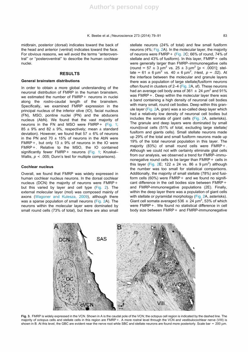
Fig. 3. FMRP is widely expressed in the VCN. Shown in A is the caudal pole of the VCN; the octopus cell region is indicated by the dashed line. The
majority of octopus cells and stellate cells in this region are FMRP+. A more rostral level through the VCN and vestibulocochlear nerve (VIII) is
shown in B. At this level, the GBC are evident near the nerve root while SBC and stellate neurons are found more posteriorly. Scale bar = 200 lm.
K. Beebe et al. / Neuroscience 273 (2014) 79–91 83

Author's personal copy
giant cell populations (2F; 545 ± 37 vs. 526 ± 31 lm2; ttest, p= .69).
In the VCN the majority of neurons were FMRP+ but
again this varied by region and cell type. In the caudal
VCN (cVCN; Fig. 3A) there was a collection of octopus
cells (OC; Fig. 4A, asterisks) intermingled with stellate
neurons (Fig. 4A, arrow). The vast majority of octopus
cells were FMRP+ (89%, Fig. 5) and FMRP+ octopus
cells had significantly larger somata compared to
FMRP-immunonegative octopus cells (466 ± 15 lm2
compared to 370 ± 21 lm2; Mann–Whitney, p= .01,
Fig. 5C). FMRP-immunolabeling was not restricted to
octopus cell somata and FMRP+ dendritic profiles
emerged from FMRP+ somata. Additionally, there were
numerous thin, FMRP+ fibers, which we interpreted as
axonal profiles, coursing through the octopus cell region
(OC) and these axons appeared to course in a
predominately anterior–posterior direction (Fig. 4A,
arrowheads).
Rostral to the OC, at the level of the entering auditory
nerve, the vast majority of somata in the VCN were
FMRP+ (Fig. 3B). Here, nearly all globular bushy cells
(GBC) are FMRP+ (95%, Fig. 5a) and the cell bodies
of these neurons measured 451 ± 18 lm2 (Figs. 4B
and 5C). There were insufficient FMRP-immunonegative
GBCs for statistical testing. In GBC, FMRP+ dendrites
could be followed out to secondary dendritic branches
(Fig. 4B, arrowheads). In the GBC region, there was
also heavy FMRP+ labeling in the neuropil, which was
absent from the layers of the DCN and OC (Fig. 4B).
In the rostral VCN (rVCN), the majority of spherical
bushy cells (SBC) and stellate neurons were FMRP+
(Fig. 4C, asterisk and arrowhead, respectively).
Specifically, 85% of SBC and 68% of stellate neurons
were FMRP+ (Fig. 5A). There were no statistical
differences in cell body size between FMRP+ and
FMRP-immunonegative SBC or stellate neurons (SBC:
278± 10 lm2 vs. 307± 21 lm2; stellate: 265± 15 lm2
compared to 278± 26 lm2, Mann–Whitney, p> .05;
Fig. 5C). However, FMRP+ SBC were significantly more
round than FMRP-immunonegative SBC (.77 ± .008 vs.
.72±.02, Mann–Whitney, p= .001, Fig. 5B). Additionally,
throughout the VCN there were occasional small, fusiform
neurons (average soma size= 163± 18 lm2). Only
42% of these fusiform neurons were FMRP+ and we
found no statistical difference in cell body size between
FMRP+ and FMRP-immunonegative fusiform neurons
(Fig. 5C). In the SBC region, there was also heavy FMRP
labeling in the neuropil (Fig. 4C).
SOC
As in the VCN, the majority of SOC neurons were
FMRP+ (Fig. 6). Within the MSO, the majority of
neuronal cell bodies were FMRP+ and we estimated
that 40% of all FMRP+ neurons in the human SOC are
within the MSO. In MSO neurons, FMRP immunolabeling
extended from the somata to reach primary and
secondary dendritic arbors. Medial and lateral to the MSO
cell column there was a halo which included dense
tangles of FMRP+ dendritic profiles (black dashed line in
Fig. 6 and black arrowheads in 7A). Closer examination
of this territory revealed that many of these profiles could
be traced back to a soma in the MSO (Fig. 7A, B). The
MSO cell column was about 200-lm wide medial–
laterally. At the mid-anteroposterior level of the cell
column, FMRP+ dendritic profiles extended both medial
and laterally up to 450 lm from MSO somata. In the
specimens examined in this study, 83% of all MSO
neurons were FMRP+ and we found no difference in
cross sectional area between FMRP+ MSO somata
and FMRP-immunonegative neurons (250 ± 6 lm2
compared to 237 ± 26 lm2, Mann–Whitney, p= .08,
Fig. 11A). We examined FMRP-immunoreactivity in
subpopulations of MSO neurons and found that 88% of
fusiform, 79% of stellate neurons and 88% of round
Fig. 4. Most VCN cell types are FMRP+. Shown in A are examples
of FMRP+ octopus cells and an FMRP-immunonegative stellate
neuron (neutral red stain; arrow). Numerous thin FMRP+ profiles are
found traversing the octopus cells region (presumed axons; arrow-
heads). Shown in B are examples of FMRP+ GBC; dendritic profiles
are indicated by arrowheads. Shown in C are examples of FMRP+
SBC (asterisk) and a large stellate neuron (arrowhead). Scale
bar = 20 lm.
84 K. Beebe et al. / Neuroscience 273 (2014) 79–91

Author's personal copy
MSO neurons were FMRP+. We found no statistical
difference in cell body size between FMRP+ and
FMRP-immunonegative subpopulations (Fig. 11A; Mann–
Whitney, fusiform p= .45; stellate p= .26; round
p= .34). Additionally, we considered that FMRP-
immunonegative MSO neurons might comprise a
topographical subset of MSO neurons. However, we
found no correlation of x and y coordinates of FMRP-
immunonegative neurons (Pearson, r2 = .01; p= .19)
and no difference in the anterior–posterior location in the
MSO cell column (Fig. 8; Mann–Whitney, p= .58).
Finally, we examined colocalization of FMRP and the
Kv3.1b potassium channel in human MSO neurons. We
found notable overlap of FMRP and Kv3.1b
immunolabeling in somata (asterisks in Fig. 9A–C) and
dendrites (arrowheads in Fig. 9D). Additionally, there
were abundant FMRP+ axon profiles in the vicinity of
the MSO (open arrowheads).
Within the lateral superior olive (LSO), the majority of
neurons were FMRP+ (87%; Fig. 10A). Of all LSO
neurons, 33% were FMRP+ stellate neurons and 58%
were FMRP+ round neurons. However, we found no
differences in soma size between fusiform, stellate or
round cell populations (Fig. 11B; Mann–Whitney, pall > .05). Additionally in the LSO, FMRP-
immunolabeling extended from the soma into primary
dendrites and heavy FMRP-immunoreactivity was
observed in the neuropil of the LSO. This heavy pattern
of neuropil labeling was not identified in any of the other
SOC nuclei (Figs. 6 and 10A).
Within the superior paraolivary nucleus (SPON), the
majority of neurons were FRMP+ (83%; Fig. 10B). Of
all SPON neurons, 34% were FMRP+ stellate neurons
and 52% were FMRP+ round neurons. FMRP+
stellate neurons were generally larger than FMRP-
immunonegative stellate neurons (Fig. 11D; 256 ± 23
vs. 162 ± 14 lm2) but this difference did not reach
statistical significance (Mann–Whitney, p= .10). There
were no differences between fusiform and round cell
populations (Mann–Whitney, p> .05; Fig. 11D).
Fig. 5. Quantification of FMRP in the VCN. In A, the percentage of VCN cell types that are FMRP+ is shown. Most neurons are FMRP+ except for
fusiform neurons of which less than 50% are FMRP+. Shown in B is a plot of circularity for each neuronal subpopulation. For SBC and stellate cells,
FMRP+ somata were more round when compared to FMRP-immunonegative cells. Shown in C is the cell body area by VCN cell type. FMRP+
octopus cells had significantly larger cell bodies when compared to FMRP-immunonegative cells. In B and C the box plots are of 5–95th percentile.
K. Beebe et al. / Neuroscience 273 (2014) 79–91 85

Author's personal copy
In the remaining SOC nuclei (medial nucleus of the
trapezoid body (MNTB), ventral nucleus of the trapezoid
body (VNTB) and lateral nucleus of the trapezoid body
(LNTB); Fig. 6), we again found that the majority of
neurons were FMRP+ (89%, 82% and 87%, respectively)
and 94% of MNTB round/principal neurons and 85%
of LNTB stellate neurons were FMRP+. As in other
nuclei, FMRP-immunolabeling extended from the cell
body into primary dendrites. There were no differences
in cell body size between FMRP+ and FMRP negative
neurons in these nuclei (Mann–Whitney, p> .05;
Fig. 11C–F). Of all MNTB neurons, 76% were FMRP+
round cells and 63% of LNTB neurons were FMRP+
round neurons (Fig. 9C). Finally, we estimated that
approximately 21% of all SOC FMRP+ neurons are
localized to the LNTB.
DISCUSSION
To the best of the authors’ knowledge, this is the first
quantitative investigation of FMRP-immunolabeling in
the human brainstem and the first characterization of
FMRP+ neurons in the human cochlear nucleus and
SOC. We have identified FMRP+ neurons in each of
the brainstem nuclei we investigated, but the proportion
varied considerably by nucleus. Specifically, we found
that nearly 85% of FN neurons but only about 13% of
neurons in the principal nucleus of the IO were
FMRP+. We interpret these observations to indicate
that FMRP is widely expressed but not ubiquitous in the
human brainstem and that FMRP may function more
prominently in some nuclei (FN, MSO) than others (IO).
Additionally, the neuronal populations which abundantly
Fig. 6. Distribution of FMRP in the SOC. This figure shows a
transverse section through the middle third of the SOC immunola-
beled for FMRP. FMRP is widely expressed in SOC neurons but the
pattern of labeling the neuropil varies. Neuropil labeling is heaviest in
the LSO (white dashed line) and very light on either side of the MSO
cell column (white dashed line), a region occupied by FMRP+ MSO
dendrites (black dashed line). FMRP-immunolabeling is largely
absent from axons in the trapezoid body (tz; arrowheads). Scale
bar = 200 lm.
Fig. 7. FMRP is localized to MSO cell bodies and dendrites. Shown
in A is a field of the MSO from a mid-rostral caudal level of the
nucleus. The dashed line indicates the boundaries of the MSO cell
column. The majority of MSO neurons are FMRP+. FMRP+
fusiform (open arrowheads) and stellate neurons (arrow) are dem-
onstrated. FMRP+ dendritic profiles are found in the fields medial
and lateral to the MSO cell column (arrowheads). Scale
bar = 100 lm. Shown in B is a tracing of an FMRP+ MSO neuron
from the caudal aspect of the nucleus. Note that FMRP-immunola-
beling extends from the soma into dendrites out to secondary
branches. Scale bar = 20 lm.
Fig. 8. FMRP-immunonegative MSO neurons have no topographic
preference. Both A (FMRP+) and B (FMRP-immunonegative) show
normalized plots, based on x and y coordinates, of MSO neurons
from the middle 1/3 of the nucleus. Note that FMRP-immunonegative
neurons span the entire anterior–posterior extent of the nucleus and
show no topographic preference.
86 K. Beebe et al. / Neuroscience 273 (2014) 79–91

Author's personal copy
express FMRP are likely to be more severely impacted in
FXS or Fmr1 KO animals that those with minimal FMRP
expression. Finally, the reader should be reminded that
the subjects used in this investigation ranged in age
from 57 to 96 years. We are open to the possibility that
FMRP expression patterns may change with age and
that these patterns may be markedly different in human
neonatal populations. Regardless, FMRP is abundantly
expressed in the auditory brainstem of the 57–96-year-
old age group and likely plays an important functional
role within these neuronal populations.
Within the cochlear nuclei and SOC, we found the
majority of neurons and neuronal subtypes to be
FMRP+. In the human DCN, we identified a group of
large stellate/fusiform neurons between the molecular
and granule layers (Wagoner and Kulesza, 2009; present
study), the majority of which are FMRP+. We believe
these stellate/fusiform neurons to be equivalent to pyra-
midal or fusiform neurons described previously in the
DCN and further, that this population is a major source
of DCN output to the inferior colliculus (Oertel and
Golding, 1997; Oertel and Young, 2004). Since the DCN
is known to integrate auditory and non-auditory informa-
tion and relay information to the inferior colliculus, our
findings suggest that FMRP plays an important role in this
function. In the human VCN, we found that nearly all GBC
and the vast majority of SBC, stellate and octopus cells
Fig. 9. FMRP colocalizes with Kv3.1b in the human MSO. Shown in
A–C is a region of the MSO, immunolabeled for FMRP (A) and
Kv3.1b (B). Three FMRP+ MSO neurons are indicated (A, empty
arrowheads). In B, neuronal cell bodies (empty arrowheads) and
punctate profiles (white arrowheads) are Kv3.1b-immunoreactive.
Both A, B are overlaid in C; regions of colocalization are indicated in
yellow. A lipofuscin artifact is indicated by the double arrow. Scale
bar = 30 lm. Shown in D is a higher magnification view of an
MSO neuron (soma indicated by asterisk; primary dendrites indicated
by open arrowheads) immunolabeled for FMRP and Kv3.1b. Regions
of colocalization in the cell body and dendrites are indicated in
yellow. A lipofuscin artifact is indicated by the double arrow. Scale
bar = 30 lm.
Fig. 10. FMRP is abundant in the SOC nuclei. Shown in A is FMRP-
immunolabeling in the LSO. The majority of LSO neurons are
FMRP+ and there is a high density of FMRP-immunoreactive linear
profiles, which we interpret as dendrites arising from LSO neurons.
Shown in B is FMRP-immunolabeling in the SPON. The majority of
SPON neurons are FMRP+ and FMRP-immunolabeled dendrites
are seen to emerge from SPON cell bodies. The neuropil labeling is
relatively light in the SPON. Scale bar = 100 lm (B); A, B are at the
same scale. Shown in C is FMRP-immunolabeling in the MNTB.
Again, the majority of MNTB neurons are FMRP+ and the neuropil
labeling is generally light. Scale bar = 100 lm (C).
K. Beebe et al. / Neuroscience 273 (2014) 79–91 87

Author's personal copy
were FMRP+. Additionally, we found that FMRP+ SBC
had more circular cell bodies compared to FMRP-
immunonegative SBC. Cell body shape and dendritic
architecture are interdependent morphometric features
and a less circular (i.e. more irregular) cell body contour
results when 2 or more primary dendrites originate from
the soma. We suggest that FMRP-immunonegative SBC
may be distinguishable by multiple primary dendrites
(see Figs. 3–5 in Cant, 1992). We also found that
FMRP+ octopus cells had larger somata compared to
FMRP-immunonegative octopus cells. If a larger soma
is required to support further-reaching or more elaborate
dendritic arbors and/or axonal projections (Friede, 1963;
Kulesza et al., 2011), FMRP+ octopus cells might be dis-
tinguished from FMRP-immunonegative octopus cells by
the complexity of dendritic arbors, axonal length and/or
size of the axons terminal field. Because of the abun-
dance of FMRP in the VCN, we suggest that FMRP plays
an important role in processing temporal information and
is essential for the relay of precisely timed action poten-
tials to the SOC, nuclei of the lateral lemniscus and infe-
rior colliculus. Within the SOC, we found the majority of
LSO neurons to be FMRP+ and within this nucleus we
encountered heavy neuropil labeling, which was also
observed in the cochlear nucleus. In these regions, FMRP
may be localized to presynaptic sites (Christie et al.,
2009) and within astrocytes (Pacey and Doering, 2007;
Higashimori et al., 2013). The vast majority of MSO neu-
rons were FMRP+ and FMRP-immunolabeling in these
neurons extended from the soma out to secondary den-
dritic branches. FMRP-immunonegative MSO neurons
were not confined to a specific region of the nucleus,
but were found throughout the MSO contour. Further-
more, we observed colocalization of FMRP and the
high-voltage-activated potassium channel Kv3.1b in
human MSO neurons. Colocalization of FMRP and
Kv3.1b suggests the possibility that in the human MSO,
FMRP may regulate Kv3.1b translation in a tonotopic
fashion as in mouse (Strumbos et al., 2010). Overall,
we have found that FMRP is abundantly expressed in
neuronal somata and dendritic profiles. This localization
suggests that FMRP plays an important role in normal
function and dendritic processing in the DCN, VCN
and SOC. Furthermore, we anticipate that with loss of
FMRP function (as in FXS), these auditory brainstem
centers will demonstrate significant errors in processing
temporal features and localization of complex sounds.
Hypersensitivity to auditory and visual stimuli is a
cardinal feature of FXS (Miller et al., 1999) and audi-
tory-processing difficulties are well documented in FXS.
Specifically, FXS is associated with increased amplitude
of early cortical auditory-evoked potentials (St Clair
et al., 1987; Rojas et al., 2001; Castren et al., 2003;
Van der Molen et al., 2012a) and patients with FXS are
less accurate, have more false alarms and delayed reac-
tion time in visual and auditory tasks (Van der Molen
et al., 2012b). Further, FXS patients are known to exhibit
language delay (Brady et al., 2006), hypersensitivity to
Fig. 11. Quantification of FMRP in the SOC. Shown in A–F are box plots of cell body area by SOC nucleus, cell body morphology (fusiform, stellate
and round) and FMRP-immunoreactivity (FMRP+ in black, FMRP-immunonegative in red). In the SPON, FMRP+ somata were significantly larger
than FMRP-immunonegative somata. All box pots are of 10–90th percentile. (For interpretation of the references to color in this figure legend, the
reader is referred to the web version of this article.)
88 K. Beebe et al. / Neuroscience 273 (2014) 79–91

Author's personal copy
auditory stimulation (Van der Molen et al., 2012a), altered
brain activation patterns during auditory discrimination
tasks (Hall et al., 2009), weak auditory-processing skills
and characteristic cluttering of speech (Hanson et al.,
1986), normal latency of early cortical responses but
increased amplitude and reduced lateralization (Knoth
and Lippe, 2012). Fmr1 KO mice also exhibit auditory
hypersensitivity and are susceptible to fatal sound-
induced seizures (Chen and Toth, 2001; Nielsen et al.,
2002). Single-unit recordings from the auditory cortex of
Fmr1 KO mice (Rotschafer and Razak, 2013) reveal
broader frequency receptive fields, pure tone stimuli elicit
significantly more spikes and there is more variability in
first-spike latency compared to control animals. Addition-
ally, in an animal model of FXS, FMRP has been shown to
play an essential role in maturation of the acoustic startle
response (Yun et al., 2006).
To restate our major findings, we have demonstrated
abundant neuronal FMRP-immunolabeling in the human
cochlear nucleus and SOC, structures which play
essential roles in processing temporal features of
speech, sound source localization and descending
modulation of the cochlea. Even though much of the
literature to date has focused on cortical aspects of
FMRP function, we suggest that auditory dysfunction in
FXS may arise, at least in part, from timing errors in the
CN and SOC. Additionally, it should be noted that
auditory brainstem responses and hearing thresholds in
young males with FXS do not differ significantly from
controls (Roberts et al., 2005). We propose that the errors
in temporal processing that are likely present in the
FMRP-deficient CN and MSO cannot be detected on rou-
tine audiological screening tests, but become evident in
vocalization and speech-based tasks.
Processing of interaural time differences (ITDs) in the
MSO requires precisely timed convergence and
segregation of both excitatory inputs from the CN and
inhibitory inputs from within the SOC (Smith et al., 2000;
Brand et al., 2002; Pecka et al., 2008; Couchman et al.,
2012). Inappropriate group 1 mGluR signaling is proposed
to contribute to the pathophysiological changes in FXS
(Bear et al., 2004). Additionally, in Fmr1 KO mice, there
is increased seizure activity and reduced expression of
GABAA receptors/subunits (Miyashiro et al., 2003; El
Idrissi et al., 2005; Gantois et al., 2006; D’Hulst et al.,
2006) and abnormal GAD65/67 expression (El Idrissi
et al., 2005; Olmos-Serrano et al., 2010). It is possible then
that in FXS, there is a disruption of the delicate balance of
excitation and inhibition required for ITD coding in the
MSO and this may lead to significant processing errors
(Brand et al., 2002; Hassfurth et al., 2010; Grothe and
Koch, 2011).
CONFLICT OF INTEREST
The authors declare that there are no conflicts of interest.
Acknowledgments—This work was supported in part by a grant
from the Lake Erie Consortium for Osteopathic Medical Training.
The authors would like to thank Jerome McGraw, Kristen Ruby
and George Grignol for technical assistance.
REFERENCES
Akins MR, Berk-Rauch HE, Fallon JR (2009) Presynaptic translation:
stepping out of the postsynaptic shadow. Front Neural Circuits 3:17.
Akins MR, Leblanc HF, Stackpole EE, Chyung E, Fallon JR (2012)
Systematic mapping of fragile X granules in the mouse brain
reveals a potential role for presynaptic FMRP in sensorimotor
functions. J Comp Neurol 520(16):3687–3706.
Bassell GJ, Warren ST (2008) Fragile X syndrome: loss of local
mRNA regulation alters synaptic development and function.
Neuron 60:201–214.
Bear MF, Huber KM, Warren ST (2004) The mGluR theory of fragile X
mental retardation. Trends Neurosci 27(7):370–377.
Berry-Kravis E (2002) Epilepsy in fragile X syndrome. Dev Med Child
Neurol 44(11):724–728.
Brady N, Skinner D, Roberts J, Hennon E (2006) Communication in
young children with fragile X syndrome: a qualitative study of
mothers’ perspectives. Am J Speech Lang Pathol 15(4):353–364.
Brand A, Behrend O, Marquardt T, McAlpine D, Grothe B (2002)
Precise inhibition is essential for microsecond interaural time
difference coding. Nature 417(6888):543–547.
Braun K, Segal M (2000) FMRP involvement in formation of synapses
among cultured hippocampal neurons. Cereb Cortex
10(10):1045–1052.
Brown MR, Kronengold J, Gazula VR, Chen Y, Strumbos JG,
Sigworth FJ, Navaratnam D, Kaczmarek LK (2010) Fragile X
mental retardation protein controls gating of the sodium-activated
potassium channel Slack. Nat Neurosci 13(7):819–821.
Cant NB (1992) The cochlear nucleus: neuronal types and their
synaptic organization. In: Webster DB, Popper AN, Fay RR, editors.
The mammalian auditory pathway: neuroanatomy. Berlin: Springer-
Verlag. p. 66–116.
Castren M, Paakkonen A, Tarkka IM, Ryynanen M, Partanen J (2003)
Augmentation of auditory N1 in children with fragile X syndrome.
Brain Topogr 15(3):165–171.
Castren M, Tervonen T, Karkkainen V, Heinonen S, Castren E,
Larsson K, Bakker CE, Oostra BA, Akerman K (2005) Altered
differentiation of neural stemcells in fragile x syndrome. Proc Natl
Acad Sci U S A 102:17834–17839.
Chen L, Toth M (2001) Fragile X mice develop sensory hyperactivity
to auditory stimuli. Neuroscience 103:1043–1050.
Christie SB, Akins MR, Schwob JE, Fallon JR (2009) The FXG: a
presynaptic fragile x granule expressed in a subset of developing
brain circuits. J Neurosci 29:1514–1524.
Comery TA, Harris JB, Willems PJ, Oostra BA, Irwin SA, Weiler IJ,
Greenough WT (1997) Abnormal dendritic spines in fragile X
knockout mice: maturation and pruning deficits. Proc Natl Acad
Sci U S A 94(10):5401–5404.
Cornish K, Turk J, Hagerman R (2008) The fragile X continuum: new
advances and perspectives. J Intellect Disabil Res 52(Pt 6):
469–482.
Couchman K, Grothe B, Felmy F (2012) Functional localization of
neurotransmitter receptors and synaptic inputs to mature neurons
of the medial superior olive. J Neurophysiol 107(4):1186–1198.
D’Hulst C, De Geest N, Reeve SP, Van Dam D, De Deyn PP, Hassan
BA, Kooy RF (2006) Decreased expression of the GABAA
receptor in fragile X syndrome. Brain Res 1121(1):238–245.
Deng PY, Rotman Z, Blundon JA, Cho Y, Cui J, Cavalli V,
Zakharenko SS, Klyachko VA (2013) FMRP regulates
neurotransmitter release and synaptic information transmission
by modulating action potential duration via BK channels. Neuron
77(4):696–711.
Devys D, Lutz Y, Rouyer N, Bellocq JP, Mandel JL (1993) The FMR1
protein is cytoplasmic, most abundant in neurons and appears
normal in carriers of fragile x permutation. Nat Genet 4:335–340.
El Idrissi A, Ding XH, Scalia J, Trenkner E, Brown WT, Dobkin C
(2005) Decreased GABA(A) receptor expression in the seizure-
prone fragile X mouse. Neurosci Lett 377(3):141–146.
Eliez S, Blasey CM, Freund LS, Hastie T, Reiss AL (2001) Brain
anatomy, gender and IQ in children and adolescents with fragile X
syndrome. Brain 124(Pt 8):1610–1618.
K. Beebe et al. / Neuroscience 273 (2014) 79–91 89

Author's personal copy
Feng Y, Gutekunst CA, Eberhart DE, Yi H, Warren ST, Hersch SM
(1997) Fragile X mental retardation protein: nucleocytoplasmic
shuttling and association with somatodendritic ribosomes. J
Neurosci 17(5):1539–1547.
Friede RL (1963) The relationship of body size, nerve cell size, axon
length, and glial density in the cerebellum. Proc Natl Acad Sci U S
A 49:187–193.
Galvez R, Gopal AR, Greenough WT (2003) Somatosensory cortical
barrel dendritic abnormalities in a mouse model of the fragile X
mental retardation syndrome. Brain Res 971(1):83–89.
Galvez R, Smith RL, Greenough WT (2005) Olfactory bulb mitral cell
dendritic pruning abnormalities in a mouse model of the Fragile-X
mental retardation syndrome: further support for FMRP’s
involvement in dendritic development. Brain Res Dev Brain Res
157(2):214–216.
Gantois I, Vandesompele J, Speleman F, Reyniers E, D’Hooge R,
Severijnen LA, Willemsen R, Tassone F, Kooy RF (2006)
Expression profiling suggests underexpression of the GABA(A)
receptor subunit delta in the fragile X knockout mouse model.
Neurobiol Dis 21(2):346–357.
Grothe B, Koch U (2011) Dynamics of binaural processing in the
mammalian sound localization pathway – the role of GABA(B)
receptors. Hear Res 279(1–2):43–50.
Hagerman RJ, Berry-Kravis E, Kaufmann WE, Ono MY, Tartaglia N,
Lachiewicz A, Kronk R, Delahunty C, Hessl D, Visootsak J, Picker
J, Gane L, Tranfaglia M (2009) Advances in the treatment of
fragile X syndrome. Pediatrics 123(1):378–390.
Hall SS, Walter E, Sherman E, Hoeft F, Reiss AL (2009) The neural
basis of auditory temporal discrimination in girls with fragile X
syndrome. J Neurodev Disord 1(1):91–99.
Hanson DM, Jackson 3rd AW, Hagerman RJ (1986) Speech
disturbances (cluttering) in mildly impaired males with the
Martin-Bell/fragile X syndrome. Am J Med Genet 23(1–
2):195–206.
Harris JC (2011) Autism spectrum disorders in neurogenic
syndromes: phenocopies of Autism. In: Adam A, editor. Textbook
of autism spectrum disorders. Washington, DC: American
Psychiatric Publishing, Inc.
Hassfurth B, Grothe B, Koch U (2010) The mammalian interaural time
difference detection circuit is differentially controlled by GABAB
receptors during development. J Neurosci 30(29):9715–9727.
Hatton DD, Sideris J, Skinner M, Mankowski J, Bailey Jr DB, Roberts
J, Mirrett P (2006) Autistic behavior in children with fragile X
syndrome: prevalence, stability, and the impact of FMRP. Am J
Med Genet 140:1804–1813.
Higashimori H, Morel L, Huth J, Lindemann L, Dulla C, Taylor A,
Freeman M, Yang Y (2013) Astroglial FMRP-dependent
translational down-regulation of mGluR5 underlies glutamate
transporter GLT1 dysregulation in the fragile X mouse. Hum Mol
Genet 22(10):2041–2054.
Hinds HL, Ashley CT, Sutcliffe JS, Nelson DL, Warren ST, Housman
DE, Schalling M (1993) Tissue specific expression of FMR-1
provides evidence for a functional role in fragile X syndrome. Nat
Genet 3:38–43.
Hinton VJ, Brown WT, Wisniewski K, Rudelli RD (1991) Analysis of
neocortex in three males with the fragile X syndrome. Am J Med
Genet 41(3):289–294.
Hoeft F, Carter JC, Lightbody AA, Cody Hazlett H, Piven J, Reiss AL
(2010) Region-specific alterations in brain development in one- to
three-year-old boys with fragile X syndrome. Proc Natl Acad Sci U
S A 107(20):9335–9339.
Irwin SA, Galvez R, Greenough WT (2000) Dendritic spine structural
anomalies in fragile-x syndrome. Nat Rev Neurosci 6:376–387.
Jacobs S, Cheng C, Doering LC (2012) Probing astrocyte function in
fragile X syndrome. Results Probl Cell Differ 54:15–31.
Kates WR, Abrams MT, Kaufmann WE, Breiter SN, Reiss AL (1997)
Reliability and validity of MRI measurement of the amygdala and
hippocampus in children with fragile X syndrome. Psychiatry Res
75(1):31–48.
Klemmer P, Meredith RM, Holmgren CD, Klychnikov OI, Stahl-Zeng
J, Loos M, van der Schors RC, Wortel J, de Wit H, Spijker S,
Rotaru DC, Mansvelder HD, Smit AB, Li KW (2011) Proteomics,
ultrastructure, and physiology of hippocampal synapses in a
fragile X syndrome mouse model reveal presynaptic phenotype.
J Biol Chem 286(29):25495–25504.
Knoth IS, Lippe S (2012) Event-related potential alterations in fragile
X syndrome. Front Hum Neurosci 6:264.
Kulesza Jr RJ (2007) Cytoarchitecture of the human superior
olivary complex: medial and lateral superior olive. Hear Res
225(1–2):80–90.
Kulesza Jr RJ (2008) Cytoarchitecture of the human superior olivary
complex: nuclei of the trapezoid body and posterior tier. Hear Res
241(1–2):52–63.
Kulesza Jr RJ (2013) Characterization of human auditory brainstem
circuits by calcium binding protein immunohistochemistry.
Neuroscience 258:318–331.
Kulesza RJ, Mangunay K (2008) Morphological features of the medial
superior olive in autism. Brain Res 1200:132–137.
Kulesza Jr RJ, Lukose R, Stevens LV (2011) Malformation of the
human superior olive in autistic spectrum disorders. Brain Res
1367:360–371.
Li J, Pelletier MR, Perez Velazquez JL, Carlen PL (2002) Reduced
cortical synaptic plasticity and GluR1 expression associated with
fragile X mental retardation protein deficiency. Mol Cell Neurosci
19(2):138–151.
Miller LJ, McIntosh DN, McGrath J, Shyu V, Lampe M, Taylor AK,
Tassone F, Neitzel K, Stackhouse T, Hagerman RJ (1999)
Electrodermal responses to sensory stimuli in individuals with fragile
X syndrome: a preliminary report. Am J Med Genet. 83(4):268–279.
Miyashiro KY, Beckel-Mitchener A, Purk TP, Becker KG, Barret T, Liu
L, Carbonetto S, Weiler IJ, Greenough WT, Eberwine J (2003)
RNA cargoes associating with FMRP reveal deficits in cellular
functioning in Fmr1 null mice. Neuron 37(3):417–431.
Moore JK, Osen KK (1979) The cochlear nuclei in man. Am J Anat
154(3):393–418.
Mostofsky SH, Mazzocco MM, Aakalu G, Warsofsky IS, Denckla MB,
Reiss AL (1998) Decreased cerebellar posterior vermis size in
fragile X syndrome: correlation with neurocognitive performance.
Neurology 50(1):121–130.
Nielsen DM, Derber WJ, McClellan DA, Crnic LS (2002) Alterations in
the auditory startle response in Fmr1 targeted mutant mouse
models of fragile X syndrome. Brain Res 927(1):8–17.
Nimchinsky EA, Oberlander AM, Svoboda K (2001) Abnormal
development of dendritic spines in FMR1 knock-out mice. J
Neurosci 21(14):5139–5146.
Oertel D, Golding NL (1997) Circuits of the dorsal cochlear nucleus.
In: Syka, editor. Acoustical signal processing in the central
auditory system. New York: Plenum Press.
Oertel D, Young ED (2004) What’s a cerebellar circuit doing in the
auditory system? Trends Neurosci 27(2):104–110.
Olmos-Serrano JL, Paluszkiewicz SM, Martin BS, Kaufmann WE,
Corbin JG, Huntsman MM (2010) Defective GABAergic
neurotransmission and pharmacological rescue of neuronal
hyperexcitability in the amygdala in a mouse model of fragile X
syndrome. J Neurosci 30(29):9929–9938.
Osen KK (1969) Cytoarchitecture of the cochlear nuclei in the cat. J
Comp Neurol 136(4):453–484.
Pacey LK, Doering LC (2007) Developmental expression of FMRP in
the astrocyte lineage: implications for fragile X syndrome. Glia
55(15):1601–1609.
Pecka M, Brand A, Behrend O, Grothe B (2008) Interaural time
difference processing in the mammalian medial superior olive: the
role of glycinergic inhibition. J Neurosci 28(27):6914–6925.
Pfeiffer BE, Huber KM (2009) The state of synapses in fragile X
syndrome. Neuroscientist 15(5):549–567.
Reiss AL, Abrams MT, Greenlaw R, Freund L, Denckla MB (1995)
Neurodevelopmental effects of the FMR-1 full mutation in
humans. Nat Med 1(2):159–167.
Roberts J, Hennon EA, Anderson K, Roush J, Gravel J, Skinner M,
Misenheimer J, Reitz P (2005) Auditory brainstem responses in
young males with Fragile X syndrome. J Speech Lang Hear Res
48(2):494–500.
90 K. Beebe et al. / Neuroscience 273 (2014) 79–91

Author's personal copy
Rogers SJ, Wehner DE, Hagerman R (2001) The behavioral
phenotype in fragile X: symptoms of autism in very young
children with fragile X syndrome, idiopathic autism, and other
developmental disorders. J Dev Behav Pediatr 22(6):409–417.
Rojas DC, Benkers TL, Rogers SJ, Teale PD, Reite ML, Hagerman
RJ (2001) Auditory evoked magnetic fields in adults with fragile X
syndrome. NeuroReport 12(11):2573–2576.
Rotschafer S, Razak K (2013) Altered auditory processing in a mouse
model of fragile X syndrome. Brain Res 1506:12–24.
Rudelli RD, Brown WT, Wisniewski K, Jenkins EC, Laure-
Kamionowska M, Connell F, Wisniewski HM (1985) Adult fragile
X syndrome. Clinico-neuropathologic findings. Acta Neuropathol
67(3–4):289–295.
Schmidt E, Wolski Jr TP, Kulesza Jr RJ (2010) Distribution of
perineuronal nets in the human superior olivary complex. Hear
Res 265(1–2):15–24.
Smith AJ, Owens S, Forsythe ID (2000) Characterisation of inhibitory
and excitatory postsynaptic currents of the rat medial superior
olive. J Physiol 529(Pt 3):681–698.
St Clair DM, Blackwood DH, Oliver CJ, Dickens P (1987) P3
abnormality in fragile X syndrome. Biol Psychiatry 22(3):303–312.
Standring S (2008) Gray’s anatomy: the anatomical basis of clinical
practice. 40th ed. Churchill Livingstone.
Strumbos JG, Brown MR, Kronengold J, Polley DB, Kaczmarek LK
(2010) Fragile X mental retardation protein is required for rapid
experience- dependent regulation of the potassium channel
Kv3.1b. J Neurosci 30:10263–10271.
Thomas CC, Combe CL, Dyar KA, Inglis FM (2008) Modest
alterations in patterns of motor neuron dendrite morphology in
the Fmr1 knockout mouse model for fragile X. Int J Dev Neurosci
26(7):805–811.
Van der Molen MJ, Van der Molen MW, Ridderinkhof KR, Hamel BC,
Curfs LM, Ramakers GJ (2012a) Auditory change detection
in fragile X syndrome males: a brain potential study. Clin
Neurophysiol 123:1309–1318.
Van der Molen MJ, Van der Molen MW, Ridderinkhof KR, Hamel BC,
Curfs LM, Ramakers GJ (2012b) Auditory and visual cortical
activity during selective attention in fragile x syndrome: a cascade
of processing deficiencies. Clin Neurophysiol 123:720–729.
Verkerk AJ, Pieretti M, Sutcliffe JS, Fu YH, Kuhl DP, Pizzuti A, Reiner
O, Richards S, Victoria MF, Zhang FP (1991) Identification of a
gene (FMR-1) containing a CGG repeat coincident with a
breakpoint cluster region exhibiting length variation in fragile X
syndrome. Cell 65(5):905–914.
Wagoner JL, Kulesza Jr RJ (2009) Topographical and cellular
distribution of perineuronal nets in the human cochlear nucleus.
Hear Res 254(1–2):42–53.
Wang H, Ku L, Osterhout DJ, Li W, Ahmadian A, Liang Z, Feng Y
(2004) Developmentally-programmed FMRP expression in
oligodendrocytes: a potential role of FMRP in regulating translation
in oligodendroglia progenitors. Hum Mol Genet 13(1):79–89.
Wang Y, Sakano H, Beebe K, Brown MR, de Laat R, Bothwell M,
Kulesza Jr RJ, Rubel EW (2013) Intense and specialized dendritic
localization of the fragile X mental retardation protein in binaural
brainstem neurons – a comparative study in the alligator, chicken,
gerbil, and human. J Comp Neurol.
Yin TC, Chan JC (1990) Interaural time sensitivity in medial superior
olive of cat. J Neurophysiol 64(2):465–488.
Yun SW, Platholi J, Flaherty MS, Fu W, Kottmann AH, Toth M (2006)
Fmrp is required for the establishment of the startle response
during the critical period of auditory development. Brain Res
1110(1):159–165.
Zarnescu DC, Jin P, Betschinger J, Nakamoto M, Wang Y,
Dockendorff TC, Feng Y, Jongens TA, Sisson JC, Knoblich JA,
Warren ST, Moses K (2005) Fragile X protein functions with lgl
and the par complex in flies and mice. Dev Cell 8(1):43–52.
Zhang Y, Brown MR, Hyland C, Chen Y, Kronengold J, Fleming MR,
Kohn AB, Moroz LL, Kaczmarek LK (2012) Regulation of neuronal
excitability by interaction of fragile X mental retardation protein
with slack potassium channels. J Neurosci 32:15318–15327.
(Accepted 2 May 2014)(Available online 15 May 2014)
K. Beebe et al. / Neuroscience 273 (2014) 79–91 91