Embed Size (px)
Citation preview

BCR-ABL Tyrosine Kinase Inhibitors: Which Mechanism(s)May Explain the Risk of Thrombosis?Hélène Haguet1,2 Jonathan Douxfils1,4 Christian Chatelain1 Carlos Graux3 François Mullier2
Jean-Michel Dogné1
1University of Namur, Namur Thrombosis and Hemostasis Center(NTHC), Namur Research Institute for Life Sciences (NARILIS),Department of Pharmacy, Namur, Belgium
2Université catholique de Louvain, CHU UCL Namur, Namur Thrombosisand Hemostasis Center, Hematology Laboratory, Yvoir, Belgium
3Université catholique de Louvain, CHU UCL Namur, NamurThrombosis and Hemostasis Center, Department of Hematology,Yvoir, Belgium
4QUALIblood s.a., Namur, Belgium
TH Open 2018;2:e68–e88.
Address for correspondence Hélène Haguet, MBS, Department ofPharmacy, Namur Thrombosis and Hemostasis Center (NTHC),Namur Research Institute for Life Sciences (NARILIS), University ofNamur, Rue de Bruxelles, 61, B-5000 Namur, Belgium(e-mail: [email protected]).
Introduction
In 2001, the approval of imatinib, the first-in-class tyrosinekinase inhibitor (TKI) targeting BCR-ABL, transformed theprognosis of patients with chronic-phase (CP) chronic mye-loid leukemia (CML) from a life-threatening condition to amanageable and chronic disease.1 Yet, despite satisfactoryoutcomes, 33% of patients did not achieved optimal responsebecause of treatment resistance or intolerance.1 The identi-
fication of the predominant resistancemechanism (i.e., pointmutations in the kinase domain of Bcr-Abl) led to thedevelopment of second-generation BCR-ABL TKIs (dasatinib,nilotinib, and bosutinib, respectively, approved in 2006,2007, and 2012) active against most of the BCR-ABL mutatedforms.2,3 Second-generation TKIs demonstrated no or littleimprovement of the overall survival compared with imati-nib,4–6 but two of these (i.e., dasatinib and nilotinib) improve
Keywords
► BCR-ABL► arterial thrombotic
events► tyrosine kinase
inhibitors► chronic myeloid
leukemia
Abstract Imatinib, the first-in-class BCR-ABL tyrosine kinase inhibitor (TKI), had been a revolution forthe treatment of chronic myeloid leukemia (CML) and had greatly enhanced patientsurvival. Second- (dasatinib, nilotinib, and bosutinib) and third-generation (ponatinib) TKIshave been developed to be effective against BCR-ABL mutations making imatinib lesseffective. However, these treatments have been associated with arterial occlusive events.This review gathers clinical data and experiments about the pathophysiology of thesearterial occlusive events with BCR-ABL TKIs. Imatinib is associated with very low rates ofthrombosis, suggesting a potentially protecting cardiovascular effect of this treatment inpatients with BCR-ABLCML. This protective effectmight bemediated by decreased plateletsecretion and activation, decreased leukocyte recruitment, and anti-inflammatory orantifibrotic effects. Clinical data have guided mechanistic studies toward alteration ofplatelet functions and atherosclerosis development, which might be secondary tometabolism impairment. Dasatinib, nilotinib, and ponatinib affect endothelial cells andmight induce atherogenesis through increased vascular permeability. Nilotinib also impairsplatelet functions and induces hyperglycemia and dyslipidemia that might contribute toatherosclerosis development. Description of the pathophysiology of arterial thromboticevents is necessary to implement risk minimization strategies.
receivedSeptember 13, 2017accepted after revisionNovember 27, 2017
DOI https://doi.org/10.1055/s-0038-1624566.ISSN 2512-9465.
© 2018 Georg Thieme Verlag KGStuttgart · New York
Review ArticleTHIEME
e68

surrogate outcomes and permit quicker and deeper achieve-ment of molecular response, which is criteria to try treat-ment cessation (i.e., MR4 or higher molecular response stablefor at least 2 years).7 Based on these results, dasatinib andnilotinibwere approved in 2010 for frontlinemanagement ofCML, whereas bosutinib is used only after failure or intoler-ance of first-line BCR-ABL TKIs. Unfortunately, these treat-ments were ineffective against a common mutation (14% ofall mutations) in the gatekeeper residue of BCR-ABL (i.e., theT315Ia mutation),8–10 requiring the development of a third-generation TKI (ponatinib), efficient against this mutation.Ponatinib is currently the only treatment active against theT315I mutation and is therefore reserved for patients withthis mutation or for patients resistant to frontlinetreatments.11
Since its approval, the first-generation TKI, imatinib, hasdemonstrated reassuring safety profile, with low rate ofgrade 3/4 adverse events and excellent tolerability.12,13
Conversely, new-generation BCR-ABL TKIs—nilotinib, dasa-tinib, bosutinib, and ponatinib—are more recent and displaydifferent safety profile. Dasatinib, nilotinib, and ponatinibare largely associated with fluid retention and dasatinibspecifically induces high rate of pleural effusions.14–18 Nilo-tinib induces metabolic disorders such as dyslipidemia andhyperglycemia, whereas bosutinib safety profile is mainlycharacterized by gastrointestinal events (i.e., diarrhea, nau-sea, vomiting).19,20 Finally, ponatinib has been rapidly asso-ciated with high rate of vascular occlusion.21
Recently, meta-analyses of randomized clinical trialsestablished that ponatinib is not the only new-generationTKI that increases the cardiovascular risk.22,23 The four new-generation BCR-ABL TKIs increase the risk of vascular occlu-sive events compared with imatinib, especially arterialocclusive diseases, and this is in accordance with clinicaltrial data.22–25 However, this cardiovascular risk is contro-versy for dasatinib because of the low incidence (1.1 per 100patient-year) of cardiovascular events in clinical trials.26,27
Recently, a large retrospective analysis of CP-CML patientstreated with BCR-ABL TKIs at theMDAnderson Cancer Centerconfirmed the increased risk of vascular occlusive eventswith dasatinib.28 Another controversial point is the effect ofimatinib on the cardiovascular system. Indeed, imatinib isassociated with low risk of cardiovascular events and it wastherefore hypothesized that imatinib prevents their occur-rence.29,30 Clinical data indicate that most patients develop-ing arterial occlusive events with new-generation BCR-ABLTKIs are high-risk patients, but cardiovascular events alsooccurred in young and healthy patients. Additional informa-tion on clinical safety of BCR-ABL TKIs is described in theSupplementary Material (►Table S1). We assume that themechanism underlying arterial thrombosis with BCR-ABLTKIs might be multiple. The predominance of arterial eventsraised concerns about the impact of BCR-ABL TKIs on platelet
functions, atherosclerosis, and metabolism, and precludedprothrombotic states to be responsible of these events.31
This review particularly focuses on the contribution ofglucose and lipid metabolism, atherosclerosis, and plateletsin the occurrence of cardiovascular events with new-gen-eration TKIs. The last section discusses relevant off-targetsthat might be implicated in the cardiovascular toxicity. Thediscovery of the mechanism(s) by which arterial occlusiveevents arose in CML patients would help in the managementof patients treated with BCR-ABL TKIs and implement riskminimizationmeasures. Discovery of the pathophysiology ofthese events in CML patients might also led to the develop-ment of predictive biomarkers or to the development of newtherapies with no or reduced cardiovascular toxicity profilewhile keeping an unaltered efficacy.
Impact on Platelet Functions
BCR-ABL TKIs are associated with both bleeding and throm-botic complications. ►Table 1 describes experiments asses-sing the impact of BCR-ABL TKIs on platelet production andfunctions. Imatinib and dasatinib inducehemorrhagic eventsin patients with CML. Interestingly, dasatinib-associatedhemorrhages occurred both in patients with and withoutthrombocytopenia.32 In vitro and in vivo investigationsdemonstrated that dasatinib affects both platelet functions(i.e., platelet aggregation, secretion, and activation) andplatelet formation by impairment of megakaryocyte migra-tion.33–36 Furthermore, dasatinib decreases thrombus for-mation in vitro, in vivo, and ex vivo,34 and decreases thenumber of procoagulant platelets (i.e., phosphatidylserine-exposing platelets).35 Several dasatinib off-targets are impli-cated in platelet signaling and functions including membersof the SFKs (e.g., Src, Lyn, Fyn, Lck, and Yes) (►Fig. 1).37,38
However, SFKs are also inhibited by bosutinib without dis-turbance of platelet aggregation and adhesion. Dasatinib alsoinhibits Syk, BTK, and members of the ephrin familyb (e.g.,EphA2), all known to be involved in platelet functions.
Experimental assessments of platelet functions with ima-tinib demonstrate less pronounced effects on platelets. Ima-tinib inhibits platelet aggregation only at high doses,34 anddoes not interfere with platelet aggregation in vivo.39 How-ever, in vitro studies also indicate decreased platelet secre-tion and activation by imatinib.34 The mechanism by whichimatinib inhibits platelet functions is unknown. Oppositelyto dasatinib, imatinib does not inhibit SFKs, ephrins, BTK, andSyk. A hypothesis also suggests that imatinib induces bleed-ing disorders because of BCR-ABL rearrangements in mega-karyocytic cell lines, leading to clonal expansion ofdysfunctional megakaryocytes.40
Even if ponatinib induces very few bleeding disorders,assessment of primary hemostasis in CML patients demon-strated that ponatinib induces defect in platelet aggregation.This impairment was found at all ponatinib dosage, inpatients with or without low platelet counts.41 These resultsa T315I: Substitution at position 315 in BCR-ABL from a threonine to
an isoleucine. This substitution alters the structure of the ATP-binding pocket and eliminates a crucial hydrogen bond required forbinding of first- and second-generation TKIs.
b Ephrin family: Members of this family are involved in plateletspreading, adhesion to fibrinogen and platelet secretion.
TH Open Vol. 2 No. 1/2018
Thrombosis with BCR-ABL Tyrosine Kinases Inhibitors Haguet et al. e69

Table
1In
vitroan
dex
vivo
inve
stigations
oftheeffectsof
BCR-ABL
TKIson
platelet
produ
ctionan
dfunc
tion
s
Endpo
ints
Method
sModels
TKIs
Findings
Ref.
Platelet
produ
ction
Platelet
coun
tMurinewho
lebloo
dDasatinib
Thrombo
cytope
nia
platelet
prod
uction
33
Flow
cytometry
(DNAploidy)
Migrationassay(D
unnch
ambe
r)Meg
akaryo
cyte
prim
arycu
lture
Dasatinib
meg
akaryo
cyte
differen
tiation
meg
akaryo
cyte
migration
prop
lateletform
ation
33
Platelet
aggreg
ation
Born
aggrego
metry;Ligh
ttran
smission
aggreg
ometry
Washe
dhu
man
platelet
Imatinib
¼CRP
-,co
llage
n-an
dthrombin-indu
ced
platelet
aggreg
ation
38,39,42
Ligh
ttran
smission
aggreg
ometry
Hum
anplatelet
(PRP)
Imatinib
ADP-indu
cedplatelet
aggreg
ation
collage
n-an
dCRP
-indu
cedplatelet
aggreg
ation
34
Ligh
ttran
smission
aggreg
ometry,im
mu-
nostaining
(PAC-1)
Hum
anplatelet
(PRP);pa
tien
tblood
Dasatinib
ADP-,co
llage
n-,thrombin-
and
CRP
-indu
cedplatelet
aggreg
ation
34,35,38
Ligh
ttran
smission
aggreg
ometry;Bo
rnag
greg
ometry
Hum
anplatelet
(PRP);Washe
dhu
man
platelet
Nilo
tinib
¼platelet
aggreg
ation
34,39,42
Born
aggrego
metry
Washe
dhu
man
platelet
Pona
tinib
CRP
-indu
cedplatelet
aggreg
ation
¼thrombin-indu
cedplatelet
aggreg
ation
42
Platelet
activa
tion
Immun
ostaining(PS)
Washe
dhu
man
platelet
Imatinib
¼PS
expo
sure
42
Western
blot
Hum
anplatelet
lysate
Imatinib
¼Src,
Lyn,
LAT,
andBT
Kac
tiva
tion
42
Immun
ostaining(PS)
Patien
tbloo
dDasatinib
PSexpo
sure
35
Immun
ostaining(PS)
Washe
dhu
man
platelet
Nilo
tinib
¼PS
expo
sure
42
Immun
ostaining(PS)
Patien
tbloo
dNilo
tinib
PSexpo
sure
35
Western
blot
Hum
anplatelet
lysate
Nilo
tinib
¼Src,
Lyn,
LATan
dBT
Kac
tiva
tion
42
Immun
ostaining(PS)
Patien
tbloo
dBo
sutinib
PSexpo
sure
35
Immun
ostaining(PS)
Washe
dhu
man
platelet,p
atient
blood
Pona
tinib
PSexpo
sure
35,42
Western
blot
Hum
anplatelet
lysate
Pona
tinib
Src,
Lyn,
LATan
dBT
Kactivation
42
Granu
lerelease
Immun
ostaining(P-selec
tin)
Hum
anplatelet
Imatinib
thrombin-,PA
R-1-
andCRP
-med
iated
α-gran
ulerelease
¼PA
R-4-med
iatedα-gran
ulerelease
34
Immun
ostaining(P-selec
tin)
Washe
dhu
man
platelet
Imatinib
¼α-granu
lerelease
42
Immun
ostaining(P-selec
tin)
Hum
anplatelet
Dasatinib
thrombin-,PA
R-1-,PA
R-4-
and
CRP
-med
iatedα-gran
ulerelease
34
Immun
ostaining(P-selec
tin)
Washe
dhu
man
platelet
Nilo
tinib
¼PA
R-4-,CRP-
andthrombin-med
iated
α-gran
ulerelease
34,42
TH Open Vol. 2 No. 1/2018
Thrombosis with BCR-ABL Tyrosine Kinases Inhibitors Haguet et al.e70

Table
1(Con
tinue
d)
Endpo
ints
Method
sModels
TKIs
Findings
Ref.
Immun
ostaining(P-selec
tin)
Murineplatelet
Nilo
tinib
CRP
-,PA
R-4-
andthrombin-med
iated
α-gran
ulerelease
34
Immun
ostaining(P-selec
tin)
Hum
anplatelet
Nilo
tinib
PAR-1-med
iatedα-gran
ulerelease
34
Immun
ostaining(P-selec
tin)
Washe
dhu
man
platelet
Pona
tinib
α-gran
ulerelease
42
Platelet
spread
ing
Microscop
y(plateletsp
read
ing)
Washe
dhu
man
platelet
Imatinib
¼platelet
spread
ingan
dlamellip
odia
form
ation
42
Microscop
y(plateletsp
read
ing)
Washe
dhu
man
platelet
Nilo
tinib
¼platelet
spread
ingan
dlamellip
odia
form
ation
42
Microscop
y(plateletsp
read
ing)
Washe
dhu
man
platelet
Pona
tinib
platelet
spread
ingan
dlamellip
odia
form
ation
42
Thrombu
sform
ation
Invitroflow
stud
y,PFA-100
Hum
anbloo
d,murinewho
lebloo
dIm
atinib
¼platelet
depo
sition
andthrombu
svo
lume
¼clos
uretime
34,36,44
Exvivo
andin
vitroflow
stud
yMurinewho
lebloo
d,hu
man
who
leblood
Imatinib
thrombu
svo
lumean
dag
greg
ate
form
ation
34,42
Invitroan
dex
vivo
flow
stud
yHum
anbloo
d,murinewho
lebloo
d,pa
tien
twho
lebloo
dDasatinib
thrombu
svo
lumean
dplatelet
depo
sition
34–3
6
PFA-100
Hum
anwho
lebloo
dDasatinib
closuretime(collage
n/ep
inep
hrine
activation
)¼
closuretime(collage
n/ADPactivation
)
44
Exvivo
flow
stud
yMurinewho
lebloo
d,pa
tien
twho
lebloo
dNilo
tinib
thrombu
svo
lume(growth
andstab
ility)
34
Invitroflow
stud
yHum
anwho
lebloo
d,m
urinewho
lebloo
dNilo
tinib
¼platelet
depo
sition
andthrombu
svo
lume
34,36,42
Invitroflow
stud
yHum
anbloo
dBo
sutinib
platelet
depo
sition
(late)
36
PFA-100
Patien
tbloo
dPo
natinib
closuretime
41
Invitroflow
stud
yHum
anwho
lebloo
dPo
natinib
aggreg
ateform
ation
42
Abb
reviations
:ADP,ad
enos
inedipho
spha
te;B
TK,B
ruton’styrosine
kina
se;C
RP,C
-rea
ctiveprotein;
DNA,d
eoxyribon
ucleicacid;LAT
,linke
rfora
ctivationof
T-cells;P
AR,p
rotease-ac
tiva
tedrece
ptor;P
FA,p
latelet
func
tion
assay;
PRP,
platelet-richplasma;
PS,ph
ospha
tidyl
serine
.
TH Open Vol. 2 No. 1/2018
Thrombosis with BCR-ABL Tyrosine Kinases Inhibitors Haguet et al. e71

were in accordance with in vitro studies which previouslydemonstrated similar characteristics than dasatinib (i.e.,decrease of platelet spreading, aggregation, P-selectin secre-tion, and phosphatidylserine exposure).35,42 However, invitro assays tested ponatinib at 1 µM, a dose far higherthan the concentration observed in patients on treatment.43
Nilotinib and bosutinib are not associated with bleedingdisorders in CMLpatients. First in vitro studies demonstratedlittle or no effect on platelet aggregation and activation withthese two TKIs.36,39,44 However, recent experimentsdescribed prothrombotic phenotype of platelets inducedby nilotinib, with increase of PAR-1c–mediated plateletsecretion, adhesion, and activation, without disturbing pla-telet aggregation.34 Additional studies demonstrated thatnilotinib increases secretion of adhesivemolecules as well asthrombus formation and stability ex vivo.34
To summarize, dasatinib and imatinib induce hemorrha-gic events through alteration of platelet functions, but the
molecular mechanism needs to be better determined. Pona-tinib also impairs platelet functions. Therefore, no currentdata involve platelets in the pathogenesis of arterial throm-bosis occurring with dasatinib and ponatinib. Oppositely,nilotinib might induce arterial thrombosis through altera-tion of platelet secretion, adhesion, and activation.
Metabolic Dysregulation
Glucose MetabolismBCR-ABL TKIs have contradictory effect on glucose metabo-lism. Imatinib and dasatinib improve glucose metabolismand type 2 diabetes management in CML patients (i.e.,decrease of antidiabetic drug dosage and reversal of type 2diabetes).14,45–49 This clinical profile is in accordancewith invivo studies in which imatinib is effective to prevent thedevelopment of type 1 diabetes in prediabetic mice, withoutimpacting the adaptive immune system.50 Therefore, imati-nib is currently tested in clinical trials for patients sufferingfrom type 1 diabetes mellitus (NCT01781975). The mechan-ism(s) by which dasatinib and imatinib improve glucose
Fig. 1 Signaling pathways supporting platelet adhesion, activation, and aggregation. Tyrosine kinases are involved in several pathways andcontribute to platelet adhesion, aggregation, and activation. Important players in platelet signaling are members of the Src family kinases;particularly Lyn, Fyn, and cSRC. These three tyrosine kinases are inhibited by dasatinib which might explain platelet dysfunction encounteredwith this treatment. Additionally, dasatinib also inhibits BTK, Syk, EphA4, and EphB1—four tyrosine kinases involved in platelet activation andaggregate stabilization. 5HT, 5-hydroxytryptamine; ADP, adenosine diphosphate; Btk, Bruton’s tyrosine kinase; Ca, calcium; Eph, ephrin; FcR, Fcreceptor; GP, glycoprotein; PAR, protease-activated receptor; PI3K, phosphoinositide 3-kinase; PLC, phospholipase C; TXA2, thromboxane A2;vWF, Von Willebrand factor.
c PAR-1: protease-activated receptor 1. PAR receptors mediatecellular effects of thrombin in platelets and endothelial cells.
TH Open Vol. 2 No. 1/2018
Thrombosis with BCR-ABL Tyrosine Kinases Inhibitors Haguet et al.e72

metabolism remains unknown. Global hypotheses suggestthat imatinib increases peripheral insulin sensitivity, pro-motes β-cell survival, or decreases hepatic glucose produc-tion (►Fig. 2).51–54 This latter hypothesis (i.e., decreasedhepatic glucose production by imatinib) is not currentlythe preferred theory, whereas it was demonstrated thatimatinib weakly affects hepatic glucose production.51 Sev-eral targetsmight be involved in thismetabolic effect. PDGFRhas already been linked with type 1 diabetes reversal.50
Hägerkvist et al hypothesized that c-Abl inhibition by ima-tinib promotes β-cell survival through activation of NF-κBsignaling and inhibition of proapoptotic pathways(►Fig. 2).53,54 Inhibition of c-Abl in β-cells might alsoincrease insulin production and contribute to the glucoseregulation by imatinib.55 It was also speculated that imatinibdecreases insulin resistance in peripheral tissues due to c-Abl-dependent JNK inactivation.d,51 Similar hypothesesmight be translated to dasatinib because of the similar off-target inhibitory profile (i.e., dasatinib also inhibits c-Abl and
PDGFR). It was hypothesized that imatinib and dasatinibimpact glucose metabolism through reduced adiposemass.51,56 However, clinical data do not demonstrate weightloss in CML patients and do not favor this hypothesis. In bothimatinib- and dasatinib-treated patients, increased circulat-ing adiponectine level correlates with decreased insulinresistance.57,58 This correlation might be explained by thetranslocation of the glucose transporter GLUT4f from thecytoplasm to the cell membrane following adiponectin sig-naling.59 Additionally, adiponectin has been related todecreased hepatic glucose production which could be anadditional mechanism by which imatinib and dasatinibimprove glucose metabolism.60 It was speculated that theraise of adiponectin level with imatinib and dasatinib is theconsequence of increased adipogenesis subsequent toPDGFR inhibition.61
Fig. 2 Effects of BCR-ABL TKIs on glucose metabolism. Imatinib and dasatinib possess hypoglycemic effects, whereas nilotinib increases bloodglucose level and diabetes development. The figure describes glucose metabolism and boxes contain emitted hypotheses for effects of imatinib,dasatinib, and nilotinib on glucose metabolism. Four major hypotheses have been emitted including impact on insulin production by β-cells, β-cell survival, peripheral insulin sensitivity, and hepatic glucose production. ABL, Abelson; FAK, focal adhesion kinase; GLUT, glucose transporter;IRS-1, insulin receptor substrate 1; JNK, c-Jun N-terminal kinases; MEKK1, MAPK/ERK kinase kinase 1; NF-κB, nuclear factor-kappa B; PDK1,pyruvate dehydrogenase kinase 1; PI3K, phosphoinositide 3-kinase; ROS, reactive oxygen species.
d JNK: c-Jun N-terminal kinases. JNK is responsive to stress stimuliand mediates insulin resistance through inhibition of insulinreceptor substrate.
e Adiponectin is a protein regulating glucose metabolism. Adipo-nectin increases peripheral insulin sensitivity by improvingglucose uptake.
f GLUT4: Glucose transporter type 4. GLUT4 is an insulin-regulatedglucose transporter expressed in peripheral tissues.
TH Open Vol. 2 No. 1/2018
Thrombosis with BCR-ABL Tyrosine Kinases Inhibitors Haguet et al. e73

Oppositely to imatinib and dasatinib, case reports andclinical trials indicate that nilotinib increases blood glucoselevel and promotes diabetes mellitus.62–65 Indeed, 20% ofnilotinib-treated patients developed diabetes after 3 years oftreatment,65 whereas 29% of patients suffer from increase offasting glucose after 1 year of therapy.64 However, no varia-tions of glycated hemoglobin were reported.64,65 Clinicaldata indicate no direct effect of nilotinib on β-cells, butsuggest fasting insulin increase, fasting C-peptide decrease,and an increase of HOMA-IR values (i.e., a model to assessinsulin resistance).64,66,67 Therefore, the preferred hypoth-esis to explain the development of hyperglycemia is themanifestation of insulin resistance. Weakened insulin secre-tion occurred sometimes, but it is likely that this impairmentis the consequence of β-cell exhaustion.68 However, in vitroexperiments demonstrated inhibitory effect of nilotinib onpancreatic cell growth.69 Breccia et al proposed an additionalhypothesis linking development of hyperglycemia and bodymass index. They suggested that the development of hyper-glycemia might be the consequence of increase fat leveltissue resulting in decrease peripheral insulin sensitivity.70
However, dietetic measures to restrict glucose exogenous
uptake in patients who developed hyperglycemia were notsuccessful,63 and nilotinib does not induce changes in patientbody weight.71 Little is known regarding the mechanism bywhich nilotinib induces insulin resistance. Racil et al sug-gested that peripheral insulin resistance is mediated by c-Ablinhibition which is involved in insulin receptor signaling(►Fig. 2).67 This hypothesis is contrary to the hypothesisdescribed with dasatinib and imatinib in which c-Ablenhances insulin sensitivity through c-Abl inhibition. Thesetwo hypotheses describe different pathways involving c-Ablbut with opposite outcomes. To date, no hypothesis is pre-ferred and additional studies are required to understand theopposite effect on glucose metabolism between TKIs,whereas both have been attributed to c-Abl inhibition.Interestingly, Frasca et al described opposite role of c-Ablin insulin signaling depending on the receptor involved, thesignaling pathway, and the cell context.72 Similar investiga-tions should be performed in the context of c-Abl inhibitionby BCR-ABL TKIs. For bosutinib and ponatinib, little is knownregarding their impact on glucosemetabolism, but no drasticchanges in glucose profile has been reported during clinicaltrials.
Fig. 3 Effects of BCR-ABL TKIs on lipid metabolism. Several hypotheses have been emitted to explain the imatinib-induced hypolipidemic effect.Imatinib regulates expression of genes involved in lipid metabolism: Apobec1 that regulates ApoB expression through the introduction of a stopcodon into ApoBmRNA (ApoB is essential for VLDL production), and LDLR that is implicated in lipid clearance. Imatinib-induced PDGFR inhibitioninfluences LPL synthesis and dysregulates LRP. Dasatinib and nilotinib increase cholesterol plasma level through an unknown mechanism. Globalhypotheses can be emitted and include increased hepatic lipid synthesis (possibly related to hyperinsulinemia) and decreased lipid clearancethrough LDLR functional defect or decreased LPL synthesis. ABC, ATP-binding cassette; C, cholesterol; CETP, cholesteryl ester transfer protein;CM, chylomicron; FA, fatty acid; HMGCoA reductase, hydroxymethylglutaryl-CoA reductase; IDL, intermediate-density lipoprotein; LDL, low-density lipoprotein; LDLR, low-density lipoprotein receptor; LPL, lipoprotein lipase; LRP, lipoprotein receptor-related protein; PDGFR, platelet-derived growth factor receptor; VLDL, very low-density lipoprotein.
TH Open Vol. 2 No. 1/2018
Thrombosis with BCR-ABL Tyrosine Kinases Inhibitors Haguet et al.e74

Lipid MetabolismSimilarly with glucose metabolism, effects on lipid metabo-lism are conflicting betweenTKIs. Oppositely to in vivo studywhich demonstrated no impact of imatinib on total choles-terol and triglycerides levels in diabetic mice,29 imatinib isassociated in CML patients with a rapid and progressivedecrease of cholesterol and triglycerides levels.66,73–75 Firsthypothesis relates the inhibition of PDGFR by imatinib(►Fig. 3). PDGFR is involved in the synthesis of the lipopro-tein lipase (LPL) and in the regulation of the lipoproteinreceptor-related protein (LRP).73,74 However, all BCR-ABLTKIs possess inhibitory activity against PDGFR but do notshare this positive impact on lipid profile. Recently, Ellis et aldescribed that imatinib impairs gene expression of proteinsinvolved in plasma lipid regulation. Indeed, in in vitro modelof CML cells, imatinib affects gene expression of four genesimplicated in lipid synthesis (HMG-CoA reductaseg gene andapobec1h), lipid clearance (LDLR genei) and in exchange oflipids from very low-density lipoprotein (VLDL) or low-density lipoprotein (LDL) to high-density lipoprotein (HDL)(CETPj gene). However, these studies were performed in amodel of CML cells and need to be confirmed in morerelevant models (e.g., primary cell lines, hepatocytes).76
Franceschino et al suggested that imatinib decreases diar-rhea-related lipid absorption due to inhibition of c-kit ininterstitial Cajal cells (i.e., c-kit signaling is critical for thesurvival and development of these cells).73 However,this hypothesis is unlikely, few patients (3.3%) developedgrade 3/4 diarrhea, and patients treated with interferon-αand cytarabine developed diarrhea at a same rate and do notpresent lipid level reduction in the phase 3 clinical trial(NCT00333840).
Oppositely, dasatinib and mostly nilotinib are associatedwith an increase of cholesterol level.26,66,77Nilotinib inducesquick rise of total cholesterol, HDL, and LDL (i.e., within 3months). Nilotinib-induced dyslipidemia are responsive tostatin and lipid level normalized after nilotinib discontinua-tion.78 To date, the mechanism by which dasatinib andnilotinib impact lipid metabolism is unknown. Futureresearches should determine how these treatments inducedyslipidemia. Global hypotheses could be formulated andinclude an increase of lipid synthesis that mightbe secondary to insulin resistance and hyperinsulinemia.This hypothesis is particularly relevant with nilotinib and itis also associated with hyperglycemia. Dasatinib and niloti-nib might also decrease blood lipid clearance (e.g., distur-bance of LDLR and LPL synthesis). The development ofdyslipidemia might contribute to the occurrence of arterialocclusive events that occurred with nilotinib and dasatinib.
However, the relationship between impaired lipid metabo-lism and cardiovascular occlusive events is unknown withBCR-ABL TKIs, and there is no indication that correct man-agement of lipidmetabolism can prevent arterial thrombosis(e.g., stenosis occurred in a nilotinib-treated patient despitethe management of its hyperlipidemia through statin treat-ment).79 On their side, bosutinib and ponatinib do notdisturb lipid metabolism.78,80
Effects on Atherosclerosis
Endothelial Dysfunction►Fig. S1 in the Supplementary Material details the role ofendothelial cells (ECs) in atherosclerosis. Several in vitro andin vivo experiments assess the impact of imatinib on ECviability and functions (►Table 2). These studies demon-strate that imatinib does not affect EC viability nor induceapoptosis but increases EC proliferation.39,81–84 Only onestudy reports a proapoptotic effect of imatinib on ECs, buttheir experiments were performed on a cell line (i.e., EA.hy926 cells),85 a model less reliable than primary cultures(e.g., HUVEC,k HCAECl). In vitro studies also assessed theeffect of imatinib on EC functions. In these studies, imatinibdoes not influence adhesion molecule expressions (i.e.,ICAM-1m and VCAM-1n), EC migration, reactive oxygenspecies (ROS) production, nor angiogenesis.81,82,85–87 Let-siou et al suggested that imatinib decreases EC inflammationby decreasing the secretion of proinflammatorymediators.86
The impact of imatinib on endothelial permeability is notclear. Indeed, in vitro studies demonstrate that imatinibincreases endothelial permeability by decreasing the levelof plasma membrane VE-cadherin,o,85,86 whereas in vivoexperiments indicate decreased vascular leak following ima-tinib treatment in a murine model of acute lung injury.88
Additionally, imatinib has been tested in patients sufferingfrom acute lung injury, a disease characterized by vascularleakage, and demonstrate promising clinical efficacy. There-fore, imatinib might positively affect atherogenesis bydecreasing endothelial inflammation and reducing vascularleakage.
Nilotinib and ponatinib reduce EC proliferation andmightimpaired endothelial regeneration.39,82,89,90 Additionally,ponatinib induces EC apoptosis, although it is well recog-nized that high glucose concentration induces EC death,91
suggesting that nilotinib might, by this intermediary, affectEC viability. Moreover, clinical data indicate that dasatinibinduces pulmonary arterial hypertension, whereas imatinibis possibly beneficial in this disease.92,93 This pathology isinitiated by dysfunction or injury of pulmonary ECs.87
g HMGCoA reductase: 3-hydroxy-3-methyl-glutaryl-coenzyme Areductase. HMGCoA reductase catalysis is the conversion ofHMG-CoA to mevalonic acid, an essential step in cholesterolsynthesis.
h Apobec1: Apolipoprotein B mRNA editing enzyme catalytic subunit1. Apobec1 introduces a stop codon into ApoB mRNA.
i LDLR: Low-density lipoprotein receptor. This cell surface receptormediates LDL endocytosis.
j CETP: Cholesteryl ester transfer protein.
k HUVEC: Human umbilical vein endothelial cells.l HCAEC: Human coronary artery endothelial cells.m ICAM-1: Intercellular adhesion molecule 1. ICAM-1 stabilizes
leukocyte-endothelial cell adhesion and facilitates leukocytetransmigration.
n VCAM-1: Vascular cell adhesion molecule 1. VCAM-1 mediatesrolling-type and firm adhesion of leukocyte.
o VE-cadherin: Vascular endothelial cadherin. VE-cadherin is a cell–cell adhesion molecules and implies in endothelial junctions.
TH Open Vol. 2 No. 1/2018
Thrombosis with BCR-ABL Tyrosine Kinases Inhibitors Haguet et al. e75

Table
2In
vivo
andin
vitroinve
stigations
oftheeffectsof
BCR-ABL
TKIs
onen
dothelialc
ellv
iabilityan
dmajor
func
tions
Endpoints
Metho
ds
Models
TKIs
Find
ings
Ref.
ECprolife
ration
/survival
Cellc
ounting;tryp
anblue
staining
EA.hy92
6cell;
HCAEC
Imatinib
¼EC
viab
ility
<10
µM84
,85
Caspaseassay;
Ann
exin
Vstaining
;Hoe
chst
staining
;TU
NEL
assay
HMEC
-1;HUVEC
;Hum
anpu
lmon
ary
EC;M
ouse
ECIm
atinib
¼EC
apoptos
is81
,82,87
TUNEL
assay;
Ann
exin
Vstaining
EA.hy92
6cell
Imatinib
ECap
optosis
85
MTT
cellprolife
ration
assay;
3H-thy
midine
inco
rporation;
WST
-1assay;
cellco
unting
HMEC
-1;HUVEC
;HCAEC
Imatinib
¼EC
proliferation
39,81,82
,84
Resazu
rinproliferationassay;
PCNA
expression
HUVEC
;BA
ECIm
atinib
ECprolife
ration
(�1.2µM
)83
Caspaseassay;
Hoe
chst
staining
;Ann
exin
Vstaining
;TU
NEL
assay
Hum
anpu
lmon
aryEC
Dasatinib
ECap
optosis
87
3H-thy
midineinco
rporation;
WST
-1assay;
MTT
assay
HUVEC
;HCAEC
;HMEC
-1;HCtA
ECNilo
tinib
ECprolife
ration
39,82,89
Ann
exin
Vstaining
HUVEC
Nilo
tinib
¼EC
apoptos
is82
Caspaseassay;
Ann
exin
Vstaining
HCAEC
;HUVEC
Pona
tinib
ECap
optosis
82,90
3H-thy
midineinco
rporation;
WST
-1assay
HUVEC
;HMEC
-1;EP
CPo
natinib
ECprolife
ration
82,90
Oxida
tive
stress
Fluo
rescen
tRO
Sde
tection;
Immun
ofluo
r-esce
nce(8-oxo
-dG)
Hum
anPu
lmon
aryEC
;Ra
tlung
Imatinib
¼en
dothe
lialR
OS
87
Fluo
rescen
tRO
Sde
tection;
Immun
ofluo
r-esce
nce(8-oxo
-dG)
Hum
anPu
lmon
aryEC
;Ra
tlung
Dasatinib
endo
thelialR
OS
87
ECmigration
Wou
ndscratchassay;
Microch
emotaxis
assay;
Tran
swellm
igration
assay
HMEC
-1;H
UVEC
;EA.hy92
6cell;HCAEC
Imatinib
¼EC
migration
81,82,84
,85
Wou
ndscratchassay
HUVEC
;HCAEC
;HMEC
-1Nilo
tinib
ECmigration
39
Tran
swellm
igration
assay
HUVEC
Nilo
tinib
¼EC
migration
82
Tran
swellm
igration
assay
HUVEC
Pona
tinib
ECmigration
82
Ang
iogen
esis
Tube
-formationassay
HMEC
-1;HUVEC
Imatinib
¼an
gioge
nesis
81,82
Tube
-formationassay
HUVEC
;HCAEC
;HMEC
-1Nilo
tinib
angiog
enesis
39
Tube
-formationassay
HUVEC
Nilo
tinib
¼an
gioge
nesis
82
Tube
-formationassay
HUVEC
Pona
tinib
angiog
enesis
82
Perm
eability
Perm
eabilityto
album
inEA
.hy92
6cell
Imatinib
endo
thelialp
ermea
bility(10µM
)85
Immun
ofluo
rescen
ce(VE-ca
dherin)
EA.hy92
6cell;
HPA
ECIm
atinib
mem
bran
eVE-cadh
erin
(10µM
)85
,86
BALproteinleve
lsMice(2-hitmod
elof
ALI)
Imatinib
BALproteinlevels
86,88
Imatinib
¼en
dothe
lialp
ermea
bility
94,147
TH Open Vol. 2 No. 1/2018
Thrombosis with BCR-ABL Tyrosine Kinases Inhibitors Haguet et al.e76

Table
2(Con
tinu
ed)
Endpoints
Method
sModels
TKIs
Findings
Ref.
Perm
eabilityto
FITC
-Dex
tran
;pe
rmea
bility
toHRP
HMEC
-1;H
UVEC
;Hum
anlung
micro-
vascular
EC
Immun
ostaining
HUVEC
Imatinib
intercellularga
ps14
7
Evan
sblue
/album
inex
trav
asation
Mice
Imatinib
Evan
sblue
extravasation
147
Pulm
onarymicrovascular
perm
eability
assay;
perm
eabilityassay(FITC-Dex
tran
)Mice;
HMEC
-1;HPA
ECDasatinib
endo
thelialp
ermea
bility
94
Perm
eabilityassay(FITC-Dex
tran
)HRM
ECDasatinib
VEG
F-indu
cedpe
rmea
bility
148
CAM
expression
Con
focalm
icroscop
y;ELISA;qR
T-PC
R;flow
cytometry
HMEC
-1;P
ulmon
aryEC
(rat
lung
);EA
.hy
926
Imatinib
¼ICAM-1,V
CAM-1
andE-selectin
expression
¼so
luble
ICAM-1,VCAM-1
andE-selectin
81,87,14
9
Immun
oblotting(VCAM-1)
Hum
anlung
ECIm
atinib
VCAM-1
expression
86
Con
focalm
icroscop
yPu
lmon
aryEC
(rat
lung
)Dasatinib
ICAM-1,V
CAM-1
andE-selectin
expression
87
ELISA
Rat
Dasatinib
solubleICAM-1,VCAM-1
andE-selectin
87
qRT-PC
R;flow
cytometry
EA.hy9
26Dasatinib
¼ICAM-1,V
CAM-1
andE-selectin
expression
149
Unk
nown
HUVEC
Nilo
tinib
ICAM-1,V
CAM-1
andE-selectin
expression
(�1µM
)
39
qRT-PC
R;flow
cytometry
EA.hy9
26Nilo
tinib
ICAM-1,V
CAM-1
andE-selectin
expression
149
Secretory
ELISA(IL-6;
IL-8)
Stim
ulated
HPA
ECIm
atinib
IL-8
andIL-6
(LPS
indu
ced)
86
qRT-PC
R;ELISA(IL-1β
;IL-6;
TNF-α)
EA.hy9
26ce
ll;HUVEC
Imatinib
¼IL-1β,
IL-6
andTN
F-αex
pression
and
prod
uction
149
qRT-PC
R;ELISA(IL-1β
;IL-6;
TNF-α)
EA.hy9
26ce
ll;HUVEC
Dasatinib
¼IL-1β,
IL-6
andTN
F-αex
pression
and
prod
uction
149
qRT-PC
R;ELISA(IL-1β
;IL-6;
TNF-α)
EA.hy9
26ce
ll;HUVEC
Nilo
tinib
¼IL-6
andTN
F-αex
pression
andprodu
ction
IL-1βexpression
andprod
uction
149
ELISA(t-PA;PA
I-1;ET
-1;vW
F;totalN
O)
HCtA
ECNilo
tinib
t-PA
PAI-1
,ET
-1,vW
Fan
dtotalN
O
89
Adhe
sion
Unk
nown
HUVEC
Pona
tinib
adhe
sion
toplasticsurfaceat
1µM
90
Abbrev
iation
s:8-ox
o-dG,8
-hyd
roxy
-2′-d
eoxygu
anosine
;ALI,a
cute
lung
injury;B
AEC
,bov
ineao
rticen
dothe
lialcell;BA
L,bron
choa
lveo
larlevel;
EC,e
ndothe
lialcell;ELISA,e
nzym
e-lin
kedim
mun
oso
rben
tassay;
EPC,e
ndothe
lialp
rogen
itor
cell;
ET-1,e
ndothe
lin1;
FITC
,fluo
resceinisothioc
yana
te;H
CAEC
,hum
anco
rona
ryartery
endo
thelialcell;HCtAEC
,hum
ancarotidartery
endo
thelialcell;HMEC-1,h
uman
microvascular
endo
thelialcell;HPA
EC,h
uman
pulm
onaryartery
endo
thelialcell;HRMEC
,hum
anretina
lmicrovascular
endo
thelialcells;H
UVEC
,hum
anum
bilica
lveinen
dothelialcell;ICAM-1,intercellu
lara
dhesionmolec
ule1;
IL,interleuk
in;LPS
,lipop
olysacch
aride;
NO,n
itricox
ide;
PAI-1
,plasm
inoge
nac
tivatorinhibitor-1;R
OS,
reac
tive
oxyg
enspec
ies;t-PA
,tissueplasminog
enac
tivator;TU
NEL,terminal
deox
ynuc
leotidyl
tran
sferase
dUTP
nick
endlabeling;VCAM-1,vascular
cellad
hesion
molec
ule1;
VE-cadh
erin,vascularen
dothe
lialc
adhe
rin;
vWF,Von
Willeb
rand
factor.
TH Open Vol. 2 No. 1/2018
Thrombosis with BCR-ABL Tyrosine Kinases Inhibitors Haguet et al. e77

Therefore, in vivo and in vitro studies investigated effect ofimatinib and dasatinib on pulmonary ECs and demonstratethat dasatinib induces apoptosis on pulmonary ECsmediatedby increased mitochondrial ROS production.87 Futureresearches should assess if this effect is also found in arterialECs and ROS production should also be tested with othernew-generation BCR-ABL TKIs.
In addition to their effect on EC viability, nilotinib andponatinib also influence EC functions, inhibit their migra-tion, and decrease angiogenesis.39,82 It was suggested thatthe antiangiogenic effect of ponatinib is the consequence ofVEGFRp inhibition, but this hypothesis cannot explain theantiangiogenic effect of nilotinib (i.e., nilotinib does notinhibit VEGFR).82 Nilotinib also increases adhesion moleculeexpressions (i.e., ICAM-1, VCAM-1, and E-selectin) in vitro,39
suggesting that nilotinib might increase leukocyte recruit-ment. However, further experiments are needed to validatethis hypothesis (e.g., assessment of endothelium permeabil-ity and transendothelial migration). Dasatinib also inducesendothelium leakage in vitro, and the RhoA-ROCKq pathwayis involved in this phenomenon.94 It was demonstrated thatRhoA activation induces the phosphorylation of myosin lightchain that increases the actomyosin contractibility and dis-rupt endothelial barrier.94 Therefore, increased endotheliumpermeability is a potential mechanism by which dasatiniband nilotinib promote atherosclerosis development andarterial thrombosis. Likewise, it is plausible that ponatinibaffects endothelium integrity because of its inhibitory activ-ity against VEGFR, which is recognized as a permeability-inducing agent. Additional hypotheses suggest that inhibi-tion of Abl kinase (i.e., Argr and c-Abl) and PDGFR might alsobe implicated in vascular leakage.85 Finally, Guignabert et aldemonstrated that both in rats and in CML patients takingdasatinib, there is an increase of soluble adhesionmolecules,which arewell-knownmarkers of endothelial dysfunction.87
Inflammation►Fig. S2 in the Supplementary Material describes the role ofimmune cells and inflammation process duringatherosclerosis. ►Table 3 summarizes in vitro studies thatinvestigate impacts of BCR-ABL TKIs on survival, prolifera-tion, andmajor functions ofmonocytes,macrophages, and T-lymphocytes. Globally, in vitro studies demonstrate thatimatinib inhibits the development and maturation of mono-cytes and alters monocyte functions.95,96 Imatinib decreasesproduction of proinflammatory cytokines (i.e., TNF-αs andIL-6t) and diminishes the potential of monocytes to phago-
cytose.97,98 These impacts on monocyte functions are pos-sibly related to c-fmsu inhibition.99 Imatinib also inhibitsmacrophage functions in vitro. Imatinib decreases lipiduptake without impacting the lipid efflux and decreasesactivity and secretion of two matrix metalloproteinases(MMPs; i.e., MMP-2 and MMP-9v) on a posttranscriptionallevel.100 Additionally, imatinib inhibits T-lymphocyte acti-vation and proliferation and decreases proinflammatorycytokines secretion (i.e., IFN-γw).101 The inhibition of mono-cyte, macrophage, and T-cell functions by imatinib mightprevent the development of atherosclerosis or reduce therisk of atherosclerotic plaque rupture.
Effects of new-generation TKIs on inflammatory cellswere less studied, but first experiments indicate similaritieswith imatinib about its impact on monocytes and macro-phages. Both dasatinib and nilotinib have similar inhibitoryprofile on macrophage-colony formation that has beenlinked to CSFR inhibition.96,102 Dasatinib also possessesanti-inflammatory functions by attenuating proinflamma-tory cytokines production (i.e., TNF-α, IL-6, and IL-12x) bymacrophages and increasing production of anti-inflamma-tory mediator (i.e., IL-10y).103 These effects are thought to bemediated by SIKz inhibition, a subfamily of three serine/threonine kinases that regulate macrophage polariza-tion.103,104 Finally, dasatinib is associated with decreasedT-cell functions and particularly it decreases the productionof proinflammatory cytokines (e.g., TNF-α, IFN-γ) and che-motactic mediators.105 Nilotinib and bosutinib also possessanti-inflammatory activity and decrease cytokine produc-tion and T-cell activation.103,106 Inhibition of Lck,ai a tyrosinekinase implicated inT-cell receptor signaling, is implicated inthe impairment of T-cell functions by dasatinib and niloti-nib.107,108 It has been hypothesized that nilotinib decreasesmast cell activity through c-kit inhibition,62,109 which mightresult in a decrease of the vascular repair system.39,62
Clinical profile of nilotinib in patients with CML consolidatesthis hypothesis and demonstrates a decreased of mast celllevel.39 However, similar decreased of mast cell is alsoreported with imatinib without high rate of arterialthrombosis.110
Globally, BCR-ABL TKIs possess reassuring profile oninflammatory cells. However, impact of new-generationTKIs on several functions of macrophages have not beenassessed (e.g., MMP secretion and activity, lipid uptake, and
p VEGFR: Vascular endothelial growth factor. This protein playsmajor roles in vasculogenesis and angiogenesis.
q RhoA-ROCK: Ras homolog gene family, member A—Rho-associatedprotein kinase. Rho-kinase regulates cytoskeletal reorganization,cell migration, cell proliferation, and survival.
r Arg: Abelson-related gene (also known as ABL2). Arg possessescytoskeletal-remodeling functions.
s TNF-α: Tumor necrosis factor alpha. This cytokine is mainlyinvolved in systemic inflammation and regulates immune cells.
t IL-6: Interleukin 6. IL-6 is a proinflammatory cytokine secreted byT-cells and macrophages to stimulate immune response.
u CSFR: Colony-stimulating factor receptor. CSFR drives growth anddevelopment of monocytes.
v MMP-2 and MMP-9 are two proteases capable of degradingextracellular matrix components. These two MMPs are the mainproteases involved in atherogenesis.
w IFN-γ: Interferon gamma. IFN-γ is involved in innate and adaptiveimmunity and activates macrophages.
x IL-12: Interleukin 12. IL-12 is involved in T-cell differentiation andfunctions.
y IL-10: Interleukin 10. IL-10 exerts immunoregulation and regulatesinflammation.
z SIK: Salt-inducible kinase. SIKs regulate production of anti- andproinflammatory cytokines.
ai Lck: Lymphocyte-specific protein tyrosine kinase. Lck is mostlyinvolved in T-cell maturation.
TH Open Vol. 2 No. 1/2018
Thrombosis with BCR-ABL Tyrosine Kinases Inhibitors Haguet et al.e78
Table
3In
vitrostud
ieson
effectsof
BCR-ABL
TKIs
onprolife
ration
,su
rvival,an
dmajor
func
tions
ofmono
cytes,
mac
roph
ages,a
ndT-lymph
ocytes
Endpo
ints
Metho
ds
Models
TKIs
Find
ings
Ref.
Mono
cytes/Mac
ropha
ges
Prolife
ration
/su
rvival
Propidium
iodidestaining
PBMC
Imatinib
¼viab
ility
150
Cellc
ounting
Ova
rian
tumor
ascitessamples
Imatinib
macroph
ageprod
uction
96
Cellc
ounting
Ova
rian
tumor
ascitessamples
Dasatinib
macroph
ageprod
uction
96
WST
-1assay
Hum
anmacroph
ages
Pona
tinib
¼macropha
geviab
ility
82
Mono
cyte
differen
tiation
Morph
ologyassessmen
tHum
anmon
ocyte
Imatinib
differen
tiationinto
macroph
ages
95
Secretion
ELISA;qP
CR
Hum
anmono
cyte
andmacropha
ge;P
BMC
Imatinib
TNF-α,
IL-6
andIL-8
prod
uction
97,150
ELISA
PBMC;H
uman
mon
ocytean
dmac
roph
age
Imatinib
¼IL-10prod
uction
150
ELISA;Biop
lexsystem
;nitriteassay
Raw
264.7;
bone
-marrow
derive
dmac
roph
age
Dasatinib
TNF-α,
IL-6,IL-12
p40an
dNO
prod
uction
103,15
1
qPCR;Biop
lexsystem
Prim
arymacroph
age(m
ice)
Dasatinib
IL-10prod
uction
103
Biop
lexsystem
Bone
-marrow
derive
dmacroph
age
Bosu
tinib
IL-6,IL-12p
40an
dTN
F-αprod
uction
103
qPCR;Biop
lexsystem
Prim
arymacroph
age(m
ice)
Bosu
tinib
IL-10prod
uction
103
Phag
ocytos
isAntigen
-uptak
eassay
Hum
anmon
ocyte
Imatinib
phag
ocytosis
97
Cho
lesterol
uptake
Cho
lesterol
uptake
assay
THP-1;
PBMC
Imatinib
LDLup
take
100
Cho
lesterol
uptake
assay
THP-1
Bosu
tinib
LDLup
take
100
MMPprod
uc-
tion
/activity
Zymog
raphy
THP-1
Imatinib
MMP-2an
dMMP-9secretionan
dactivity
100
TLympho
cytes
Prolife
ration
/su
rvival
3H-TdR
inco
rporation;
CFS
Estaining
;titrated
thym
idine
Naïve
CD4þ
Tcell;
Hum
anTcell
Imatinib
T-cellprolife
ration
101,15
2,15
3
Ann
exin
Vstaining
;Caspa
seassay
Hum
anTce
llIm
atinib
¼T-ce
llap
optosis
101,15
2,15
3
Ann
exin
Vstaining
Hum
anTce
llIm
atinib
¼Tcellap
optosis
CFS
Edy
eHum
anTce
llDasatinib
T-cellprolife
ration
107
Ann
exin
Vstaining
PBMC;Hum
anTcell
Dasatinib
¼Tcellviab
ility
105,10
7
CFS
Edy
eCD8þ
Tcell;
PBMC
Nilo
tinib
Tcellprolife
ration
106,15
4
Secretion
ELISA
Hum
anTce
ll;CD8þ
andCD4þ
Tce
llIm
atinib
IFN-γ
prod
uction
101,10
7
ELISA;proteo
meprofi
learray
Hum
anTce
ll;PB
MC
Dasatinib
TNF-α,
IFN-γ,IL-2,IL-6,IL-17prod
uction
105,10
7
Proteo
meprofile
array
PBMC
Dasatinib
chem
otacticfactorssecretion
105
(Con
tinue
d)
TH Open Vol. 2 No. 1/2018
Thrombosis with BCR-ABL Tyrosine Kinases Inhibitors Haguet et al. e79

foam cell formation), whereas effect of ponatinib on inflam-matory cells is unknown. The assessment of lipid uptake andfoam cell formation is particularly relevant with new-gen-eration TKIs because there are numerous interactionsbetween TKIs and ABC transporters.aii,111,112
Fibrous Cap Thickness►Fig. S3 in the Supplementary Material describes themechanism by which atherosclerotic plaque ruptures andinduces arterial thrombosis. ►Table 4 summarizes in vitroand in vivo experiments performed on VSMCs and fibro-blasts. Imatinib decreases VSMC proliferation and growthbut results are conflicting about its impact on apoptosis.Some studies demonstrate no impact on SMC apoptosis,whereas others indicate increased SMC death.83,113–116 Ima-tinib also affects VSMC functions and decreases their migra-tion and LDL binding, inducing decreased LDL retention bythe sub-endothelium.113,117 Imatinib also exerts negativeeffect on the synthesis of major ECM components (type Icollagen and fibronectin A) by fibroblasts, correlating todecreased ECM accumulation in vivo.118 The impact ofimatinib on SMCs is thought to be mediated by PDGFRinhibition,114 which is involved in several VSMC functionsincluding VSMC survival and plasticity.113 Subsequent to thehypothesis that imatinib inhibits PDGFR signaling, preventsabundant SMC and fibroblast proliferation, and inhibitsabundant ECM accumulation, imatinib has been tested forthemanagement of several fibrotic diseases (e.g., dermal andliver pulmonary fibrosis, systemic sclerosis).30,118,119 Imati-nib successfully acts on pulmonary fibrosis and pulmonaryarterial hypertension (i.e., a disease involving vascular remo-deling mediated by pulmonary SMC proliferation),93,114 andhas beneficial activity in sclerotic chronic graft-versus-hostdisease.120 Finally, imatinib was tested in vivo for the pre-vention of cardiovascular diseases and demonstrates efficacyfor the treatment of myocardial fibrosis by reducing ECMcomponent synthesis (i.e., procollagen I and III).30 In a ratmodel, imatinib successfully inhibits stenosis after ballooninjury and presents interest in intimal hyperplasia andstenosis after bypass grafts.115,116,121–123 Imatinib also suc-cessfully prevents arterial thrombosis following microvas-cular surgery in rabbits.124 Imatinibwas also encompassed ina stent but do not demonstrate efficacy in restenosisprevention.84
Impact of new-generation TKIs on fibrosis was less stu-died but demonstrate similar inhibitory effect on VSMCs andfibroblasts. Indeed, dasatinib inhibits PDGFR more potentlythan imatinib,113 and the hypothesis that dasatinib preventsrestenosis similarly with imatinib was emitted. Therefore, apatent has been filed claiming the use of dasatinib for theprevention of stenosis and restenosis.125 Compared withimatinib, dasatinib has additional off-targets and is able to
Table
3(Con
tinue
d)
Endpoints
Metho
ds
Mod
els
TKIs
Find
ings
Ref.
(SDF-1,
MIP-1α,
MIP-1β,
MCP-1,
CXC
L-1)
ELISPO
Tassay
CD8þ
Tcell
Nilo
tinib
IFN-γ
prod
uction
154
Activation
Immun
ofluo
rescen
ceHum
anTce
llIm
atinib
Tcellactivation
101
Flow
cytometry
(CD25
,CD69
)Hum
anTce
llIm
atinib
¼Tcellac
tiva
tion
153
Flow
cytometry
(CD25
,CD69
)Hum
anTce
ll;PB
MC
Dasatinib
Tcellactivation
105,10
7
Flow
cytometry
(CD25
,CD69
)Hum
anTce
llNilo
tinib
Tcellactivation
154
Abbrev
iation
s:CFS
E,carbox
yfluo
resceinsuccinim
idylester;CXC
L1,(C-X-C
motif)ligan
d1;
ELISA,e
nzym
e-lin
kedim
mun
osorbe
ntassay;
ELISPO
T,en
zyme-lin
kedim
mun
ospo
t;IFN,interferon;
IL,interleuk
in;M
CP,
mon
ocytech
emoa
ttractan
tprotein-1;M
IP-1,m
acroph
ageinflam
matoryprotein1;
NO,n
itricox
ide;
PBMC,p
eriphe
ralb
lood
mon
onuc
lear
cell;
qPCR,q
uantitativepo
lymerasech
ainreac
tion;
SDF-1,strom
alcell-
derive
dfactor
1;TN
F,tumorne
cros
isfactor.
aii ABC transporter: ATP-binding cassette transporters. ABCG1 andABCA1 are implicated in macrophage reverse cholesterol transport.Their deficiency leads to foam cell formation and atherosclerosisdevelopment.
TH Open Vol. 2 No. 1/2018
Thrombosis with BCR-ABL Tyrosine Kinases Inhibitors Haguet et al.e80

Table
4In
vitroan
din
vivo
stud
ieson
effectsof
BCR-ABL
TKIs
onprolife
ration
,su
rvival,a
ndmajor
func
tion
sof
smoo
thmus
clecells
andfibroblasts
Endpoints
Metho
ds
Mod
els
TKIs
Findings
Ref.
Prolife
ration
/su
rvival
Resazu
rinassay;
immun
ofluo
rescen
ce;
3H-thy
midineinco
rporation;
BrdU
inco
rporation;
MTT
assay
HVSM
C;B
AoS
MC;P
ASM
C;A
SMC;
VSM
C;H
AoS
MC;HCASM
C;Ra
bbit
Imatinib
SMCprolife
ration
83,84,114
–116
,123
,155
Caspaseassay;
PARP(W
estern
blot);JC-
1dy
e;Ann
exin
Vstaining
BAoSM
C;D
ermal
fibrob
last;PA
SMC
Imatinib
¼SM
C/fibrob
last
apop
tosis
83,118
,155
TUNEL;c
aspaseassay
PASM
C;HAoS
MC;Ra
bbit
Imatinib
SMCap
optosis(PDGF-stim
ulated
)11
4,11
6,12
3
Tryp
anblue
exclus
ion
HCASM
C;A
10ce
lllin
eIm
atinib
¼SM
Cviab
ility
84
Cellc
ounting;Prop
idium
iodide
staining
A10
celllin
e,HAoSM
CDasatinib
SMCprolife
ration
113,12
5
Migration
Tran
swellc
ellm
igration
assay
HAoS
MC;PA
SMC;HCASM
C;A10
cell
Imatinib
SMCmigration
84,116
,155
Tran
swellc
ellm
igration
assay
HAoS
MC;A10
cell
Dasatinib
SMCmigration
113,12
5
Secretion/
synthe
sis
Rad
iolabe
linc
orpo
ration
Hum
anVSM
CIm
atinib
proteo
glycan
synthe
sis
117
RT-PCR;
Western
blot;Sircol
colla
gen
assay
Dermal
fibrob
last
Imatinib
COL1
A1,
COL1
A2,
fibrone
ctin
1synthe
sis
collage
nsynthe
sis
118
RT-PCR
Dermal
fibrob
last
Imatinib
¼MMP-1,
MMP-2,
TIMP-1,
TIMP-2,
TIMP-3an
dTIMP-4
118
qRT-PC
RHum
anfibroblast
Nilo
tinib
Dec
reases
COL1
A1an
dCOL1
A2
synthe
sis
127
Fibrosis
Sirius
redstaining
Rat
Imatinib
myo
cardialfibrosis,
liver
fibrosis
30,119
Intima/med
iaratio
Rat(Ballooninjury
mod
el)
Imatinib
sten
osis
121,12
2
Intima/med
iaratio
Rabb
itIm
atinib
intimal
thickn
ess
124
Hyd
roxyproline,
colla
genco
nten
tRa
tliver
Imatinib
hydrox
yprolin
ean
dco
llage
nco
nten
t12
8
Hyd
roxyproline,
colla
genco
nten
tRa
tliver
Nilo
tinib
hydrox
yprolin
ean
dco
llage
nco
nten
t12
8
Sirius
redstaining
Ratliver
Nilo
tinib
liver
fibrosis
128
Abbrev
iation
s:ASM
C,a
rterialsmoo
thmus
clecell;
BAoS
MC,b
ovineao
rticsm
ooth
mus
clecell;
BrdU
,bromod
eoxyuridine;
COL,co
llage
n;HaO
SMC,h
uman
aorticsm
ooth
mus
clecell;
HCASM
C,h
uman
corona
ryartery
smoo
thmus
clecell;
HVSM
C,hu
man
vascularsm
ooth
musclecell;
MMP,
matrixmetalloproteinase;
PARP,
poly(ADP-ribo
se)po
lymerase;
PASM
C,pu
lmon
arysm
ooth
musclecell;
PDGF,
platelet-derived
grow
thfactor;q
RT-PCR,q
uantitativereve
rsetran
scriptionpo
lymerasech
ainreac
tion
;SMC,smoothmus
clecell;
TIMP,tissue
inhibitorof
metalloproteina
se;T
UNEL,terminal
deox
ynuc
leotidyltran
sferasedU
TPnick
endlabe
ling;VSM
C,vascular
smoo
thmus
clecell.
TH Open Vol. 2 No. 1/2018
Thrombosis with BCR-ABL Tyrosine Kinases Inhibitors Haguet et al. e81

inhibit Src,aiii a kinase involved in dermal fibrosis in additionto PDGFR.126 Therefore, dasatinibwas tested in patients withscleroderma-like chronic graft-versus-host disease, a diseaseresulting from inflammation and progressive fibrosis of thedermis and subcutaneous tissues, and first results areencouraging.126 Nilotinib also appears to be clinically effi-cient in scleroderma-like graft-versus-host disease by redu-cing collagen expression.127 Finally, nilotinib was tested invivo for the treatment of liver fibrosis and demonstratesdecreased fibrotic markers and inflammatory cytokines (IL-1α, IL-1β, IFN-γ, IL-6).128 However, only low-dose nilotinibwas found to be efficient against fibrosis and normalizedcollagen content.128 This lack of antifibrotic effect at higherdoses might be explained by inhibition of additional off-targets by nilotinib that affect the benefit of low-dosenilotinib against fibrosis. Arterial thrombosis occurringwith dasatinib and nilotinib are probably not the conse-quence of VSMC impairment, but investigations should beperformed on VSMCs rather than on fibroblasts. Additionalinvestigations are warranted to complete impact of BCR-ABLTKIs on VSMC functions (e.g., VSMC apoptosis, proliferation,and migration) and confirm their safety toward VSMCs.
Off-targets
BCR-ABL TKIs bind the highly conserved ATP binding site andarethereforenot veryspecific toBCR-ABL andpossessmultiplecellular targets (kinases and nonkinase proteins).129,130 Thisallowed the possibility to exploit them in other indications(e.g., PDGFR inhibitionby imatinib isused inBCR-ABL-negativechronic myeloid disorders),131 but this may also induce toxi-cities and side effects.129 The development of arterial throm-botic events with new-generation BCR-ABL TKIs is likely to berelated to inhibition of off-targets, as described throughoutthis review. ►Fig. 4 describes inhibitory profiles of imatinib,dasatinib, nilotinib, bosutinib, and ponatinib. Globally, imati-nib is the most selective BCR-ABL TKIs, whereas dasatinib andponatinib inhibit numerous off-targets.
However, inhibitory profiles are difficult to determine andseveral researches published discrepancies. For conflictingresults, a conservative approach has been applied in ►Fig. 4,but supplementary information (►Table S2) describes the tyr-osine kinase selectivity profile of the five BCR-ABL TKIs andindicates divergences between studies.43,130,132–134 These dis-crepancies can be explained by the difference in drug concentra-tionandmethodologies.Todate, severalmethodshavebeenusedtodetermine inhibitoryprofile of BCR-ABLTKIs including invitrokinase assay,133–135 kinase expression in bacteriophages,136 and
Fig. 4 Specificity of imatinib, dasatinib, nilotinib, and ponatinib toward tyrosine kinases. Green, yellow, red, and blue circles contain tyrosinekinase inhibited by dasatinib, nilotinib, bosutinib, and ponatinib, respectively. Tyrosine kinases in white represent imatinib off-targets. This figuresummarizes results from 13 experiments.39,43,130,132–137,156–159 In case of conflictual results between studies, a conservative approach hasbeen applied. Additional information is provided in the Supplementary Material.
aiiiSrc is involved in angiogenesis and cell survival and proliferation.
TH Open Vol. 2 No. 1/2018
Thrombosis with BCR-ABL Tyrosine Kinases Inhibitors Haguet et al.e82
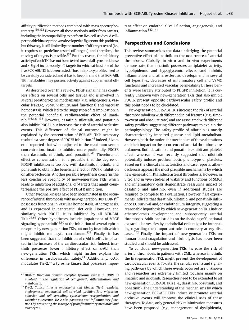
affinity purification methods combined with mass spectropho-tometry.130,132 However, all these methods suffer from caveats,including the incompatibility to perform live-cell studies. A cell-permeablekinaseprobewasdevelopedtofigureoutthisproblem,butthisassayisstill limitedbythenumberofoff-targettested(i.e.,it requires to predefine tested off-targets) and therefore, themissing of targets is possible.137 For this reason, the inhibitoryactivityofeachTKIhasnotbeen tested towardall tyrosinekinaseand►Fig. 4 includes onlyoff-targets forwhich at least oneof thefiveBCR-ABLTKIhasbeentested.Thus, inhibitoryprofilesneedtobe carefully considered and it has to keep in mind that BCR-ABLTKI metabolites may possess activity against supplemental off-targets.
As described over this review, PDGF signaling has count-less effects on several cells and tissues and is involved inseveral proatherogenic mechanisms (e.g., adipogenesis, vas-cular leakage, VSMC viability, and functions) and vascularhomeostasis, which led to the suggestion of its implication inthe potential beneficial cardiovascular effect of imati-nib.116,123,138 However, dasatinib, nilotinib, and ponatinibalso inhibit PDGFR but increase the risk of arterial occlusiveevents. This difference of clinical outcome might beexplained by the concentration of BCR-ABL TKIs necessaryto obtain a same degree of PDGFR inhibition.43 Indeed, Riveraet al reported that when adjusted to the maximum serumconcentration, imatinib inhibits more profoundly PDGFRthan dasatinib, nilotinib, and ponatinib.43 Therefore, ateffective concentration, it is probable that the degree ofPDGFR inhibition is too low with dasatinib, nilotinib, andponatinib to obtain the beneficial effect of PDGFR inhibitionon atherosclerosis. Another possible hypothesis concerns theless conclusive specificity of new-generation TKIs whichleads to inhibition of additional off-targets that might coun-terbalance the positive effect of PDGFR inhibition.
Other tyrosine kinases have been incriminated in the occur-renceof arterial thrombosiswithnew-generationTKIs. DDR-1aiv
possesses functions in vascular homeostasis, atherogenesis,and is expressed in pancreatic islet cells. However, andsimilarly with PDGFR, it is inhibited by all BCR-ABLTKIs.26,62 Other hypotheses include impairment of VEGFsignaling by ponatinib43,90 or the inhibition of several ephrinreceptors by new-generation TKIs but not by imatinib whichmight inhibit monocyte recruitment.139 Finally, it hasbeen suggested that the inhibition of c-Abl itself is implica-ted in the increase of the cardiovascular risk. Indeed, ima-tinib possesses lower inhibitory effect on c-Abl thannew-generation TKIs, which might further explain thedifference in cardiovascular safety.43 Additionally, c-Ablmodulates Tie-2,av a tyrosine kinase that possesses impor-
tant effect on endothelial cell function, angiogenesis, andinflammation.140,141
Perspectives and Conclusions
This review summarizes the data underlying the potentialpreventive effect of imatinib on the occurrence of arterialthrombosis. Globally, in vitro and in vivo experimentsdemonstrate that imatinib possesses antiplatelet activity,hypolipidemic and hypoglycemic effects, and inhibitsinflammation and atherosclerosis development in severalcell types (i.e., decreases of inflammatory cell and VSMCfunctions and increased vascular permeability). These ben-efits were largely attributed to PDGFR inhibition. It is cur-rently unknown why new-generation TKIs that also inhibitPDGFR present opposite cardiovascular safety profile andthis point needs to be elucidated.
New-generation BCR-ABL TKIs increase the risk of arterialthromboembolismwith different clinical features (e.g., time-to-event and absolute rate) and are associated with differentsafety profiles, suggesting different pathways to explain thepathophysiology. The safety profile of nilotinib is mostlycharacterized by impaired glucose and lipid metabolism.However, both themolecularmechanism of these alterationsand their impact on the occurrence of arterial thrombosis areunknown. Both dasatinib and ponatinib exhibit antiplateleteffect, whereas it was recently suggested that nilotinibpotentially induces prothrombotic phenotype of platelets.Based on the clinical characteristics and case reports, ather-osclerosis appears the most plausible mechanisms by whichnew-generationTKIs induce arterial thrombosis. However, invitro and in vivo studies of viability and functions of SMCsand inflammatory cells demonstrate reassuring impact ofdasatinib and nilotinib, even if additional studies arerequired to complete this evaluation. However, first experi-ments indicate that dasatinib, nilotinib, and ponatinib influ-ence EC survival and/or endothelium integrity, suggesting areasonable hypothesis by which new-generation TKIs induceatherosclerosis development and, subsequently, arterialthrombosis. Additional studies on the shedding of functionalextracellular vesicles by endothelial cells might be interest-ing regarding their important role in coronary artery dis-eases.142 Finally, the impact of new-generation TKIs onhuman blood coagulation and fibrinolysis has never beenstudied and should be addressed.
To conclude, new-generation TKIs increase the risk ofarterial thrombosis in patients with CML, whereas imatinib,the first-generation TKI, might prevent the development ofcardiovascular events. To date, the cellular events and signal-ing pathways by which these events occurred are unknownand researches are extremely limited focusing mainly onimatinib and nilotinib. Researches need to be extended to allnew-generation BCR-ABL TKIs (i.e., dasatinib, bosutinib, andponatinib). The understanding of the mechanisms by whichnew-generation BCR-ABL TKIs induce or promote arterialocclusive events will improve the clinical uses of thesetherapies. To date, only general risk minimization measureshave been proposed (e.g., management of dyslipidemia,
aivDDR-1: Discoidin domain receptor tyrosine kinase 1. DDR1 isinvolved in the regulation of cell growth, differentiation, andmetabolism.
av Tie-2: Tunica interna endothelial cell kinase. Tie-2 regulatesangiogenesis, endothelial cell survival, proliferation, migration,adhesion and cell spreading, cytoskeleton reorganization, andvascular quiescence. Tie-2 also possesses anti-inflammatory func-tions by preventing the leakage of proinflammatory mediators andleukocytes.
TH Open Vol. 2 No. 1/2018
Thrombosis with BCR-ABL Tyrosine Kinases Inhibitors Haguet et al. e83

diabetes, arterial hypertension following standard ofcare).14,22,23,143–146 The understanding of the pathophysiol-ogy is required to implement the most appropriate riskminimization strategies for thrombotic events and to selectpatients to whom the prescription of these drugs should beavoided when applicable. Finally, the understanding of thepathophysiology will help in the design of new BCR-ABLinhibitors sparing the toxic targets.
Review Criteria
Relevant articles published from the database inception toJuly 11, 2017, were identified from an electronic database(PubMed) using the keywords “vascular,” “thrombosis,”“atherosclerosis,” “arteriosclerosis,” “venous,” “arterial,”“hemostasis,” “metabolic,” “metabolism,” “glycemia,” “gly-caemia,” “cholesterol,” “triglycerides,” and “platelet” com-bined with the five approved BCR-ABL TKIs. The searchstrategy is presented in supplementary files. Articles pub-lished in languages other than English were excluded fromthe analysis. Primary criteriawere pathophysiological expla-nation of arterial thrombotic events. Abstracts and full-textarticles were reviewedwith a focus on atherogenesis, plaquerupture, platelet functions, and their link with the develop-ment of arterial thrombosis with BCR-ABL TKIs. The refer-ence section of identified articles was also examined.
Authors’ ContributionsH.H. was responsible for the first draft of the manuscript.F.M., C.C., C.G., J.M.D., and J.D. contributed to thefinal draftof the manuscript.
Conflicts of InterestJ.D. reports personal fees from Roche Diagnostics, StagoDiagnostica, Bayer Healthcare, and Daiichi-Sankyo; travelgrants from Bayer Healthcare, Boehringer Ingelheim, CSLBehring, and Stago Diagnostica outside the submittedwork.F.M. reports personal fees from Boehringer Ingelheim,Bayer Healthcare, and Bristol-Myers Squibb-Pfizer out-side the submitted work.C.G. reports personal fees from Novartis, Celgene, andAmgen outside the submitted work.The other authors have no conflicts of interest to disclose.
References1 Bhamidipati PK, Kantarjian H, Cortes J, Cornelison AM, Jabbour E.
Management of imatinib-resistant patients with chronic mye-loid leukemia. Ther Adv Hematol 2013;4(02):103–117
2 Gorre ME, Mohammed M, Ellwood K, et al. Clinical resistance toSTI-571 cancer therapy caused by BCR-ABL gene mutation oramplification. Science 2001;293(5531):876–880
3 Bixby D, Talpaz M. Mechanisms of resistance to tyrosine kinaseinhibitors in chronic myeloid leukemia and recent therapeuticstrategies to overcome resistance. Hematology (Am Soc HematolEduc Program) 2009;2009(01):461–476
4 Cortes JE, Saglio G, Kantarjian HM, et al. Final 5-year study resultsof DASISION: the Dasatinib Versus Imatinib Study in Treatment-Naïve Chronic Myeloid Leukemia Patients Trial. J Clin Oncol 2016;34(20):2333–2340
5 Hochhaus A, Saglio G, Hughes TP, et al. Long-term benefits andrisks of frontline nilotinib vs imatinib for chronic myeloidleukemia in chronic phase: 5-year update of the randomizedENESTnd trial. Leukemia 2016;30(05):1044–1054
6 Brümmendorf TH, Cortes JE, de Souza CA, et al. Bosutinib versusimatinib in newly diagnosed chronic-phase chronic myeloidleukaemia: results from the 24-month follow-up of the BELAtrial. Br J Haematol 2015;168(01):69–81
7 Mealing S, Barcena L, Hawkins N, et al. The relative efficacy ofimatinib, dasatinib and nilotinib for newly diagnosed chronicmyeloid leukemia: a systematic review and network meta-analysis. Exp Hematol Oncol 2013;2(01):5
8 Lamontanara AJ, Gencer EB, Kuzyk O, Hantschel O. Mechanismsof resistance to BCR-ABL and other kinase inhibitors. BiochimBiophys Acta 2013;1834(07):1449–1459
9 Pagnano KB, Bendit I, Boquimpani C, et al; Latin AmericanLeukemia Net (LALNET). BCR-ABL mutations in chronic myeloidleukemia treated with tyrosine kinase inhibitors and impact onsurvival. Cancer Invest 2015;33(09):451–458
10 Ursan ID, Jiang R, Pickard EM, Lee TA, Ng D, Pickard AS. Emer-gence of BCR-ABL kinase domain mutations associated withnewly diagnosed chronic myeloid leukemia: a meta-analysis ofclinical trials of tyrosine kinase inhibitors. J Manag Care SpecPharm 2015;21(02):114–122
11 National Comprehensive Cancer Network. NCCNClinical PracticeGuidelines in Oncology: Chronic Myeloid Leukemia. Version1.2017. 2016
12 Thanopoulou E, Judson I. The safety profile of imatinib in CML andGIST: long-term considerations. Arch Toxicol 2012;86(01):1–12
13 Kalmanti L, Saussele S, Lauseker M, et al. Safety and efficacy ofimatinib in CML over a period of 10 years: data from therandomized CML-study IV. Leukemia 2015;29(05):1123–1132
14 Moslehi JJ, Deininger M. Tyrosine kinase inhibitor-associatedcardiovascular toxicity in chronic myeloid leukemia. J Clin Oncol2015;33(35):4210–4218
15 Breccia M, Alimena G. Pleural/pericardic effusions during dasa-tinib treatment: incidence, management and risk factors asso-ciated to their development. Expert Opin Drug Saf 2010;9(05):713–721
16 European Medicines Agency. Sprycel - Summary of Product Char-acteristics2017. Available at: http://www.ema.europa.eu/docs/en_GB/document_library/EPAR_-_Product_Information/human/000709/WC500056998.pdf. Accessed December 22, 2017
17 European Medicines Agency. Iclusig - Summary of ProductCharacteristics 2017. Available at: http://www.ema.europa.eu/docs/en_GB/document_library/EPAR_-_Product_Informa-tion/human/002695/WC500145646.pdf. AccessedDecember 22,2017
18 European Medicines Agency. Tasigna - Summary of Product Char-acteristics 2017. Available at: http://www.ema.europa.eu/docs/en_GB/document_library/EPAR_-_Product_Information/human/000798/WC500034394.pdf. Accessed December 22, 2017
19 Kantarjian HM, Cortes JE, Kim DW, et al. Bosutinib safety andmanagement of toxicity in leukemia patients with resistance orintolerance to imatinib and other tyrosine kinase inhibitors.Blood 2014;123(09):1309–1318
20 Saglio G, Kim D-W, Issaragrisil S, et al; ENESTnd Investigators.Nilotinib versus imatinib for newly diagnosed chronic myeloidleukemia. N Engl J Med 2010;362(24):2251–2259
21 Poch Martell M, Sibai H, Deotare U, Lipton JH. Ponatinib in thetherapy of chronic myeloid leukemia. Expert Rev Hematol 2016;9(10):923–932
22 Douxfils J, Haguet H, Mullier F, Chatelain C, Graux C, Dogné JM.Association between BCR-ABL tyrosine kinase inhibitors forchronic myeloid leukemia and cardiovascular events, majormolecular response, and overall survival: a systematic reviewand meta-analysis. JAMA Oncol 2016; doi: 10.1001/jamaoncol.2015.5932
TH Open Vol. 2 No. 1/2018
Thrombosis with BCR-ABL Tyrosine Kinases Inhibitors Haguet et al.e84

23 Haguet H, Douxfils J, Mullier F, Chatelain C, Graux C, Dogné JM.Risk of arterial and venous occlusive events in chronic myeloidleukemia patients treated with new generation BCR-ABL tyro-sine kinase inhibitors: a systematic review and meta-analysis.Expert Opin Drug Saf 2017;16(01):5–12
24 Cortes J, Kim D-W, Pinilla-Ibarz J, et al. Ponatinib efficacy andsafety in heavily pretreated leukemia patients: 3-year results ofthe PACE trial. 20th Congress of EHA; Copenhagen, Denmark,June 12, 2015
25 Pasvolsky O, Leader A, Iakobishvili Z, Wasserstrum Y, KornowskiR, Raanani P. Tyrosine kinase inhibitor associated vascular toxi-city in chronicmyeloid leukemia. Cardio-Oncology 2015;1(01):5
26 Gora-Tybor J, Medras E, Calbecka M, et al. Real-life comparison ofsevere vascular events and other non-hematological complicationsin patients with chronic myeloid leukemia undergoing second-linenilotinib or dasatinib treatment. Leuk Lymphoma 2015;56(08):2309–2314
27 le Coutre PD, Hughes TP, Mahon FX, et al. Low incidence ofperipheral arterial disease in patients receiving dasatinib inclinical trials. Leukemia 2016;30(07):1593–1596
28 Sam PY, Ahaneku H, Nogueras-Gonzalez GM, et al. Cardiovas-cular events among patients with chronic myeloid leukemia(CML) treatedwith tyrosine kinase inhibitors (TKIs). Blood 2016;128(22):1919
29 Lassila M, Allen TJ, Cao Z, et al. Imatinib attenuates diabetes-associated atherosclerosis. Arterioscler Thromb Vasc Biol 2004;24(05):935–942
30 Ma LK, Li Q, He LF, et al. Imatinib attenuates myocardial fibrosisin association with inhibition of the PDGFRα activity. Arq BrasCardiol 2012;99(06):1082–1091
31 Fossard G, Blond E, Balsat M, et al. Hyperhomocysteinemia andhigh doses of nilotinib favor cardiovascular events in chronicphase chronic myelogenous leukemia patients. Haematologica2016;101(03):e86–e90
32 Kostos L, Burbury K, Srivastava G, Prince HM. Gastrointestinalbleeding in a chronic myeloid leukaemia patient precipitated bydasatinib-induced platelet dysfunction: case report. Platelets2015;26(08):809–811
33 Mazharian A, Ghevaert C, Zhang L, Massberg S, Watson SP.Dasatinib enhances megakaryocyte differentiation but inhibitsplatelet formation. Blood 2011;117(19):5198–5206
34 Alhawiti N, Burbury KL, Kwa FA, et al. The tyrosine kinaseinhibitor, nilotinib potentiates a prothrombotic state. ThrombRes 2016;145:54–64
35 Lotfi K, Deb S, Sjöström C, Tharmakulanathan A, Boknäs N,Ramström S. Individual variation in hemostatic alterationscaused by tyrosine kinase inhibitors: a way to improve perso-nalized cancer therapy? Blood 2016;128(22):1908
36 Li R, Grosser T, Diamond SL. Microfluidic whole blood testing ofplatelet response to pharmacological agents. Platelets 2017;28(05):457–462
37 Senis YA,Mazharian A, Mori J. Src family kinases: at the forefrontof platelet activation. Blood 2014;124(13):2013–2024
38 GratacapMP,Martin V, ValéraMC, et al. The new tyrosine-kinaseinhibitor and anticancer drugdasatinib reversibly affects plateletactivation in vitro and in vivo. Blood 2009;114(09):1884–1892
39 Albrecht-Schgoer K, Huber K, Grebien F, et al. Nilotinib exertsdirect pro-atherogenic and anti-angiogenic effects on vascularendothelial cells: a potential explanation for drug-induced vas-culopathy in CML. Blood 2013;122(21):257
40 Shimabukuro-Vornhagen A, Rothe A, Nogova L, Kochanek M,Scheid C, von Bergwelt-Baildon M. Improvement of plateletdysfunction in chronic myelogenous leukemia following treat-ment with imatinib: a case report. J Med Case Reports 2011;5:215
41 Neelakantan P, Marin D, Laffan M, Goldman J, Apperley J,Milojkovic D. Platelet dysfunction associated with ponatinib, anew pan BCR-ABL inhibitor with efficacy for chronic myeloid
leukemia resistant to multiple tyrosine kinase inhibitor therapy.Haematologica 2012;97(09):1444
42 Loren CP, Aslan JE, Rigg RA, et al. The BCR-ABL inhibitor ponatinibinhibits platelet immunoreceptor tyrosine-based activationmotif (ITAM) signaling, platelet activation and aggregate forma-tion under shear. Thromb Res 2015;135(01):155–160
43 Rivera VM, Pritchard JR, Gonzalvez F, Baker T, Gozgit JM, HodgsonG. Comparative TKI profiling analyses to explore potentialmechanisms of Ponatinib-associated arterial thrombotic events.Blood 2014;124(21):1783
44 Quintás-Cardama A, Han X, Kantarjian H, Cortes J. Tyrosinekinase inhibitor-induced platelet dysfunction in patients withchronic myeloid leukemia. Blood 2009;114(02):261–263
45 Breccia M, Muscaritoli M, Aversa Z, Mandelli F, Alimena G.Imatinibmesylate may improve fasting blood glucose in diabeticPhþ chronicmyelogenous leukemia patients responsive to treat-ment. J Clin Oncol 2004;22(22):4653–4655
46 Breccia M, Alimena G. The metabolic consequences of imatinibmesylate: changes on glucose, lypidic and bone metabolism.Leuk Res 2009;33(07):871–875
47 Agostino NM, Chinchilli VM, Lynch CJ, et al. Effect of the tyrosinekinase inhibitors (sunitinib, sorafenib, dasatinib, and imatinib) onbloodglucose levels indiabetic andnondiabetic patients ingeneralclinical practice. J Oncol Pharm Pract 2011;17(03):197–202
48 Breccia M, Muscaritoli M, Cannella L, Stefanizzi C, Frustaci A,Alimena G. Fasting glucose improvement under dasatinib treat-ment in an accelerated phase chronic myeloid leukemia patientunresponsive to imatinib and nilotinib. Leuk Res 2008;32(10):1626–1628
49 Ono K, Suzushima H, Watanabe Y, et al. Rapid amelioration ofhyperglycemia facilitated by dasatinib in a chronic myeloidleukemia patient with type 2 diabetes mellitus. Intern Med2012;51(19):2763–2766
50 Louvet C, Szot GL, Lang J, et al. Tyrosine kinase inhibitors reversetype 1 diabetes in nonobese diabetic mice. Proc Natl Acad Sci U SA 2008;105(48):18895–18900
51 Hägerkvist R, Jansson L, Welsh N. Imatinib mesylate improvesinsulin sensitivity and glucose disposal rates in rats fed a high-fatdiet. Clin Sci (Lond) 2008;114(01):65–71
52 Han MS, Chung KW, Cheon HG, et al. Imatinib mesylate reducesendoplasmic reticulum stress and induces remission of diabetesin db/db mice. Diabetes 2009;58(02):329–336
53 Hägerkvist R, Makeeva N, Elliman S, Welsh N. Imatinib mesylate(Gleevec) protects against streptozotocin-induced diabetes andislet cell death in vitro. Cell Biol Int 2006;30(12):1013–1017
54 Hägerkvist R, Sandler S, Mokhtari D, Welsh N. Amelioration ofdiabetes by imatinib mesylate (Gleevec): role of beta-cell NF-kappaB activation and anti-apoptotic preconditioning. FASEB J2007;21(02):618–628
55 Xia CQ, Zhang P, Li S, et al. C-Abl inhibitor imatinib enhancesinsulin production by β cells: c-Abl negatively regulates insulinproduction via interfering with the expression of NKx2.2 andGLUT-2. PLoS One 2014;9(05):e97694
56 Sadiq S, Owen E, Foster T, et al. Nilotinib-induced metabolicdysfunction: insights from a translational pilot study using invitro adipocytemodels and patient cohorts. 22nd EHA congress;June 22–25, 2017; Madrid, Spain
57 Iizuka K, Niwa H, Kato T, Takeda J. Dasatinib improves insulinsensitivity and affects lipidmetabolism in a patient with chronicmyeloid leukaemia. BMJ Case Rep 2016;2016:201
58 Fitter S, Vandyke K, Schultz CG, White D, Hughes TP, ZannettinoAC. Plasma adiponectin levels aremarkedly elevated in imatinib-treated chronic myeloid leukemia (CML) patients: a mechanismfor improved insulin sensitivity in type 2 diabetic CML patients?J Clin Endocrinol Metab 2010;95(08):3763–3767
59 Hosch SE, Olefsky JM, Kim JJ. APPLied mechanics: uncoveringhow adiponectin modulates insulin action. Cell Metab 2006;4(01):5–6
TH Open Vol. 2 No. 1/2018
Thrombosis with BCR-ABL Tyrosine Kinases Inhibitors Haguet et al. e85

60 Lihn AS, Pedersen SB, Richelsen B. Adiponectin: action, regula-tion and association to insulin sensitivity. Obes Rev 2005;6(01):13–21
61 Fitter S, Vandyke K, Gronthos S, Zannettino AC. Suppression ofPDGF-induced PI3 kinase activity by imatinib promotes adipo-genesis and adiponectin secretion. J Mol Endocrinol 2012;48(03):229–240
62 Aichberger KJ, Herndlhofer S, Schernthaner GH, et al. Progressiveperipheral arterial occlusive disease and other vascular eventsduring nilotinib therapy in CML. Am J Hematol 2011;86(07):533–539
63 Breccia M, Muscaritoli M, Gentilini F, et al. Impaired fastingglucose level as metabolic side effect of nilotinib in non-diabeticchronic myeloid leukemia patients resistant to imatinib. LeukRes 2007;31(12):1770–1772
64 Castagnetti F, Breccia M, Gugliotta G, et al; GIMEMA CMLWork-ing Party. Nilotinib 300 mg twice daily: an academic single-armstudy of newly diagnosed chronic phase chronic myeloid leuke-mia patients. Haematologica 2016;101(10):1200–1207
65 Delphine R, Gautier J-f, Breccia M, et al. Incidence of hypergly-cemia by 3 years in patients (Pts) with newly diagnosed chronicmyeloid leukemia in chronic phase (CML-CP) treated withNilotinib (NIL) or Imatinib (IM) in ENESTnd. Blood 2012;120(21):1686
66 Iurlo A, Orsi E, Cattaneo D, et al. Effects of first- and second-generation tyrosine kinase inhibitor therapyon glucose and lipidmetabolism in chronic myeloid leukemia patients: a real clinicalproblem? Oncotarget 2015;6(32):33944–33951
67 Racil Z, Razga F, Drapalova J, et al. Mechanism of impairedglucose metabolism during nilotinib therapy in patients withchronic myelogenous leukemia. Haematologica 2013;98(10):e124–e126
68 Ito Y, Miyamoto T, Chong Y, Maki T, Akashi K, Kamimura T.Nilotinib exacerbates diabetes mellitus by decreasing secretionof endogenous insulin. Int J Hematol 2013;97(01):135–138
69 Hadzijusufovic E, Herndlhofer S, Aichberger KJ, et al. Nilotinibexerts direct effects on vascular endothelial cells andmayact as aco-trigger of atherosclerosis in patients with Phþ CML. Blood2011;118(21):2753
70 Breccia M, Loglisci G, Salaroli A, Serrao A, Alimena G. Nilotinib-mediated increase in fasting glucose level is reversible, does notconvert to type 2 diabetes and is likely correlated with increasedbody mass index. Leuk Res 2012;36(04):e66–e67
71 Saglio G, Larson RA, Hughes TP, et al. Efficacy and safety ofNilotinib in chronic phase (CP) chronic myeloid leukemia (CML)patients (Pts) with type 2 diabetes in the ENESTnd trial. Blood2010;116(21):3430
72 Frasca F, Pandini G, Malaguarnera R, et al. Role of c-Abl indirecting metabolic versus mitogenic effects in insulin receptorsignaling. J Biol Chem 2007;282(36):26077–26088
73 Franceschino A, Tornaghi L, Benemacher V, Assouline S, Gam-bacorti-Passerini C. Alterations in creatine kinase, phosphateand lipid values in patients with chronic myeloid leukemiaduring treatment with imatinib. Haematologica 2008;93(02):317–318
74 Gottardi M, Manzato E, Gherlinzoni F. Imatinib and hyperlipi-demia. N Engl J Med 2005;353(25):2722–2723
75 Gologan R, Constantinescu G, Georgescu D, et al. Hypolipemiantbesides antileukemic effect of imatinibmesylate. Leuk Res 2009;33(09):1285–1287
76 Ellis M, Krashin E, Hamburger-Avnery O, Gan S, Elis A, Ashur-Fabian O. The anti-leukemic and lipid lowering effects of ima-tinib are not hindered by statins in CML: a retrospective clinicalstudy and in vitro assessment of lipid-genes transcription. LeukLymphoma 2017;58(05):1172–1177
77 Franklin M, Burns L, Perez S, Yerragolam D, Makenbaeva D.Incidence of type II diabetes mellitus and hyperlipidemia inpatients prescribed Dasatinib or Nilotinib as first or second line
therapy for chronic myelogenous leukemia (CML). Blood 2016;128(22):4766
78 Rea D, Mirault T, Cluzeau T, et al. Early onset hypercholester-olemia induced by the 2nd-generation tyrosine kinase inhibitornilotinib in patients with chronic phase-chronic myeloid leuke-mia. Haematologica 2014;99(07):1197–1203
79 Maurizot A, Beressi JP, Manéglier B, et al. Rapid clinical improve-ment of peripheral artery occlusive disease symptoms afternilotinib discontinuation despite persisting vascular occlusion.Blood Cancer J 2014;4:e247
80 Breccia M, Molica M, Alimena G. How tyrosine kinase inhibitorsimpair metabolism and endocrine system function: a systematicupdated review. Leuk Res 2014;38(12):1392–1398
81 Venalis P, Maurer B, Akhmetshina A, et al. Lack of inhibitoryeffects of the anti-fibrotic drug imatinib on endothelial cellfunctions in vitro and in vivo. J Cell Mol Med 2009;13(10):4185–4191
82 Gover-Proaktor A, Granot G, Shapira S, et al. Ponatinib reducesviability, migration, and functionality of human endothelialcells. Leuk Lymphoma 2017;58(06):1455–1467
83 Vallières K, Petitclerc E, Laroche G. On the ability of imatinibmesylate to inhibit smooth muscle cell proliferation withoutdelaying endothelialization: an in vitro study. Vascul Pharmacol2009;51(01):50–56
84 Hacker TA, Griffin MO, Guttormsen B, Stoker S, Wolff MR.Platelet-derived growth factor receptor antagonist STI571 (ima-tinib mesylate) inhibits human vascular smooth muscle prolif-eration and migration in vitro but not in vivo. J Invasive Cardiol2007;19(06):269–274
85 Vrekoussis T, Stathopoulos EN, De Giorgi U, et al. Modulation ofvascular endothelium by imatinib: a study on the EA.hy 926endothelial cell line. J Chemother 2006;18(01):56–65
86 Letsiou E, Rizzo AN, Sammani S, et al. Differential and opposingeffects of imatinib on LPS- and ventilator-induced lung injury.Am J Physiol Lung Cell Mol Physiol 2015;308(03):L259–L269
87 Guignabert C, Phan C, Seferian A, et al. Dasatinib induces lungvascular toxicity and predisposes to pulmonary hypertension.J Clin Invest 2016;126(09):3207–3218
88 Rizzo AN, Sammani S, Esquinca AE, et al. Imatinib attenuatesinflammation and vascular leak in a clinically relevant two-hitmodel of acute lung injury. Am J Physiol Lung Cell Mol Physiol2015;309(11):L1294–L1304
89 Katgı A, Sevindik ÖG, Gökbulut AA, et al. Nilotinib does not alterthe secretory functions of carotid artery endothelial cells in aprothrombotic or antithrombotic fashion. Clin Appl ThrombHemost 2015;21(07):678–683
90 Hadzijusufovic E, Kirchmair R, Theurl M, et al. Ponatinib exertsmultiple effects on vascular endothelial cells: possiblemechanisms and explanations for the adverse vascular eventsseen in CML patients treated with Ponatinib. Blood 2016;128(22):1883
91 Lorenzi M, Cagliero E, Toledo S. Glucose toxicity for humanendothelial cells in culture. Delayed replication, disturbed cellcycle, and accelerated death. Diabetes 1985;34(07):621–627
92 Shah NP, Wallis N, Farber HW, et al. Clinical features of pulmon-ary arterial hypertension in patients receiving dasatinib. Am JHematol 2015;90(11):1060–1064
93 Frost AE, Barst RJ, Hoeper MM, et al. Long-term safety andefficacy of imatinib in pulmonary arterial hypertension.J Heart Lung Transplant 2015;34(11):1366–1375
94 Dasgupta SK, Le A, Vijayan KV, Thiagarajan P. Dasatinib inhibitsactin fiber reorganization and promotes endothelial cell perme-ability through RhoA-ROCK pathway. Cancer Med 2017;6(04):809–818
95 Dewar AL, Domaschenz RM, Doherty KV, Hughes TP, Lyons AB.Imatinib inhibits the in vitro development of the monocyte/macrophage lineage from normal human bone marrow progeni-tors. Leukemia 2003;17(09):1713–1721
TH Open Vol. 2 No. 1/2018
Thrombosis with BCR-ABL Tyrosine Kinases Inhibitors Haguet et al.e86

96 Brownlow N, Mol C, Hayford C, Ghaem-Maghami S, Dibb NJ.Dasatinib is a potent inhibitor of tumour-associated macro-phages, osteoclasts and the FMS receptor. Leukemia 2009;23(03):590–594
97 Dewar AL, Doherty KV, Hughes TP, Lyons AB. Imatinib inhibitsthe functional capacity of cultured human monocytes. ImmunolCell Biol 2005;83(01):48–56
98 Dewar AL, Cambareri AC, Zannettino ACW, et al. Macrophagecolony-stimulating factor receptor c-fms is a novel target ofimatinib. Blood 2005;105(08):3127–3132
99 Metharom P, Martin K, Kumar AH, et al. Pleiotropic role formonocyte C-fms protein in response to vascular injury: potentialtherapeutic target. Atherosclerosis 2011;216(01):74–82
100 Gacic J, Vorkapic E, Olsen RS, et al. Imatinib reduces cholesteroluptake and matrix metalloproteinase activity in human THP-1macrophages. Pharmacol Rep 2016;68(01):1–6
101 Cwynarski K, Laylor R, Macchiarulo E, et al. Imatinib inhibits theactivation and proliferation of normal T lymphocytes in vitro.Leukemia 2004;18(08):1332–1339
102 Brownlow N, Russell AE, Saravanapavan H, et al. Comparison ofnilotinib and imatinib inhibition of FMS receptor signaling,macrophage production and osteoclastogenesis. Leukemia2008;22(03):649–652
103 Ozanne J, Prescott AR, Clark K. The clinically approved drugsdasatinib and bosutinib induce anti-inflammatorymacrophagesby inhibiting the salt-inducible kinases. Biochem J 2015;465(02):271–279
104 Darling NJ, Toth R, Arthur JS, Clark K. Inhibition of SIK2 and SIK3during differentiation enhances the anti-inflammatory pheno-type of macrophages. Biochem J 2017;474(04):521–537
105 Schade AE, Schieven GL, Townsend R, et al. Dasatinib, a small-molecule protein tyrosine kinase inhibitor, inhibits T-cell acti-vation and proliferation. Blood 2008;111(03):1366–1377
106 Blake SJ, Lyons AB, Hughes TP. Nilotinib inhibits the Src-familykinase LCK and T-cell function in vitro. J Cell Mol Med 2009;13(03):599–601
107 Blake S, Hughes TP, Mayrhofer G, Lyons AB. The Src/ABL kinaseinhibitor dasatinib (BMS-354825) inhibits function of normalhuman T-lymphocytes in vitro. Clin Immunol 2008;127(03):330–339
108 Lee KC, Ouwehand I, Giannini AL, Thomas NS, Dibb NJ, BijlmakersMJ. Lck is a key target of imatinib and dasatinib in T-cellactivation. Leukemia 2010;24(04):896–900
109 Farha S, Dweik R, Rahaghi F, et al. Imatinib in pulmonary arterialhypertension: c-Kit inhibition. Pulm Circ 2014;4(03):452–455
110 Juurikivi A, Sandler C, Lindstedt KA, et al. Inhibition of c-kittyrosine kinase by imatinib mesylate induces apoptosis inmast cells in rheumatoid synovia: a potential approach tothe treatment of arthritis. Ann Rheum Dis 2005;64(08):1126–1131
111 Sun YL, Kumar P, Sodani K, et al. Ponatinib enhances anticancerdrug sensitivity in MRP7-overexpressing cells. Oncol Rep 2014;31(04):1605–1612
112 Yvan-Charvet L,WangN, Tall AR. Role of HDL, ABCA1, and ABCG1transporters in cholesterol efflux and immune responses. Arter-ioscler Thromb Vasc Biol 2010;30(02):139–143
113 Chen Z, Lee FY, Bhalla KN, Wu J. Potent inhibition of platelet-derived growth factor-induced responses in vascular smoothmuscle cells by BMS-354825 (dasatinib). Mol Pharmacol 2006;69(05):1527–1533
114 Nakamura K, Akagi S, Ogawa A, et al. Pro-apoptotic effects ofimatinib on PDGF-stimulated pulmonary artery smooth musclecells frompatients with idiopathic pulmonary arterial hyperten-sion. Int J Cardiol 2012;159(02):100–106
115 Li L, Blumenthal DK, Masaki T, Terry CM, Cheung AK. Differentialeffects of imatinib on PDGF-induced proliferation and PDGFreceptor signaling in human arterial and venous smooth musclecells. J Cell Biochem 2006;99(06):1553–1563
116 Rocha A, Azevedo I, Soares R. Anti-angiogenic effects of imatinibtarget smooth muscle cells but not endothelial cells. Angiogen-esis 2007;10(04):279–286
117 Ballinger ML, Osman N, Hashimura K, et al. Imatinib inhibitsvascular smoothmuscle proteoglycan synthesis and reduces LDLbinding in vitro and aortic lipid deposition in vivo. J Cell MolMed2010;14(6B):1408–1418
118 Distler JH, Jüngel A, Huber LC, et al. Imatinib mesylate reducesproduction of extracellular matrix and prevents development ofexperimental dermal fibrosis. Arthritis Rheum 2007;56(01):311–322
119 Yoshiji H, Noguchi R, Kuriyama S, et al. Imatinib mesylate (STI-571) attenuates liver fibrosis development in rats. Am J PhysiolGastrointest Liver Physiol 2005;288(05):G907–G913
120 Salhotra A, Tsai N, Thomas SH, et al. Sclerodermatous chronicGVHD in patients receiving tyrosine kinase inhibitors afterallogeneic hematopoietic cell transplantation. Bone MarrowTransplant 2015;50(01):139–141
121 Makiyama Y, Toba K, Kato K, et al. Imatinib mesilate inhibitsneointimal hyperplasia via growth inhibition of vascular smoothmuscle cells in a rat model of balloon injury. Tohoku J Exp Med2008;215(04):299–306
122 Park D, Kim SM,Min SI, Ha J, Kim IG,Min SK. Inhibition of intimalhyperplasia by local perivascular application of rapamycin andimatinib mesilate after carotid balloon injury. J Korean Surg Soc2013;85(06):296–301
123 Leppänen O, Rutanen J, Hiltunen MO, et al. Oral imatinibmesylate (STI571/Gleevec) improves the efficacy of local intra-vascular vascular endothelial growth factor-C gene transfer inreducing neointimal growth in hypercholesterolemic rabbits.Circulation 2004;109(09):1140–1146
124 Sevim KZ, Silistreli O, Gorgu M, Sevim O, Ergur B. Short-termvasculoprotective effects of imatinib mesylate on intimal hyper-plasia of arterial anastomosis: An experimental study using arabbit model. Can J Plast Surg 2012;20(04):223–228
125 Wu J, Chen Z, Bhalla KN. Treatment of restenosis and stenosiswith dasatinib (2014). USF Patents. 152
126 Sánchez-Ortega I, Servitje O, Arnan M, et al. Dasatinib as salvagetherapy for steroid refractory and imatinib resistant or intolerantsclerotic chronic graft-versus-host disease. Biol Blood MarrowTransplant 2012;18(02):318–323
127 Marinelli Busilacchi E, Olivieri J, Viola N, et al. The exaggeratedcollagen expression in Gvhd-fibroblasts is effectively inhibitedby therapeutic concentration of Nilotinib. Blood 2016;128(22):4597
128 Shiha GE, Abu-Elsaad NM, Zalata KR, Ibrahim TM. Tracking anti-fibrotic pathways of nilotinib and imatinib in experimentallyinduced liver fibrosis: an insight. Clin Exp Pharmacol Physiol2014;41(10):788–797
129 Giles FJ, O’Dwyer M, Swords R. Class effects of tyrosine kinaseinhibitors in the treatment of chronic myeloid leukemia. Leuke-mia 2009;23(10):1698–1707
130 Rix U, Hantschel O, Dürnberger G, et al. Chemical proteomicprofiles of the BCR-ABL inhibitors imatinib, nilotinib, and dasa-tinib reveal novel kinase and nonkinase targets. Blood 2007;110(12):4055–4063
131 Pardanani A, Tefferi A. Imatinib targets other than bcr/abl andtheir clinical relevance in myeloid disorders. Blood 2004;104(07):1931–1939
132 Bantscheff M, Eberhard D, Abraham Y, et al. Quantitativechemical proteomics reveals mechanisms of action of clinicalABL kinase inhibitors. Nat Biotechnol 2007;25(09):1035–1044
133 Weisberg E, Manley PW, Breitenstein W, et al. Characterizationof AMN107, a selective inhibitor of native and mutant Bcr-Abl.Cancer Cell 2005;7(02):129–141
134 Buchdunger E, Cioffi CL, Law N, et al. Abl protein-tyrosine kinaseinhibitor STI571 inhibits in vitro signal transduction mediated
TH Open Vol. 2 No. 1/2018
Thrombosis with BCR-ABL Tyrosine Kinases Inhibitors Haguet et al. e87

by c-kit and platelet-derived growth factor receptors.J Pharmacol Exp Ther 2000;295(01):139–145
135 Manley PW, Drueckes P, FendrichG, et al. Extended kinase profileand properties of the protein kinase inhibitor nilotinib. BiochimBiophys Acta 2010;1804(03):445–453
136 Fabian MA, Biggs WH III, Treiber DK, et al. A small molecule-kinase interaction map for clinical kinase inhibitors. Nat Bio-technol 2005;23(03):329–336
137 Shi H, Zhang CJ, Chen GY, Yao SQ. Cell-based proteome profilingof potential dasatinib targets by use of affinity-based probes.J Am Chem Soc 2012;134(06):3001–3014
138 Liu Y,Wang Z, Kwong SQ, et al. Inhibition of PDGF, TGF-β, and Ablsignaling and reduction of liver fibrosis by the small moleculeBcr-Abl tyrosine kinase antagonist Nilotinib. J Hepatol 2011;55(03):612–625
139 Sakamoto A, Sugamoto Y, Tokunaga Y, et al. Expression profilingof the ephrin (EFN) and Eph receptor (EPH) family of genes inatherosclerosis-related human cells. J Int Med Res 2011;39(02):522–527
140 Khan AA, Sandhya VK, Singh P, et al. Signaling network map ofendothelial TEK tyrosine kinase. J Signal Transduct 2014;2014:173026
141 Chislock EM, Ring C, Pendergast AM. Abl kinases are required forvascular function, Tie2 expression, and angiopoietin-1-mediatedsurvival. Proc Natl Acad Sci U S A 2013;110(30):12432–12437
142 Boulanger CM, Loyer X, Rautou PE, Amabile N. Extracellularvesicles in coronary artery disease. Nat Rev Cardiol 2017;14(05):259–272
143 Aghel N, Delgado DH, Lipton JH. Cardiovascular toxicities of BCR-ABL tyrosine kinase inhibitors in chronic myeloid leukemia:preventive strategies and cardiovascular surveillance. VascHealth Risk Manag 2017;13:293–303
144 Damrongwatanasuk R, Fradley MG. Cardiovascular complica-tions of targeted therapies for chronic myeloid leukemia. CurrTreat Options Cardiovasc Med 2017;19(04):24
145 Valent P, Hadzijusufovic E, Hoermann G, et al. Risk factors andmechanisms contributing to TKI-induced vascular events inpatients with CML. Leuk Res 2017;59:47–54
146 García-Gutiérrez V, Jiménez-Velasco A, Gómez-Casares MT, Sán-chez-Guijo F, López-Sendón JL, Steegmann Olmedillas JL. [Car-diovascular management of patients with chronic myeloidleukemia from a multidisciplinary perspective, and proposingaction protocol by consensus meeting]. Med Clin (Barc) 2016;146(12):561.e1–561.e8
147 Aman J, van Bezu J, Damanafshan A, et al. Effective treatment ofedema and endothelial barrier dysfunction with imatinib. Cir-culation 2012;126(23):2728–2738
148 Kim SR, Suh W. Beneficial effects of the Src inhibitor, dasatinib,on breakdown of theblood-retinal barrier. Arch PharmRes 2017;40(02):197–203
149 Sukegawa M, Wang X, Nishioka C, et al. The BCR/ABL tyrosinekinase inhibitor, nilotinib, stimulates expression of IL-1β invascular endothelium in association with downregulation ofmiR-3p. Leuk Res 2017;58:83–90
150 Wolf AM,Wolf D, Rumpold H, et al. The kinase inhibitor imatinibmesylate inhibits TNF-alpha production in vitro and preventsTNF-dependent acutehepatic inflammation. ProcNatl Acad Sci US A 2005;102(38):13622–13627
151 Fraser CK, Lousberg EL, Kumar R, Hughes TP, Diener KR, HayballJD. Dasatinib inhibits the secretion of TNF-alpha following TLRstimulation in vitro and in vivo. Exp Hematol 2009;37(12):1435–1444
152 Seggewiss R, Loré K, Greiner E, et al. Imatinib inhibits T-cellreceptor-mediated T-cell proliferation and activation in a dose-dependent manner. Blood 2005;105(06):2473–2479
153 Dietz AB, Souan L, Knutson GJ, Bulur PA, LitzowMR, Vuk-PavlovicS. Imatinib mesylate inhibits T-cell proliferation in vitro anddelayed-type hypersensitivity in vivo. Blood 2004;104(04):1094–1099
154 Chen J, Schmitt A, Chen B, et al. Nilotinib hampers the pro-liferation and function of CD8þ T lymphocytes through inhibi-tion of T cell receptor signalling. J Cell Mol Med 2008;12(5B):2107–2118
155 Perros F, Montani D, Dorfmüller P, et al. Platelet-derived growthfactor expression and function in idiopathic pulmonary arterialhypertension. Am J Respir Crit Care Med 2008;178(01):81–88
156 Canning P, Tan L, Chu K, Lee SW, Gray NS, Bullock AN. Structuralmechanisms determining inhibition of the collagen receptorDDR1 by selective and multi-targeted type II kinase inhibitors.J Mol Biol 2014;426(13):2457–2470
157 Hantschel O, Rix U, Superti-Furga G. Target spectrum of the BCR-ABL inhibitors imatinib, nilotinib and dasatinib. Leuk Lymphoma2008;49(04):615–619
158 Remsing Rix LL, Rix U, Colinge J, et al. Global target profile of thekinase inhibitor bosutinib in primary chronic myeloid leukemiacells. Leukemia 2009;23(03):477–485
159 Green MR, Newton MD, Fancher KM. Off-Target Effects of BCR-ABL and JAK2 Inhibitors. Am J Clin Oncol 2016;39(01):76–84
TH Open Vol. 2 No. 1/2018
Thrombosis with BCR-ABL Tyrosine Kinases Inhibitors Haguet et al.e88