Embed Size (px)
Citation preview

Bazooka is required for localization of determinantsand controlling proliferation in the sensory organprecursor cell lineage in DrosophilaFabrice Roegiers, Susan Younger-Shepherd, Lily Yeh Jan, and Yuh Nung Jan*
Howard Hughes Medical Institute, Departments of Physiology and Biochemistry, University of California, San Francisco, CA 94143-0725
Contributed by Yuh Nung Jan, October 18, 2001
Asymmetric divisions with two different division orientationsfollow different polarity cues for the asymmetric segregation ofdeterminants in the sensory organ precursor (SOP) lineage. Thefirst asymmetric division depends on frizzled function and hasthe mitotic spindle of the pI cell in the epithelium oriented alongthe anterior–posterior axis, giving rise to pIIa and pIIb, which dividein different orientations. Only the pIIb division resembles neuro-blast division in daughter-size asymmetry, spindle orientationalong the apical–basal axis, basal Numb localization, and require-ment for inscuteable function. Because the PDZ domain proteinBazooka is required for spindle orientation and basal localizationof Numb in neuroblasts, we wondered whether Bazooka plays asimilar role in the pIIb in the SOP lineage. Surprisingly, Bazookacontrols asymmetric localization of the Numb-anchoring proteinPon, but not spindle orientation, in pI and all subsequent divisions.Bazooka also regulates cell proliferation in the SOP lineage; loss ofbazooka function results in supernumerary cell divisions and apo-ptotic cell death.
The sensory organ precursor (SOP) cell lineage unfolds witheach division exhibiting a stereotyped orientation and seg-
regating Numb, a cell-fate determinant protein, to one of the twodaughter cells (1), giving rise to a complete external sensory (ES)organ composed of a hair, socket, sheath, neuron, and glial cell.The pI and pIIa divisions occur along the anterior–posterior axis,whereas the pIIb and pIIIb divisions proceed along the apical–basal axis of the developing fly notum. Different polarity cuesdetermine the asymmetric localization of cell-fate determinantsin different divisions (2, 3). In pI, the correct spindle orientationand anterior localization of Numb crescent depend on frizzledfunction (3–5), which is also required for planar polarity in thesurrounding pupal epithelium. The pI division generates aposterior daughter, pIIa, and an anterior daughter, pIIb. Al-though both pIIb and its daughter pIIIb divide along theapical–basal axis, only the pIIb division resembles the embryonicneuroblast division in its dependence on inscuteable for properspindle orientation and Numb localization at the basal cortex (2,3). Thus, frizzled affects the pI but not the pIIb division, whereasinscuteable is required for the correct orientation of the pIIb butnot the pI cell division.
In embryonic mitotic neuroblasts, Inscuteable is localized toan apical crescent in a complex containing Pins, DaPKC, Dm-Par-6, and Bazooka (6–12). This apical complex is required fororienting neuroblast mitosis along the apical–basal axis andpositioning cell-fate determinants at the basal cortex. Given thesimilar requirement for Inscuteable in the asymmetric divisionsof embryonic neuroblasts and the pIIb cell of the SOP lineage,one might expect that Bazooka be involved in correct positioningof spindle and Numb crescent for the pIIb, the cell that dividesin a neuroblast-like manner, but not for the pI. To our surprise,we found no Pon�Numb crescent formation during mitosis in theentire SOP lineage in bazooka mutant clones. In the absence ofBazooka the SOP lineage also exhibits ectopic mitosis, cell-fatetransformation, and apoptosis of ES cell clusters.
MethodsFly Lines and in Vivo Imaging. yw bazxi106 P[mini-w�, FRT]9–2�FM6(kindly provided by A. Wodarz, University of Dusseldorf, Ger-many) and bazEH171 P[mini-w�, FRT]9–2�FM6;pr pwn P[hsFLP]�Cyo females were crossed to P[w�, Gal80], P[mini-w�,FRT]9–2;GAL4sca P[w�, UASt-Pon-GFP], and P[w�, UASt-Tau-GFP] (3). For antibody staining, GAL4109–68 (a PNS-specificGAL4 line) UAS-mCD8-GFP (13) and w� were used. mCD8-GFP, a membrane marker, allowed us to stage SOP lineage cellsbefore fixation. XZ images were acquired by using the verticalsection scan mode on a Bio-Rad 1024 confocal microscope.Larvae were heat-shocked for 1 h at 37°C during first�secondinstar stages. Larvae were allowed to grow for 3–4 days untilpupation and were mounted for live imaging or prepared forfixation (see below) 15 h after puparium formation (APF).Complete absence of Bazooka protein in bazooka mutant cloneswas confirmed by antibody staining with the Bazooka antibody(data not shown). Pupae were mounted and prepared forimaging as in ref. 3. Nota of pherate adults were fixed in 80%isopropanol, dissected, mounted in Hoyer’s media, and observedin differential-interference contrast (DIC) optics.
Immunohistochemistry and Terminal Deoxynucleotidyltransferase-Mediated dUTP Nick End Labeling (TUNEL). Control and mutantclone pupae were fixed and stained by using standard protocols(14). Antibodies used were rabbit anti-Prospero (1�1,000), rab-bit anti-Inscuteable (1�1,500; kindly provided by W. Chia,IMCB, Singapore), rabbit anti-Bazooka (1�1,500; kindly pro-vided by A. Wodarz), rat anti-mCD8 (1�100; Caltag, South SanFrancisco, CA), guinea pig anti-Numb (1�1,000), and mouseanti-�-tubulin (1�1,000; Sigma). TUNEL staining was done byusing the Apoptag kit (Intergen).
Results and DiscussionFor Bazooka to be involved in every asymmetric division of theadult SOP lineage one might expect Bazooka to be expressed inevery precursor cell. Indeed, we found that Bazooka is asym-metrically localized in every dividing cell of the SOP lineage(Figs. 1 and 2). Starting with a strong accumulation at the apicalsurface of interphase pI cells, specifically at junctions withneighboring epithelial cells (n � 10, Fig. 1 A, inter, which showsan apical section), Bazooka becomes enriched at the posteriorcortex during mitosis (n � 12, Fig. 1 A, pro and meta) and showsno overlap with the anterior Numb crescent at metaphase (n �9, Fig. 1 B and B�). By early anaphase, Bazooka forms a smoothposterior crescent (Fig. 1 A, pro and anaA). At anaphase B,Bazooka is localized to the posterior cortex, although a signif-
Abbreviations: SOP, sensory organ precursor; ES, external sensory; APF, after pupariumformation.
*To whom reprint requests should be addressed. E-mail: [email protected].
The publication costs of this article were defrayed in part by page charge payment. Thisarticle must therefore be hereby marked “advertisement” in accordance with 18 U.S.C.§1734 solely to indicate this fact.
www.pnas.org�cgi�doi�10.1073�pnas.261555598 PNAS � December 4, 2001 � vol. 98 � no. 25 � 14469–14474
DEV
ELO
PMEN
TAL
BIO
LOG
Y
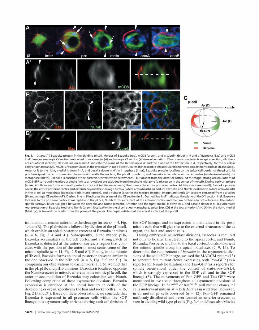
icant amount remains anterior to the cleavage furrow (n � 4, Fig.1A, anaB). The pI division is followed by division of the pIIb cell,which exhibits an apical-posterior crescent of Bazooka at mitosis(n � 6, Fig. 2 A and A�). Subsequently, in the mitotic pIIa,Bazooka accumulates in the cell cortex and a strong patch ofBazooka is detected at the anterior cortex, a region that coin-cides with the position of the anterior-most centrosome of themitotic spindle (n � 11, Fig. 2 B and B�). Finally, in the mitoticpIIIb cell, Bazooka forms an apical-posterior crescent similar tothe one observed in the pIIb cell (n � 8, Fig. 2 C and C�). Incomparing our observations to earlier work (1, 3, 5), we find thatin the pI, pIIb, and pIIIb divisions, Bazooka is localized oppositethe Numb crescent in mitosis; whereas in the mitotic pIIa cell, theanterior accumulation of Bazooka may colocalize with Numb.Following completion of these asymmetric divisions, Bazookaexpression is enriched at the apical borders in cells of thedeveloping es organ, specifically the hair and socket cells (n � 15,Fig. 2 D and D�). Based on these observations, we conclude thatBazooka is expressed in all precursor cells within the SOPlineage; it is asymmetrically enriched during each cell division of
the SOP lineage, and its expression is maintained in the post-mitotic cells that will give rise to the external structures of the esorgan, the hair and socket cells.
During embryonic neuroblast divisions, Bazooka is requirednot only to localize Inscuteable to the apical cortex and Numb,Miranda, Prospero, and Pon to the basal cortex, but also to orientthe mitotic spindle along the apical–basal axis (7, 9, 15). Todetermine the requirement of bazooka in the asymmetric divi-sions of the adult SOP lineage, we used the MARCM system (13)to generate baz mutant clones expressing both Pon-GFP (as areporter for Numb localization) and Tau-GFP (as a reporter forspindle orientation) under the control of scaberous-GAL4,which is strongly expressed in the SOP cell and in the SOPlineage (3). The movements of Pon-GFP and Tau-GFP weremonitored in live tissue throughout all asymmetric divisions ofthe SOP lineage. In bazxi106 or bazEH171 null mutant clones, pIcells underwent mitosis at �15 h APF as in wild type. However,in all mutant pI cells observed (n � 12), Pon-GFP remaineduniformly distributed and never formed an anterior crescent asseen in dividing wild-type pI cells (Fig. 3 A and B; see also Movies
Fig. 1. (A and A�) Bazooka protein in the dividing pI cell. Merges of Bazooka (red), mCD8 (green), and �-tubulin (blue) in A and of Bazooka (Baz) and mCD8in A�. Images are single XY sections extracted from a z-series (A) and a single XZ section (A�) (see schematic in C for orientation; inter is an apical section, all othersare equatorial sections). Dashed lines in A and A� indicate the plane of the XZ section in A� and the plane of the XY section in A, respectively, for the pI cell inearly anaphase (anaA). mCD8-GFP accumulates in the cytoplasm in tube-like structures that resemble intracellular membrane compartments such as ER and Golgi.Anterior is to the right, medial is down in A, and basal is down in A�. In interphase (inter), Bazooka protein localizes to the apical cell border of the pI cell. Atprophase (pro) the centrosomes (white arrows) straddle the nucleus, the pI cell rounds up, and Bazooka accumulates at the cell cortex (white arrowheads). Bymetaphase (meta), Bazooka is enriched at the posterior cortex (white arrowheads), but absent from the anterior cortex. At this stage, strong accumulations ofmCD8-GFP surround the mitotic spindle (white arrows) but are excluded from the spindle mid-zone (dark region in the center of the cell). During early anaphase(anaA, A�), Bazooka forms a smooth posterior crescent (white arrowheads) that covers the entire posterior cortex. At late anaphase (anaB), Bazooka proteincovers the entire posterior cortex and extends beyond the cleavage furrow (white arrowheads). (B and B�) Bazooka and Numb localization (white arrowheads)in the pI cell at metaphase [Bazooka (red), Numb (green), and �-tubulin (blue) in the merged images]. Images are single XY sections extracted from a z-series(B) and a single XZ section (B�). Dashed line in B indicates the plane of the XZ section in B�. Dashed line in B� indicates the plane of the XY section in B. Bazookalocalizes to the posterior cortex at metaphase in the pI cell, Numb forms a crescent of the anterior cortex, and the two proteins do not colocalize. The mitoticspindle (arrows, blue) is aligned between the Bazooka and Numb crescent. Anterior is to the right, medial is down in B, and basal is down in B�. (C) Schematicrepresentation of Bazooka (red) and Numb (green) localization in the pI cell at early anaphase, apical [Ap. (Z)] at the top, anterior [Ant. (X)] to the right, medial(Med. (Y)] is toward the reader from the plane of the paper. The pupal cuticle is at the apical surface of the pI cell.
14470 � www.pnas.org�cgi�doi�10.1073�pnas.261555598 Roegiers et al.

1–7, which are published as supporting information on the PNASweb site, www.pnas.org). Nor did Pon-GFP crescents form in thesubsequent divisions in the lineage (n � 9, Fig. 3B; see alsoMovies 4–7). Thus, although only the pIIb resembles the em-bryonic neuroblast in its orientation of division and requirementfor Inscuteable (2, 3), Bazooka is required for the asymmetricPon�Numb localization in the pI division, as well as all subse-quent divisions.
Because Numb functions as an asymmetrically localized cell-fate determinant in the SOP lineage, the absence of Numbcrescents in baz mutant clones could lead to cell-fate transfor-mations in the daughters of the pI cell. Thus we will refer to theanterior daughter cell of the pI in bazooka mutant clones (thepIIb cell in the wild type) as pIIbb, and the posterior daughtercell as pIIab. It is worth noting, however, that either loss-of-function or misexpression of numb only causes cell-fate trans-
Fig. 2. Bazooka protein localization in the cells of the SOP lineage including mitotic pIIb (A and A�), pIIa (B and B�), pIIIb (C and C�), and post-mitotic sensoryorgan cells (D and D�). Bazooka labeling (column 1, A–D�) is red in the merged images. The cells of the SOP lineage are marked with mCD8-GFP expressed underthe control of GAL4109–68 (column 2; A–D�) are green in the merged images, and �-tubulin (column 3, B and B�) in blue in the merged images, which are shownin column 3 (A and A�, C and C�, and D and D�) and column 4 (B and B�). The XY sections in A–D are extracted from a z-series (anterior is to the right, medial isdown), and images in A�–D� are single XZ sections of the same cells shown in A–D (anterior is to the right, apical is up). A–D are single XY sections (dashed linesindicate plane of the XZ section in A�–D�). Dashed lines in A–D indicate plane of the XZ section in A�–D�. Dashed lines in A�–D� and in schematic E indicate planeof the XY section in A–D. Dashed circles indicate outlines of cells that are out of the plane of focus. (Bar, 5 �m.) (A and A�) Bazooka forms an apical-posteriorcrescent (white arrowheads) in the dividing pIIb (small dashed circle), and is localized apically on the cortex of the pIIa cell (large dashed circle). Some cytoplasmicsignal is also detected in basal portion of the pIIb and pIIa cells (A�). (B and B�) In the dividing pIIa cell, Bazooka exhibits a strong localization to an anterior spot(white arrowhead), which closely coincides with the anterior centrosome at metaphase (white arrow), as well as uniform cortical localization. (C and C�) Bazookaforms an apical-posterior crescent (white arrowheads) in the dividing pIIIb, similar to the pIIb. At this stage, Bazooka in not detected in the daughters of the pIIa(*), or the basal-most glial cell (g). (D and D�) Bazooka protein in the cells of the es organ following divisions (26 h APF). Cell types were inferred from their relativepositions after the divisions (1). Bazooka is enriched in the apical portion of the es cluster composed of a hair (h), socket (so), sheath (sh), neuron (n), and glial(g) cell. A stalk of Bazooka is localized to the apical portion of the hair cell (white arrowhead). A ring of Bazooka forms around the apical boundary of the socketcell (yellow arrow). (E) Schematic representation of Bazooka localization in the mitotic pIIb, pIIa, and pIIIb, and in cells of the es organ; apical is at the top, anterioris to the right, and medial is above the plane of the paper. Hair (h), socket (so), sheath (sh), neuron (n), and glial (g) cell. Mitotic spindle orientation is based onrefs. 1 and 3.
Roegiers et al. PNAS � December 4, 2001 � vol. 98 � no. 25 � 14471
DEV
ELO
PMEN
TAL
BIO
LOG
Y
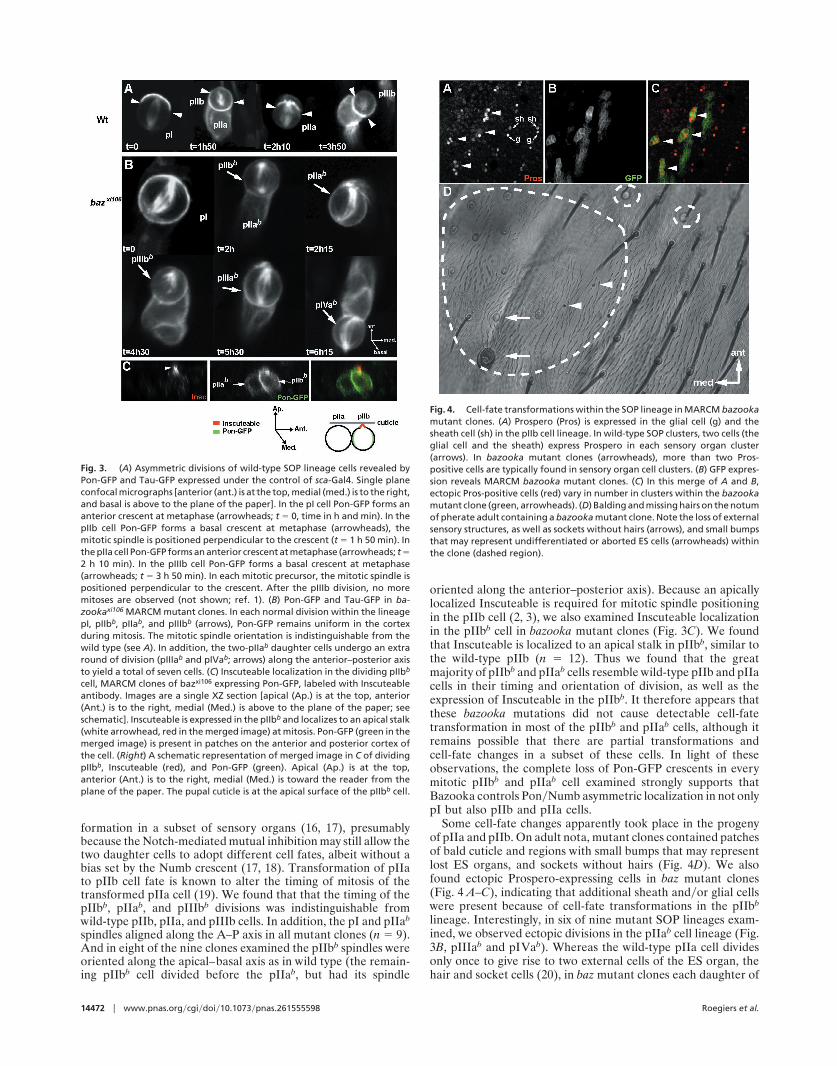
formation in a subset of sensory organs (16, 17), presumablybecause the Notch-mediated mutual inhibition may still allow thetwo daughter cells to adopt different cell fates, albeit without abias set by the Numb crescent (17, 18). Transformation of pIIato pIIb cell fate is known to alter the timing of mitosis of thetransformed pIIa cell (19). We found that that the timing of thepIIbb, pIIab, and pIIIbb divisions was indistinguishable fromwild-type pIIb, pIIa, and pIIIb cells. In addition, the pI and pIIab
spindles aligned along the A–P axis in all mutant clones (n � 9).And in eight of the nine clones examined the pIIbb spindles wereoriented along the apical–basal axis as in wild type (the remain-ing pIIbb cell divided before the pIIab, but had its spindle
oriented along the anterior–posterior axis). Because an apicallylocalized Inscuteable is required for mitotic spindle positioningin the pIIb cell (2, 3), we also examined Inscuteable localizationin the pIIbb cell in bazooka mutant clones (Fig. 3C). We foundthat Inscuteable is localized to an apical stalk in pIIbb, similar tothe wild-type pIIb (n � 12). Thus we found that the greatmajority of pIIbb and pIIab cells resemble wild-type pIIb and pIIacells in their timing and orientation of division, as well as theexpression of Inscuteable in the pIIbb. It therefore appears thatthese bazooka mutations did not cause detectable cell-fatetransformation in most of the pIIbb and pIIab cells, although itremains possible that there are partial transformations andcell-fate changes in a subset of these cells. In light of theseobservations, the complete loss of Pon-GFP crescents in everymitotic pIIbb and pIIab cell examined strongly supports thatBazooka controls Pon�Numb asymmetric localization in not onlypI but also pIIb and pIIa cells.
Some cell-fate changes apparently took place in the progenyof pIIa and pIIb. On adult nota, mutant clones contained patchesof bald cuticle and regions with small bumps that may representlost ES organs, and sockets without hairs (Fig. 4D). We alsofound ectopic Prospero-expressing cells in baz mutant clones(Fig. 4 A–C), indicating that additional sheath and�or glial cellswere present because of cell-fate transformations in the pIIbb
lineage. Interestingly, in six of nine mutant SOP lineages exam-ined, we observed ectopic divisions in the pIIab cell lineage (Fig.3B, pIIIab and pIVab). Whereas the wild-type pIIa cell dividesonly once to give rise to two external cells of the ES organ, thehair and socket cells (20), in baz mutant clones each daughter of
Fig. 3. (A) Asymmetric divisions of wild-type SOP lineage cells revealed byPon-GFP and Tau-GFP expressed under the control of sca-Gal4. Single planeconfocal micrographs [anterior (ant.) is at the top, medial (med.) is to the right,and basal is above to the plane of the paper]. In the pI cell Pon-GFP forms ananterior crescent at metaphase (arrowheads; t � 0, time in h and min). In thepIIb cell Pon-GFP forms a basal crescent at metaphase (arrowheads), themitotic spindle is positioned perpendicular to the crescent (t � 1 h 50 min). Inthe pIIa cell Pon-GFP forms an anterior crescent at metaphase (arrowheads; t �2 h 10 min). In the pIIIb cell Pon-GFP forms a basal crescent at metaphase(arrowheads; t � 3 h 50 min). In each mitotic precursor, the mitotic spindle ispositioned perpendicular to the crescent. After the pIIIb division, no moremitoses are observed (not shown; ref. 1). (B) Pon-GFP and Tau-GFP in ba-zookaxi106 MARCM mutant clones. In each normal division within the lineagepI, pIIbb, pIIab, and pIIIbb (arrows), Pon-GFP remains uniform in the cortexduring mitosis. The mitotic spindle orientation is indistinguishable from thewild type (see A). In addition, the two-pIIab daughter cells undergo an extraround of division (pIIIab and pIVab; arrows) along the anterior–posterior axisto yield a total of seven cells. (C) Inscuteable localization in the dividing pIIbb
cell, MARCM clones of bazxi106 expressing Pon-GFP, labeled with Inscuteableantibody. Images are a single XZ section [apical (Ap.) is at the top, anterior(Ant.) is to the right, medial (Med.) is above to the plane of the paper; seeschematic]. Inscuteable is expressed in the pIIbb and localizes to an apical stalk(white arrowhead, red in the merged image) at mitosis. Pon-GFP (green in themerged image) is present in patches on the anterior and posterior cortex ofthe cell. (Right) A schematic representation of merged image in C of dividingpIIbb, Inscuteable (red), and Pon-GFP (green). Apical (Ap.) is at the top,anterior (Ant.) is to the right, medial (Med.) is toward the reader from theplane of the paper. The pupal cuticle is at the apical surface of the pIIbb cell.
Fig. 4. Cell-fate transformations within the SOP lineage in MARCM bazookamutant clones. (A) Prospero (Pros) is expressed in the glial cell (g) and thesheath cell (sh) in the pIIb cell lineage. In wild-type SOP clusters, two cells (theglial cell and the sheath) express Prospero in each sensory organ cluster(arrows). In bazooka mutant clones (arrowheads), more than two Pros-positive cells are typically found in sensory organ cell clusters. (B) GFP expres-sion reveals MARCM bazooka mutant clones. (C) In this merge of A and B,ectopic Pros-positive cells (red) vary in number in clusters within the bazookamutant clone (green, arrowheads). (D) Balding and missing hairs on the notumof pherate adult containing a bazooka mutant clone. Note the loss of externalsensory structures, as well as sockets without hairs (arrows), and small bumpsthat may represent undifferentiated or aborted ES cells (arrowheads) withinthe clone (dashed region).
14472 � www.pnas.org�cgi�doi�10.1073�pnas.261555598 Roegiers et al.

the pIIab cell underwent another round of division, causing theSOP lineage to produce a cluster of seven cells, as opposed to thenormal five cells.
In the wild-type SOP lineage, shortly after the last division, ofpIIIb, at �24 h after pupa formation (1), one of the pIIIbdaughters forms a neuron and extends an axon. Within an hourafter completion of the pIIIb mitosis, the small glial cell migratesaway from the cluster along the axon (1). By following thedevelopment of the ES organs in vivo in baz mutant clones toobserve their morphogenesis, we found no axon extension orglial cell migration in 16 of 20 ES organs examined (see Movies8 and 9). Moreover, within 3–6 h after the last mitosis, clustersof cells underwent apoptosis in ten of twenty clones examined(Fig. 5; see also Movies 8 and 9). Apoptotic bodies formed anddispersed rapidly. We have confirmed apoptosis in clones byusing immunohistochemistry and terminal deoxynucleotidyl-transferase-mediated dUTP nick end labeling (TUNEL) (Fig.5C). Cell death of ES organ cells is specific to the bazookamutants, because we have never observed apoptosis in wild-typeES cells (data not shown).
Bazooka and its homologue Par-3 in C. elegans are known tobe required for asymmetric divisions, in embryonic neuroblastsin Drosophila (7, 9, 15) and in the zygote and early blastomeresof the worm embryo (21), respectively. In Drosophila neuro-blasts, Bazooka forms a complex with Pins and localizes Inscute-able, which coordinates the asymmetric localization of Numband spindle orientation (7–9, 12, 22). In the mitotic pI cell, ananterior Pins�Dlg complex has been shown to be required forBazooka localization, and that Bazooka is required for Numblocalization (23). Our study of the adult SOP lineage revealsseveral functions for Bazooka. First, we show that Bazooka is thefirst molecule to be required for the asymmetric localization ofPon, the adapter protein for Numb, in every division of the SOPlineage, even though only the pIIb division resembles embryonicneuroblast division both in its orientation along the apical–basalaxis and its dependence on Inscuteable (2, 3). It thus appears thatBazooka may localize Pon and Numb in a pathway independent
of Inscuteable. Second, Bazooka is not required for properspindle orientation in the asymmetric divisions of the SOPlineage. The function of Bazooka in the SOP lineage, therefore,concerns only determinant localization but not spindle orienta-tion. Unlike the neuroblast, in the pIIb cell Inscuteable orientsspindles along the apical–basal axis in the absence of Bazooka,indicating that pIIb cells may have a Bazooka-independentmechanism for Inscuteable localization. Third, although bazookamutations did not cause detectable cell-fate transformation inmost pIIb and pIIa cells, there is apparent cell-fate transforma-tion occurring in the pIIbb lineage, and possibly partial cell-fatetransformations of the pIIab lineage leading to formation of cellsof indeterminate cell fates, such as sockets without hairs or thebumps in bazooka mutant clones. Fourth, loss of Bazookafunction leads to apoptosis. We cannot rule out the possibilitythat inadequate cell-fate specification results in apoptosis. How-ever, the cell-fate transformations in the SOP lineage in variousmutants reported thus far have not been associated with apo-ptosis, indicating that cell-fate transformation per se does notnecessarily lead to apoptosis. The terminal fates of mutant es cellclusters are difficult to determine with certainty, because mostundergo apoptosis rather than differentiation. Fifth, Bazookaappears to limit the number of cell cycles of the pIIa to one; thepIIab daughters in bazooka mutant clones often proceed withmitosis instead of differentiating into hair and socket cells.Similarly, antiproliferative activity has been found in follicle cellsof the ovary (24). Finally, loss of Bazooka function leads tofailure of ES neuron axonal outgrowth and glial cell migration.These defects could reflect cell-fate changes in the pIIbb lineageor a requirement for bazooka in differentiation of ES organ cells.
In summary, we found Bazooka has a much broader spec-trum of function in the SOP lineage than previously suspected.In bazooka mutant clones the Numb-anchoring protein Ponfailed to form a crescent in every division of the SOP lineage,regardless of the requirement for Inscuteable. The function ofBazooka in the SOP lineage also differs from that in embry-onic neuroblasts because Bazooka controls spindle orientationin neuroblasts but not in the SOP lineage. The pI, pIIb, andpIIa cells show little evidence of cell-fate transformation inbazooka mutant clones, and yet exhibit a total loss of Pon-GFPcrescent formation. It thus appears that Bazooka controlsPon�Numb crescent formation in these precursors with dif-ferent planes of division. Although in bazooka mutant clonesthere appears to be partial cell-fate transformation in laterdivisions in the lineage, it is striking that asymmetric local-ization of determinants is abolished in all divisions. The lossof Pon-GFP crescent is fully penetrant, in contrast to thevariable and partially penetrant cell-fate transformation phe-notype. These observations suggest that Bazooka is the generallink between polarity cue and the localization of cell-fatedeterminants in all asymmetric cell divisions. Other previouslyuncharacterized functions uncovered in this study include theability of Bazooka to restrict the number of divisions in theSOP lineage and to promote differentiation instead of apo-ptosis. It is worth noting that the function of Bazooka in thecentral nervous system (CNS) has been previously studied onlyfor the neuroblast division. Based on our findings in the SOPlineage, it will be interesting to learn whether in the CNSBazooka also has an Inscuteable-independent role in control-ling asymmetry of subsequent divisions, as well as in regulatingproliferation and apoptosis. Given that Bazooka�Par-3 is partof an evolutionarily conserved gene cassette (25), our findingsof a myriad of previously uncharacterized functions of Ba-zooka in the sensory organ lineage raise the possibility thatBazooka�Par-3 may have a similarly wide range of functions invertebrates.
Fig. 5. (A) Clusters of ES cells undergo apoptosis following symmetricdivisions in bazooka mutant clones. A MARCM bazooka clone undergoes fourrounds of division (t � 0–3 h 30 min, arrows, beginning 17 h APF). Within 4 hfollowing the divisions, several cells form apoptotic bodies that are rapidlycleared (t � 7 h 30 min to 8 h, arrowheads). The remaining cells of the clusterdrift out of the plane (*, t � 8 h). The neighboring mutant cluster (�)undergoes apoptosis 1 h later (not shown). (B) An entire cluster of ES cells (t �9 h, arrow) forms apoptotic bodies simultaneously (t � 9 h 20 min, arrow-heads). (C) TUNEL labeling (red) confirms presence of apoptotic nuclei in an EScluster at 27 h APF within a bazooka mutant clone (marked with GFP, green).
Roegiers et al. PNAS � December 4, 2001 � vol. 98 � no. 25 � 14473
DEV
ELO
PMEN
TAL
BIO
LOG
Y
We thank A. Wodarz, W. Chia, and the Bloomington Stock center forproviding reagents. We thank J. Knoblich and D. Cox for helpful discussionsand M. Rothenberg for critical reading of the manuscript. F.R. is supported
by the National Institutes of Health Neuroscience Training Grant and theHuman Frontiers Science Program (HFSP) postdoctoral fellowship. L.Y.J.and Y.N.J. are Investigators at the Howard Hughes Medical Institute.
1. Gho, M., Bellaiche, Y. & Schweisguth, F. (1999) Development (Cambridge,U.K.) 126, 3573–3584.
2. Orgogozo, V., Schweisguth, F. & Bellaiche, Y. (2001) Development (Cambridge,U.K.) 128, 631–643.
3. Roegiers, F., Younger-Shepherd, S., Jan, L. Y. & Jan, Y. N. (2001) Nat. CellBiol. 3, 58–67.
4. Gho, M. & Schweisguth, F. (1998) Nature (London) 393, 178–181.5. Bellaiche, Y., Gho, M., Kaltschmidt, J., Brand, A. & Schweisguth, F. (2001)
Nat. Cell Biol. 3, 50–57.6. Kuchinke, U., Grawe, F. & Knust, E. (1998) Curr. Biol. 8, 1357–1365.7. Schober, M., Schaefer, M. & Knoblich, J. (1999) Nature (London) 402,
548–551.8. Schaefer, M., Shevchenko, A., Shevchenko, A. & Knoblich, J. (2000) Curr. Biol.
10, 353–362.9. Wodarz, A., Ramrath, A., Kuchinke, U. & Knust, E. (1999) Nature (London)
402, 544–547.10. Wodarz, A., Ramrath, A., Grimm, A. & Knust, E. (2000) J. Cell Biol. 150,
1361–1374.11. Petronczki, M. & Knoblich, J. (2001) Nat. Cell Biol. 3, 43–49.12. Yu, F., Morin, X., Cai, Y., Yang, X. & Chia, W. (2000) Cell 100, 399–409.13. Lee, T. & Luo, L. (1999) Neuron 22, 451–461.
14. Wang, S., Younger-Shepherd, S., Jan, L. Y. & Jan, Y. N. (1997) Development(Cambridge, U.K.) 124, 4435–4464.
15. Lu, B., Roegiers, F., Jan, L. Y. & Jan, Y. N. (2001) Nature (London) 409,522–525.
16. Ohshiro, T., Yagami, T., Zhang, C. & Matsuzaki, F. (2000) Nature (London)408, 593–596.
17. Rhyu, M., Jan, L. & Jan, Y. (1994) Cell 76, 477–491.18. Zeng, C., Younger-Shepherd, S., Jan, L. & Jan, Y. (1998) Genes Dev. 12,
1086–1091.19. Manning, L. & Doe, C. (1999) Development (Cambridge, U.K.) 126, 2063–2071.20. Hartenstein, V. & Posakony, J. (1989) Development (Cambridge, U.K.) 107,
389–405.21. Etemad-Moghadam, B., Guo, S. & Kemphues, K. (1995) Cell 83, 743–752.22. Kraut, R., Chia, W., Jan, L. Y., Jan, Y. N. & Knoblich, J. A. (1996) Nature
(London) 383, 50–55.23. Bellaiche, Y., Radovic, A., Woods, D., Hough, C., Parmentier, M., O’Kane, C.,
Bryant, P. & Schweisguth, F. (2001) Cell 106, 355–366.24. Cox, D. N., Abdelilah-Seyfried, S., Jan, L. Y. & Jan, Y. N. (2001) Proc. Natl.
Acad. Sci. USA 98, 14475–14480.25. Doe, C. (2001) Nat. Cell Biol. 3, E7–E9.
14474 � www.pnas.org�cgi�doi�10.1073�pnas.261555598 Roegiers et al.