Embed Size (px)
Citation preview

Bastiaan Brouwer
Umeå Plant Science CentreFysiologisk BotanikUmeå 2012

Shedding Lighton
Shade- and Dark-InducedLeaf Senescence
Bastiaan Brouwer
Akademisk avhandling
Som med vederbörligt tillstånd av Rektor vid Umeå universitet för avläggande av filosofie doktorsexamen i Växters cell- och
molekylärbiology, framläggs till offentligt försvar i KB3A9, KBC-huset, Fredagen den 25 Maj, kl. 13:00.
Avhandlingen kommer att försvaras på engelska.
Fakultetsopponent: Professor Karin Krupinska, Botanisches Institute und Botanischer Garten, Christian-Albrechts-Universität zu Kiel,
Germany.
Umeå Plant Science Centre, Department of Plant PhysiologyUmeå UniversityUmeå, Sweden, 2012

Organization Document type Date of publicationUmeå University Doctoral thesis 04th of May 2012Fysiologisk Botanik
AuthorBastiaan Brouwer
TitleShedding Light on Shade- and Dark-Induced Leaf Senescence
AbstractLeaf senescence is the final stage of leaf development, during which the leaf relocates most of its
valuable nutrients to developing or storing parts of the plant. As this process progresses, leaves lose
their green color and their capacity to perform photosynthesis. Shade and darkness are well-known
as factors inducing leaf senescence and it has been proposed that senescence can be initiated by
reductions in photosynthesis, photomorphogenesis and transpiration. However, despite the fact that
the signaling mechanisms regulating each of these processes have been extensively described,
particularly in seedlings, their contribution to the initiation of senescence in mature leaves still
remains unclear. Furthermore, the use of different experimental systems to study shade-induced
leaf senescence has yielded several divergent results, which altogether complicate the overall
understanding of leaf senescence.
To address this, darkened plants and individually darkened leaves, which show different rates of leaf
senescence, were studied. Comparing the transcriptome and metabolome of these two dark-
treatments revealed that they differed distinctly with regard to their metabolic strategies. Whole
darkened plants were severely carbohydrate-starved, accumulated amino acids and slowed down
their metabolism. In contrast, individually darkened leaves showed continued active metabolism
coupled to senescence-associated degradation and relocation of amino acids.
This knowledge was used to set up a new system to study how shade affects leaf senescence in the
model plant Arabidopsis thaliana. Use of this system revealed that different senescence-associated
hallmarks appeared in response to different intensities of shade. Some of these hallmarks were
further shown to be part of both leaf senescence and photosynthetic acclimation to low light.
Finally, using this system on phytochrome mutants revealed that loss of phytochrome A increased
the loss of chlorophyll under shade, without increasing the expression of senescence-associated
genes.
Together, these findings suggest that shade-induced leaf senescence, which is generally perceived as
a single process, is actually an intricate network of different processes that work together to
maintain an optimal distribution of nutrients within the plant.
KeywordsArabidopsis, darkness, light, photosynthesis, phytochrome, shade, senescence
Language ISBN Number of pagesEnglish 978-91-7459-437-9 52 + 3 papers

Shedding Light on
Shade- and Dark-Induced
Leaf Senescence
Bastiaan Brouwer
Umeå Plant Science Centre
Fysiologisk Botanik
Umeå 2012

This work is protected by the Swedish Copyright Legislation (Act 1960:729)
ISBN: 978-91-7459-437-9
Front cover by: Bastiaan Brouwer
Electronic version available at http://umu.diva-portal.org/
Printed by: KBC, Umeå University
Umeå, Sweden 2012

iii
Abstract
Leaf senescence is the final stage of leaf development, during which the
leaf relocates most of its valuable nutrients to developing or storing parts of
the plant. As this process progresses, leaves lose their green color and their
capacity to perform photosynthesis. Shade and darkness are well-known as
factors inducing leaf senescence and it has been proposed that senescence
can be initiated by reductions in photosynthesis, photomorphogenesis and
transpiration. However, despite the fact that the signaling mechanisms
regulating each of these processes have been extensively described,
particularly in seedlings, their contribution to the initiation of senescence in
mature leaves still remains unclear. Furthermore, the use of different
experimental systems to study shade-induced leaf senescence has yielded
several divergent results, which altogether complicate the overall
understanding of leaf senescence.
To address this, darkened plants and individually darkened leaves, which
show different rates of leaf senescence, were studied. Comparing the
transcriptome and metabolome of these two dark-treatments revealed that
they differed distinctly with regard to their metabolic strategies. Whole
darkened plants were severely carbohydrate-starved, accumulated amino
acids and slowed down their metabolism. In contrast, individually darkened
leaves showed continued active metabolism coupled to senescence-
associated degradation and relocation of amino acids.
This knowledge was used to set up a new system to study how shade
affects leaf senescence in the model plant Arabidopsis thaliana. Use of this
system revealed that different senescence-associated hallmarks appeared in
response to different intensities of shade. Some of these hallmarks were
further shown to be part of both leaf senescence and photosynthetic
acclimation to low light.
Finally, using this system on phytochrome mutants revealed that loss of
phytochrome A increased the loss of chlorophyll under shade, without
increasing the expression of senescence-associated genes.
Together, these findings suggest that shade-induced leaf senescence,
which is generally perceived as a single process, is actually an intricate
network of different processes that work together to maintain an optimal
distribution of nutrients within the plant.

iv
Sammanfattning
Senescens är det sista steget i ett blads livscykel och leder till att
värdefulla näringsämnen överförs till växande eller lagrande delar av växten.
Allt eftersom denna process fortskrider, förlorar bladet sin gröna färg samt
sin förmåga att fotosyntetisera. Skuggning och mörkläggning är välkända
faktorer som initierar bladsenescens och detta kan triggas genom
långsammare fotosyntes, fotomorfogenes och transpiration. Trots att
signaleringen bakom dessa processer är utförligt studerade och beskrivna i
groddplantor, finns många oklarheter om hur detta leder till initiering av
senescensen hos fullt utvecklade blad. Att olika experimentella system
använts för att studera skugg-inducerad bladsenescens har dessutom lett till
varierande resultat, vilket försvårar en övergripande förståelse av processen.
För att utreda detta vidare studerades hela växter i mörker vilka
jämfördes med enskilt mörklagda blad, behandlingar som uppvisar olika
hastigheter av senescens. Genom att jämföra transkriptom och metabolom
under sådana mörkerexperiment visades att de metaboliska strategierna klart
skilde sig åt mellan behandlingarna. När hela växten mörklades uppvisade
bladen klara tecken på kolhydratsvält och ackumulering av aminosyror
samtidigt som metabolismen gick på sparlåga. I individuellt mörklagda blad
fortsatte däremot en aktiv metabolism kopplad till degradering och
borttransport av aminosyror.
Denna information användes sedan för att sätta upp ett nytt system för
att studera hur skuggning av blad påverkar bladsenescens i modellväxten
Arabidopsis thaliana. Resultaten från detta visade på många typiska
kännetecken för bladscenescens som en följd av olika grader av skuggning.
Några av dessa faktorer var gemensamma för senescens och acklimering av
fotosyntesen till lågt ljus.
Användandet av fytokrommutanter i detta experimentella system visade
dessutom att signallering via fytokrom kan bidra till processen. I en mutant
utan fytokrom A fördröjdes skugg-inducerad minskning av klorofyll men
utan att uttrycket av senesces-associerade gener ökade.
Sammantaget tyder dessa upptäckter på att bladsenescens som initieras
av beskuggning, vilket oftast betraktas som en enhetlig process, i
verkligheten är ett komplicerat nätverk av olika processer som opererar
tillsammans för att upprätthålla en optimal fördelning av näringsämnen inom
växten.

v
List of Papers
I. Abdul Ahad*, Olivier Keech*, Andreas Sjödin, Pernilla Lindén,
Bastiaan Brouwer , H Stenlund, Thomas Moritz, Stefan Jansson
and Per Gardeström
Comparisons between leaves from darkened plants and individually
darkened leaves reveal differential metabolic strategies in response
to darkness
Manuscript
II. Bastiaan Brouwer, Agnieszka Ziolkowska, Matthieu Bagard,
Olivier Keech and Per Gardeström
The impact of light intensity on shade-induced leaf senescence
Plant, Cell & Environment, 2012, DOI: 10.1111/j.1365-
3040.2011.02474.x
III. Bastiaan Brouwer, Per Gardeström and Olivier Keech
Far-red light reduces senescence-associated chlorophyll loss under
low light via a Phytochrome A-mediated Far-red High Irradiance
Response
Manuscript
* These authors contributed equally
Paper II has been reproduced with kind permission of the publisher.
The papers will be referred to by their Roman numbers in the text.

vi

vii
Table of Contents
Abstract iii
Sammanfattning iv
List of Papers v
Preface ix
Abbreviations x
INTRODUCTION 1
A Leaf's Life 1
Leaf Senescence 1
Why study leaf senescence? 1
The process of leaf senescence 2
Chlorophyll degradation 2
Protein degradation 4
Nitrogen relocation 6
Lipid degradation 8
Mitochondria and loss of cellular organization 9
What induces and inhibits leaf senescence? 10
Shade- and Dark-induced leaf senescence 12
Shade and Darkness 12
Photosynthesis 12
Photomorphogenesis 14
Phytochromes 15
Transpiration 17
SUMMARY 18
AIM 19
RESULTS AND DISCUSSION 20
Different experimental systems 20
Darkened plants versus individually darkened leaves 21
Darkened leaves are sugar-starved 22
Darkened plants differ from individually darkened leaves 23
Shade-induced leaf senescence depends on the light intensity 25
Different light intensities induce different hallmarks of senescence 26

viii
PRP as part of the senescence process 27
Leaf senescence overlaps with PA 28
The LCP as a threshold to enhance expression of SAGs 28
Shade-induced chlorophyll loss depends on phytochromes 29
FR does not enhance chlorophyll loss under low light 29 PHYA inhibits chlorophyll loss 29
PHYA inhibits chlorophyll loss via the FR-HIR 30
PHYA does not directly inhibit the expression of SAGs 31
Could PHYA inhibit leaf senescence via chlorophyll biosynthesis? 32
Shedding light on leaf senescence 34
Summary 37
CONCLUSIONS AND FUTURE PERSPECTIVES 39
ACKNOWLEDGEMENTS 40
REFERENCES 42

ix
Preface
If you have plants in your house, you have undoubtedly observed the
occasional yellowing leaf. Most people remove such leaves and provide the
plant with some extra fertilizer to prevent further yellowing. Some of you
will have observed that such leaves most often form on the side of the plant
that receives the least light. The process causing such yellowing of leaves is
called shade-induced leaf senescence. Leaf senescence is a process that the
plant uses to redirect limiting nutrients (often nitrogen) from older and
shaded leaves to younger and growing leaves or storage-tissues. Because of
this feature, understanding how shade and light initiate and postpone leaf
senescence, respectively, could lead to increased crop productivity and
product shelf-life. However, this understanding is still fragmented and the
grand aim of this thesis therefore is to connect these fragments and shed
more light on the process of shade-induced leaf senescence.
Bastiaan Brouwer, 2012

x
Abbreviations
Abbreviations are explained when they first appear in the text.

1
INTRODUCTION
A Leaf's Life
When the temperature is high enough, light stimulates imbibed plant
seeds to germinate and grow upwards. Upon reaching an illuminated surface,
the light will promote seedlings to undergo photomorphogenesis, during
which they develop functional chloroplasts and become green
photoautotrophic organisms. Photoautotrophic organisms use light to
assimilate carbon dioxide (CO2) into sugars and other carbohydrates via the
process of photosynthesis. Within a plant canopy, plants compete for light by
elongating their stems and developing new leaves. These new leaves often
overshadow the older ones, which as a result experience continuous changes
in their light environment. In response to these changes, shaded leaves
acclimate their photosynthetic machinery to optimize their function. This
acclimation occurs on two levels: within leaves and between leaves. Within
leaves, the ratios between the light harvesting antennae and the photosystems
are adjusted through a process termed Photosynthetic Acclimation (PA).
Between leaves, whole-plant photosynthesis is optimized by re-allocating
limiting nutrients like nitrogen from older and shaded leaves to younger and
well-illuminated plant parts through a process termed Photosynthetic
Resource Partitioning (PRP). Leaves that cannot acclimate sufficiently or
that receive cues signaling the end of plant development undergo leaf
senescence. During this final process, the structure of the leaf is slowly
degraded, while limiting nutrients, such as nitrogen, sulfur and phosphate,
are relocated to developing and storing organs such as new leaves, roots and
seeds.
Leaf Senescence
Why study leaf senescence?
Derived from the latin verb 'senescere', which means 'to grow old', leaf
senescence is generally defined as the developmental stage that follows leaf
maturation and has leaf death as the inevitable outcome (Leopold, 1961;
Smart, 1994; Gan & Amasino, 1997; van Doorn & Woltering, 2004). During
this process, leaves turn yellow as the photosynthetic machinery is degraded
and the resulting nutrients are transported to other parts of the plant
(Gregersen et al., 2008; Masclaux-Daubresse et al., 2008).
While leaf senescence is best known for causing the idyllic scenery in
deciduous forests during autumn, it actually constitutes a major part of our
everyday lives. Stress-induced senescence reduces both the yield
(Hörtensteiner, 2009) and the shelf-life of green plant produce (King &
Morris, 1994). Additionally, major food-crops such as wheat and barley
2
employ leaf senescence to remobilize most of their leaf nutrients during seed
filling (Gregersen et al., 2008). During these processes, the rate of
senescence influences the nutrient-composition of both leaves and seeds and
thereby affects the desired quality of the produce (Gregersen et al., 2008). A
better understanding of the senescence process would therefore allow us to
modulate both plants and growing conditions to selectively postpone or
induce leaf senescence, depending on our needs with respect to growth
season, seed filling, nutrient composition or resistance to different stresses.
The process of leaf senescence
Leaf senescence is a genetically controlled process (Yoshida, 1963),
during which chloroplasts, which contain the photosynthetic machinery, are
converted into gerontoplasts (Thomas et al., 2003). This conversion is
realized by the degradation of chlorophyll and chloroplastic protein
(Krupinska, 2006) and ultrastructural changes such as the loss of chloroplast
grana and thylakoids, increased formation of plastoglobuli and decreases in
both the number and the size of the plastids (Zavaleta-Mancera et al., 1999;
Keech et al., 2007; Wada et al., 2009). Alongside these plastidial changes,
mitochondria increase in size and show a relative increase in respiration
activity, most likely to support the active metabolism accompanying leaf
senescence (Keech et al., 2007). Meanwhile, enzymes involved in protein
degradation and the synthesis of transport amino acids become relatively
abundant (Masclaux-Daubresse et al., 2008). Finally, as these processes are
completed, cells lose their structure and collapse.
Chlorophyll degradation
The most commonly observed aspect of leaf senescence is the loss of
chlorophyll, which as such is considered to be a good marker to assess the
progression of leaf senescence (Leopold, 1961; Ougham et al., 2008).
Senescence-associated degradation of chlorophyll is under nuclear control
(Thomas & Stoddart, 1980) and has been intensively studied over the last
decade (Ougham et al., 2008; Schelbert et al., 2009; Sakuraba et al., 2012).
While chlorophyll was initially thought to be degraded solely via
chlorophyllase (CLH; Tsuchiya et al., 1999), this view has recently been
questioned as mutants in the CLH-genes are still able to degrade chlorophyll
during leaf senescence (Schenk et al., 2007) and expression of CLH1 and
CLH2 is positively regulated by light (Banas et al., 2011). Instead, the
degradation of chlorophyll during leaf senescence occurs mainly via the
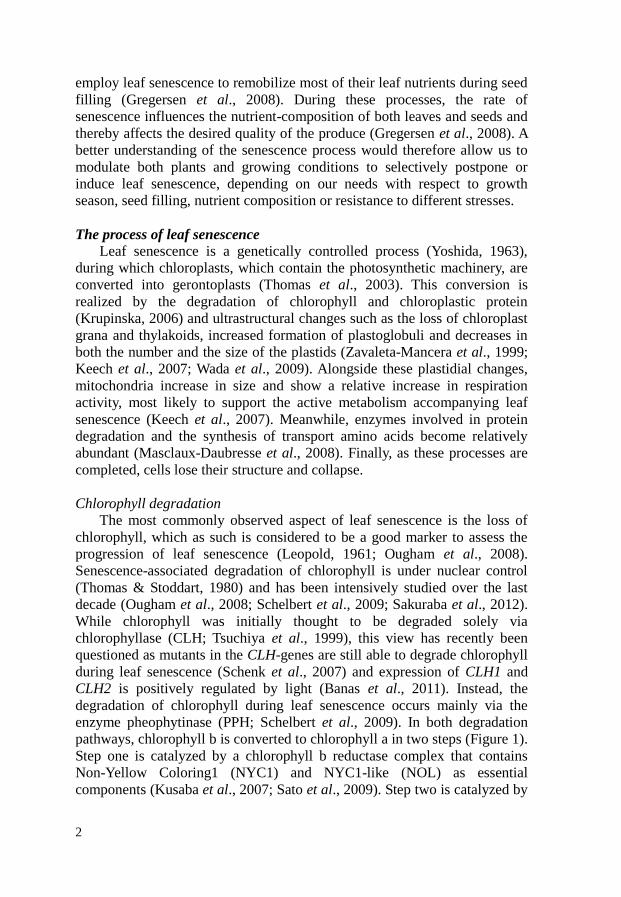
enzyme pheophytinase (PPH; Schelbert et al., 2009). In both degradation
pathways, chlorophyll b is converted to chlorophyll a in two steps (Figure 1).
Step one is catalyzed by a chlorophyll b reductase complex that contains
Non-Yellow Coloring1 (NYC1) and NYC1-like (NOL) as essential
components (Kusaba et al., 2007; Sato et al., 2009). Step two is catalyzed by

3
7-HydroxyMethyl Chlorophyll a Reductase (HMCR) and yields chlorophyll
a (Meguro et al. 2011). In case of CLH-based degradation (Figure 1, blue
arrows), chlorophyll a is converted to chlorophyllide a by CLH and
subsequently stripped of its Mg by a metal chelating substance (MCS;
Suzuki & Chioi, 2002). In PPH-based degradation (Figure 1; pink arrows),
chlorophyll a is stripped of its Mg by MCS and the resulting pheophytin a
(phein a) converted to pheophorbide a (pheide a) by PPH (Schelbert et al.,
2009). After the formation of pheide a, the two pathways converge and the
risk for photoxic reaction products is removed by the concerted action of
Pheide A Oxygenase (PAO) and Red Chlorophyll Catabolite Reductase
(RCCR; Rodoni et al., 1997; Wüthrich et al., 2000; Pružinská et al., 2003).
The resulting primary fluorescent Chl catabolyte (pFCC) is modified in the
cytosol and transported to the vacuole, where it is converted to and stored as
non-fluorescing chlorophyll catabolites (NCCs; Oberhuber et al., 2003).
Figure 1: Pathways describing chlorophyll (Chl) degradation during leaf
development (via CLH; blue arrows) and leaf senescence (via PPH; pink Arrows).
Chl biosynthesis during leaf development is represented by its final steps (CS and
CAO). Conversions of Chlide a between and Chlide b occur by the same enzymes
mediating the conversions between Chl a and Chl b. Arrow thickness indicates either
minor (thin) or major (thick) contribution to chlorophyll degradation associated to
leaf senescence. Abbreviations: CAO, Chl a oxygenase; Chlide, chlorophyllide; CS,
Chl synthase; RCC, red Chl catabolite. Other abbreviations are mentioned in the
text. Modified after Schelbert et al., (2009) and Sakuraba et al., (2012).
Recently, it was discovered that most of the chloroplastic enzymes
related to the PPH-pathway interact with the protein Stay-GReen (SGR;
Ougham et al., 2008; Figure 1), which is thought to act as a scaffold to
optimize the concerted action of these enzymes (Sakuraba et al., 2012).

4
Disconcerted action of these enzymes causes two opposing effects:
accelerated cell death and cosmetic stay-green phenotypes. Accelerated cell
death occurs when the function of HCMR, PAO or RCCR is impaired and
photoxic intermediates such as pheide a or RCC accumulate. In contrast,
lack of SGR, NYC1, NOL or PPH results in a cosmetic stay-green
phenotype, in which the light harvesting complexes are retained as well
(reviewed in Ougham et al., 2008). These observations suggest that
chlorophyll degradation is related to its stabilizing influence on light
harvesting complex (LHC) protein (Apel & Kloppstech, 1980) and that
removal of chlorophyll allows proteases to access this significant amount
(20%) of cellular protein (Hörtensteiner, 2006).
Protein degradation
While loss of chlorophyll is the most visible aspect of leaf senescence,
its onset is preceded by that of protein loss (Hensel et al., 1993; Keskitalo et
al., 2005). As much as 70% of the leaf protein is located within the
chloroplast (Gan & Amasino, 1997; Hörtensteiner & Feller, 2002), most of
which is in the form of proteins such as Rubisco and LHC-protein (50 and
20% of the leaf protein, respectively; Mae et al., 1983; Hörtensteiner, 2006).
To degrade these proteins, chloroplasts contain a number of proteases such
as Clp, FtsH, DegP and Lon (Adam & Clarke, 2002; Figure 2; chloroplast).
Many of these proteases and their subunits, e.g. the Clp protease system and
FtsH proteases, are constitutively expressed during leaf development and
serve in the turnover and repair of the photosynthetic machinery. Some of
the genes associated to these proteases show increased expression during
leaf senescence (Lin & Wu, 2004), suggesting that the corresponding
proteases contribute to this final developmental stage (Martínez et al.,
2008a). However, isolated chloroplasts from senescing leaves accumulate
specific Rubisco protein fragments (Kokubun et al., 2002), suggesting that
senescence-associated protein degradation is not completed within the
chloroplasts, but relies on the export of cleaved protein fragments to other
cellular compartments for further degradation (Martínez et al., 2008a).
Over the last decade, two types of senescence-associated bodies that
contain partially degraded plastid protein have been identified: Rubisco-
containing bodies (RCBs; Chiba et al., 2003) and Senescence-associated
vacuoles (SAVs; Otegui et al., 2005). The first type, RCBs, are small
spherical bodies (0.4–1.2 µm in diameter) that are found in both the
cytoplasm and the vacuole during the early phases of natural leaf senescence
(Chiba et al., 2003). These bodies contain stromal proteins such as Rubisco
and chloroplastic glutamine synthetase (GS2) and their double membrane, in
addition to their content, suggests that they originate from the chloroplast
(Figure 2, RCB pathway). Additional membrane and tubular structures
surrounding these bodies (Chiba et al., 2003) and the lack of RCBs in

5
AuTophaGy (ATG) mutants such as atg4 or atg5 indicate that the autophagic
system is involved (Ishida et al., 2008; Wada et al., 2009; Figure 2, bottom
pathway). While the ATG-dependent autophagy is required to reduce the
chloroplast size, which remains constant in atg4a4b-1 mutant plants (Wada
et al., 2009), it is not the only system able to reduce stromal chloroplast
protein, as exemplified by similar declines in the Rubisco content in wild-
type and atg4a4b-1 mutant plants. Furthermore, disabling the autophagic
system causes enhanced leaf senescence under conditions of limiting
nutrients, when nutrient relocation is promoted (Doelling et al., 2002;
Hanaoka et al., 2002; Thompson et al., 2005; Phillips et al., 2008). This
implies that although autophagy seems to be involved during regular leaf
senescence, it can be replaced or supplemented by other, more aggressive,
degradation processes (Martínez et al., 2008a).
Figure 2: Representation of protein degradation pathways during leaf senescence, in
particular those involving Rubisco containing bodies (RCB) and senescence
associated vacuoles (SAV). Other abbreviations are explained in the text. Modified
after Gregersen et al., (2008).
A strong candidate for such process is the senescence-specific formation
of the second type of bodies; SAVs, which are small (0.55–0.70 µm) lytic
vacuoles with a low pH (5.2) that do not depend on a functional autophagic
system (Otegui et al., 2005; Figure 2, SAV pathway). Besides partially
degraded chloroplast stromal protein, they may contain chlorophyll a, but
lack thylakoid proteins such as photosystem II reaction center protein D1 or
light harvesting complex protein II (Martínez et al., 2008b). In contrast to
isolated chloroplasts, isolated SAVs continue to degrade their protein via
proteases such as Senescence-Associated Gene 12 (SAG12; Otegui et al.,
2005; Martínez et al., 2008b), the gene of which is expressed late during leaf
senescence (Weaver et al., 1998). Both RCBs and SAVs are believed to

6
deliver their contents to the central vacuole for storage and further
degradation (Martínez et al., 2008a; Wada et al., 2009), as a number of genes
associated to vacuolar protease activity, such as SAG2, RD21 and γVPE,
show enhanced expression during leaf senescence (Kinoshita et al., 1999;
Buchanan-Wollaston et al., 2003; Gepstein et al., 2003).
In addition to chloroplast and vacuole-based protease systems, the
cytosolic/nuclear ubiquitin proteosome pathway appears to be enhanced
during leaf senescence (Park et al., 1998; Masclaux-Daubresse et al., 2008).
Important demonstrations of the involvement of this pathway involve
mutations of genes such as ORESARA 9 (ORE9) and Nitrogen Limitation
Adaptation (NLA). The ORE9 protein contains an F-box domain and has
been shown to be able to form a ubiquitin E3 ligase complex, suggesting that
it can ubiquitinate specific substrates to target them for degradation by the
26S proteasome and the ore9-1 mutant shows delayed leaf senescence (Woo
et al., 2001). The NLA protein interacts with an ubiquitin conjugase in the
nucleus and mutation of the NLA gene leads to enhanced senescence under
nitrogen-limiting conditions (Peng et al., 2007).
Altogether, senescence-associated protein degradation is carried out in
different cellular compartments by the concerted action of several protease-
systems, some of which are constitutive and some of which are enhanced
during leaf senescence.
Nitrogen relocation
One of the main functions attributed to leaf senescence is the relocation
of nitrogen from senescing to developing leaves (Smart, 1994; Lim et al.,
2007). To this end, the breakdown products of the degraded proteins are
incorporated into different amino acids for transport through the phloem
(Masclaux-Daubresse et al., 2008; figure 3, upper part). Transcript and
metabolite profile studies have shown that during leaf senescence, a number
of enzymes involved in the biosynthesis and transamination of amino acids
are enhanced consistently in different plant species (Masclaux-Daubresse et
al., 2008). Among these, two enzymes have been extensively studied:
Glutamate DeHydrogenase (GDH) and cytosolic Glutamine Synthase (GS1).
Within the mitochondria, GDH converts glutamate, originating from
transamination reactions, to α-ketoglutarate and ammonium. While α-
ketoglutarate can be used as a respiratory substrate via the tricarboxylic acid
(TCA) cycle, ammonium is exported to the cytoplasm, where it can be used
by GS1 to convert glutamate into glutamine (Masclaux-Daubresse et al.,
2008). In turn, glutamine can be used to convert aspartate into asparagine by
Asparagine Synthetase (AS), whose transcripts increase during leaf
senescence in both sunflower (Herrera-Rodriguez et al., 2006) and
Arabidopsis (Lin & Wu, 2004). Lin & Wu (2004) further proposed, based on

7
Figure 3: The concerted action of nitrogen remobilization (upper part),
galactolipid mobilization (insert) and lipid degradation (lower part). Abbreviations:
ACX, acyl CoA oxidase; α-gal, α-galactosidase; α-KG, α-ketoglutarate; AS,
asparagine synthase; Asn, asparagine; Asp, aspartate; Asp AT, aspartate
aimontransferase; AT, amino transferases; β-gal, β-galactosidase; Chl, chlorphyll;
CoA, coenzyme A; DAG, diacylglycerol; DGAT, DAG acyltransferase; GDH,
glutamate dehydrogenase; g-lip, galactolipase; Gln, glutamine; Glu, glutamate; GS1,
cytosolic glutamine synthase; ICL, isocitrate lyase; LACS, long-chain acyl CoA
lyase; Mal, malate; MDH, malate dehydrogenase; MS, malate synthase; OAA,
oxaloacetate; PEP, phosphoenol pyruvate; PEPCK, PEP carboxykinase; PPDK, PEP
dikinase; SAVs, senescence-associated vacuoles; Suc, succinate; TAG,
triacylglycerol; TCA cycle, tricarboxylic acid cycle. Modified after Thompson et al.,
(1998), Masclaux-Daubresse et al., (2008) and Dörmann (2010).

8
the senescence-associated increase in several gene transcripts in darkened
plants, that the aspartate required for asparagine synthesis is derived from
pyruvate through the combined action of Phosphoenol Pyruvate DiKinase
(PPDK), PhosphoEnol Pyruvate CarboxyKinase (PEPCK) and aspartate
aminotransferase. Additionally, PEPCK increases in response to increased
ammonium and is present specifically in phloem companion cells (Chen et
al., 2004), further supporting a role for this enzyme during the generation
and export of nitrogen-rich amino acids from senescing leaves.
Lipid degradation
As chloroplasts are converted into gerontoplasts, thylakoid lipid
degradation becomes a prominent feature (figure 3, lower part), providing
both carbon backbones for the formation of amino acids as well as
respiratory carbon (Buchanan-Wollaston, 1997; Krupinska, 2006). When
thylakoid membranes are dismantled, their breakdown products are stored in
plastoglobuli, which increase in both number and size (Krupinska, 2006).
However, in contrast to other membranes, thylakoid membranes contain a
large fraction of galactolipids (Lee, 2000), which are not directly stored in
the plastoglobuli (Tevini & Steinmüller, 1985). Instead, galactolipids are first
degraded inside the chloroplasts via a pathway involving α-galactosidases, β-
galactosidases and galactolipases (Krupinska, 2006; Dörmann, 2010) (figure
3, insert). These enzymes are upregulated during leaf senescence in various
plant species (Thompson et al., 1998; Lee et al., 2004; Krupinska, 2006) and
increased activity of an α-galactosidase in rice was recently shown to
directly influence the abundance of thylakoid membranes (Lee et al., 2009).
The resulting diacylglycerols (DAGs) are subsequently converted into
triacylglycerols (TAGs) by DAG acyltransferase (DGAT), which is strongly
increased during leaf senescence (Kaup et al., 2002). Meanwhile, free fatty
acids originating from galactolipase action and phytol, a byproduct from
chlorophyll degradation, are converted into fatty acid phytyl esters and
together with TAGs sequestered in the plastoglobuli (Kaup et al., 2002;
Dörmann, 2010). Plastoglobuli are subsequently extruded from the
chloroplast into the cytoplasm, through which they gain access to
glyoxysomes (Guiamet et al., 1999) (Figure 3, lower part). Glyoxysomes are
peroxisomes (Pracharoenwattana & Smith, 2008) that specialize in the β-
oxidation of TAG-derived fatty acids into acetyl coenzyme A to form
succinate via the glyoxylate cycle (Thompson et al., 1998). During leaf
senescence, various genes in these pathways, such as isocitrate lyase, malate
synthase, long-chain acyl CoA lyases and acyl CoA oxidases are upregulated
(Yang & Ohlrogge, 2009). Succinate is subsequently exported to the
mitochondria, where it enters the Krebs cycle (Thompson et al., 1998). From
this cycle, malate is exported into the cytosol and by NAD+-dependent
malate dehydrogenase converted into oxaloacetate, which can be used either

9
as an amino acid backbone (Lin & Wu, 2004) or via PhosphoEnolPyruvate
(PEP) converted into sugars for export or ATP-production (Buchanan-
Wollaston, 1997; Thompson et al., 1998).
As senescence progresses, non-thylakoid membranes are also degraded
through the concerted action of phospholipase D, phosphatidic acid
phosphatase, lytic acyl hydrolase and lipoxygenase (Thompson et al., 1998).
It appears that this is facilitated by the action of an acyl hydrolase encoded
by senescence-associated gene SAG101, as inhibition of this gene delays
leaf senescence somewhat and its overexpression enhances it (He & Gan,
2002).
Mitochondria and loss of cellular organization
As previously mentioned, mitochondria have a central role in the
remobilization of nutrients and providing the energy to establish this
(Buchanan-Wollaston, 1997). As senescence progresses, mitochondria
undergo distinct morphological changes as they increase in size, become
rounder in shape and decrease in number (Keech et al., 2007). Despite these
alterations and a considerable loss of their protein, mitochondria maintain a
relatively high activity, likely to sustain energy levels and carbohydrate
backbones for the relocation of nutrients (Thompson et al., 1998, Keech et
al., 2007; 2010). In addition, despite the early disruption of the microtubule
network (Keech et al., 2010), mitochondria retain a residual mobility
(Keech, 2011). This mobility indicates that cytoplasmic streaming is at least
partially retained until the end of leaf senescence via the conservation of
actin filaments (Keech, 2011).
When the above-described processes have progressed towards a critical
state, tonoplast rupture and the spill of lytic vacuolar content into the
cytoplasm cause a rapid loss of the remaining cellular structure and
inevitably lead to cell death (Hörtensteiner & Feller, 2002; van Doorn &
Woltering, 2004).

10
What induces and inhibits leaf senescence?
There are various types of plant senescence, each induced by specific
factors. An early classification of the process (Leopold, 1961) described
senescence to be either somatic (whole plant), top (stem and leaves),
deciduous (leaves only) or progressive (part by part). Nowadays, with the
increasing knowledge of different factors that can induce leaf senescence, it
is more common to specify leaf senescence based on its inducing factor. The
most recognized factor in this respect is autumn (Keskitalo et al., 2005),
which has recently been shown to induce senescence via the accompanied
reduction in daylength (Fracheboud et al., 2009). Other factors include a
number of biotic and abiotic factors, such as pathogens (Smart et al., 1994),
drought (Rivero et al., 2007), ozone (Miller et al., 1999), shade (Rousseaux
et al., 1996) and darkness (Weaver & Amasino, 2001). In addition, leaf
senescence is also modulated by plant hormones such as ethylene, abscissic
acid and cytokinins (Smart, 1994).
Ethylene is well known as an enhancer of various forms of senescence
(Abeles et al., 1988; Grbic & Bleecker,1995; Weaver et al., 1998) and the
genes related to its biosynthesis, such as 1-aminocyclopropane-1-carboxylic
acid (ACC) synthase or ACC oxidase are upregulated in senescing leaves
(van der Graaff et al., 2006; Lim et al., 2007). Inhibition of ethylene
biosynthesis correspondingly results in enhanced leaf longevity and
continued production of chlorophyll in tomato (John et al., 1995). In
Arabidopsis, leaf senescence is also delayed in ethylene perception mutants
such as ethylene resistant 1 (etr1; Grbic & Bleecker, 1995), and ethylene
signal transduction mutants such as ethylene insensitive 2 (ein2; Oh et al.,
1997). However, plants that are grown continuously under high ethylene or
mutants that have a constitutive ethylene signal transduction (ctr1) show no
premature leaf senescence (Kieber et al., 1993). The senescence-associated
response to ethylene appears to be related to leaf age, since mutants that
exhibit a different onset of leaf death (old) show altered responses to
ethylene (Jing et al., 2002; 2005).
Abscissic acid is well known for causing stomatal closure and enhancing
leaf abscission and senescence (Weaver et al., 1998; Dodd, 2003; Lee et al.,
2011). During leaf senescence, the level of ABA increases (Gepstein &
Thimann, 1980) and an ABA-inducible Receptor-like Protein Kinase, RPK1,
has recently been identified as a positive regulator of age-dependent leaf
senescence (Lee et al., 2011).
Cytokinins are well known inhibitors of leaf senescence (Mothes, 1960;
Zavaleta-Mancera et al., 1999). Presence of cytokinin causes relocation of
amino acids towards the application site (Mothes, 1960) and enhances
transcription of factors that are important in nitrate metabolism, carbon
metabolism and protein synthesis (Sakakibara et al., 2006). Additionally, in
Chenopodium rubrum L., cytokinins promote the expression of extracellular

11
invertase CIN1 and hexokinase transporters CST2 and 3, which correlated
with an increased uptake of sugars (Ehneß & Roitsch, 1997). Further
research in tobacco has shown that the inhibition of leaf senescence by
cytokinins actually depends on the enhanced expression of extracellular
invertase and the unloading of sugars from the phloem (Balibrea-Lara et al.,
2004).
Besides hormonal influences, the availability of additional nutrients such
as nitrogen often delays or even reverses leaf senescence (Mothes, 1960;
Ono & Watanabe, 1997; Schildhauer et al., 2008). Some species, which
maintain a symbiosis with nitrogen-fixing bacteria, show altered leaf
senescence. Black alder trees, for example, have a high nitrogen content and
show delayed autumn leaf senescence (Côtté & Dawson, 1986). These
observations suggest that, as long as leaves have access to sufficient
metabolizable nitrogen, leaf senescence and the accompanying nitrogen
relocation can be postponed.
On the other hand, the influence of sugars (glucose, fructose and
sucrose) on leaf senescence is a more complicated story, as sugars have been
shown to both inhibit and induce leaf senescence, depending on the
conditions of the senescing material (van Doorn, 2008). For example, while
in detached senescing leaves, addition of sugars can reduce the senescence-
specific expression of SAG12 (Noh & Amasino, 1999), accumulation of
sugars by means of disrupting phloem export via steam-girdling is
accompanied by leaf senescence (Parrot et al., 2005). Leaf senescence is also
induced when plants are grown under low nitrogen conditions and either
high light (Ono et al., 1996) or in the presence of additional sugars (Pourtau
et al., 2004). Interestingly, both the enhanced sugar levels and the leaf
senescence are reduced by addition of nitrogen to the system (Ono and
Watanabe, 1997). Finally, in senescing leaves of whole plant systems, sugars
increase around the same time as chlorophyll decreases (Masclaux et al.,
2000; Wingler et al., 2006), after a considerable decrease in levels of both
protein and amino acids (Masclaux et al., 2000). Together, these
observations suggest that the ability of sugars to enhance leaf senescence is
tightly connected to the reduced availability of nitrogen.
Over the years, this large variety of factors that influence leaf senescence
has been studied using many different experimental systems. Among these
systems, darkening plants or leaves has proven to be a convenient method to
induce a leaf senescence that exhibits similar features as natural senescence
(Buchanan-Wollaston et al., 2005, van der Graaff et al., 2006, Keech et al.,
2010). Additionally, inhibition of dark-induced leaf senescence by light has
been used extensively to draw conclusions with regard to shade-induced leaf
senescence (for review, see Biswal & Biswal, 1984).

12
Shade- and Dark-induced leaf senescence
As mentioned earlier, both shade and darkness can induce leaf
senescence. However, the rate at which these types of leaf senescence
progress depends on whether the plant is completely or partially shaded or
darkened. Completely shaded or darkened plants show a reduced rate of leaf
senescence (Mae et al., 1993; Weaver & Amasino, 2001, Keech et al., 2007),
whereas partial darkening induces a high rate of leaf senescence in the
darkened parts (Weaver & Amasino, 2001; Keech et al., 2007). This
difference in rates appears to be caused by the reduced metabolism of
completely shaded or darkened plants (Ono et al., 1996; Keech et al., 2007).
Shade and Darkness
While complete darkness occurs rarely in natural plant habitats, shade is
very common as plants overshadow each other. Leaf chlorophyll absorbs red
(R) and blue light and both transmits and reflects the photosynthetically
inactive green and far-red light (FR). As a result, leaf shade consists of
decreases in both the light intensity and the ratio between red and far-red
light (R/FR ratio). These decreases are detected by plants through three light-
dependent mechanisms, each of which has been connected to leaf
senescence: photosynthesis, photomorphogenesis and transpiration.
Photosynthesis
Photosynthesis is the process in which light is absorbed by the plant and
converted to chemical energy in the form of carbohydrates. The
photosynthetic process can be divided into two parts: a) light harvesting and
electron transport in the chloroplast thylakoids, which yield ATP and
NADPH and b) the Calvin cycle in the chloroplast stroma, which uses the
ATP and NADPH to fix CO2 into carbohydrates (Figure 4a).
Light harvesting occurs when light induces charge-separation in
chlorophyll molecules of the light harvesting antennae, followed by electron
transport that results in the generation of ATP and NADPH. To optimize this
process, chlorophyll is organized into light harvesting antennae that funnel
the electrons via other chlorophyll molecules into the reactions centers of
two different photosystems. Photosystem I (PSI) absorbs light of 700nm
(far-red light) and is located in the thylakoid lamellae exposed to the
chloroplast stroma, while photosystem II (PSII) absorbs light of 680nm (red
light) and is located in the thylakoid grana stacks (Anderson et al., 1988,
Wagner et al., 2008, Figure 4b). Concerted action of both PSI and PSII is
necessary to establish an electron transfer chain that splits water into oxygen,
protons and electrons. While oxygen is a side product, the electrons are used
to increase the proton gradient and eventually reduce NADP+ to NADPH,
while the proton gradient is used to generate ATP (Figure 4a).
The products of light harvesting, ATP and NADPH, are subsequently

13
used in the Calvin cycle, in which RibUlose-1,5-BISphosphate Carboxylase
Oxygenase (Rubisco) and other enzymes work together to assimilate CO2
into carbohydrates. Most of these carbohydrates are stored as starch in the
chloroplast or exported to form sucrose in the cytosol. Besides sucrose and
starch, a fraction of carbohydrates is also used to generate cellular building
blocks such as fatty acids or to assimilate nitrate into amino acids. During
the night, when photosynthesis does not occur, starch is used for respiration
or reconverted into sucrose and exported via the phloem.
Figure 4: a) Schematic representation of the process of photosynthesis, divided in
light reactions (left) and Calvin cycle (right). b) Chloroplast organization and
location of PSI and PSII within thylakoid lamellae and grana stacks, respectively
(insert).
However, before a net carbohydrate production can be established,
carbohydrate production by photosynthesis needs to exceed its consumption
by respiration. The light intensity where the two processes are in balance is
called the light compensation point (LCP). To maintain a positive
carbohydrate production in response to shade, plants can optimize their
photosynthesis through different acclimation strategies: Photosynthetic
Acclimation (PA) and Photosynthetic Resource Partitioning (PRP; Evans,
1993). Photosynthetic acclimation is induced by reductions in light intensity
to increase the size of the light harvesting antennae relative to the
photosystem cores (Anderson et al., 1995; Walters, 2005). Furthermore,
reductions in the R/FR ratio cause PA to adjust the relative abundances of
PSI and PSII so that the altered activity of PSI balances that of PSII
(Anderson et al., 1995, Wagner et al., 2008). Whereas shading of whole
plants induces only PA, shading parts of plants will additionally induce PRP,
which relocates nutrients from shaded to young, non-shaded plant parts
(Evans, 1993; Hikosaka et al., 2005). Although PRP decreases the
photosynthetic capacity of the shaded leaves, it effectively increases the
photosynthetic capacity of the whole plant (Evans, 1993).

14
Over the past decades, photosynthesis has been mentioned intermittently
as a process by which light could inhibit leaf senescence and a number of
observations suggest that a functional photosynthesis can inhibit leaf
senescence. First, inhibition of electron transport by 3-(3,4-
DiChlorophenyl)-1,1-diMethylUrea (DCMU) or 2,5-DiBromo-3-Methyl-6-
Isopropyl-p-Benzoquinone (DBMIB) tends to induce senescence in
illuminated leaves (Goldwaithe & Laetsch, 1967; Thimann et al., 1977;
Okada & Katoh, 1998). Although contradicting results have been published
(Haber et al., 1969, Ono & Watanabe, 1997), both the induction (Thimann et
al., 1977) and inhibition (Ono & Watanabe., 1997) of leaf senescence in
response to DCMU were accompanied by a decrease in leaf carbohydrate
content, which is well known to influence leaf senescence in different ways
(van Doorn, 2008). Second, light requires CO2 (Satler & Thimann, 1977)
and light intensities above the LCP to inhibit leaf senescence (Veierskov,
1987; Boonman et al., 2006). Finally, photosynthetic protein and activity
show a marked decline prior to the enhanced expression of senescence-
associated genes SAG2 and SAG4, suggesting the decline in photosynthesis
to precede leaf senescence (Hensel et al., 1993).
Regarding the acclimation responses, leaf senescence and PA have been
shown to occur alongside each other and PA has been discussed to interfere
with senescence by reducing the degradation of both light harvesting
complex proteins and chlorophyll (Mae et al., 1993). Degradation of
chlorophyll and protein occurs as part of both leaf senescence and PRP.
Nevertheless, PRP has been argued to differ from leaf senescence on the
bases of the chloroplast-specific degradation during PRP (Hikosaka et al.,
2005) and the specific initiation of leaf senescence by FR (Pons & de Jong
van Berkel, 2004).
Photomorphogenesis
Photomorphogenesis refers to the collection of processes regulating
plant development in response to light signals (Franklin & Quail, 1020).
Photomorphogenic processes are best known for their role during seedling
development and in establishing the photosynthetic machinery (Franklin &
Quail, 2010; Shin et al., 2009). In plants, there are several known types of
light receptors: phytochromes, which absorb red and far-red light (Franklin
& Quail, 2010), cryptochromes and phototropins, which absorb blue light,
UVR8, which absorbs UV-B (Brown and Jenkins, 2008) and an unknown
receptor that absorbs green light (Zhang et al., 2011). Of these light
receptors, phytochromes have been studied most extensively with regard to
leaf senescence.

15
Phytochromes
Phytochromes (PHY) are a class of light receptors that mainly absorb R
and FR light (Franklin & Quail, 2010). They are synthesized in an inactive
Pr form and upon absorption of R undergo photoconversion into their active
Pfr form (Figure 5a). This active form can absorb FR, which converts them
back into their inactive Pr form, something that also occurs passively in
darkness through the process of dark reversion (Rausenberger et al., 2010).
While in their active Pfr form, most phytochromes, like PHYB in figure 5a,
expose a nuclear localization sequence that allows the Pfr to be translocated
into the nucleus (Chen et al., 2005). In the nucleus, Pfr can either bind to
phytochrome interaction factors (PIFs) and initiate their degradation
(Franklin & Quail, 2010) or repress the COnstitutive Photomorphogenic/
De-ETiolated/ FUSca (COP/DET/FUS) system (Lau & Deng, 2010) (Figure
5b). Both PIFs and the COP/DET/FUS system negatively influence
photomorphogenesis; PIFs by being transcription factors that repress
photomorphogenesis-related genes and the COP/DET/FUS system by
mediating the degradation of transcription factors, such as HYpocotyl
elongated 5 (HY5), that promote photomorphogenesis (Saijo et al., 2003,
Lau & Deng, 2010). Thus, by reducing the levels of PIFs and promoting
those of HY5, phytochromes indirectly promote photomorphogenesis.
Figure 5: Light signaling by phytochromes. a) Activation of light-stable
(represented by PHYB) and light labile (PHYA) phytochromes and the mechanisms
that translocate them to the nucleus. Modified after Rausenberg et al., (2011). b)
Simplified phytochrome signaling pathways within the nucleus, involving different
photomorphogenesis-related transcription factors. Modified after Lau & Deng
(2010).

16
Plants contain multiple phytochromes that can be subdivided into two
types: light-stable and light-labile phytochromes (Franklin & Quail, 2010).
In light-grown plants, the main type is light-stable phytochrome, which in
Arabidopsis consists of four members: PHYB, C, D and E. Of these
members, PHYB is the most abundant (Sharrock & Clack, 2002) and shows
redundant functions with PHYD and PHYE (Franklin & Quail, 2010). Light-
stable phytochromes mediate Low Fluence Responses (LFR), the strength of
which depends on the relative amount of active phytochrome. Short periods
of light, even pulses, can be sufficient to induce a response. For instance, a
pulse of R can induce de-etiolation in a dark-grown plant, whereas a
subsequent FR pulse can inhibit this signal. Examples of photoreversible
responses regulated by light-stable phytochromes are de-etiolation and
shade-avoidance (Franklin & Quail, 2010).
The other type, light-labile phytochrome, consists solely of PHYA and
while signal transduction of light-stable phytochromes is relatively
straightforward, that of PHYA is more complex. First of all, while PHYA is
very abundant in seeds and dark-grown seedlings, it is rapidly degraded
under light conditions (Clough & Vierstra, 1997). However, despite this
degradation, a small quantity of PHYA forms a small, but stable pool that
contributes to a variety of functions such as shade avoidance, photoperiod
detection and internode elongation (Franklin et al., 2007; Franklin & Quail,
2010). Second, in contrast to light-stable phytochromes, PHYA lacks a
localization sequence. Instead, activated PHYA depends on binding to Far-
red elongated HYpocotyl 1 (FHY1) or FHY1-Like (FHL) for transport to the
nucleus (Rausenberger et al., 2011) (Figure 5a). Once inside the nucleus,
PHYA requires inactivation by FR to be released from these shuttling
proteins and reactivation by R before light signals can be transduced. These
unique properties cause PHYA-signaling to be stronger under low light
conditions that are enriched in FR (Rausenberger et al., 2011) and thereby
reduce the inhibiting effect of FR on the other phytochromes (Yanovsky et
al., 1995; Smith et al., 1997). Third, PHYA mediates two distinct response
modes: the Very Low Fluence Response (VLFR) and the High Irradiance
Response (HIR) (Casal et al., 1998; Zhou et al., 2002). Of these modes, the
VLFR is extremely sensitive and can be triggered by pulses of either R or
FR at extremely low light fluences, while the HIR requires continuous light.
Phytochromes have been implicated with leaf senescence based on
observations that R inhibits loss of chlorophyll and protein and that
subsequent FR can negate this inhibition (Sugiura, 1963, Tucker, 1981,
Biswal & Biswal 1984). Since effective inhibition of leaf senescence by R is
accomplished by R pulses of a mere few minutes long, it is unlikely that the
inhibition is caused by photosynthesis (de Greef & Fredericq, 1971; Okada
& Katoh, 1998). Recent observations in phytochrome quintuple mutants,

17
which develop green leaves under white or blue light and turn yellow after
transfer to red light, indicate that the combination of an established
photosynthetic machinery and photosynthetic light irradiance is not
sufficient to inhibit leaf yellowing (Strasser et al., 2010). Furthermore,
prolonged addition of FR to white light has been reported to induce leaf
senescence (Rousseaux et al 1996, 1997), whereas plants that overexpress
either PHYA or PHYB exhibit delayed leaf senescence (Cherry et al 1991,
Rousseaux et al 1997, Thiele et al 1999).
However, phytochrome signaling is well-known to affect expression of
photosynthesis-related genes (Kaufman et al., 1984; Shin et al., 2009).
Concomitantly, plants overexpressing either PHYA or PHYB contain
enhanced levels of chlorophyll and a number of photosynthetic proteins
(Cherry et al., 1991, Sharkey et al., 1992, Rousseaux et al., 1997, Thiele et
al., 1999). Moreover, the loss of chlorophyll in PHYB-overexpressing potato
starts at the same time as the wild type (Thiele et al., 1999). These
observations suggest that the enhanced levels of chlorophyll and protein may
not be due to enhanced leaf senescence, but are rather a consequence of the
phytochrome overexpression phenotype that interferes with the markers for
visual leaf senescence, namely chlorophyll and protein. Altogether, while
phytochromes have been shown to be important for retaining the
photosynthetic machinery and delaying leaf senescence, little information is
available regarding the link between the phytochrome signaling and its
inhibiting effect on leaf senescence.
Transpiration
The third light-dependent mechanism by which plants can detect
changes in both light quality and light intensity is transpiration (Pieruschka
et al., 2010). Most transpiration occurs when water in the leaf apoplast
evaporates after absorbing light energy. The rate of evaporation by light
depends on both the intensity and the wavelength of the light; since higher
light intensities and lower wavelengths contain more energy and thus
evaporate more water than lower light intensities and higher wavelengths
(Pieruschka et al., 2010). The evaporated water is evacuated from the leaf
through open stomata, thus reducing leaf temperature. Besides regulating
leaf temperature, evaporation also drives xylem-mediated transport of water,
mineral nutrients and root-synthesized cytokinins (Sakakibara, 2006).
Cytokinins can stimulate transpiration by promoting the opening of
stomata (Dodd, 2003). Open stomata, besides stimulating transpiration, have
further been suggested to delay leaf senescence in two other ways; by
enhancing the CO2-concentration within the leaf and thereby photosynthesis
(Biswal & Biswal, 1984) and by preventing accumulation of senescence-
stimulating hormone ABA, which occurs after stomatal closure (Gepstein &
Thimann, 1980).

18
Reducing transpiration by increasing the relative humidity around
individual leaves has been shown to reduce the amount of cytokinins and
cytokinin-responsive factors in the leaf and to result in enhanced leaf
senescence (Boonman et al., 2007). However, contradictory results were
obtained when reducing transpiration by application of petroleum-ether on
both sides of attached leaves, which did not particularly induce leaf
senescence (Weaver & Amasino, 2001).
SUMMARY
Understanding the process of shade-induced leaf senescence is important
with regard to improve the cultivation of crops. When cultivating crops at
high density, crop yields may be reduced by senescence of the lower leaves.
Such leaf senescence is induced by relative changes in the light conditions
lower in the leaf canopy, which are caused by shade from the upper leaves.
As mentioned previously, research regarding shade-induced leaf senescence
has been conducted using a variety of experimental setups, ranging from
detached leaves to whole plants at different developmental stages.
Physiologically different systems have shown to cause considerable
differences in the rate of leaf senescence, as exemplified in darkened plants
and individually darkened leaves. Therefore, comparing observations
between different experimental systems should be done with care, taking
into account the limitations of the experimental systems, as well as the
consequential implications with regard to leaf physiology. As previously
mentioned, light has been shown to inhibit leaf senescence through different
processes such as photosynthesis, photomorphogenesis and transpiration.
However, the signaling and molecular mechanisms by which these processes
can inhibit leaf senescence are still unclear. In order to address these
questions, it would be convenient to use an experimental system based on
the model plant Arabidopsis thaliana that resembles the physiology of a
plant participating in a crop-canopy.

19
AIM
With regard to the above, the aim of this project was to establish an
experimental system in Arabidopsis to study shade-induced leaf senescence,
similar to that in canopies, and to use this system to increase the
understanding of how changes in light affect leaf senescence. To achieve this
aim, the following questions were asked:
1) How does the type of darkening, whole plants or individual leaves, affect
the senescence process?
2) How do changes in light intensity and R/FR ratio affect leaf senescence in
individually shaded leaves?
3) To what extent do phytochromes contribute to shade-induced leaf
senescence?

20
RESULTS AND DISCUSSION
Integrating the three mechanisms by which changes in the light
environment control shade-induced leaf senescence, photosynthesis,
photomorphogenesis and transpiration, is difficult due to their simultaneous
occurrence. Additionally, the use of different experimental systems, growth
conditions and methods to assess the progression of leaf senescence may
have affected one or more components of these mechanisms and thereby
affected the senescence-process. I will therefore start this discussion by
comparing different experimental systems that have been used to study
shade- and dark-induced leaf senescence. Then I will relate the basics of the
experimental system and how it was used to study how changes in light
intensity and light quality affect leaf physiology and senescence. After a
discussion on the overlap between leaf senescence and photosynthetic
acclimation responses, the focus will shift to the role that phytochrome A
plays under shade. Finally, I will discuss how these results integrate in the
present knowledge and how this knowledge suggests that photosynthesis,
photomorphogenesis and transpiration work together to regulate shade-
induced leaf senescence.
Different experimental systems
Shade-induced leaf senescence naturally occurs in plant canopies, where,
from top to bottom, light intensity and R/FR ratio decrease and relative
humidity (RH) and leaf age increase. These parameters differ per canopy,
depending on variables such as growth conditions, plant species, and density
of the plant populations within that canopy. To reduce the number of
variables and to identify important components of the senescence-process,
shade-induced leaf senescence has been studied using different experimental
setups:
Earlier studies on shade- and dark-induced leaf senescence often used
detached leaves or leaf segments floating in aqueous solutions (Figure 6a),
which allowed studies regarding the effects of light (Sugiura, 1963), air-
composition (Satler & Thimann, 1983) and exogenous application of
chemical substances (Goldthwaite & Laetsch, 1967; Gepstein & Thimann,
1980) on the senescence-process. While such a system is relatively simple to
handle, detached leaves lack the physiological interactions with the rest of
the plant, such as regulated supplies of water, nitrogen and cytokinins via the
xylem and sugars and other nutrients via the phloem. Physiologically
regulated transport of these nutrients to the apoplast can to some extent be
bypassed by adding supplements to the aqueous solution, which through the
stomata can directly access the apoplast (van Doorn, 2005; van Doorn,
2008).

21
Other studies resorted to shading or darkening whole plants (Mae et al.,
1993; Ono et al., 1996, Weaver & Amasino, 2001; Figure 6b).
Physiologically, this type of shading mimics plants that spend part or all of
their development underneath a closed canopy. The advantage of darkening
plants instead of detached leaves is its convenience and the fact that the
communication between leaves and plant is kept intact. However, during
shade-induced leaf senescence in natural conditions, such a system does not
subject a plant to a light-gradient such as in a crop-canopy. In crops, the light
gradient regulates relocation of nitrogen from shaded to non-shaded leaves
(Hikosaka et al., 2005). Furthermore, the rate of chlorophyll degradation in
this system is reduced compared to that of detached leaves (Ono et al., 1996;
Weaver et al., 1998), suggesting that these systems differ in leaf senescence
(Weaver et al., 1998).
Figure 6: Three experimental systems to study shade- and dark-induced leaf
senescence; detached leaves floating on an aqueous solution (a); darkened plants:
the cover is shown semitransparent to reveal the setup underneath, which is
ventilated through holes in the floor (b); individually darkened leaves: darkening
envelopes reduce both light and heat, while the rest of the plant remains under
growth light conditions (c).
A more recent experimental system that better approaches the partial
plant shading in a crop canopy involves darkening only a few leaves per
plant (Figure 6c). This system allows the rest of the plant to produce
carbohydrates and to act as a sink for senescence-associated relocation of
nitrogen (Weaver & Amasino 2001). As a result, the darkened leaves in this
system senesce at a faster rate than leaves of darkened plants and this has
been connected to a reduced metabolic status in shaded and darkened plants
(Ono et al., 1996, Keech et al., 2007).
Darkened plants versus individually darkened leaves
To improve our understanding of why darkened plants show a reduced
rate of leaf senescence compared to individually darkened leaves, we
22
investigated the metabolomes and the transcriptomes of the darkened leaves.
Profiling these metabolomes and transcriptomes using principal component
analysis showed that leaves from the two dark treatments differed
considerably from illuminated leaves after up to 6 days of treatment (Paper I,
Figure 1a,c). Further analysis of the metabolomes, using supervised
Orthogonal Projection to Latent Structures - Discriminant Analysis (OPLS-
DA; Wiklund et al., 2008), revealed that leaves of darkened plants became
increasingly different from individually darkened leaves as the dark-
treatments progressed (Paper I, Figure 1b). Much like the metabolome, the
transcriptome also showed the two dark-treatments to be different (Paper I,
Figure 1c). However, in contrast to the metabolomes of the darkened leaves,
their transcriptomes did not diverge as the treatment progressed. Because of
these characteristics, further transcript analyzes were based on the average of
the different time points.
Darkened leaves are sugar-starved
Comparing the metabolomes of leaves from both dark-treatments to
those of illuminated leaves was done using a Shared and Unique Structure
(SUS) plot (Paper I, Figure 2a). This comparison revealed that, compared to
illuminated leaves, darkened leaves contained less metabolites related to
photosynthesis and photorespiration, such as sucrose, fructose and glycine.
Meanwhile, darkened leaves, in particular those from darkened plants,
showed increased levels of amino acids (Table 2). Additional comparison of
the transcriptomes of the respective treatments revealed that both dark-
treatments caused a reduced expression of genes related to photosynthesis
and photorespiration, while expression of genes related to cell wall
degradation, gluconeogenesis and biosynthesis of nitrogen-rich amino acids
was increased. These changes in the metabolomes and transcriptomes of the
dark-treatments indicated that the darkened leaves were carbon-starved. To
investigate this further, the transcript profiles of both dark-treatments were
compared to those of sucrose-starved cell suspension cultures (Contento et
al., 2004). In individually darkened leaves, 28% of the up- and 59% of the
down-regulated genes behaved similar to those in the cell suspension
cultures. In darkened plants, these values were 75% and 65%, respectively,
indicating that at least the darkened plants exhibited sucrose starvation.
Sucrose starvation has been described to relieve the inhibition of protein
KINases KIN10 and KIN11 (Baena-Gonzales et al., 2007). These kinases are
both members of the Starvation-induced sucrose Non-fermenting-1-Related
protein Kinase-1 (SnRK1) family and promote a variety of processes related
to catabolism (Baena-Gonzales et al., 2007, Baena-Gonzales & Sheen 2008).
To assess whether KIN10 played a role during the starvation response in
darkened plants, the transcript profile of darkened plants was compared to a
list of genes that were specifically up- or down-regulated in protoplasts

23
overexpressing KIN10. Many of the KIN10-related changes in transcription
matched the direction of expression in darkened plants. Changes that did not
match, involved genes related to DNA-synthesis and photosynthesis and are
likely caused by the KIN10-overexpressing material being a cell suspension
culture grown under light. Interestingly, the transcript profile of individually
darkened leaves showed the same directional changes in KIN10-regulated
genes, albeit at a reduced amplitude. These parallels suggest that both
darkened plants and individually darkened leaves are starving for sugars,
even though the starvation in darkened plants appears to be more severe.
Darkened plants differ from individually darkened leaves
Even though both darkened plants and individually darkened leaves
showed comparable changes in their metabolomes and transcriptomes
compared to illuminated leaves, they showed considerable differences when
compared to each other. As mentioned earlier, the metabolome of darkened
plants became increasingly different from that of individually darkened
leaves as the dark-treatments progressed (Paper I, Figure 1b). Compared to
darkened plants, individually darkened leaves contained more sugars,
galactosyl glycerol and phytol, while the darkened plants contained more
nitrogen-rich amino acids, glycerol and xylose (Paper I, Table 1). While
these differences were relatively small after the first day, they became
increasingly pronounced as the dark-treatments progressed (Paper I, Table
S2). Metabolically, the most obvious difference between the two dark-
treatments was the accumulation of amino acids, in particular asparagine,
which was verified using High Performance Liquid Chromatography
(HPLC; Paper I, Table 2). Additionally, these results showed that while
darkened plants accumulated almost all detected amino acids compared to
light, individually darkened leaves only accumulated minor amounts of
aromatic and branch-chain amino acids. To see whether this difference was
due to a lack of amino acid transport, the metabolomes of petiole-sap
extracts from leaves of both dark-treatments were compared. Compared to
individually darkened leaves, darkened plants showed enhanced levels of
amino acids in their petiole sap (Paper I, Table 3), which suggests that the
accumulation of amino acids in darkened plants was not caused by reduced
transport of those amino acids.
Together with the metabolomes, transcriptomes of the two dark-
treatments also showed considerable differences (Paper I, Figure 3, Table
S1). Individually darkened leaves showed increased transcript abundance
related to nitrogen metabolism, sulphur assimilation, metabolic regulation
and gluconeogenesis, in particular PEPCK. Combined with the metabolic
changes, these findings indicate that individually darkened leaves use the
galactolipid-containing thylakoid membranes as a source for carbohydrate-
production via gluconeogenesis. While these leaves remain carbon-starved,

24
the relatively increased availability of carbohydrates seemed to reduce the
starvation response and enhance their energy metabolism compared to
darkened plants.
Darkened plants, on the other hand, showed increased abundance of
transcripts related to cell wall expansion and biosynthesis of tetrapyrroles for
both phytochromes and chlorophyll. Along with the metabolic changes, the
transcriptional differences suggest that darkened plants did not use thylakoid
membranes as a carbon source, but instead degraded regular membranes and
cell wall components. Interestingly, many of the cell wall degrading
enzymes specific to darkened plants, such as expansins, pectin lyases, pectin
methylesterases and xyloglucan endotransglucosylase/ hydrolases, have been
connected to elongation responses during the shade avoidance syndrome
(Devlin et al., 2003; Sasidharan et al., 2008; Franklin, 2008). Earlier reports
have mentioned that, compared to individually darkened leaves, darkened
plants show reductions in mitochondrial respiration (Keech et al., 2007) and
degradation of chlorophyll- and protein (Weaver & Amasino 2001). Our
observations additionally show that darkened plants are more carbon-starved
and show strong accumulation of amino acids, enhanced cell wall
degradation and reduced degradation of chlorophyll, protein, and thylakoid
membranes.
With regard to what causes these differences between the two dark-
treatments, two main differences stand out: individually darkened leaves
contain more carbon, which likely accounts for their enhanced metabolism,
and darkened plants contain more amino acids (Paper I, Table 1). The
enhanced carbohydrate content in the petiole sap of individually darkened
leaves could originate from the illuminated parts of the plant and could
possibly supply the darkened leaves. However, our observations that
individually darkened leaves are carbon-starved do not support this idea.
Instead, it seems more likely that the enhanced degradation of thylakoid
membranes and phytol, in combination with gluconeogenesis, causes the
enhanced carbohydrate content in individually darkened leaves. A plausible
reason for why darkened plants do not share this enhanced degradation can
be found in the accumulation of amino acids. Reduced degradation of
photosynthetic machinery, at the cost of forfeiting degradation of thylakoid
membranes and phytol as sources for carbohydrates, might limit further
accumulation of amino acids in the darkened plants. Accumulation of amino
acids in both leaves and petiole-sap of darkened plants suggests that these
plants exhibit a reduced uptake of amino acids from the phloem by nutrient
sinks. Most types of leaf senescence occur in the presence of nutrient sinks,
such as bark during autumn senescence, seeds or roots during somatic
senescence or developing leaves during senescence of individual leaves.
Therefore, the reduced sink strength in darkened plants questions whether
these plants undergo leaf senescence.

25
The results presented above indicate that darkened plants and
individually darkened leaves exhibit different responses: Individually
darkened leaves show a strong degradation of chlorophyll, protein and
thylakoid membranes, as well as a continuous relocation of amino acids,
which all indicate that individually darkened leaves exhibit an accelerated
leaf senescence. Darkened plants on the other hand, exhibit a reduced
relocation of amino acids, which accumulate in the leaves and appear to
delay further degradation of protein and thylakoid membranes. As a result,
these plants become increasingly carbon-starved, slowing down their
metabolism to a 'stand-by mode'.
Based on these conclusions, an experimental setup that is similar to that
of individually darkened leaves appears to be the more appropriate system to
study shade-induced leaf senescence.
Shade-induced leaf senescence depends on the light intensity
A common criticism on the use of darkened leaves as an experimental
system is that sudden shifts from light to complete darkness for several days
are uncommon in nature. Leaf shading, on the other hand, occurs
continuously; both between plants and between individual leaves from the
same plant. To assess whether the individually darkened leaf system would
also work for shading, individual leaves of Arabidopsis ecotype Columbia-0
(Col-0) were covered with envelopes constructed of light filters with
different transmission spectra (Figure 7a). Removing these envelopes after 8
days revealed that the filters transmitting blue, red and green light had
reduced the chlorophyll content by 36, 42 and 57%, respectively (figure 7b).
Combination of the red and blue filters, which together transmitted mainly
FR, reduced the chlorophyll content by 55%, similar to green filters.
Combination of the red and green filters, which together transmitted light of
wavelengths longer than FR, reduced the chlorophyll content by 97%,
comparable to individual leaf darkening (Paper II, Figure 1). These results
suggest that although FR is considered to induce leaf senescence (Rousseaux
et al 1996, 1997), it can significantly reduce the rate of leaf senescence
compared to darkness. Furthermore, the loss in chlorophyll appeared to
depend more on the decrease in light intensity than to the decline in the
R/FR ratio (Figure 7a & b).
To study the effects of light intensity and R/FR ratio in more detail, two
new types of envelopes were constructed using water-resistant paper and
Neutral Density Filter (NDF). Paper was used as it reduced the light intensity
and affecting the R/FR relatively little compared to other filter material
(Figure 7a). On the other hand, while NDF reduces the intensity of visible
light, it fully transmits FR wavelengths, thus greatly reducing the R/FR ratio.
By using multiple layers of these materials, we obtained envelopes that
transmitted visible light of equal light intensities (both 14 and 3.0 µmol m-2

26
s-1
), but with different R/FR ratios (Paper II, figure 3a). Using these
envelopes to cover individual leaves for 6 days showed that, under the tested
light intensities, the loss of chlorophyll was not enhanced by enriched FR,
but by the reduction in light intensity (Paper II, figure 3b).
Figure 7: a) Light transmission spectra of different filter materials used to construct
filter envelopes. Additional transmission spectra of paper and neutral density filter
(NDF) are shown in grey and black, respectively. b and c) Chlorophyll contents after
shading Arabidopsis ecotype Col-0 for 8 days using different filter envelopes,
plotted as a function of either the light intensity (b) or the R/FR ratio (c) that was
transmitted by the filters. Values are means, n > 2. Statistical notations (Spearman
correlation): rs, spearman's ρ; *, p < 0.05; ns, not significant.
Different light intensities induce different hallmarks of senescence
To investigate how light intensity regulates leaf senescence, individual
leaves were subjected to a range of reduced light intensities by covering
them for 6 days with shading envelopes constructed from multiple layers of
paper (Paper II, Figure 3a). Analysis of the shaded leaves revealed that the
different senescence-associated parameters, e.g. loss of chlorophyll, loss of
protein and enhanced expression of SAGs appeared in response to different
shade light intensities. As expected, the chlorophyll content decreased with
the light intensity, but only at shading of moderate and stronger levels of
shade (14 µmol m-2
s-1
and lower), reaching its minimum under complete
darkness (Paper II, Figure 2c). Loss of protein was already visible under
mild shading (37 µmol m-2
s-1
), increased with stronger shade and reached its
minimum (30% of the original content) at strong shade (3 µmol m-2
s-1
;
Paper II, figure 4a). Looking at individual protein levels, protein related to
PSII only degraded under stronger shade (3 µmol m-2
s-1
and below), while
other proteins, including Rubisco, PSI, GS1, GS2 and COXII, degraded also

27
under milder shade (Paper II, Figure 4b). Expression of senescence-
enhanced genes, such as SAG2 and PAO, showed increased expression
under stronger shade (Paper II, Figure 6). Senescence-specific SAG12 was
mildly expressed under very strong shade (0.5 µmol m-2
s-1
) and strongly
induced under darkness.
Differential appearance of hallmarks related to leaf senescence is not
specific to our experimental setup. Similar observations have been made
regarding natural leaf senescence, where protein loss also occurred before
loss of chlorophyll and enhanced expression of SAGs (Batt & Woolhouse,
1975; Hensel et al., 1993, Masclaux et al., 2000). Furthermore, the
expression of individual SAGs has been shown to be initiated at different
time-points during leaf development (Weaver et al., 1998). With these
observations in mind, it appears that shade induces leaf senescence at a rate
that depends on the residual light intensity.
PRP as part of the senescence process
Interestingly, loss of protein without the visible loss of chlorophyll and
expression of SAGs has also been suggested to be caused by PRP, the
photosynthetic acclimation response that relocates nitrogen from shaded to
unshaded young leaves (Evans 1993, Hikosaka et al., 2005, Terashima et al.,
2005). In theory, PRP differs from leaf senescence based on two suggestions:
PRP selectively degrades photosynthetic protein (Terashima et al., 2005) and
PRP is not induced by FR (Pons & de Jong van Berkel, 2004). Although the
protein lost under mild shade mostly consisted of photosynthesis-related
protein, other proteins, such as COXII and GS1 decreased as well (Paper II,
Figure 4b). This, as well as the inability of FR to enhance chlorophyll loss
under low light and the lack of other senescence hallmarks suggested that the
loss of protein under mild and moderate shade could be classified as PRP.
Such classification implies that similar losses of protein, such as under
stronger shade and darkness and which can encompass more than 50% of the
cellular protein (Paper II, Figure 4a), are a result of PRP as well.
Degradation of other proteins, especially those related to PSII, occurred only
under stronger shade (Paper II, Figure 4b) and was accompanied by the
expression of SAGs (Paper II, Figure 6) and other senescence-hallmarks
(Paper II, Figure 2c, 4d). However, PSII-related protein only encompasses a
minor part of the cellular protein, which disagrees with the notion that
protein degradation is one of the main hallmarks of leaf senescence. The
question thus becomes whether PRP-associated protein degradation could be
considered to be a part of leaf senescence. Indeed, by definition, the two
processes overlap almost completely, except for PRP being an acclimation
response and thus, in theory, reversible. Furthermore, as mentioned
previously, the two proposed differences between leaf senescence and PRP
are not particularly clear. For one, additional FR does not enhance

28
chlorophyll loss under low light (Paper II, Figure 3a), which implies that leaf
senescence is not affected specifically by FR. For the other, protein
degradation associated to PRP is not restricted to photosynthetic protein
(Paper II, Figure 4b). Moreover, large losses of protein are generally not
reversed under natural conditions (van Doorn & Woltering, 2004) and are
eventually followed by leaf death. Since this sequence of events is generally
considered part of the senescence-process (Leopold, 1961, Smart, 1994, Gan
& Amasino, 1997, van Doorn & Woltering, 2004), one could argue that PRP
is a senescence-related process. In this regard, while PRP comprises the bulk
of the protein degradation, the expression of SAGs appears to act as an
auxiliary mechanism to degrade the final part of the photosynthetic protein
and complete the senescence-process.
Leaf senescence overlaps with PA
In addition to losses of chlorophyll and protein, the chlorophyll a/b ratio
decreased as well (Paper II, figure 2e). Chlorophyll b is specific to light
harvesting antennae and the decrease in chl a/b ratio therefore indicates a
relative increase in these antennae, which is a hallmark of PA (Walters,
2005). Additional hallmarks of PA, such as a relative increase in light
harvesting antennae protein compared to photosystem core protein (Walters
2005), a relative increase in PSII over PSI (Walters, 2005, Wagner et al.,
2008) and increased thylakoid stacking (Melis & Harvey 1981, Wagner et
al., 2008) were visible as well (Paper II, figures 5 and S2). Appearing at all
shade light intensities, these hallmarks were visible even as the relative
abundance of photosynthesis-related proteins decreased (Paper II, figure S2)
and expression of SAGs increased (Paper II, figure 6). Additionally, the
increase in both number and size of plastoglobuli under both strong and very
strong shade (Paper II, figure 5) indicated enhanced degradation of thylakoid
membranes, which has been reported to be associated with leaf senescence
(Haber et al., 1969, Wagstaff et al., 2009). However, plastoglobuli have also
been shown to appear in response to PA (Wagner et al., 2008), which
suggests that this hallmark is not specific to leaf senescence. Rather, the
appearance of plastoglobuli and the selective loss of protein and chlorophyll
under shade suggest that PA overlaps with leaf senescence.
The LCP as a threshold to enhance the expression of SAGs
Degradation of photosynthetic protein, prior to the expression of SAGs,
has been suggested to induce leaf senescence by decreasing photosynthesis
(Hensel et al., 1993). The data presented above show that loss of protein and
photosynthetic activity, without expression of SAGs, also occurs in response
to shade and that SAGs are expressed depending on the intensity of the
shade (Paper II, figures 4b, 6 and 7a). Furthermore, the onsets of both age-
and shade-induced leaf senescence have been suggested to relate to the LCP.

29
While shade reduces the light intensity that is required to establish a positive
carbohydrate balance (Veierskov, 1987, Boonman et al., 2006), age-related
loss of photosynthetic protein reduces the capacity to establish this (Hensel
et al., 1993). Under our growth conditions, uncovered leaves had a LCP
around 8 µmol m-2
s-1
(Paper II, figure 7c), which coincided with the light
intensity below which the expression of SAG2 and PAO was enhanced
(figure 6). However, after 6 days of shade, the LCP had decreased to 2-3
µmol m-2
s-1
(Paper III, Figure 7d-f), which likely results from the 50%
decrease in respiration (figure 7b), in combination with PA. This is an
important result, as it shows that leaves acclimate to extend their range of
light intensities at which they have a positive carbon balance. However, this
'acclimated LCP' was below the light level where SAG2 and PAO showed
increased expression, which suggests that the LCP is not the clear-cut border
below which leaf senescence is initiated. On the other hand, expression of
SAG12 and the subsequent occurrence of leaf death were only observed at
light intensities below the acclimated LCP. These results indicate that
parameters associated to visual leaf senescence appear in response to both
mild and strong shading as part of acclimation responses such as PA and
PRP. However, parameters such as SAGs, which are generally correlated to
degradation, showed up only when the intensity of the shade light was lower
than the LCP at growth light and particularly when the shade light intensity
was lower than the acclimated LCP. Such relation between the initiation of
leaf senescence and the LCP suggests that under shade, leaf senescence is
inversely related to the photosynthetic production of carbohydrates.
Shade-induced chlorophyll loss depends on phytochromes
FR does not enhance chlorophyll loss under low light
As mentioned previously, FR could inhibit leaf senescence compared to
darkness (Figure 7a). At first sight, these results on FR-enriched light
inhibiting chlorophyll loss differed from earlier reports that showed FR-
enriched light to enhance chlorophyll loss of individually shaded sunflower
leaves (Rousseaux et al., 1996, 1997). However, the results on sunflower
leaves were obtained at light intensities above 200 µmol m-2
s-1
. At high
growth light intensities (250 µmol m-2 s-1), additional FR (300 µmol m-2
s-1
)
decreased the chlorophyll content of individual Arabidopsis leaves (ecotpye
Wassilewskija, Ws) by 20% after 12 days (Paper III, figure 2c), similar to
results in sunflower (Rousseaux et al., 1997). In contrast, at lower light
intensities, such as within filter envelopes or dense maize crops, decreased
R/FR ratios did not seem to enhance leaf senescence (Borrás et al., 2003,
figure 7). These results indicate that FR induces chlorophyll loss via
different signaling pathways that depend on the intensity of the visible light.

30
PHYA inhibits chlorophyll loss
In sunflower, FR-induced chlorophyll loss has been shown to be
inhibited by overexpression of PHYA (Rousseaux et al., 1997). As mentioned
in the introduction, PHYA is labile in light, but shows enhanced signaling at
low light intensities and low R/FR ratios (Rausenberger et al., 2011),
conditions under which light-stable phytochromes (PHYB-E) are inactivated
(Franklin & Quail 2010). Based on this, PHYA is a good candidate to explain
the lack of FR-induced chlorophyll loss at low light.
To investigate whether PHYA plays a role during shade-induced leaf
senescence, individual leaves of different phytochrome mutants were shaded
using paper envelopes (Paper III, figure 1). In this chapter, Arabidopsis
ecotype Wassilewskija (Ws) was used as it is a natural phyD-mutant that
showed less sensitivity to pathogens, which could cause unrelated leaf
senescence. The results showed that while the phyD-mutation had no effect
during shade (Paper III, Figure 1a), the phyA-mutant exhibited an enhanced
loss of chlorophyll under low light (Paper III, Figure 1b). Under darkness,
this mutant showed a similar loss of chlorophyll as the wt, consistent with
earlier results from Weaver & Amasino (2001). In contrast to the phyA-
mutant, the phyB-mutant showed a reduced chlorophyll content at higher
light intensities that was slightly increased under low light (Paper III, Figure
1c). However, while the phyA-mutant behaved like their respective wt, a
phyAphyB double-mutant showed reduced chlorophyll contents under all
tested light intensities compared to the phyB-mutant (Paper III, Figure 1d).
Earlier reports have mentioned that phyB-mutant seedlings show enhanced
expression of PHYA (Devlin et al., 2003) and an increased stability of the
protein (Franklin et al., 2007). The enhanced chlorophyll content in phyB-
mutant plant compared to phyAphyB double-mutant plant could be caused by
enhanced levels of PHYA protein. Nevertheless, the similar response of
phyA- and phyAphyB double-mutants under low light suggested that PHYA is
involved in delaying senescence-associated chlorophyll loss under low light.
PHYA inhibits chlorophyll loss via the FR-HIR
As mentioned in the introduction, PHYA-signaling can occur via two
signaling pathways: the VLFR and the FR-HIR, which can be separated by
pulsed FR (pFR) and continuous FR (cFR), respectively (Casal et al., 2000).
To estimate which of these signaling pathways was responsible for the
PHYA-specific inhibition of chlorophyll loss, wt and phyA-mutant plants
were partially covered using a dark box that allowed illumination with FR
LED-arrays (Paper III, Figure S1a). These arrays were used to illuminate the
darkened parts of the plants with either pFR or cFR. While pulsed FR
induced a similar loss of chlorophyll loss as darkness, continuous FR
induced an enhanced loss of chlorophyll in the phyA-mutant leaves (Paper

31
III, figure 2a). However, continuous FR did not reduce the loss of
chlorophyll to a level that was significantly different from darkness, which
disputes an inhibitory action of FR. In response to both shade and darkness,
Ws showed a reduced loss of chlorophyll compared to the ecotypes Col-0
(Paper II, figure 2c) and Landsberga erecta (Paper III, figure 3d), which
suggested ecotype-specific differences may have masked the inhibitory
response of FR in Ws. Repetition of this experiment in Ler revealed that FR
can effectively reduce dark-induced chlorophyll loss depending on the
presence and activation of PHYA via the FR-HIR (Paper II, figure 2b).
To see how the stability and functionality of PHYA affects the FR-HIR
during the shading treatments, a logical next step would have been to study
the PHYA protein contents under these conditions. However, the PHYA
protein content decreases rapidly to undetectable levels as plants are
transferred to light (Franklin et al., 2007) and could not be detected in wt
protein extracts (data not shown). To bypass the low amount of PHYA
protein, while still being able to study PHYA protein-related effects, a
number of phyA missense-mutation lines were used. Each of these lines
contained a point-mutation in the PHYA-gene causing a single amino acid
change in the PHYA protein that led to specific alterations with regard to
PHYA-stability and -signaling. For example, the PHYA in phyA-401 has
been reported to have an increased stability, leading to a stronger HIR
(Dieterle et al., 2005), whereas the PHYA in phyA-402 has a reduced
persistence of both its Pfr and HIR, particularly in darkness (Müller et al.,
2009). The HIR-related changes described in these lines were also visible
when their leaves were individually shaded; compared to their respective
wildtypes, chlorophyll loss in phyA-401 was reduced in response to both
shade and darkness, whereas in phyA-402 it was enhanced in response to
darkness (Paper III, Figure 3a,b). Additionally, in a third missense line
(phyA-302), the mutated PHYA fails to form nuclear spots in the nucleus and
as a result has no detectable HIR, while retaining its VLFR (Yanovsky et al.,
2002). Shade-induced chlorophyll loss in this line was similar to that of the
null mutant phyA-201 (Paper III, figure 3b), suggesting that the mechanisms
disrupted in phyA-302 were essential for PHYA-mediated inhibition of
chlorophyll loss during shading. These results further support the role of the
PHYA-mediated HIR in reducing the loss of chlorophyll loss in response to
shade and darkness.
Altogether, the results described above indicate that the differences in
chlorophyll retention between wt and phyA-mutants in response to shade are
mediated via a PHYA-mediated FR-HIR.

32
PHYA does not directly inhibit the expression of SAGs
Loss of chlorophyll after shading or illuminating mature leaves with FR
has often been attributed as leaf senescence as it usually ends in leaf death
(Rousseaux et al., 1996, 1997). However, additional FR has also been
described to decrease the accumulation of chlorophyll and protein, as well as
the photosynthetic activity in developing primary leaves of soybean, as part
of a light acclimation process (Barreiro et al., 1992). To assess whether the
PHYA-mediated retention of chlorophyll was accompanied by an actual
change in the senescence process, individually shaded leaves of wt and
phyA-mutant plants were compared for changes in both protein and
expression of senescence-associated genes. Interestingly, the phyA-mutant
showed no significant changes in either total protein or the expression of
SAG2, SAG12 and PAO (Paper III, Figure 4a-d). While under very strong
shade (0.5 µmol m-2
s-1
) and darkness these senescence-associated
parameters seemed to increase slightly in the phyA-mutants, they did not
increase significantly (Paper III, Figure 4a-d). Since these increases occurred
below the acclimated LCP of Col-0 wt (Paper II, Figure 7) and reductions in
chlorophyll could affect photosynthesis, the LCP of Ws wt and the phyA-
mutant was determined. Under strong shade, photosynthesis in phyA-mutants
had decreased, whereas the acclimated LCP had significantly increased
(Paper III, Figure S2). Such decrease in photosynthesis and increase in LCP
implies that the phyA-mutants had a reduced production of carbohydrates
and therefor underwent increased starvation under very strong shade. Since
carbohydrate starvation has earlier been connected to leaf senescence (Paper
I), it could explain the enhanced expression of senescence-associated genes
in the phyA-mutant under very strong shade and be an indirect consequence
of the lack of PHYA. However, since the added loss of chlorophyll in the
phyA-mutants also occurred under milder shading conditions, it appears that
PHYA does not directly inhibit the expression of SAGs.
Could PHYA inhibit leaf senescence via chlorophyll biosynthesis?
Besides inhibiting shade-induced leaf yellowing, phytochromes are
mainly known to regulate the expression of various components of the
photosynthetic machinery during photomorphogenesis (Moon et al., 2008;
Shin et al 2009; Stephenson et al., 2009). As a result, plants that overexpress
PHYA or PHYB show enhanced abundance of photosynthetic proteins and
chlorophyll (Sharkey et al., 1991; Thiele et al., 1999). Two phytochrome-
regulated genes related to the biosynthesis of chlorophyll are HEMA1 and
PORB. In response to shading of individual leaves, both of these genes were
strongly down-regulated (Paper III, Figure 4ef) and PORB was significantly
reduced in the phyA-mutant compared to the wt (Paper III, figure 4f). To see
whether the reduction in PORB-expression related to the additional
chlorophyll loss in the phyA-mutants compared to the wt, the chlorophyll

33
content was plotted against the expression of PORB, which revealed two
overlapping lines (Paper III, figure 5c). This overlap indicated that the
difference in chlorophyll loss between wt and the phyA-mutant could be
explained by the reduced expression of PORB in the phyA-mutant. Similar
correlations, using the expression of HEMA1 and PAO instead of PORB,
revealed seemingly overlapping correlation patterns for HEMA1, while they
diverged for PAO (Paper III, figure 5c and a, respectively). These data show
that the additional chlorophyll loss in the phyA-mutant is not met by an
increase in the expression of PAO. Together, these data suggest that the
difference in chlorophyll loss between wt and the phyA-mutant during
shading may be caused by the decreased expression of chlorophyll
biosynthesis-related genes such as HEMA1 and PORB, independently of the
expression of chlorophyll degradation-related genes such as PAO. However,
limited expression data by themselves provide insufficient proof to claim
this.
To provide further confirmation that chlorophyll biosynthesis contributes
during shading, chlorophyll intermediates were preliminary analyzed. Since
the chlorophyll biosynthesis in mature leaves was expected to be relatively
low, mature leaves that had been shaded for 6 days were fed with
aminolevunilic acid (ALA), a precursor of chlorophyll biosynthesis. Hereto,
the shaded leaves were detached under green safe-light just before the dark-
period and placed overnight with their petioles in an ALA solution. In the
ALA-fed leaves, chlorophyll intermediates were detectable for both
illuminated and moderately shaded leaves, but not for strongly shaded
leaves. This indicates that the chlorophyll biosynthesis machinery is still
functional in mature leaves. However, further analysis is required to check
whether the ALA-processing capacity differs between wt and phyA-mutant
leaves after shading.
In addition, a comparison of the re-greening capacity between darkened
wt and mutants in a type 5 serine/threonine protein phosphatase (papp5)
seems to support that phytochromes influence chlorophyll biosynthesis in
mature leaves. PAPP5 is a protein that stabilizes the active form of
phytochrome and stimulates its signal transduction (Ryu et al., 2005).
Darkening papp5-mutants for 6 days showed a detectable loss in
chlorophyll, but yielded no visible differences compared to wt. However,
when left to re-green for 2 weeks, the younger wt leaves re-greened slowly,
while those of the papp5-mutant re-greened at a visibly lower rate (data not
shown).
Further experimentation is required to confirm the preliminary
observations that phytochrome A, and potentially other phytochromes, delay
visible senescence by stimulating the biosynthesis of chlorophyll.
Nevertheless, the presented data support the extensive literature on how

34
phytochrome action promotes biosynthesis of the photosynthetic machinery.
The reduced photosynthetic capacity and 'acclimated LCP' in phyA-mutants,
which is accompanied by enhanced leaf senescence under very strong shade,
further suggests that the effect of PHYA on leaf senescence operates via
photosynthesis.
Shedding light on leaf senescence
The overall aim of this thesis is to increase the understanding of how
photosynthesis, photomorphogenesis and transpiration work together to
decide when leaf senescence should occur. Hereto these processes were
combined into a single scheme, which was modified based on the earlier
presented data and information from other experimental systems to predict
the interaction between the different processes (Figure 8).
In this scheme, leaf senescence (Figure 8, in red) is represented by the
degradation of protein, chlorophyll, cell wall and lipids that provide amino
acids and carbohydrates for relocation to sink tissues via the phloem. Since
most of the degraded protein and chlorophyll is part of the photosynthetic
machinery, leaf senescence has a direct inhibitory effect on photosynthesis.
In turn, photosynthesis (Figure 8, in green) has been described to
regulate leaf senescence. In this regulation, photosynthetic light reactions
seem to play a non-essential part, as their inhibition by DCMU and DBMIB
in attached leaves does not induce leaf senescence (Okada & Katoh, 1998).
Carbon fixation on the other hand, plays an important role for two reasons:
first, CO2 is essential for light to inhibit senescence in detached leaves
floating on water (Satler & Thimann, 1983). Second, most senescence
markers show significant up-regulation around the acclimated LCP, when
CO2-fixation yields no net carbohydrates (Veierskov, 1989, Paper II). These
observations suggest that photosynthesis may inhibit leaf senescence through
the production of carbohydrates (sugars).
Photomorphogenesis (Figure 8, in blue) has been connected to leaf
senescence based on the role of phytochromes in inhibiting the loss of
photosynthetic machinery (Biswal & Biswal 1984; Rousseaux et al., 1997).
However, phytochromes and cryptochromes have not been shown to reduce
the expression of SAGs. Instead, they are well-known for regulating
photosynthesis-related genes (Kaufman et al., 1984; McCormac & Terry,
2002; Shin et al., 2009) and promoting assimilation of nitrate into amino
acids (Rousseaux et al., 1997, Lillo et al., 2008) in seedlings. The results
regarding FR and PHYA (Paper III; previous section in this thesis) support
this notion. Together, this knowledge suggests that photomorphogenesis
promotes the biosynthesis of photosynthetic machinery and amino acids in
mature leaves as well (Figure 8, blue dotted arrows). Promotion of such

35
biosynthesis would therefore contribute to a stable photosynthetic system
and carbohydrate production.
Figure 8: Framework presenting how light affects leaf senescence. Transpiration
and phytochromes influence the biosynthesis of the photosynthetic machinery and
thereby exert control over the visual aspects of leaf senescence. Photosynthesis,
nitrate and cytokinins on the other hand influence the levels of both carbohydrates
and amino acids in the leaf by regulating both their production and their unloading
from the phloem. With regard to the degradative aspects of leaf senescence, in
particular the levels of carbohydrates are important, as their over-abundance inhibits
the biosynthesis of the photosynthetic machinery via HXK1, while their limitation
relieves the inhibition of SnRK1-regulated catabolic processes to restore
carbohydrate levels, with amino acids as a by-product that is relocated via the
phloem to sinks elsewhere in the plant. Dashed arrows indicate pathways that have
not been published in mature and senescing leaves. Abbreviations: Chl, chlorophyll;
HXK1, hexokinase 1; SAGs, senescence associated genes; SnRK1, Starvation-
induced sucrose Non-fermenting-1-Related protein Kinase-1.
Transpiration (Figure 8, in yellow) mainly regulates the xylem-mediated
flow of nitrate and cytokinins into the leaf, both of which have been shown
to inhibit leaf senescence (Mothes, 1960; Zavaleta-Mancera et al., 1999,
Boonman et al., 2007). While it remains unclear how nitrate inhibits leaf
senescence, nitrate is known to serve as a substrate for the production of
amino acids and to stimulate the production of cytokinins (Sakakibara et al.,
2006; Figure 8, in black). Cytokinin action is very diverse and influences

36
photosynthesis, photomorphogenesis and transpiration. Photosynthesis and
transpiration are positively affected as cytokinins promote stomatal opening
and gas exchange (Dodd, 2003; Figure 8, black dotted arrows). Furthermore,
cytokinins promote both the abundance of various photosynthetic proteins
(Paul & Foyer, 2001) and the assimilation of nitrate into amino acids
(Sakakibara et al., 2006). These effects overlap with photomorphogenesis,
which in seedlings is enhanced by the stabilizing effect of cytokinins on
HY5 (Sweere et al., 2001; vandenBussche et al., 2007; Figure 8, black
dotted arrows). With regard to leaf senescence, cytokinins have an inhibitory
effect, which has been connected to cytokinins promoting the unloading of
carbohydrates and amino acids from the phloem (Mothes 1960; Balibrea-
Lara et al., 2004; Figure 8, black arrows). Thus, cytokinins appear to inhibit
leaf senescence in two ways; by stimulating photosynthesis,
photosmorphogenesis and transpiration and by promoting the unloading of
sugars to the apoplast, even when the photosynthesis within that leaf is
inhibited.
As outlined above, all three mechanisms by which light has been
described to inhibit shade-induced leaf senescence converge on the
availability of carbohydrates and nitrate (Figure 8, center, black). Reductions
in light, such as under shade, result in reduced carbohydrate levels that may
lead to starvation, as occurs in both darkened detached leaves and
individually darkened leaves, which enhances leaf senescence (Noh &
Amasino, 1999, van Doorn, 2008, Paper I). Reduced carbohydrate levels
relieve the inhibition on SnRK1-signaling (Baena-Gonzales et al., 2007;
Figure 8; red, SnRK1) and thereby stimulate degradation pathways to restore
the carbohydrate content to a functional level (Baena-Gonzales & Sheen,
2008). However, the different degradation strategies employed in darkened
plants and individually darkened leaves suggest that the SnRK1-dependent
regulation is modified by the metabolic state of the plant, possibly related to
the abundance of amino acids (Paper I).
On the other hand, when carbohydrate availability is relatively high
compared to that of nitrogen, such as during girdling, sugar-feeding or strong
illumination, leaf senescence is enhanced (Parrot et al., 2005; Pourteau et al.,
2004; Ono et al., 2001). High contents of the carbohydrate glucose have
been shown to repress photosynthetic genes such as RbcS and Lhcb1 via the
sugar sensor HeXoKinase 1 (HXK1; Moore et al., 2003; Figure 8, red,
HXK1). This is further illustrated by the overexpression of HXK1, which
enhances both sugar-sensitivity and leaf senescence in tomato (Dai et al.,
1999; Swartzberg et al., 2006). Additionally, leaf senescence that is
enhanced by overexpression of HXK1 is not inhibited by senescence-directed
production of cytokinin (Swartzberg et al., 2011). These observations
suggest that HXK1 contributes to the senescence phenotype separately from

37
the starvation-induced leaf senescence.
Besides repressing photosynthetic genes, high carbohydrate availability
has furthermore been indicated to enhance assimilation of nitrate and
production of amino acids (Stitt & Krapp 1999, Wingler et al., 2006).
Correspondingly, addition of nitrate can reduce enhanced sugar contents
(Ono & Watanabe 1997). Thus, carbohydrate levels are balanced by nitrate
in two ways: nitrate can reduce carbohydrate levels, as well as its own, by
stimulating amino acid production and enhance carbohydrate levels by
stimulating the synthesis of cytokinins. These abilities of nitrate further
contribute to the central role carbohydrates appear to play in the induction of
senescence-associated symptoms.
Summary
The scheme presented above illustrates that the decision of the plant to
initiate leaf senescence depends on a variety of factors that are
interdependent and mostly regulated by light. Photomorphogenesis has been
shown to be essential to maintain green leaves, possibly via biosynthesis of
photosynthetic machinery. However, photosynthesis and transpiration are
important as well, since they regulate the actual availability of
carbohydrates. In this regard it is also extremely important to consider the
experimental systems behind the results. While detached leaves floating on
top of a solution may close their phloem near the wound site, their apoplast
remains in direct contact with the solution. This could act as a non-selective
sink and prevent accumulation of nutrients in the leaf. Darkened plants, as a
result of their starvation, accumulate nitrogen-rich amino acids, which
appears to discourage further degradation of protein and inhibits visible leaf
senescence. Individually darkened leaves on the other hand, do contain sinks
in the form of developing leaves on the illuminated part of the plant. These
sinks prevent amino acids to accumulate and thereby promote the
degradation of the photosynthetic machinery, including thylakoid
membranes as a carbon source.
Darkening of individual leaves results in the simultaneous increase of
different characteristics of leaf senescence. However, shading individual
leaves revealed that these hallmarks can be initiated depending on the light
intensity. Furthermore, many hallmarks of senescence overlap with those of
photosynthetic acclimation responses. One of these, PRP, can actually be
considered part of the senescence-process, despite the absence of enhanced
expression of SAGs. The switch between PRP and enhanced SAG-
expression appears to be related to the LCP. The LCP itself can acclimate to
shade and, once acclimated, can act as a threshold below which SAG12 is
expressed and leaf death occurs. By being a threshold for the net-production
of carbohydrates, the LCP indicates that reduced carbohydrate levels control
the enhanced senescence-associated degradation.

38
Besides separating senescence-associated hallmarks, the experimental
setup of shading individual leaves is the first system in which phytochrome
mutants have been shown to have an altered visual leaf senescence. While
mutations in light-stable phytochromes result in light-green or yellow
phenotypes under growth light, these differences disappear with increasing
shade treatments. Mutations in light labile PHYA, on the other hand,
enhance leaf yellowing under shade, indicating that the action of PHYA
inhibits visible leaf senescence. Interestingly, it does so without affecting the
expression of SAGs and several observations suggest that PHYA instead
stimulates chlorophyll biosynthesis, as it has been shown to do in seedlings.
Together, these findings suggest that shade-induced leaf senescence is
not merely a straightforward process of degradation, but an intricate network
of different processes, that work together to maintain an optimal distribution
of nutrients within the plant.

39
CONCLUSIONS AND FUTURE PERSPECTIVES
The work presented in this thesis contributes to the understanding of
how changes in light affect leaf senescence and associated phenotypes.
During the last decades, the process of shade-induced leaf senescence has
been studied using a variety of experimental setups, yielding many and
sometimes conflicting results. Because of this, research within the field was
examined critically, especially with regard to the experimental conditions
and the methods used to assess leaf senescence. Based on this examination,
an experimental setup was designed that allowed attached Arabidopsis leaves
to be shaded in a controlled manner that mimics canopy dynamics.
As a part this examination, the comparison between darkened plants and
individually leaves suggested that both dark-treatments are accompanied by
sucrose-starvation, possibly via SnRK1-related signaling. This is an
interesting notion and should be further studied using SnRK1-mutants and
over-expressing lines. However, such lines can be expected to exhibit
significant alterations in both phenotype and metabolism, which may affect
leaf senescence indirectly from the SnRK1-alteration. In this regard,
studying the effects of SnRK1 in an inducible manner would provide more
direct information concerning a role of SnRK1 in leaf senescence. Using a
similar method, the role of HXK1 during leaf senescence in response to high
sugar levels can be elucidated as well.
While both darkened plants and individually darkened leaves exhibited
sugar starvation, their metabolisms differed significantly. Most obvious was
the accumulation of amino acids in darkened plants, which was suggested to
discourage senescence-associated degradation of the photosynthetic
machinery. To confirm this suggestion, the leaf amino acid content should be
increased in senescing leaves by other methods, such as feeding. However,
transport of amino acids away from the senescing leaf and the capacity of
plants to inter-convert amino acids should be considered. Steam-girdling
petioles of senescing leaves, while feeding either plant or leaf using a
cocktail of certain amino acids, could bypass these issues.
Traditionally, leaf senescence is viewed as a process that enhances
degradation of chlorophyll and protein. However, our results have shown
that large losses of protein could occur despite the absence of other typical
hallmarks of leaf senescence. This is interesting with regard to two things.
First, since the protein loss occurred in absence of other senescence
hallmarks, transcriptomics analysis would be required to estimate whether
the loss of certain proteins under mild shading is accompanied by similar
reductions in gene-expression. Second, the mechanism by which the protein

40
loss occurs have not been established. It would be interesting to see whether
protein degradation under mild shade is accompanied by RCBs or SAVs or
whether these bodies only appear at stronger shade, together with other
hallmarks of senescence.
Studying mutants using the shade-induced system has been successfully
demonstrated using phyA-mutants. Furthermore, this was the first
demonstration of an enhanced senescence-like response in a phytochrome
mutant. However, this enhance in response was not accompanied by
enhanced expression of SAGs, but suggested to be caused by a decreased
chlorophyll biosynthesis. While research on photomorphogenesis in
seedlings has shown that light-receptor signaling influences the biosynthesis
of the photosynthetic machinery, this remains to be confirmed for mature
leaves. To this end, the recently reported phytochrome quintuple mutants,
which turn yellow when shifted from white to red light, would provide very
interesting material. Although these mutations lead to considerable changes
in phenotype, the redundancy between phytochromes and cryptochromes
does allow a functional photosynthetic system to develop. A controlled shift
from white light to red light, both at equal photosynthetically active
irradiance, should keep photosynthesis and transpiration constant, while
disabling photomorphogenesis. While this treatment is already known to
cause leaf yellowing in the phytochrome quintuple mutants, the underlying
cause of this yellowing remains speculative. The questions that now beg to
be answered are whether photosynthesis is still functional under these light
conditions and what changes in gene-expression accompany the leaf
yellowing.
In conclusion, this work illustrates that shade-induced leaf senescence is
not merely a program, but that it is a collection of processes that are closely
tied to the environmental conditions of both plant and leaf. Combining the
progress on different signaling pathways with that of leaf senescence, while
keeping in mind the specifics of the experimental conditions, a hypothetical
model was created (Figure 8). Besides providing interesting new angles with
regard to how leaf senescence appears to be regulated, it also emphasizes the
conflicts that may arise due to overlaps between the light-sensing processes
that influence leaf senescence. By doing so, this work sheds new light on the
processes underlying shade-and dark-induced leaf senescence.

41
ACKNOWLEDGEMENTS
Finally we've made it to the end, although this particular section is probably
the first thing many of you will read. It has been a very interesting journey
since I came to Sweden five and a half years ago and there are a lot of people
I am very grateful to have met. Therefore I would like to say up front:
Thank you for the great times I have had with you all!
However, there are some people who I'd like to thank in particular:
My supervisor, Per Gardeström. Thank you for providing me the possibility
to do my PhD in Sweden, for encouraging me to pursue my own project and
for letting me learn things the hard way. I greatly admire your uncanny
patience, your acceptance and also your seemingly effortless skiing
technique. Thank you very much for this experience!
Olivier Keech. Man, thank you for helping me get started at the beginning
of my thesis: for the nice discussions while potting plants and instructing me
how to work with Arabidopsis and dark-induced leaf senescence. But most
of all, thank you for being there during the final year, scrutinizing my
writing and helping my stubborn head recognize how to improve it. Without
your feedback this thesis would have been a lot harder to digest.
Wouter van Doorn. Your critical review on how sugars affect leaf
senescence opened my eyes and made me start looking for answers beyond
the field of leaf senescence. I sincerely hope that you keep writing such
reviews, as they are ever inspiring.
Gunilla. For the nice talks about the garden lot, for the help with finding
chemicals and equipment and for pushing people to clean up their mess.
Siv, Karin, Monica, Inger, Eva-Maria and the cleaning ladies. You make
UPSC a breathable place and cannot get enough credit. Special thanks also
to Britt-Marie, for all her good advice on plants, soil and bugs.
Jan. For being an awesome office-member and the nice discussions on
movies, music and the intricate workings of Sweden. Also, many thanks for
the excellent help and suggestions on a variety of software that contributed
to the visual aspects of this thesis.
Eva. For all the support and always being there to talk about teaching,
learning, life, chlorophyll, Sweden and the garden lot. It is always nice to
talk to you.

42
Krisztina, Agnieszka, Agnieszka, Boszena and Sabine. For being nice
colleagues in both Lab and Office.
Lars, Matthieu B. and Raik. For all our discussions on photosynthetic
acclimation and leaf senescence, they really helped.
Catherine B. For reeling me into the PhD social life when I had just arrived
and for explaining me what IKSU represents.
Hanna and Anders. For being excellent friends, for allowing me to dig
holes in the garden lot and for the skiing, both in Umeå and in Gällivare.
Special thanks as well to Per-Erik and Lisbeth, for making it possible for
me to experience Dundret Runt.
Peter and Micke. For being good friends and colleagues and for all the nice
times at conferences and parties. Melodiefestivalen will never look like it did
before.
Kristina and Barbara. My personal stylists and etiquette trainers. You
proved that there is hope for everyone.
Catherine and Mark. For the help with Bert and Ernie, for being wonderful
friends and for the excellent dinners and discussions.
Henrik, Ye, Sacha, Franziska, Vicky and Daniel. For all our nice game-
evenings; you are always welcome!
Moniek and Arjan. Who needs good friends when you've got faraway
neighbors? Thank you so very much for visiting so often in Sweden and for
ever providing an excellent stay in Wageningen. Special thanks as well to the
rest of clan 4C; Linda, Thomas and Gerben for always being open to
spontaneously drop by.
My family, for all your support, the many visits in Sweden, the packages
with coffee and pepernoten, and of course for Sinterklaas! It is great to be
around you!
Finally, Márcia. Meu Amor, obrigado por ser quem você é e mais que
obrigado por me proporcionando o equilíbrio que me da força para
continuar. Amo-te tambem.

43
REFERENCES
Adam Z, Clarke AK. 2002. Cutting edge of chloroplast proteolysis. Trends in Plant
Science 7: 451–456.
Anderson JM, Chow WS, Goodchild DJ. 1988. Thylakoid Membrane
Organisation in Sun/Shade Acclimation. Functional Plant Biology 15: 11–26.
Anderson JM, Chow WS, Park Y-I. 1995. The grand design of photosynthesis:
Acclimation of the photosynthetic apparatus to environmental cues. Photosynthesis
Research 46: 129–139.
Apel K, Kloppstech K. 1980. The effect of light on the biosynthesis of the light-
harvesting chlorophyll a/b protein. Planta 150: 426–430.
Batt T, Woolhouse HW. 1975. Changing Activities During Senescence and Sites of
Synthesis of Photosynthetic Enzymes in Leaves of the Labiate, Perilla frutescens
(L.) Britt. Journal of Experimental Botany 26: 569 –579.
Baena-Gonzalez E, Rolland F, Thevelein JM, Sheen J. 2007. A central integrator
of transcription networks in plant stress and energy signalling. Nature 448: 938–942.
Baena-González E, Sheen J. 2008. Convergent energy and stress signaling. Trends
in Plant Science 13: 474–482.
Banaś AK, Łabuz J, Sztatelman O, Gabryś H, Fiedor L. 2011. Expression of
Enzymes Involved in Chlorophyll Catabolism in Arabidopsis Is Light Controlled.
Plant Physiology 157: 1497 –1504.
Barreiro R, Guiamét JJ, Beltrano J, Montaldi ER. 1992. Regulation of the
photosynthetic capacity of primary bean leaves by the red:far‐ red ratio and
photosynthetic photon flux density of incident light. Physiologia Plantarum 85: 97–
101.
Biswal UC, Biswal B. 1984. Photocontrol of Leaf Senescence. Photochemistry and
Photobiology 39: 875–879.
Boonman A, Anten NPR, Dueck TA, Jordi WJRM, van der Werf A, Voesenek
LACJ, Pons Thijs L. 2006. Functional significance of shade-induced leaf
senescence in dense canopies: an experimental test using transgenic tobacco. The
American Naturalist 168: 597–607.
Boonman A, Prinsen E, Gilmer F, Schurr U, Peeters AJM, Voesenek LACJ,
Pons Thijs L. 2007. Cytokinin Import Rate as a Signal for Photosynthetic
Acclimation to Canopy Light Gradients. Plant Physiology 143: 1841 –1852.
Borrás L, Maddonni GA, Otegui ME. 2003. Leaf senescence in maize hybrids:
plant population, row spacing and kernel set effects. Field Crops Research 82: 13–
26.
Brown BA, Jenkins GI. 2008. UV-B Signaling Pathways with Different Fluence-
Rate Response Profiles Are Distinguished in Mature Arabidopsis Leaf Tissue by
Requirement for UVR8, HY5, and HYH. Plant Physiology 146: 576 –588.
Buchanan-Wollaston V. 1997. The molecular biology of leaf senescence. Journal
of Expermental Botany 48: 181–199.
Buchanan-Wollaston V, Earl S, Harrison E, Mathas E, Navabpour S, Page T,
Pink D. 2003. The molecular analysis of leaf senescence - a genomics approach.
Plant Biotechnology Journal 1: 3–22.
Casal JJ, Yanovsky MJ, Luppi J P. 2000. Two photobiological pathways of
Phytochrome A activity, only one of which shows dominant negative suppression by

44
Phytochrome B. Photochemistry and Photobiology 71: 481–486.
Casal JJ., Sánchez RA., Botto JF. 1998. Modes of action of phytochromes.
Journal of Experimental Botany 49: 127 –138.
Chen M, Tao Y, Lim J, Shaw A, Chory J. 2005. Regulation of Phytochrome B
Nuclear Localization through Light-Dependent Unmasking of Nuclear-Localization
Signals. Current Biology 15: 637–642.
Chen Z-H, Walker RP, Técsi LI, Lea PJ, Leegood RC. 2004.
Phosphoenolpyruvate carboxykinase in cucumber plants is increased both by
ammonium and by acidification, and is present in the phloem. Planta 219: 48–58.
Cherry JR, Hershey HP, Vierstra RD. 1991. Characterization of Tobacco
Expressing Functional Oat Phytochrome : Domains Responsible for the Rapid
Degradation of Pfr Are Conserved between Monocots and Dicots. Plant Physiology
96: 775–785.
Chiba A, Ishida H, Nishizawa NK, Makino A, Mae T. 2003. Exclusion of
Ribulose-1,5-bisphosphate Carboxylase/oxygenase from Chloroplasts by Specific
Bodies in Naturally Senescing Leaves of Wheat. Plant and Cell Physiology 44: 914
–921.
Clough RC, Vierstra RD. 1997. Phytochrome degradation. Plant, Cell and
Environment: 713–721.
Contento AL, Kim S-J, Bassham DC. 2004. Transcriptome Profiling of the
Response of Arabidopsis Suspension Culture Cells to Suc Starvation. Plant
Physiology 135: 2330–2347.
Dai N, Schaffer A, Petreikov M, Shahak Y, Giller Y, Ratner K, Levine A,
Granot D. 1999. Overexpression of Arabidopsis Hexokinase in Tomato Plants
Inhibits Growth, Reduces Photosynthesis, and Induces Rapid Senescence. The Plant
Cell 11: 1253–1266.
Devlin PF, Yanovsky MJ, Kay SA. 2003. A Genomic Analysis of the Shade
Avoidance Response in Arabidopsis. Plant Physiology 133: 1617 –1629.
Dieterle M, Bauer D, Büche C, Krenz M, Schäfer E, Kretsch T. 2005. A new
type of mutation in phytochrome A causes enhanced light sensitivity and alters the
degradation and subcellular partitioning of the photoreceptor. The Plant Journal 41:
146–161.
Dodd I. 2003. Hormonal Interactions and Stomatal Responses. Journal of Plant
Growth Regulation 22: 32–46.
Doelling JH, Walker JM, Friedman EM, Thompson AR, Vierstra RD. 2002. The
APG8/12-Activating Enzyme APG7 Is Required for Proper Nutrient Recycling and
Senescence in Arabidopsis Thaliana. Journal of Biological Chemistry 277: 33105–
33114.
van Doorn WG. 2005. Plant programmed cell death and the point of no return.
Trends in Plant Science 10: 478–483.
van Doorn WG. 2008. Is the Onset of Senescence in Leaf Cells of Intact Plants Due
to Low or High Sugar Levels? Journal of Experimental Botany 59: 1963–1972.
van Doorn WG, Woltering EJ. 2004. Senescence and programmed cell death:
substance or semantics? Journal of Experimental Botany 55: 2147 –2153.
Dörmann P. 2010. Synthesis and Function of the Galactolipid
Digalactosyldiacylglycerol. In: Rebeiz CA, Benning C, Bohnert HJ, Daniell H,
Hoober JK, Lichtenthaler HK, Portis AR, Tripathy BC, eds. The Chloroplast: Basics

45
and Applications. Dordrecht: Springer Netherlands, 203–211.
Evans J. 1993. Photosynthetic Acclimation and Nitrogen Partitioning Within a
Lucerne Canopy. II. Stability Through Time and Comparison With a Theoretical
Optimum. Functional Plant Biology 20: 69–82.
Franklin KA. 2008. Shade avoidance. New Phytologist 179: 930–944.
Franklin KA, Allen T, Whitelam GC. 2007. Phytochrome A is an
irradiance‐ dependent red light sensor. The Plant Journal 50: 108–117.
Franklin KA., Quail PH. 2010. Phytochrome functions in Arabidopsis
development. Journal of Experimental Botany 61: 11 –24.
Gan S, Amasino RM. 1997. Making Sense of Senescence (Molecular Genetic
Regulation and Manipulation of Leaf Senescence). Plant Physiology 113: 313–319.
Gepstein S, Sabehi G, Carp M, Hajouj T, Nesher MFO, Yariv I, Dor C, Bassani
M. 2003. Large‐ scale identification of leaf senescence‐ associated genes. The Plant
Journal 36: 629–642.
Gepstein S, Thimann KV. 1980. Changes in the Abscisic Acid Content of Oat
Leaves During Senescence. Proceedings of the National Academy of Sciences 77:
2050–2053.
Goldthwaite JJ, Laetsch WM. 1967. Regulation of Senescence in Bean Leaf Discs
by Light and Chemical Growth Regulators 1. Plant Physiology 42: 1757–1762.
De Greef J, Butler WL, Roth TF, Fredericq H. 1971. Control of Senescence in
Marchantia by Phytochrome. Plant Physiology 48: 407–412.
Gregersen PL, Holm PB, Krupinska K. 2008. Leaf senescence and nutrient
remobilisation in barley and wheat. Plant Biology 10: 37–49.
Guiamét JJ, Pichersky E, Noodén LD. 1999. Mass Exodus from Senescing
Soybean Chioroplasts. Plant and Cell Physiology 40: 986 –992.
Haber AH, Thompson PJ, Walne PL, Triplett LL. 1969. Nonphotosynthetic
Retardation of Chloroplast Senescence by Light. Plant Physiology 44: 1619–1628.
Hanaoka H, Noda T, Shirano Y, Kato T, Hayashi H, Shibata D, Tabata S,
Ohsumi Y. 2002. Leaf Senescence and Starvation-Induced Chlorosis Are
Accelerated by the Disruption of an Arabidopsis Autophagy Gene. Plant Physiology
129: 1181–1193.
He Y, Gan S. 2002. A Gene Encoding an Acyl Hydrolase Is Involved in Leaf
Senescence in Arabidopsis. The Plant Cell 14: 805 –815.
Hensel LL, Grbic V, Baumgarten DA, Bleecker AB. 1993. Developmental and
Age-Related Processes That Influence the Longevity and Senescence of
Photosynthetic Tissues in Arabidopsis. The Plant Cell 5: 553–564.
Herrera-Rodríguez MB, Pérez-Vicente R, Maldonado J-M. 2007. Expression of
asparagine synthetase genes in sunflower (Helianthus annuus) under various
environmental stresses. Plant Physiology and Biochemistry 45: 33–38.
Hikosaka K. 2005. Leaf Canopy as a Dynamic System: Ecophysiology and
Optimality in Leaf Turnover. Annals of Botany 95: 521 –533.
Hörtensteiner S. 2006. Chlorophyll degradation during senescence. Annual Review
of Plant Biology 57: 55–77.
Hörtensteiner S. 2009. Stay-green regulates chlorophyll and chlorophyll-binding
protein degradation during senescence. Trends in Plant Science 14: 155–162.
Hörtensteiner S, Feller U. 2002. Nitrogen metabolism and remobilization during
senescence. Journal of Experimental Botany 53: 927–937.

46
Ishida H, Yoshimoto K, Izumi M, Reisen D, Yano Y, Makino A, Ohsumi Y,
Hanson MR, Mae T. 2008. Mobilization of Rubisco and Stroma-Localized
Fluorescent Proteins of Chloroplasts to the Vacuole by an ATG Gene-Dependent
Autophagic Process. Plant Physiology 148: 142 –155.
Kaufman LS, Thompson WF, Briggs WR. 1984. Different Red Light
Requirements for Phytochrome-Induced Accumulation of cab RNA and rbcS RNA.
Science 226: 1447 –1449.
Kaup MT, Froese CD, Thompson JE. 2002. A Role for Diacylglycerol
Acyltransferase during Leaf Senescence. Plant Physiology 129: 1616 –1626.
Keech O. 2011. The conserved mobility of mitochondria during leaf senescence
reflects differential regulation of the cytoskeletal components in Arabidopsis
thaliana. Plant Signaling & Behavior 6: 147–150.
Keech O, Pesquet E, Ahad A, Askne A, Nordvall D, Vodnala SM, Tuominen H,
Hurry V, Dizengremel P, Gardeström P. 2007. The different fates of mitochondria
and chloroplasts during dark-induced senescence in Arabidopsis leaves. Plant, Cell
& Environment 30: 1523–1534.
Keech O, Pesquet E, Gutierrez L, Ahad A, Bellini C, Smith SM, Gardestrom P.
2010. Leaf Senescence Is Accompanied by an Early Disruption of the Microtubule
Network in Arabidopsis. Plant Physiology 154: 1710–1720.
King GA, Morris SC. 1994. Physiological Changes of Broccoli during Early
Postharvest Senescence and through the Preharvest-Postharvest Continuum. Journal
of the American Society for Horticultural Science 119: 270 –275.
Kinoshita T, Yamada K, Hiraiwa N, Kondo M, Nishimura Mikio,
Hara‐ Nishimura I. 1999. Vacuolar processing enzyme is up‐ regulated in the lytic
vacuoles of vegetative tissues during senescence and under various stressed
conditions. The Plant Journal 19: 43–53.
Kokubun N, Ishida H, Makino A, Mae T. 2002. The Degradation of the Large
Subunit of Ribulose-1,5-Bisphosphate Carboxylase/Oxygenase into the 44-kDa
Fragment in the Lysates of Chloroplasts Incubated in Darkness. Plant and Cell
Physiology 43: 1390–1395.
Krupinska K. 2007. Fate and Activities of Plastids During Leaf Senescence. In:
Wise RR, Hoober JK, eds. The Structure and Function of Plastids. Dordrecht:
Springer Netherlands, 433–449.
Kusaba M, Ito H, Morita R, Iida S, Sato Y, Fujimoto M, Kawasaki S, Tanaka R,
Hirochika H, Nishimura M, et al. 2007. Rice NON-YELLOW COLORING1 is
involved in light-harvesting complex II and grana degradation during leaf
senescence. The Plant Cell 19: 1362–1375.
Lau OS, Deng XW. 2010. Plant hormone signaling lightens up: integrators of light
and hormones. Current Opinion in Plant Biology 13: 571–577.
Lee AG. 2000. Membrane lipids: It’s only a phase. Current Biology 10: R377–R380.
Lee R-H, Hsu J-H, Huang H-J, Lo S-F, Chen S-C Grace. 2009. Alkaline
α‐ galactosidase degrades thylakoid membranes in the chloroplast during leaf
senescence in rice. New Phytologist 184: 596–606.
Leopold AC. 1961. Senescence in Plant Development: The death of plants or plant
parts may be of positive ecological or physiological value. Science 134: 1727–1732.
Lillo C. 2008. Signalling cascades integrating light-enhanced nitrate metabolism.
Biochemical Journal 415: 11.

47
Lim PO, Kim HJ, Nam HG. 2007. Leaf senescence. Annual Review of Plant
Biology 58: 115–136.
Lin J, Wu S. 2004. Molecular events in senescing Arabidopsis leaves. The Plant
Journal 39: 612–628.
Mae T, Makino A, Ohira K. 1983. Changes in the Amounts of Ribulose
Bisphosphate Carboxylase Synthesized and Degraded during the Life Span of Rice
Leaf (Oryza sativa L.). Plant and Cell Physiology 24: 1079 –1086.
Mae T, Thomas H, Gay AP, Makino A, Hidema J. 1993. Leaf Development in
Lolium Temulentum: Photosynthesis and Photosynthetic Proteins in Leaves
Senescing Under Different Irradiances. Plant and Cell Physiology 34: 391–399.
Martínez DE, Costa ML, Guiamet JJ. 2008. Senescence‐ associated degradation
of chloroplast proteins inside and outside the organelle. Plant Biology 10: 15–22.
Martínez DE, Costa ML, Gomez FM, Otegui MS, Guiamet JJ. 2008.
“Senescence‐ associated vacuoles” are involved in the degradation of chloroplast
proteins in tobacco leaves. The Plant Journal 56: 196–206.
Masclaux-Daubresse C, Reisdorf-Cren M, Orsel M. 2008. Leaf nitrogen
remobilisation for plant development and grain filling. Plant Biology 10: 23–36.
Maxwell K, Johnson GN. 2000. Chlorophyll fluorescence—a practical guide.
Journal of Experimental Botany 51: 659 –668.
McCormac AC, Terry MJ. 2002. Light-signalling pathways leading to the co-
ordinated expression of HEMA1 and Lhcb during chloroplast development in
Arabidopsis thaliana. The Plant Journal 32: 549–559.
Meguro M, Ito H, Takabayashi A, Tanaka R, Tanaka A. 2011. Identification of
the 7-Hydroxymethyl Chlorophyll a Reductase of the Chlorophyll Cycle in
Arabidopsis. The Plant Cell 23: 3442–3453.
Melis A, Harvey GW. 1981. Regulation of photosystem stoichiometry, chlorophyll
a and chlorophyll b content and relation to chloroplast ultrastructure. Biochimica et
Biophysica Acta (BBA) - Bioenergetics 637: 138–145.
Moon J, Zhu L, Shen H, Huq E. 2008. PIF1 directly and indirectly regulates
chlorophyll biosynthesis to optimize the greening process in Arabidopsis.
Proceedings of the National Academy of Sciences 105: 9433 –9438.
Moore B, Zhou L, Rolland F, Hall Q, Cheng W-H, Liu Y-X, Hwang I, Jones T,
Sheen J. 2003. Role of the Arabidopsis Glucose Sensor HXK1 in Nutrient, Light,
and Hormonal Signaling. Science 300: 332 –336.
Müller R, Fernández AP, Hiltbrunner A, Schäfer E, Kretsch T. 2009. The
Histidine Kinase-Related Domain of Arabidopsis Phytochrome A Controls the
Spectral Sensitivity and the Subcellular Distribution of the Photoreceptor. Plant
Physiology 150: 1297 –1309.
Oberhuber M, Berghold J, Breuker K, Hörtensteiner S, Kraütler B. 2003.
Breakdown of chlorophyll: A nonenzymatic reaction accounts for the formation of
the colorless “nonfluorescent” chlorophyll catabolites. Proceedings of the National
Academy of Sciences 100: 6910 –6915.
Okada K, Katoh S. 1998. Two Long-Term Effects of Light that Control the
Stability of Proteins Related to Photosynthesis during Senescence of Rice Leaves.
Plant and Cell Physiology 39: 394 –404.
Ono K, Nishi Y, Watanabe A, Terashima I. 2001. Possible Mechanisms of
Adaptive Leaf Senescence. Plant Biology 3: 234–243.

48
Ono K, Terashima I, Watanabe A. 1996. Interaction between Nitrogen Deficit of a
Plant and Nitrogen Content in the Old Leaves. Plant and Cell Physiology 37: 1083 –
1089.
Ono K, Watanabe A. 1997. Levels of Endogenous Sugars, Transcripts of rbcS and
rbcL, and of RuBisCO Protein in Senescing Sunflower Leaves. Plant and Cell
Physiology 38: 1032 –1038.
Otegui MS, Noh Y-S, Martínez DE., Petroff MGV, Staehelin LA, Amasino RM.,
Guiamet JJ. 2005. Senescence-associated vacuoles with intense proteolytic activity
develop in leaves of Arabidopsis and soybean. The Plant Journal 41: 831–844.
Ougham H, Hörtensteiner S, Armstead I, Donnison I, King I, Thomas H, Mur
L. 2008. The control of chlorophyll catabolism and the status of yellowing as a
biomarker of leaf senescence. Plant Biology (Stuttgart, Germany) 10 Suppl 1: 4–14.
Park J-H, Oh SA, Kim YH, Woo HR, Nam HG. 1998. Differential expression of
senescence-associated mRNAs during leaf senescence induced by different
senescence-inducing factors in Arabidopsis. Plant Molecular Biology 37: 445–454.
Paul MJ, Foyer CH. 2001. Sink regulation of photosynthesis. Journal of
Experimental Botany 52: 1383–1400.
Peng M, Hannam C, Gu H, Bi Y, Rothstein SJ. 2007. A mutation in NLA, which
encodes a RING‐ type ubiquitin ligase, disrupts the adaptability of Arabidopsis to
nitrogen limitation. The Plant Journal 50: 320–337.
Phillips AR, Suttangkakul A, Vierstra RD. 2008. The ATG12-Conjugating
Enzyme ATG10 Is Essential for Autophagic Vesicle Formation in Arabidopsis
thaliana. Genetics 178: 1339–1353.
Pieruschka R, Huber G, Berry JA. 2010. Control of transpiration by radiation.
Proceedings of the National Academy of Sciences 107: 13372 –13377.
Pons TL, de Jong-van Berkel YEM. 2004. Species-specific Variation in the
Importance of the Spectral Quality Gradient in Canopies as a Signal for
Photosynthetic Resource Partitioning. Annals of Botany 94: 725 –732.
Pracharoenwattana I, Smith SM. 2008. When is a peroxisome not a peroxisome?
Trends in Plant Science 13: 522–525.
Pružinská A, Tanner G, Anders I, Roca M, Hörtensteiner S. 2003. Chlorophyll
breakdown: Pheophorbide a oxygenase is a Rieske-type iron–sulfur protein, encoded
by the accelerated cell death 1 gene. Proceedings of the National Academy of
Sciences 100: 15259 –15264.
Rausenberger J, Hussong A, Kircher S, Kirchenbauer D, Timmer J, Nagy F,
Schäfer E, Fleck C. 2010. An Integrative Model for Phytochrome B Mediated
Photomorphogenesis: From Protein Dynamics to Physiology. PLoS ONE 5: e10721.
Rausenberger J, Tscheuschler A, Nordmeier W, Wüst F, Timmer J, Schäfer E,
Fleck C, Hiltbrunner A. 2011. Photoconversion and Nuclear Trafficking Cycles
Determine Phytochrome A’s Response Profile to Far-Red Light. Cell 146: 813–825.
Rodoni S, Muhlecker W, Anderl M, Krautler B, Moser D, Thomas H., Matile P,
Hortensteiner S. 1997. Chlorophyll Breakdown in Senescent Chloroplasts
(Cleavage of Pheophorbide a in Two Enzymic Steps). Plant Physiology 115: 669 –
676.
Rousseaux MC, Ballare CL, Jordan ET, Vierstra RD. 1997. Directed
overexpression of PHYA locally suppresses stem elongation and leaf senescence
responses to far-red radiation. Plant, Cell & Environment 20: 1551–1558.

49
Rousseaux MC, Hall AJ, Sánchez RA. 1996. Far-red enrichment and
photosynthetically active radiation level influence leaf senescence in field-grown
sunflower. Physiologia Plantarum 96: 217–224.
Ryu JS, Kim J-I, Kunkel T, Kim BC, Cho DS, Hong Sung Hyun, Kim S-H,
Fernández AP, Kim Y, Alonso JM, et al. 2005. Phytochrome-Specific Type 5
Phosphatase Controls Light Signal Flux by Enhancing Phytochrome Stability and
Affinity for a Signal Transducer. Cell 120: 395–406.
Saijo Y, Sullivan JA, Wang H, Yang J, Shen Y, Rubio V, Ma L, Hoecker U, Deng
XW. 2003. The COP1–SPA1 interaction defines a critical step in phytochrome A-
mediated regulation of HY5 activity. Genes & Development 17: 2642 –2647.
Sakakibara H, Takei K, Hirose N. 2006. Interactions between nitrogen and
cytokinin in the regulation of metabolism and development. Trends in Plant Science
11: 440–448.
Sakakibara H. 2006. Cytokinins: Activity, Biosynthesis, and Translocation. Annual
Review of Plant Biology 57: 431-449.
Sakuraba Y, Balazadeh S, Tanaka R, Mueller-Roeber B, Tanaka A. 2012.
Overproduction of Chlorophyll B Retards Senescence Through Transcriptional Re-
Programming in Arabidopsis. Plant and Cell Physiology.
Sasidharan R, Chinnappa CC, Voesenek LAC., Pierik R. 2008. The Regulation
of Cell Wall Extensibility During Shade Avoidance: A Study Using Two Contrasting
Ecotypes of Stellaria Longipes. Plant Physiology 148: 1557–1569.
Satler SO, Thimann KV. 1983. Metabolism of Oat Leaves during Senescence.
Plant Physiology 71: 67–70.
Sato Y, Morita R, Katsuma S, Nishimura M, Tanaka A, Kusaba M. 2008. Two
short‐ chain dehydrogenase/reductases, NON‐ YELLOW COLORING 1 and
NYC1‐ LIKE, are required for chlorophyll b and light‐ harvesting complex II
degradation during senescence in rice. The Plant Journal 57: 120–131.
Schelbert S, Aubry S, Burla B, Agne B, Kessler F, Krupinska Karin,
Hortensteiner S. 2009. Pheophytin Pheophorbide Hydrolase (Pheophytinase) Is
Involved in Chlorophyll Breakdown during Leaf Senescence in Arabidopsis. The
Plant Cell 21: 767–785.
Schenk N, Schelbert S, Kanwischer M, Goldschmidt EE, Dörmann P,
Hörtensteiner S. 2007. The chlorophyllases AtCLH1 and AtCLH2 are not essential
for senescence-related chlorophyll breakdown in Arabidopsis thaliana. FEBS
Letters 581: 5517–5525.
Sharkey T, Vassey T, Vanderveer P, Vierstra R. 1991. Carbon metabolism
enzymes and photosynthesis in transgenic tobacco (Nicotiana tabacum L.) having
excess phytochrome. Planta 185: 287–296.
Sharrock RA, Clack T. 2002. Patterns of Expression and Normalized Levels of the
Five Arabidopsis Phytochromes. Plant Physiology 130: 442 –456.
Shin J, Kim K, Kang H, Zulfugarov IS, Bae G, Lee C-H, Lee D, Choi G. 2009.
Phytochromes promote seedling light responses by inhibiting four negatively-acting
phytochrome-interacting factors. Proceedings of the National Academy of Sciences
106: 7660 –7665.
Smart CM. 1994. Gene expression during leaf senescence. New Phytologist 126:
419–448.
Smith H, Xu Y, Quail PH. 1997. Antagonistic but Complementary Actions of

50
Phytochromes A and B Allow Optimum Seedling De-Etiolation. Plant Physiology
114: 637 –641.
Stephenson PG, Fankhauser C, Terry MJ. 2009. PIF3 is a repressor of chloroplast
development. Proceedings of the National Academy of Sciences 106: 7654 –7659.
Stitt M, Krapp A. 1999. The interaction between elevated carbon dioxide and
nitrogen nutrition: the physiological and molecular background, The interaction
between elevated carbon dioxide and nitrogen nutrition: the physiological and
molecular background. Plant, Cell & Environment 22: 583–621.
Strasser B, Sánchez-Lamas M, Yanovsky MJ, Casal JJ, Cerdán PD. 2010.
Arabidopsis thaliana life without phytochromes. Proceedings of the National
Academy of Sciences 107: 4776 –4781.
Sugiura M. 1963. Effect of red and far-red light on protein and phosphate
metabolism in tobacco leaf discs. Botanical Magazine Tokyo 76: 174–180.
Suzuki T, Shioi Y. 2002. Re-examination of Mg-dechelation reaction in the
degradation of chlorophylls using chlorophyllin a as a substrate. Photosynthesis
Research 74: 217–223.
Swartzberg D, Dai N, Gan S, Amasino R, Granot D. 2006. Effects of Cytokinin
Production under Two SAG Promoters on Senescence and Development of Tomato
Plants. Plant Biology 8: 579–586.
Swartzberg D, Hanael R, Granot D. 2011. Relationship between hexokinase and
cytokinin in the regulation of leaf senescence and seed germination. Plant Biology
13: 439–444.
Sweere U, Eichenberg K, Lohrmann J, Mira-Rodado V, Bäurle I, Kudla J,
Nagy F, Schäfer E, Harter K. 2001. Interaction of the Response Regulator ARR4
with Phytochrome B in Modulating Red Light Signaling. Science 294: 1108 –1111.
Terashima I, Araya T, Miyazawa S-I, Sone K, Yano S. 2005. Construction and
Maintenance of the Optimal Photosynthetic Systems of the Leaf, Herbaceous Plant
and Tree: an Eco-developmental Treatise. Annals of Botany 95: 507–519.
Tevini M, Steinmüller D. 1985. Composition and function of plastoglobuli. Planta
163: 91–96.
Thiele A, Herold M, Lenk I, Quail PH, Gatz C. 1999. Heterologous Expression of
Arabidopsis Phytochrome B in Transgenic Potato Influences Photosynthetic
Performance and Tuber Development. Plant Physiology 120: 73 –82.
Thimann KV, Tetley RM, Krivak BM. 1977. Metabolism of Oat Leaves during
Senescence. Plant Physiology 59: 448–454.
Thomas H, Stoddart JL. 1980. Leaf Senescence. Annual Review of Plant
Physiology 31: 83–111.
Thomas H, Ougham Helen J, Wagstaff C, Stead AD. 2003. Defining Senescence
and Death. Journal of Experimental Botany 54: 1127–1132.
Thompson AR, Doelling JH, Suttangkakul A, Vierstra RD. 2005. Autophagic
Nutrient Recycling in Arabidopsis Directed by the ATG8 and ATG12 Conjugation
Pathways. Plant Physiology 138: 2097–2110.
Thompson JE, Froese CD, Madey E, Smith MD, Hong Y. 1998. Lipid
metabolism during plant senescence. Progress in Lipid Research 37: 119–141.
Tsuchiya T, Ohta H, Okawa K, Iwamatsu A, Shimada H, Masuda T, Takamiya
K. 1999. Cloning of chlorophyllase, the key enzyme in chlorophyll degradation:
Finding of a lipase motif and the induction by methyl jasmonate. Proceedings of the

51
National Academy of Sciences 96: 15362 –15367.
Tucker DJ. 1981. Phytochrome regulation of leaf senescence in cucumber and
tomato. Plant Science Letters 23: 103–108.
Vandenbussche F, Habricot Y, Condiff AS, Maldiney R, Straeten DVD, Ahmad
M. 2007. HY5 is a point of convergence between cryptochrome and cytokinin
signalling pathways in Arabidopsis thaliana. The Plant Journal 49: 428–441.
Veierskov B. 1987. Irradiance-dependent senescence of isolated leaves. Physiologia
Plantarum 71: 316–320.
Wada S, Ishida H, Izumi M, Yoshimoto K, Ohsumi Y, Mae T, Makino A. 2009.
Autophagy Plays a Role in Chloroplast Degradation during Senescence in
Individually Darkened Leaves. Plant Physiology 149: 885–893.
Wagner R, Dietzel L, Bräutigam K, Fischer W, Pfannschmidt T. 2008. The long-
term response to fluctuating light quality is an important and distinct light
acclimation mechanism that supports survival of Arabidopsis thaliana under low
light conditions. Planta 228: 573–587.
Wagstaff C, Yang TJW, Stead AD, Buchanan-Wollaston V, Roberts JA. 2009. A
molecular and structural characterization of senescing Arabidopsis siliques and
comparison of transcriptional profiles with senescing petals and leaves. The Plant
Journal 57: 690–705.
Walters RG. 2005. Towards an understanding of photosynthetic acclimation.
Journal of Experimental Botany 56: 435–447.
Weaver LM, Amasino RM. 2001. Senescence Is Induced in Individually Darkened
Arabidopsis Leaves, but Inhibited in Whole Darkened Plants. Plant Physiology 127:
876–886.
Weaver LM, Gan S., Quirino B, Amasino R. M. 1998. A comparison of the
expression patterns of several senescence-associated genes in response to stress and
hormone treatment. Plant Molecular Biology 37: 455–469.
Wiklund S, Johansson E, Sjostrom L, Mellerowicz EJ, Edlund U, Shockcor JP,
Gottfries J, Moritz T, Trygg J. 2007. Visualization of GC/TOF-MS-Based
Metabolomics Data for Identification of Biochemically Interesting Compounds
Using OPLS Class Models. Analytical Chemistry 80: 115–122.
Woo HR, Chung KM, Park J-H, Oh SA, Ahn T, Hong Sung Hyum, Jang SK,
Nam HG. 2001. ORE9, an F-Box Protein That Regulates Leaf Senescence in
Arabidopsis. The Plant Cell 13: 1779–1790.
Wüthrich KL, Bovet L, Hunziker PE, Donnison IS, Hörtensteiner S. 2000.
Molecular cloning, functional expression and characterisation of RCC reductase
involved in chlorophyll catabolism. The Plant Journal 21: 189–198.
Yang Z, Ohlrogge JB. 2009. Turnover of Fatty Acids during Natural Senescence of
Arabidopsis, Brachypodium, and Switchgrass and in Arabidopsis β-Oxidation
Mutants. Plant Physiology 150: 1981 –1989.
Yanovsky MJ, Luppi JP, Kirchbauer D, Ogorodnikova OB, Sineshchekov VA,
Adam E, Kircher S, Staneloni RJ, Schäfer E, Nagy F, et al. 2002. Missense
Mutation in the PAS2 Domain of Phytochrome A Impairs Subnuclear Localization
and a Subset of Responses. The Plant Cell 14: 1591 –1603.
Yanovsky MJ, Casal JJ, Whitelam GC. 1995. Phytochrome A, phytochrome B
and HY4 are involved in hypocotyl growth responses to natural radiation in
Arabidopsis: weak de‐ etiolation of the phyA mutant under dense canopies. Plant,

52
Cell & Environment 18: 788–794.
Yoshida Y. 1962. Nuclear control of chloroplast activity in Elodea leaf cells.
Protoplasma 54: 476–492.
Zavaleta-Mancera HA, Franklin KA, Ougham HJ, Thomas H, Scott IM. 1999.
Regreening of senescent Nicotiana leaves I: Reappearance of NADPH-
protochlorophyllide oxidoreductase and light-harvesting chlorophyll a/b binding
protein. Journal of Experimental Botany 50: 1677 –1682.
Zhang T, Maruhnich SA, Folta KM. 2011. Green Light Induces Shade Avoidance
Symptoms. Plant Physiology 157: 1528 –1536.
Zhou Y-C, Dieterle M, Buche C, Kretsch T. 2002. The Negatively Acting Factors
EID1 and SPA1 Have Distinct Functions in Phytochrome A-Specific Light
Signaling. Plant Physiology 128: 1098–1108.

Fysiologisk Botanik, UPSCUmeå Universitet, 901 87 Umeåwww.umu.se / www.upsc.seISBN: 978-91-7459-437-9