Embed Size (px)
Citation preview

Basics of Flap Design

Reconstructive Ladder (Mathes & Nahai 1982)■ Consider the defect,
systematically ■ Move from simple to complex ■ Occam’s Razor ■ Sutton’s Law ■ Ladder is simple, emphasizes
closure over form and function ■ Consider the goals:
■ Form, Function, Safety

Flap: Definition■ Tissue transferred from a donor site to a recipient
site while maintaining its own blood supply ■ Used when a defect cannot be closed primarily,
should be allowed to heal secondarily, and cannot support a skin graft
■ Donor site may closed primarily or with a STSG ■ Flaps are created by raising 3 sides and using the
4th as a pedicle ■ Length:width ratio should not exceed 2:1 to avoid
vascular comprimise

Evolution of Flaps■ All flaps were skin flaps or “random” flaps ■ Raised without regard to known blood
supply, only maintain the subdermal plexus ■ Classification was easy, due to inherent
limited blood supply ■ Rigid length-to-width ratios


Milton(1970)■ Flap viability directly proportional to circulatory
pattern ■ Mcgregor & Morgan:(1973) some regions of the
body have discrete and relatively large subcutaneous vessels that pierce the deep fascia, predictably
■ Axial flaps: comparatively huge cutaneous flaps, oriented along the axis of the vascular pathway

Musculocutaneous flaps■ Orticochea:(1972) clinical application that flat
muscles could “carry” their overlying skin as composite flaps
■ McCraw:(1977) studied vascular territories of several musculocutaneous units, flap dimensions and arcs of rotation
■ Ponten:(1981) described a novel technique; skin flap based on vascular plexus of the deep fascia

Classifications■ Mathes & Nahai (1979)
■ Muscle flap research ■ Described 5 types of muscle based on circulatory pattern ■ Categorized on the type of deep fascial perforator
■ Cormack & Lamberty(1986) ■ Fasciocutaneous flaps differentiated by origin of circulation of vascular
plexus (3 types) ■ Nakajima (1986)
■ Fasciocutaneous flaps (6 types) each based on a distinctly different perforator of the adipofascial layer
■ Looked at 3D recons of angiograms of 28 segmental arteries

Mathes & Nahai(1979)

Nakajima (1986)

Common Source, Different paths

Angiosomes (Taylor & Palmer, 1987) ■ 2 theories of blood supply to soft tissues ■ #1: The angiosome is a composite unit of skin and
underlying deep tissue supplied by the source artery
■ #2: Two routes of supply to the integument, direct and indirect ■ Direct: vessels primarily directed to the skin, whether they
pierce intermuscular septum or muscle ■ Indirect: vessels whose main supply is to muscle or
another deep tissue, only secondarily supply the skin


Axial Pattern■ Supplied by direct cutaneous vessels

Axial Pattern Flaps■ Based on an anatomically defined
configuration of vessels ■ Unlike random pattern flaps, the
defined blood supply means they can be local regional, or distant; pedicled or free

Classification systems■ No system can perfectly categorize all flaps ■ None would ever be accepted


6 C’s of Cormack & Lamberty (1986)■ Constituents (composition) ■ Circulation ■ Conformation (form/shape) ■ Contiguity (destination) ■ Construction (type of pedicle) ■ Conditioning (preparation) ■ Update to the atomic classification
system (right)

Composition■ Cutaneous flap - skin and variable amount of subcutaneous
tissue ■ Fasciocutaneous - skin, fascia, intervening subcutaneous
tissue ■ Muscle flap - muscle only ■ Myocutaneous flap - muscle, skin, & intervening tissue ■ Osseous flap - vascularized bone only ■ Osteomyocutaneous flap - muscle, skin, subcutaneous
tissue

Contiguity■ ‘The Source’ ■ Local flaps - from adjacent to the defect ■ Regional flaps - from the same anatomic
region of the body as the defect (e.g. low extr, head and neck)
■ Distant flaps - transferred from different anatomic region

Contiguity■ Flaps may be pedicled (remain attached to
the blood supply at their source) ■ Distant flaps may also be transferred as
Free flaps using microsurgical techniques

Conditioning■ Increasing the reliability of the flap ■ The ‘delay phenomenon’
■ 2 weeks prior to pedicled TRAM flap, divide the deep inferior epigastric artery; the superior epigastric supplies via choke vessels, flap is ‘preconditioned’

6C’s: Table

Terminology: Conclusion■ Emphasis on the vascular anatomy ■ All cutaneous flaps are either ‘direct or
indirect perforator flaps’ ■ Terms ‘axial’ ‘fasciocutaneous’ and
‘musculocutaneous’ are so entrenched ■ Complete classification of flaps is elusive

Random Pattern Flaps■ Based on small,
unnamed vessels ■ Originating in the dermal-
subdermal plexus ■ Limited by geometry ■ Differ in mobility and
geometry

Fundamental vascular patterns of subcutaneous vessels■ Reticular ■ Segmental ■ Axial

Technique■ Local
■ Advancement, Pivot, Rotation, Transposition ■ Distant
■ Direct, tube, free flap

Upper Extremity: local, regional and distant flap options

Advancement Flaps■ Slide forward or
backward along the flap’s long axis
■ Rectangular advancement flap
■ V-Y advancement flap

Atasoy V-Y Volar advancement flap■ Indicated for Transverse fingertip amputation, or
amputation with more dorsal tissue loss ■ Key steps
■ Skin cut leaving subq tissue intact for vascular supply ■ V-cut points to DIP flexion crease ■ Distal flat edge mobilized and pulled over the top ■ Proximal V segment closed primarily ■ Useful when sensation is vital (e.g. musicians)

Atasoy V-Y

Atasoy V-Y

Kutler Paired Lateral V-Y advancement flap■ Similar to Atasoy’s ■ V-Y advancement performed on both sides of the
digit ■ Key Steps
■ Flat distal portions sutured together in the midline of fingertip
■ Cleland ligaments must be freed dorsally ■ Anterior neurovascular supply must be protected ■ Best for transverse fingertip amputations ■ Scar at the tip of the finger may be problematic

Kutler

Moberg Volar advancement flap■ Typically used for thumb pulp defects ■ Key steps
■ 2 midlateral incisions are made, dorsal to neurovascular bundles ■ Flap dissected off the flexor tendon sheath ■ Advanced approximately 2.0 cm ■ Secondary site closed primarily or STSG ■ Flap includes all volar skin, subctaneous tissue, both
neurovascular bundles from the tip injury to the MCP ■ IP joint immobilized for 10 days

Moberg

Moberg

Dorsal Metacarpal Artery Island Flap■ Neurovascular island flap is raised from dorsum of
the index proximal phalanx ■ Most useful for: defect on thumb, first web space,
proximal long and ring fingers ■ Pedicle contains:
■ 1st or 2nd metacarpal artery, veins,branches of the radial nerve
■ Interosseous fascia included in the flap (vessel runs deep)
■ Doppler u/s can be used to identify the vessel course

Dorsal MC Artery Island Flap

Axial Flag flap■ Like a flag on a pole ■ Based on the dorsal digital artery at the webspace
of the donor finger ■ Raised over the dorsum of the proximal phalanx,
near the interdigital crease ■ Index and long fingers most common donor fingers ■ Flap is very mobile ■ Flap is particularly useful for coverage of defects
with exposed tendon

Axial Flag flap

Regional Flaps■ Posterior Interosseous Artery Flap
■ Covers dorsal hand defects; no major artery is sarificed but posterior interosseous nerve limits flap elevation, short pedicle
■ Radial Forearm flap ■ Workhorse for coverage of large soft tissue defects of hand, forearm, elbow; based on radial
A, rotated at radial styloid, allen test prior ■ Lateral arm flap
■ Pedicle for elbow coverage, free tissue transfer for hand; based on posterior collateral artery ■ Thenar Flap
■ Palmar fingertip defects; sewing the fingertip down into the thenar eminence; raised from proximal radial aspect of the thumb; fingertip sewn into flap for 2 weeks…potential for stiffness in adults because of immobilization, better tolerated in youth
■ Dorsal Cross- Finger flap ■ Random pattern, dorsal skin and subcutaneous tissues elevated and transferred to the
palmar surface of involved adjacent finger; fingers held together for 2 weeks until sectioning

Radial Forearm Flap

Thenar Flap

Dorsal Cross Finger Flap

Rotational Flaps■ Similar to transposition
flaps ■ Semicircular, rotate on a
pivot point into the defect ■ Base can be ‘back-cut’ at
the pivot point ■ Triangle of skin can be
excised ‘burow’s triangle’

Rotational flap, Mucocyst excision
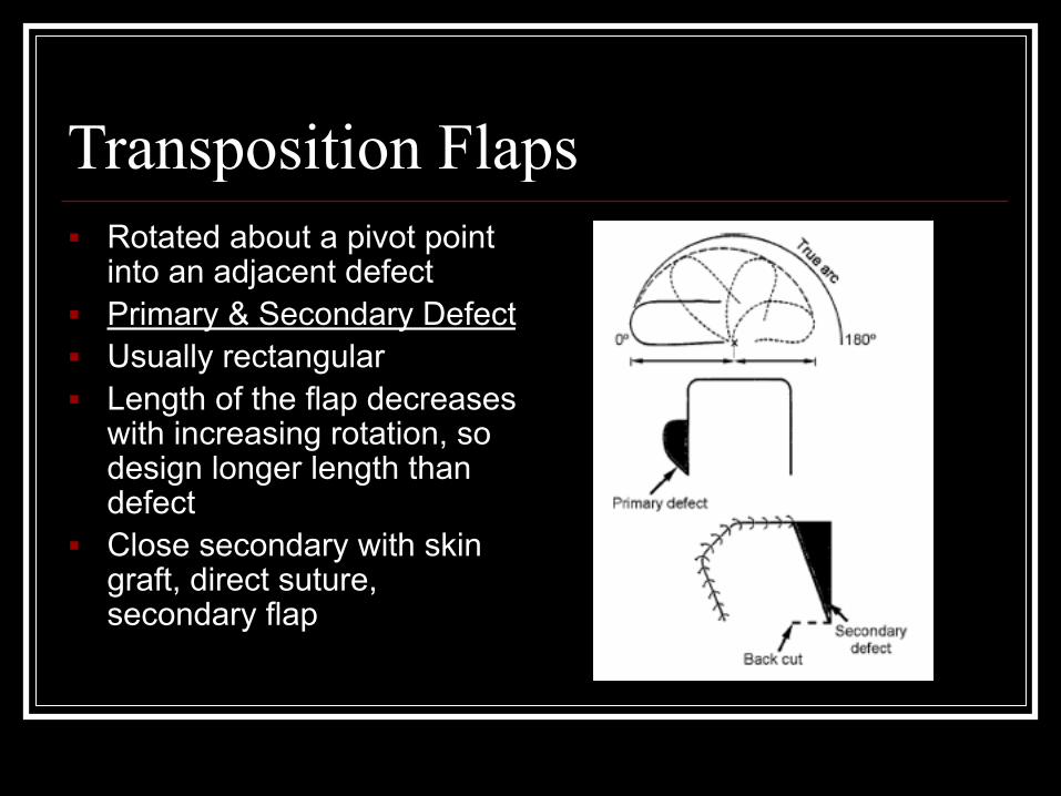
Transposition Flaps■ Rotated about a pivot point
into an adjacent defect ■ Primary & Secondary Defect ■ Usually rectangular ■ Length of the flap decreases
with increasing rotation, so design longer length than defect
■ Close secondary with skin graft, direct suture, secondary flap

Z-plasty■ Type of transposition flap ■ Random ■ Used to lengthen contractures or
scars ■ Two triangular flaps interchanged ■ Three limbs (Z) must be equal in
length ■ Amount of length gained related to
the degree of angles of the Z ■ Inside angles 60 degrees ■ Single large Z-plasty more effective
than multiple smaller ones

Z-plasty■ Keep angle >30
degrees to avoid tip necrosis
■ All limbs equal length ■ Increase scar length by
25% for 30, 50% with 45, and 75% with 60

M-plasty■ Reduces the amount of skin excised (e.g. lip, forehead) ■ Can advance tip with closure

Rhomboid (Limberg) Flap■ Transposition flap ■ Longitudinal axis -
line of minimal skin tension
■ Designed around angles of 60 degrees

Rhomboid design

Rhomboid Design

Bipedicle flap■ Two mirror image
transposition flaps ■ Share their distal,
undivided margin (pedicle)

Interpolation flaps■ Rotate about a pivot
point (like transposition flaps)
■ Defects near, not adjacent to the donor site
■ Littler (neurovascular island flap)

Neurovascular Island Flaps■ Axial flap, transfer of skin, subq, digital artery and nerve to a defect
that requires durable skin and sensation ■ Ulnar side of the long or ring fingers is typically used as donor site
because these digits have codominant digital vessels ■ Digital allen test preoperatively ■ Best axial flap for digital amputation can be used for severe thumb
pulp defects ■ Key Steps
■ Extensive Brunner incision to mobilize a neurovascular bundle to mid palmar origin
■ Reroute flap to the defect theough a subcutaneous tunner

Neurovascular Island Flaps
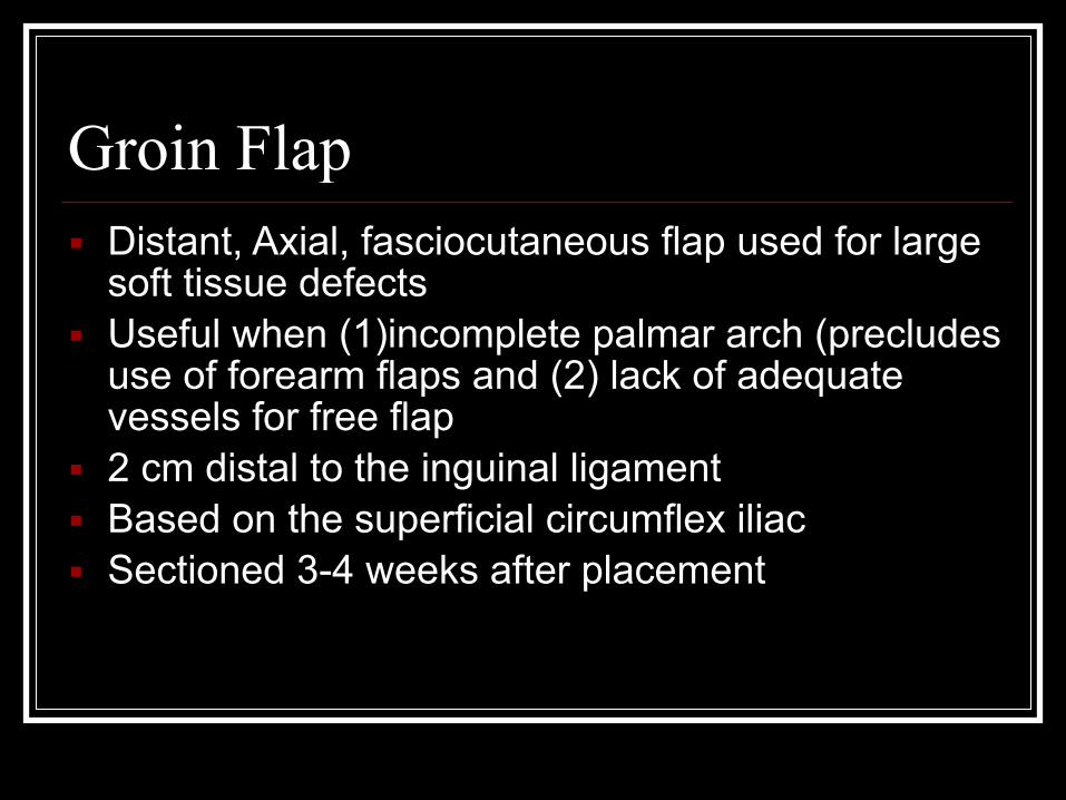
Groin Flap■ Distant, Axial, fasciocutaneous flap used for large
soft tissue defects ■ Useful when (1)incomplete palmar arch (precludes
use of forearm flaps and (2) lack of adequate vessels for free flap
■ 2 cm distal to the inguinal ligament ■ Based on the superficial circumflex iliac ■ Sectioned 3-4 weeks after placement

Groin Flap

Groin flap
■ Combined pedicled superficial inferior epigastric & groin flap for reconstruction of a dorsal & volar hand injury

Free Tissue Transfer■ Transfer of autologous tissue from one location to
another using techniques of microvascular surgery for small vessel anastomoses
■ Three (main) steps: ■ Complete detachment of the flap ■ Revascularization with anastomoses to BV ■ Intervening period of flap ischemia

Microvascular anastomoses■ Most flap pedicle vessels 0.8 - 4.0mm ■ Operative microscope between 6 - 40x ■ Success rates have increased to 95% (no
longer considered ‘last-ditch’) ■ Most important factor in free flap failure is
thrombosis

Virchow’s Triad■ Factors altering laminar flow, causing
endothelial damage,or hypercoagulability

Free Flap: Planning■ Pedicle length & size ■ Which recipient vessel to use ■ Orientation of anastomosis (e.g. end-to-side) ■ Deal with mismatched vessel size ■ Overcome unhealthy vessels ■ How to inset flap tissues for function & cosmesis ■ Routing the pedicle (restore blood flow, avoid kinking, etc) ■ Patient positioning ■ Postop dressings (avoid compression) ■ Backup plan (e.g. interposition vein graft for length)

Conclusions: Technical points ■ Check for joint movement
■ Ensure this does not compromise the stability or place tension on the flap
■ Consider the zone of injury and scarring ■ these zones may be unsuitable for re-
vascularization of a free flap or for basing a pedicle flap (the stiffness of the tissue may prevent transposition)

Conclusions: The Pedicle■ Vascular pedicle must be protected from ■ 1) exposure ■ 2) tension ■ 3) internal compression ■ 4) external pressure